Non cancerous and pre cancerous skin lesions are often overlooked, but understanding them is crucial for early detection and effective management. These skin changes, ranging from benign moles to potentially precancerous growths, can vary significantly in appearance and location. Early diagnosis is paramount to preventing further complications and ensuring proper treatment. This comprehensive guide will explore the different types, risk factors, diagnosis, prevention, and treatment strategies associated with these conditions.
This post delves into the complexities of non-cancerous and pre-cancerous skin lesions, providing a detailed overview of their characteristics, causes, and potential risks. We’ll cover everything from identifying different lesion types based on their appearance to discussing the importance of regular check-ups and preventive measures. By understanding these lesions, you can take proactive steps towards maintaining healthy skin and well-being.
Introduction to Non-Cancerous and Pre-Cancerous Skin Lesions

Skin conditions manifest in various forms, some benign and others potentially harmful. Understanding the nuances between non-cancerous and pre-cancerous skin lesions is crucial for early detection and appropriate management. Early intervention can prevent progression to more serious forms of skin cancer. This exploration delves into the characteristics, risk factors, and potential complications of these conditions.Non-cancerous skin lesions are harmless growths or changes in the skin’s appearance.
Pre-cancerous lesions, on the other hand, are alterations in skin cells that, if left untreated, can potentially develop into cancerous growths. Recognizing these distinctions is vital for prompt medical attention and effective treatment strategies.
Definition of Non-Cancerous and Pre-Cancerous Lesions
Non-cancerous skin lesions are harmless growths or alterations in the skin’s texture or appearance. Pre-cancerous lesions are changes in skin cells that have the potential to become cancerous if left untreated. They represent an intermediate stage between normal skin cells and cancerous cells.
Importance of Early Detection and Diagnosis
Early detection and prompt diagnosis are critical in managing non-cancerous and pre-cancerous skin lesions. Early intervention can prevent progression to more serious conditions. This approach significantly improves treatment outcomes and reduces the risk of complications. Regular self-exams and professional checkups are essential tools in this process.
Risk Factors Associated with These Lesions
Several factors increase the likelihood of developing non-cancerous and pre-cancerous skin lesions. Exposure to ultraviolet (UV) radiation from sunlight or tanning beds is a significant risk factor. Fair skin, a history of sunburns, and a family history of skin cancer are additional factors. Age, certain medications, and immune deficiencies also play a role.
Characteristics of Different Lesion Types
This table summarizes the key features of various non-cancerous and pre-cancerous skin lesions. Knowing the typical appearance, location, and potential complications is vital for recognizing these conditions early.
| Lesion Type | Appearance | Location | Potential Complications |
|---|---|---|---|
| Actinic Keratosis | Rough, scaly patches, often reddish-pink or brown, may feel rough or gritty | Sun-exposed areas, such as face, ears, scalp, hands, and forearms | Can progress to squamous cell carcinoma if left untreated. |
| Seborrheic Keratosis | Warty, raised, brown, or black lesions, often appear stuck on the skin | Scalp, face, chest, and back | Rarely become cancerous. May be mistaken for other conditions. |
| Benign Nevi (moles) | Small, pigmented spots, can vary in color, shape, and size. | Anywhere on the body | Rarely become cancerous, but regular monitoring is important. |
| Solar Lentigines (liver spots) | Flat, brown spots, usually occur in sun-exposed areas | Hands, face, arms, and back | Rarely become cancerous, but regular monitoring is important. |
Types of Non-Cancerous Skin Lesions
Understanding non-cancerous skin lesions is crucial for early detection and appropriate management. These lesions, while not malignant, can vary significantly in appearance and characteristics. Differentiating between benign and cancerous growths is essential for accurate diagnosis and treatment.Various factors influence the development of non-cancerous skin lesions, including genetics, sun exposure, and underlying health conditions. Some are harmless and self-resolving, while others may require medical intervention for cosmetic reasons or to rule out potential pre-cancerous changes.
Careful observation and prompt consultation with a dermatologist are vital for proper management.
Moles, Non cancerous and pre cancerous skin lesions
Moles are common pigmented skin growths, typically appearing as dark spots. They are often present at birth or develop later in life. Variations in size, shape, and color exist, and they can be flat or raised. A mole’s color can range from light brown to dark brown, and sometimes even black. Some moles might have a slightly uneven texture.
Regular self-examination is key for early detection of potential changes that might indicate a need for further evaluation.
Warts
Warts are skin growths caused by human papillomavirus (HPV) infections. They are characterized by rough, bumpy surfaces, often appearing as small, flesh-colored or slightly darker nodules. Warts can occur on various parts of the body, including the hands, feet, and face. Some warts are flat, while others are raised. Warts can appear in various shapes, sizes, and densities, ranging from tiny, smooth bumps to larger, rough, and clustered growths.
Knowing the characteristics of warts helps differentiate them from other skin conditions.
Skin Tags
Skin tags are small, fleshy, benign growths that appear on the skin’s surface. They often arise from the skin’s surface, with a stalk connecting them to the surrounding skin. Skin tags are typically soft to the touch, and their size can range from a few millimeters to a centimeter or more. They can appear on the neck, armpits, eyelids, or other areas where skin folds or creases occur.
Skin tags are generally harmless and often require no treatment unless they are bothersome or become infected.
Table of Non-Cancerous Skin Lesions
| Lesion Type | Examples | Common Locations |
|---|---|---|
| Moles | Common mole, congenital mole, atypical mole | Anywhere on the body, but commonly found on sun-exposed areas |
| Warts | Common wart, plantar wart, flat wart | Hands, feet, face, and other areas with friction or trauma |
| Skin Tags | Skin tag, acrochordon | Neck, armpits, eyelids, groin, and other areas with skin folds |
Types of Pre-Cancerous Skin Lesions: Non Cancerous And Pre Cancerous Skin Lesions
Pre-cancerous skin lesions are changes in the skin that, if left untreated, have the potential to develop into skin cancer. Recognizing these early warning signs is crucial for timely intervention and preventing the progression to malignancy. Understanding the different types of pre-cancerous lesions and their characteristics empowers individuals to take proactive steps in their skin health management.
Actinic Keratosis
Actinic keratosis (AK) is a common pre-cancerous skin condition typically arising from sun exposure. It presents as rough, scaly, or crusted patches of skin, often appearing red or pink. These lesions frequently develop on sun-exposed areas, such as the face, ears, scalp, and back of the hands. Early detection is vital as AK can progress to squamous cell carcinoma, a type of skin cancer.
- Appearance: Actinic keratoses typically appear as small, rough, and slightly raised patches. The surface may be red, pink, or skin-colored, and often feels rough or slightly scaly to the touch. They can range in size from a few millimeters to a centimeter or more.
- Location: Frequently found on sun-exposed areas, including the face, ears, scalp, and back of the hands. They can also appear on the forearms and the backs of the knees.
- Potential for Malignant Transformation: While most actinic keratoses do not progress to skin cancer, some can develop into squamous cell carcinoma. Factors like persistent sun exposure, chronic inflammation, and individual susceptibility influence the risk of malignant transformation. For example, a patient with a history of multiple AKs and intense sun exposure might have a higher risk compared to someone with few AKs and minimal sun exposure.
Bowen’s Disease
Bowen’s disease is a type of squamous cell carcinoma in situ (SCCIS). It manifests as a persistent, scaly, and red or pink patch on the skin. The lesion often appears as a flat, slightly raised, and rough area, with irregular borders. Early diagnosis and treatment are crucial to prevent the disease from spreading to deeper layers of the skin.
- Appearance: Bowen’s disease lesions typically present as a flat, slightly raised, and rough area with irregular borders. The surface might appear scaly or crusted, with variations in color ranging from red to pink, or even a brownish hue. The size can range from a few millimeters to several centimeters.
- Location: Bowen’s disease can develop on various parts of the body, including the genitals, lower legs, and hands. It’s important to note that while it often occurs on sun-exposed areas, it can also appear on areas not typically exposed to the sun.
- Potential for Malignant Transformation: Bowen’s disease is considered a pre-cancerous condition. If left untreated, it has the potential to progress to invasive squamous cell carcinoma. Early diagnosis and treatment are vital in preventing this progression. A specific example is a patient diagnosed with Bowen’s disease on their leg. Early intervention and treatment with topical therapies effectively halted the progression, preventing the development of invasive squamous cell carcinoma.
Dysplastic Nevi
Dysplastic nevi are atypical moles that exhibit unusual features compared to ordinary moles. They can be flat or slightly raised, with irregular borders, varied colors (often containing shades of brown, black, and red), and sometimes asymmetrical shapes. Monitoring these lesions is essential to detect any changes and address them promptly.
- Appearance: Dysplastic nevi are characterized by irregular borders, varying colors (often a combination of brown, black, and red), and an asymmetrical shape. They can be flat or slightly raised and often larger than ordinary moles. A key feature is the uneven distribution of color within the lesion.
- Location: Dysplastic nevi can appear on any part of the body, although they are more frequently found on sun-exposed areas like the back, shoulders, and legs.
- Potential for Malignant Transformation: Dysplastic nevi are considered pre-cancerous because they have an increased risk of developing into melanoma, the most dangerous form of skin cancer. Regular monitoring by a dermatologist is crucial for early detection and management.
Table of Pre-Cancerous Skin Lesions
| Lesion Type | Key Characteristics | Potential Progression | Associated Risks |
|---|---|---|---|
| Actinic Keratosis | Rough, scaly patches on sun-exposed skin | Squamous cell carcinoma | Sun exposure, genetic predisposition |
| Bowen’s Disease | Persistent, scaly, red/pink patch | Invasive squamous cell carcinoma | Genetic factors, immunosuppression |
| Dysplastic Nevi | Atypical moles with irregular borders and varied colors | Melanoma | Family history of melanoma, multiple nevi |
Diagnosis and Evaluation
Accurately distinguishing between non-cancerous and pre-cancerous skin lesions is crucial for timely and appropriate treatment. A precise diagnosis relies on a combination of clinical evaluation, laboratory tests, and sometimes specialized imaging techniques. This process helps determine the nature and extent of the lesion, guiding treatment decisions and potentially preventing the progression to malignancy.
Dermatological Examinations
Dermatological examinations are the cornerstone of initial assessment. A skilled dermatologist visually examines the lesion, noting its size, shape, color, border definition, and any associated symptoms like itching or bleeding. Careful observation of the lesion’s evolution over time is also critical. This includes assessing the lesion’s location, symmetry, border regularity, color uniformity, and diameter (the ABCD rule).
The dermatologist considers factors like the patient’s medical history, sun exposure patterns, and family history of skin cancer.
Importance of Biopsies and Other Tests
While dermatological examinations provide valuable initial information, biopsies and other tests often become necessary for definitive diagnosis. A skin biopsy involves removing a small sample of the suspicious lesion for microscopic examination. This procedure allows pathologists to assess the cellular structure and characteristics of the lesion, enabling a definitive diagnosis. Other tests, such as dermoscopy (using a dermatoscope to view the skin’s surface in detail) or specialized imaging techniques (like confocal microscopy), may also be employed to provide additional insights.
Diagnostic Procedures and Accuracy
The accuracy of diagnostic procedures varies. A well-performed dermatological examination, coupled with a thorough patient history, can often suggest the nature of the lesion. However, when the clinical presentation is unclear or when there’s a suspicion of malignancy, a biopsy is often necessary for definitive diagnosis.
| Diagnostic Procedure | Description | Accuracy | Limitations |
|---|---|---|---|
| Dermatological Examination | Visual assessment of the lesion by a dermatologist | High, but not definitive | Subjectivity, requires expert interpretation |
| Dermoscopy | Magnified visualization of the lesion using a dermatoscope | High, can aid in early detection | Requires specialized training, not always conclusive |
| Skin Biopsy | Removal of a small tissue sample for microscopic analysis | High, definitive diagnosis | Invasive procedure, potential for scarring |
| Molecular Testing | Analysis of specific genetic or molecular markers | High, can aid in early diagnosis and prognosis | May not be available in all settings, costly |
A table summarizing various diagnostic procedures and their respective accuracy, limitations, and relevance in the diagnostic process. This table highlights the importance of a multi-faceted approach to diagnosis, integrating clinical examination with supporting laboratory tests.
Prevention and Management
Protecting your skin from non-cancerous and pre-cancerous lesions involves a proactive approach. Understanding the risk factors and implementing preventative measures can significantly reduce the likelihood of developing these conditions. Early detection and appropriate management are crucial for effective treatment and minimizing potential complications.Effective management often involves a combination of preventative measures and appropriate treatment for existing lesions. This approach aims to reduce the risk of progression to cancerous skin conditions and improve the overall health and well-being of individuals.
Strategies for Preventing Skin Lesions
Preventive strategies focus on minimizing exposure to harmful environmental factors and promoting healthy skin habits. A comprehensive approach includes avoiding excessive sun exposure, using protective clothing, and applying broad-spectrum sunscreen.
- Sun Protection: Limiting exposure to ultraviolet (UV) radiation is paramount. Seek shade during peak sun hours, particularly between 10 a.m. and 4 p.m. This is when the sun’s rays are most intense. Prolonged sun exposure significantly increases the risk of developing skin lesions.
- Protective Clothing: Wearing protective clothing, such as long-sleeved shirts, pants, and wide-brimmed hats, can offer an additional layer of defense against UV radiation. This is especially important for individuals with fair skin or a history of skin cancer.
- Sunscreen Application: Regular application of broad-spectrum sunscreen with an SPF of 30 or higher is essential. Sunscreen should be applied liberally and reapplied every two hours, or more frequently if swimming or sweating. This is a crucial step in protecting the skin from the harmful effects of UV radiation.
- Healthy Lifestyle: Maintaining a healthy lifestyle, including a balanced diet and regular exercise, can contribute to overall skin health and resilience. A diet rich in antioxidants can help protect against free radical damage.
Protective Measures Against UV Radiation Exposure
UV radiation is a significant contributor to skin damage and the development of skin lesions. Understanding the different types of UV radiation and their effects is crucial for effective protection.
- UVB Radiation: UVB radiation is primarily responsible for sunburn and plays a role in the development of skin cancer. It has a shorter wavelength and is more readily absorbed by the skin’s outer layers.
- UVA Radiation: UVA radiation penetrates deeper into the skin, causing cumulative damage over time. It is associated with premature aging and plays a role in the development of various skin conditions, including skin lesions.
- Sun Protection Strategies: Individuals should avoid prolonged sun exposure, particularly during peak hours. Wearing protective clothing and applying broad-spectrum sunscreen are vital for shielding the skin from both UVB and UVA radiation.
Common Treatment Options for Skin Lesions
Treatment options for non-cancerous and pre-cancerous skin lesions vary depending on the specific type and severity of the lesion. Some lesions may require topical therapies, while others may necessitate surgical intervention.
- Cryotherapy: This treatment involves freezing the lesion with liquid nitrogen. It is often used for small, superficial lesions and is considered a relatively simple procedure. It is a widely used method for treating benign skin conditions.
- Surgical Excision: This procedure involves surgically removing the lesion, often under local anesthesia. Surgical excision is typically employed for larger or deeper lesions and for lesions that may be concerning for malignancy.
- Topical Medications: Topical medications, such as 5-fluorouracil (5-FU) or imiquimod, can be used to treat certain types of non-cancerous and pre-cancerous lesions. These medications work by targeting the abnormal cells in the lesion.
Comparison of Treatment Modalities
The following table summarizes common treatment options for various lesion types, highlighting the advantages and disadvantages of each approach.
Learning about non-cancerous and pre-cancerous skin lesions is crucial for early detection and prevention. While physical therapy after a total ankle replacement is a whole other ballgame, physical therapy after a total ankle replacement can significantly improve mobility and recovery, similar to how understanding these skin conditions can lead to better overall health. Ultimately, recognizing these subtle changes on your skin is key to maintaining your well-being.
| Lesion Type | Treatment Modality | Advantages | Disadvantages |
|---|---|---|---|
| Actinic Keratosis | Cryotherapy | Relatively simple procedure | May not be suitable for large or deep lesions |
| Actinic Keratosis | Surgical Excision | Effective for larger lesions | More invasive procedure |
| Actinic Keratosis | Topical 5-FU | Can be effective for superficial lesions | May cause skin irritation |
| Seborrheic Keratosis | Cryotherapy | Simple and effective for small lesions | Potential for scarring |
| Seborrheic Keratosis | Surgical Excision | Removal of the lesion | More invasive |
Clinical Presentation and Appearance

Spotting skin changes early is crucial for preventing complications and ensuring prompt treatment. Knowing the visual characteristics of non-cancerous and pre-cancerous lesions can significantly aid in early detection. A thorough understanding of their appearance, including color, shape, size, and borders, empowers individuals to recognize potential problems and seek professional evaluation.Recognizing these subtle changes in the skin’s appearance is vital.
Early detection allows for timely intervention, potentially preventing progression to more serious conditions. A systematic approach to evaluating skin lesions based on their visual characteristics is essential.
Non-Cancerous Lesion Appearances
Non-cancerous skin lesions exhibit a wide array of appearances, often varying based on their specific type. Recognizing these variations can help differentiate them from pre-cancerous or cancerous lesions.
- Freckles: Small, flat, brownish macules, typically appearing clustered on sun-exposed skin. They are usually symmetrical and uniform in color, with well-defined edges.
- Moles (Melanocytic Nevi): Varied in color, ranging from light brown to dark brown or black. They can be flat or raised, with different shapes, such as round, oval, or irregular. Moles usually have a smooth texture and well-defined borders. Some moles may have a slightly rough surface. Their size can vary from a few millimeters to a centimeter or more.
They typically have a consistent color throughout.
- Seborrheic Keratoses: Warty, often raised lesions, with a rough, scaly surface. They typically present as brownish, black, or light tan plaques. They are often found on the face, chest, or back. Their shape can vary from flat to slightly raised and can be quite irregular in shape. They are usually well-defined and can vary in size from a few millimeters to several centimeters.
Their appearance often resembles a stuck-on patch of skin.
Pre-Cancerous Lesion Appearances
Pre-cancerous lesions, often referred to as actinic keratoses, can exhibit subtle changes in their appearance, sometimes mimicking benign lesions. A careful examination and recognition of these specific features are essential for early detection.
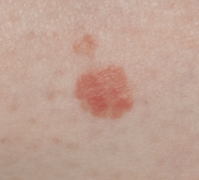
- Actinic Keratoses: These lesions frequently appear as rough, scaly patches on sun-exposed skin, often on the face, scalp, ears, or hands. They may be red, pink, or flesh-colored. They are usually slightly raised and have a rough, sandpaper-like texture. Their size can range from a few millimeters to several centimeters. The borders are often irregular, and the surface may appear crusted or thickened.
They can sometimes look like a rough patch of skin with tiny, red bumps. The color can vary from pale red to dark brown.
- Solar Lentigines: These are flat, brown or dark brown spots that often develop on sun-exposed skin. They can be larger than freckles, and their borders may be irregular or ill-defined. The shape can vary from round to oval, but they are typically well-defined, with a smooth surface.
Comparative Table of Lesion Appearances
| Lesion Type | Color | Shape | Size | Borders | Texture | Location | Examples |
|---|---|---|---|---|---|---|---|
| Freckle | Brownish | Round to oval | Small (few mm) | Well-defined | Smooth | Sun-exposed skin | Small, flat spots on the face |
| Mole | Brown to black | Round, oval, irregular | Variable (few mm to cm) | Well-defined | Smooth or slightly rough | Anywhere on the body | Dark brown spot on the back |
| Seborrheic Keratosis | Brownish, black, tan | Varied | Variable (few mm to cm) | Well-defined | Warty, rough | Face, chest, back | Rough, raised patch on the chest |
| Actinic Keratosis | Red, pink, flesh-colored | Irregular | Variable (few mm to cm) | Irregular | Rough, scaly | Sun-exposed skin | Rough patch on the ear |
| Solar Lentigo | Brown, dark brown | Round to oval | Variable (larger than freckles) | Irregular or ill-defined | Smooth | Sun-exposed skin | Large, flat brown spot on the hand |
Risk Factors and Associated Conditions
Understanding the factors that increase the likelihood of developing non-cancerous and pre-cancerous skin lesions is crucial for prevention and early detection. These factors range from environmental exposures to underlying health conditions, highlighting the importance of proactive skin health management.Skin lesions, both benign and potentially problematic, aren’t randomly distributed. Certain individuals are more predisposed due to a combination of genetic predisposition, environmental influences, and associated medical conditions.
This section delves into the key risk factors and conditions linked to the development of these skin changes.
Sun Exposure
Prolonged and unprotected sun exposure is a significant contributor to skin damage. Ultraviolet (UV) radiation from the sun penetrates the skin, causing cellular changes that can lead to the development of both non-cancerous and pre-cancerous lesions. Sunburns, even mild ones, are a clear indicator of UV damage and an increased risk. Fair-skinned individuals, those with a family history of skin cancer, and those with reduced melanin production are particularly susceptible.
Repeated sun exposure over time can result in cumulative damage and increase the likelihood of developing lesions.
Genetics
Genetic predisposition plays a substantial role in skin health. Individuals with a family history of skin cancer, especially melanoma, have a higher risk of developing various skin lesions. This genetic component may influence the skin’s natural defenses against UV damage and contribute to a faster rate of cell proliferation. For instance, specific gene mutations or variations can increase the risk of developing certain types of pre-cancerous lesions.
Other Risk Factors
Several other factors can increase the risk of skin lesions. These include:
- Immunosuppression: Individuals with weakened immune systems, such as those undergoing organ transplantation or taking immunosuppressant medications, may be more prone to developing skin lesions due to their reduced ability to fight off abnormal cell growth. This includes people with HIV/AIDS.
- Chronic skin conditions: Certain chronic skin conditions, like eczema or psoriasis, can increase the risk of developing skin lesions. The inflammation and damage associated with these conditions can create an environment conducive to abnormal cell growth.
- Exposure to certain chemicals and substances: Prolonged or high-level exposure to specific chemicals or substances, such as arsenic, certain industrial chemicals, or even certain medications, can increase the risk of developing skin lesions.
- Age: As we age, our skin undergoes changes that make it more vulnerable to damage and potentially lead to skin lesions. This includes reduced collagen production, decreased skin elasticity, and increased sun damage.
Conditions Associated with Increased Risk
Certain medical conditions can increase the likelihood of skin lesions. These include:
- Atopic dermatitis: This chronic inflammatory skin condition can lead to skin thickening and inflammation, increasing the risk of precancerous changes.
- Chronic inflammatory skin conditions: Psoriasis, lichen planus, and other chronic inflammatory skin disorders can create conditions conducive to the development of skin lesions.
- Immunodeficiency syndromes: Individuals with immunodeficiency disorders, either genetic or acquired, are more susceptible to various skin conditions, including the development of lesions due to impaired immune response.
Summary Table
| Risk Factor | Impact on Lesion Development |
|---|---|
| Sun Exposure | Increased UV radiation damages skin cells, potentially leading to DNA mutations and precancerous changes. |
| Genetics | Family history of skin cancer increases predisposition to various skin lesions. Specific gene mutations can influence susceptibility. |
| Immunosuppression | Weakened immune response allows abnormal cell growth to go unchecked, increasing lesion risk. |
| Chronic Skin Conditions | Inflammation and damage associated with conditions like eczema and psoriasis can create an environment for abnormal cell growth. |
| Exposure to Chemicals | Certain chemicals can damage skin cells and increase the risk of precancerous and cancerous changes. |
| Age | Skin changes associated with aging increase vulnerability to damage and abnormal cell growth. |
| Medical Conditions (e.g., Atopic Dermatitis, Immunodeficiency) | Specific medical conditions can increase inflammation, weaken immune response, and create an environment more conducive to lesion development. |
Treatment and Management Strategies
Treating non-cancerous and pre-cancerous skin lesions often involves a multifaceted approach tailored to the specific lesion type, its location, and the patient’s overall health. The goal is to effectively remove or manage the lesion while minimizing potential harm to surrounding healthy tissue. This involves careful consideration of various treatment options, including surgical removal, cryotherapy, and topical medications.Effective treatment of skin lesions depends on accurate diagnosis and a thorough understanding of the lesion’s characteristics.
Choosing the most appropriate treatment method requires balancing the potential benefits and risks for each individual patient. This approach emphasizes patient safety and long-term well-being.
Surgical Removal
Surgical removal is a common and often effective treatment option for a wide range of skin lesions, particularly those that are easily accessible and well-defined. Surgical excision involves removing the lesion along with a small margin of surrounding healthy tissue. This procedure is typically performed by a dermatologist or surgeon using local anesthesia. This approach is often considered the gold standard for removing lesions that are suspected to be cancerous or have a high risk of becoming cancerous.
Precise excision minimizes the risk of recurrence and allows for histopathological examination of the removed tissue, confirming the diagnosis and guiding further management.
Learning about non-cancerous and pre-cancerous skin lesions is super important for self-care. While these lesions aren’t typically life-threatening, early detection is key. This knowledge is similar to understanding how concussions are diagnosed, a completely different but equally crucial medical process. For example, a thorough evaluation, including physical exams and neurological tests, is essential in diagnosing concussions how concussions are diagnosed.
Ultimately, regular skin checks and knowing the warning signs for these lesions are essential for staying healthy.
Cryotherapy
Cryotherapy uses extreme cold to destroy abnormal skin cells. Liquid nitrogen is commonly used in this method, freezing and damaging the lesion. Cryotherapy is often suitable for smaller, superficial lesions and can be a less invasive alternative to surgical removal. The procedure is generally quick and relatively painless, with minimal recovery time. However, it can cause some temporary discomfort and skin discoloration or scarring in certain cases.
The effectiveness of cryotherapy can vary depending on the lesion’s size and depth.
While non-cancerous and pre-cancerous skin lesions can be concerning, it’s important to remember that other health issues can also cause discomfort. For example, hip, knee, and joint pain can be a significant symptom in individuals with multiple sclerosis, as discussed in more detail here: hip knee and joint pain in ms. Thankfully, recognizing these skin changes early is key to appropriate treatment and preventing potential problems, just as proactive management is important for overall well-being.
Topical Medications
Topical medications play a crucial role in managing certain types of non-cancerous and pre-cancerous lesions. These medications often target specific cellular processes involved in lesion development. Creams, ointments, or solutions containing ingredients like 5-fluorouracil (5-FU) or imiquimod are commonly used. Topical therapies can be effective for treating superficial lesions, and they are often well-tolerated with minimal side effects.
However, they may require repeated applications over a period of weeks or months to achieve optimal results, and some patients may experience mild skin irritation or redness. Proper use of topical medications and patient adherence are essential for treatment success.
Treatment Selection
Choosing the most suitable treatment depends on several factors. The size, depth, and location of the lesion are important considerations. The patient’s overall health, any existing medical conditions, and their preferences are also taken into account. For example, a large, deep lesion may require surgical removal, while a small, superficial lesion might be suitable for cryotherapy or topical treatment.
The potential risks and benefits of each treatment method should be carefully weighed against the specific characteristics of the lesion and the patient’s individual needs.
Comparison of Treatment Options
| Treatment | Effectiveness | Side Effects | Suitability |
|---|---|---|---|
| Surgical Removal | High | Potential for scarring, bleeding, infection | Larger, deeper lesions, suspected malignancy |
| Cryotherapy | Moderate to High (depending on lesion) | Temporary discomfort, skin discoloration, scarring | Small, superficial lesions, good alternative to surgery |
| Topical Medications | Moderate to High (depending on lesion) | Mild skin irritation, redness, infrequent severe reactions | Superficial lesions, good for chronic management |
Monitoring and Follow-up Care
Staying vigilant about skin lesions, whether benign or suspicious, is crucial for early detection and effective management. Proactive monitoring allows for prompt intervention if a lesion progresses or changes. Regular follow-up appointments and self-examination are essential tools in this process.
Importance of Regular Follow-up Appointments and Skin Checks
Regular check-ups with a dermatologist are vital for monitoring skin lesions. These appointments provide a structured environment for professional evaluation, allowing the dermatologist to assess any changes in the lesions’ appearance, size, or texture. Furthermore, these visits facilitate early detection of potentially problematic developments, enabling timely intervention and potentially preventing more serious complications. Dermatologists can offer personalized guidance based on the specific lesion and individual risk factors.
Monitoring Lesions for Changes
Regular self-examination is a critical component of ongoing care. Pay close attention to changes in size, shape, or color of any skin lesion. Note any new growths, ulcerations, or bleeding. Documenting these observations using photographs or sketches can be helpful for comparison during follow-up appointments. Detailed records can aid in identifying subtle changes that might otherwise go unnoticed.
Role of Dermatological Professionals in Long-Term Management
Dermatologists play a pivotal role in the long-term management of skin lesions. Their expertise in diagnosing and treating various skin conditions allows them to tailor a monitoring plan that best suits the individual patient’s needs and circumstances. They can provide guidance on self-examination techniques, and offer recommendations for lifestyle modifications that may reduce risk factors. Dermatologists are crucial for coordinating care with other specialists if necessary.
Follow-up Appointment Schedule
This table Artikels a sample follow-up schedule, which should be tailored to individual circumstances and the nature of the lesion. Frequency of follow-ups can vary greatly based on the lesion’s characteristics and the individual patient’s risk factors. A dermatologist will determine the optimal schedule.
| Lesion Type | Initial Follow-up | Subsequent Follow-ups |
|---|---|---|
| Non-cancerous, stable | 6-12 months | Annually or as clinically indicated |
| Pre-cancerous, showing slow growth | 3-6 months | Every 3-6 months until stable, then annually |
| Pre-cancerous, rapid growth | 2-4 weeks | Weekly to monthly, depending on progression |
| Suspicious lesions (with concern for malignancy) | Immediately | Weekly or bi-weekly, as necessary, with possible referral to specialists |
Last Word
In summary, recognizing the nuances of non-cancerous and pre-cancerous skin lesions is essential for proactive health management. By understanding the various types, their appearances, risk factors, and appropriate treatment strategies, individuals can significantly reduce the risk of complications and ensure timely interventions. Regular skin checks and consultations with dermatologists are crucial in preventing potentially serious conditions. This guide offers a comprehensive resource for anyone seeking to gain a deeper understanding of skin health.
Leave a Reply