Blog

  • Black Specks in Stool Causes and Solutions

    Black Specks in Stool Causes and Solutions

    Black specks in stool can be alarming, but understanding the potential causes is crucial for appropriate action. This exploration delves into various factors, from dietary indiscretions to more serious medical conditions, to help you decipher the meaning behind these dark specks. We’ll investigate the physiological mechanisms, common diagnostic procedures, and available treatment options.

    From mild digestive issues to more severe health concerns, this comprehensive guide provides a structured approach to understanding the causes, symptoms, and necessary steps to take when encountering black specks in your stool. We’ll navigate the complexities of dietary factors and medical conditions, equipping you with the knowledge to make informed decisions.

    Possible Causes of Black Specks in Stool

    Black specks in stool

    Black specks in stool, a seemingly minor issue, can sometimes signal underlying medical conditions. Understanding the potential causes, ranging from benign to severe, is crucial for prompt diagnosis and treatment. Proper evaluation by a healthcare professional is essential for accurate assessment and personalized management.

    Potential Medical Conditions

    Various medical conditions can lead to the appearance of black specks in stool. These conditions vary in severity, from mild digestive issues to more serious gastrointestinal or systemic problems. Careful consideration of associated symptoms and medical history is vital for accurate diagnosis.

    Mild Conditions

    Iron-rich foods and medications are common causes of black stool that is not indicative of a serious condition. Consuming foods rich in iron, such as red meat and leafy greens, can sometimes lead to dark-colored stools. Certain medications, particularly iron supplements, can also result in temporary discoloration. These occurrences are usually benign and resolve on their own or with dietary adjustments.

    Moderate Conditions

    Upper gastrointestinal bleeding, a more concerning cause of black stool, involves bleeding in the esophagus, stomach, or duodenum. This bleeding can result from various factors, including ulcers, gastritis, or esophageal varices. Patients often experience symptoms like nausea, vomiting, and dizziness. The blood in the upper digestive tract breaks down into smaller particles, creating the characteristic black discoloration in stool.

    This is due to the chemical process of digestion, converting the hemoglobin in the blood to a substance called melena.

    Severe Conditions

    Conditions such as esophageal varices, tumors, or severe gastritis can cause significant bleeding in the upper digestive tract. Symptoms may include severe abdominal pain, vomiting blood (hematemesis), and signs of shock, such as rapid pulse and low blood pressure. The mechanism is the same as with moderate cases, with a large amount of blood in the upper GI tract resulting in the formation of black, tarry stool.

    Condition Symptoms Mechanism Severity
    Iron-rich foods/medications Dark-colored stool only Digestion of iron-rich substances Mild
    Upper GI bleeding (ulcers, gastritis) Nausea, vomiting, dizziness, black, tarry stool (melena) Blood in upper digestive tract digested to form melena Moderate
    Esophageal varices, tumors, severe gastritis Severe abdominal pain, vomiting blood (hematemesis), shock Significant blood loss in upper GI tract, resulting in formation of melena Severe

    Dietary Factors

    Sometimes, the culprit behind those dark specks in your stool isn’t a serious medical condition, but rather what you’ve been eating. Certain foods can temporarily alter the color of your stool, leading to a surprising appearance. Understanding these dietary influences can help distinguish between a cause for concern and a simple dietary adjustment.Dietary factors can significantly impact stool color, ranging from a subtle change to a noticeable darkness.

    This often occurs due to the presence of pigments or compounds in foods that are processed differently within the digestive system. The intensity of the darkening effect depends on individual factors, including the quantity of the food consumed, the overall diet, and individual digestive processes. In some cases, the temporary discoloration might be accompanied by other symptoms like bloating or discomfort.

    Hey everyone, ever noticed black specks in your stool? It can be a little alarming, right? Sometimes, these specks could be related to something completely different, like issues with digestion. But, it’s also important to consider that similar symptoms, like a clear sticky discharge, can have various causes, and when to get treatment for these is really important.

    For more insight into the potential causes of clear sticky discharge and when it’s time to seek medical attention, check out this helpful resource: clear sticky discharge causes and when to get treatment. Ultimately, if you’re concerned about black specks in your stool, it’s always best to consult a doctor for a proper diagnosis and personalized advice.

    Foods Causing Black Stool

    A variety of foods can lead to black stool, primarily due to their iron content or other substances that react with stomach acids. These pigments can then be excreted through the stool. The duration of this effect depends on how quickly the body processes the food.

    • Iron-Rich Foods: Foods like red meat, spinach, and iron-fortified cereals are excellent sources of iron. Iron supplements can also contribute to black stool. The dark color results from the iron reacting with stomach acid and being passed through the digestive tract.
    • Foods Containing Certain Pigments: Some foods, though not as common as iron-rich ones, contain pigments that can affect stool color. These pigments can sometimes bind with other substances in the digestive system to cause a dark coloration.
    • Foods High in Melanin: Melanin, the pigment responsible for skin color, is also present in certain foods, such as dark berries. Though not a direct cause of black stool, the presence of melanin in food can contribute to the overall color.

    Duration of Effect

    The time it takes for dietary factors to affect stool color varies. It typically ranges from a few hours to a couple of days, depending on the food, the amount consumed, and individual digestion. A meal rich in iron, for example, might lead to dark stool within a few hours to a day. Conversely, a more gradual effect might be seen with foods containing pigments.

    Food Approximate Duration of Effect
    Red meat Few hours to 1-2 days
    Iron-fortified cereals Few hours to 1-2 days
    Spinach Few hours to 1-2 days
    Iron supplements Few hours to 1-2 days
    Dark berries Few hours to 2-3 days

    Comparison to Medical Conditions

    Distinguishing between dietary and medical causes of black stool can be challenging. While certain foods can lead to temporary discoloration, persistent black stool or black stool accompanied by other symptoms, such as abdominal pain, vomiting, or dizziness, could indicate a more serious medical issue. It’s crucial to consult a healthcare professional if the dark stool persists or is accompanied by other concerning symptoms.

    A medical evaluation is essential to rule out conditions such as bleeding in the upper gastrointestinal tract. The appearance of black stool due to dietary factors typically involves a more uniform darkening of the stool, unlike the possible tarry or sticky consistency that might be associated with bleeding.

    Diagnostic Considerations

    Plain Black HD Wallpapers - Top Free Plain Black HD Backgrounds ...

    Pinpointing the cause of black specks in stool requires a systematic approach. These dark particles, while sometimes benign, can also signal underlying health issues. A thorough evaluation, combining patient history, physical examination, and appropriate diagnostic tests, is crucial to accurately determine the root cause.

    Initial Evaluation and History Taking

    Gathering a comprehensive medical history is the first step. This includes details about dietary habits, recent medications, travel history, family history of digestive disorders, and any other relevant symptoms. The doctor will inquire about the frequency, duration, and consistency of the black stool, as well as any accompanying symptoms like abdominal pain, nausea, or vomiting. This initial assessment helps narrow down the potential causes and guide the subsequent diagnostic procedures.

    Physical Examination

    A physical examination is essential to evaluate the patient’s overall health status. This may include checking for signs of anemia, such as paleness, or signs of bleeding disorders. The doctor will also examine the abdomen for tenderness, masses, or other abnormalities. This step provides valuable clues about the potential cause of the black specks, helping to direct further diagnostic investigations.

    Endoscopy Procedures

    Endoscopy procedures are often used to visualize the upper digestive tract, particularly the esophagus, stomach, and duodenum. Esophagogastroduodenoscopy (EGD) is a common procedure. In EGD, a flexible tube with a camera is inserted through the mouth and into the digestive tract. This allows the doctor to directly view the lining of the digestive tract and identify any abnormalities, ulcers, or bleeding sources.

    A biopsy may be taken to obtain tissue samples for further analysis if needed. A key point is that this procedure is more invasive than other tests.

    Blood Tests

    Blood tests are routinely performed to assess various aspects of the patient’s health. These tests may include complete blood counts (CBC) to evaluate red blood cell levels, looking for signs of anemia. Other tests might be conducted to check for clotting factors, liver function, and other relevant parameters to rule out bleeding disorders or other underlying conditions. The blood test results are instrumental in evaluating the overall health status and potential bleeding issues.

    Imaging Studies

    Imaging studies, such as X-rays and CT scans, are often used to visualize the digestive system. X-rays can help detect foreign bodies, tumors, or structural abnormalities. CT scans provide a more detailed view of the internal organs, allowing for a more comprehensive evaluation of the digestive tract and potentially identifying sources of bleeding. These procedures help assess the extent of any abnormalities and the underlying cause of the black stool.

    Comparison of Diagnostic Procedures

    Diagnostic Procedure Accuracy Invasiveness Cost
    History Taking Moderate Low Low
    Physical Examination Moderate Low Low
    Endoscopy (EGD) High Moderate Moderate to High
    Blood Tests Moderate to High Low Low to Moderate
    Imaging Studies (X-ray, CT) Moderate to High Moderate Moderate to High

    Treatment Options

    Dealing with black specks in your stool can be concerning, but understanding the potential causes and appropriate treatments is crucial. The treatment approach will vary significantly depending on the underlying reason for the discoloration. Proper diagnosis is paramount to ensure the chosen treatment effectively addresses the root cause and prevents potential complications.Effective treatment for black stools often involves addressing the specific cause, whether it’s dietary, medication-related, or a more serious condition.

    This necessitates a thorough evaluation by a healthcare professional to pinpoint the source of the issue. Self-treating can be risky and may mask or worsen the underlying problem.

    Dietary Modifications

    Dietary changes can be effective for some causes of black stools. If the cause is related to food consumption, adjusting your diet can resolve the problem. For instance, if iron-rich foods are consumed excessively or improperly combined with other ingredients, dietary modifications can be an effective solution. Iron-rich foods, while essential, need to be balanced with other nutrients to prevent digestive issues.

    • Reducing or eliminating iron-rich foods for a period of time, and reintroducing them gradually, allows your body to adjust and potentially resolve the issue.
    • Increasing fiber intake through fruits, vegetables, and whole grains can improve digestion and promote regular bowel movements.
    • Avoiding foods known to cause digestive distress, such as spicy or greasy foods, can alleviate symptoms.

    Medication Adjustments

    Certain medications can cause black stools. If the black stools are related to medication, working with a doctor to adjust the dosage or switch to an alternative medication is important. For instance, if a patient taking iron supplements experiences black stools, a lower dose or a different form of iron may resolve the problem.

    • Adjusting the dosage of medications that may be contributing to the black stools.
    • Switching to alternative medications if possible.
    • Following prescribed medication instructions carefully.

    Medical Interventions

    For more serious causes of black stools, such as bleeding in the upper gastrointestinal tract, medical interventions might be necessary. This could range from endoscopy to identify the source of the bleeding to more invasive procedures if necessary. The approach will depend on the severity and location of the bleeding.

    • Endoscopy: A procedure where a thin, flexible tube with a camera is inserted into the esophagus, stomach, and duodenum to visualize the lining and identify any bleeding sources. Expected outcomes are identifying the cause and potentially treating it during the procedure.
    • Angiography: A procedure using X-rays to visualize blood vessels and treat bleeding in the gastrointestinal tract. Outcomes may include stopping bleeding and treating the underlying cause, with potential complications including allergic reactions or infections.
    • Surgical interventions: For severe cases of bleeding ulcers or tumors, surgery may be necessary. Expected outcomes include resolving the source of bleeding, but risks are significant, including potential complications during and after surgery.

    Comparison of Treatment Options, Black specks in stool

    The best treatment option depends on the underlying cause. Dietary modifications are generally the safest and least invasive option for simple cases. Medication adjustments require close collaboration with a physician to avoid adverse effects. Medical interventions are reserved for more severe conditions requiring specialized procedures.

    Treatment Option Potential Benefits Potential Risks Typical Duration
    Dietary Modifications Relatively safe, inexpensive, and can resolve the issue in simple cases. May not be effective for all causes, requires patient adherence. Variable, often weeks to months.
    Medication Adjustments Can resolve the issue related to medications. Potential side effects from altered medications, requires careful monitoring by a physician. Variable, dependent on the medication and its effects.
    Medical Interventions Effective for severe cases, addresses the underlying cause. Higher risk of complications, more invasive procedures. Variable, from days to weeks, dependent on the severity and type of procedure.

    Preventive Measures

    Preventing black specks in stool often hinges on proactive dietary and lifestyle choices. By understanding the potential causes and adopting healthy habits, you can significantly reduce the risk of experiencing this unsettling issue. Early detection through regular checkups plays a crucial role in addressing underlying problems before they escalate.Maintaining a healthy digestive system is a multifaceted process. A balanced diet, combined with mindful lifestyle choices, can significantly impact the overall health of your digestive tract.

    Understanding these connections is key to preventative measures.

    Dietary Habits for Digestive Health

    A balanced diet is paramount for maintaining a healthy digestive system. Focus on incorporating a variety of nutrient-rich foods, including fruits, vegetables, whole grains, and lean proteins. These foods provide essential vitamins, minerals, and fiber, which are crucial for proper digestion.

    Hey everyone, Have you ever noticed black specks in your stool? It can be a bit concerning, but sometimes it’s just harmless. However, if it’s persistent, it’s definitely worth checking in with a doctor. Interestingly, donating blood can actually have some surprising benefits for your overall health, such as boosting your iron levels. Consider looking into the benefits of donating blood if you’re healthy enough.

    Either way, if you’re experiencing black specks in your stool, consulting a healthcare professional is key to getting to the bottom of it and ensuring you’re taking care of your health.

    • Fiber-Rich Foods: Increasing fiber intake promotes regular bowel movements and helps prevent constipation, a factor that can contribute to the appearance of black specks. Examples include whole grains, fruits (like berries and apples), and vegetables (like broccoli and spinach). Adequate fiber consumption can help maintain regularity.
    • Hydration: Drinking plenty of water is essential for proper digestion and bowel movements. Water helps soften stool and aids in its passage through the digestive tract. Dehydration can lead to constipation and potentially contribute to the appearance of black specks.
    • Limit Processed Foods and Red Meat: Reducing consumption of processed foods, especially those high in saturated fats and unhealthy additives, can be beneficial for overall digestive health. Similarly, excessive consumption of red meat may also impact digestion. Moderation in these areas can help prevent digestive issues.

    Lifestyle Choices for Digestive Well-being

    Beyond diet, lifestyle choices play a significant role in maintaining a healthy digestive system. Stress management techniques and regular exercise are crucial components of a holistic approach.

    • Stress Management: Chronic stress can negatively impact the digestive system, potentially leading to irregularities. Incorporating stress-reducing activities, such as yoga, meditation, or spending time in nature, can promote digestive health.
    • Regular Exercise: Physical activity is beneficial for overall health and can aid in promoting regular bowel movements. A moderate exercise routine can help maintain a healthy weight and support digestive function.
    • Adequate Sleep: Getting sufficient sleep is crucial for overall health, including digestive health. Lack of sleep can disrupt the body’s natural processes, potentially impacting digestion.

    Importance of Regular Checkups and Screenings

    Regular medical checkups and screenings are essential for early detection of potential issues. These checkups allow for early identification of underlying conditions that might be causing black specks in stool.

    • Routine Colonoscopies: For individuals with risk factors or a family history of digestive disorders, routine colonoscopies are crucial for early detection of polyps or other abnormalities that could be associated with black specks in stool. These procedures can help identify potential issues before they become serious.
    • Regular Blood Tests: Routine blood tests can reveal potential underlying conditions that might be contributing to digestive problems, such as anemia or other medical issues. These tests provide crucial diagnostic information.

    Situations Requiring Increased Preventative Measures

    Certain individuals may require heightened attention to preventative measures due to specific circumstances or risk factors.

    • Individuals with a Family History of Digestive Disorders: Those with a family history of conditions like inflammatory bowel disease (IBD) or colorectal cancer should prioritize regular checkups and screenings to detect any potential issues early.
    • Individuals with Existing Digestive Issues: If you have a history of digestive problems, maintaining a healthy diet and lifestyle is particularly important. This includes managing stress, staying hydrated, and following a balanced eating plan.

    Additional Information

    Persistent black specks in your stool, while often benign, warrant attention. Ignoring these specks could potentially mask underlying conditions requiring prompt medical intervention. Understanding when to seek immediate help and where to find reliable resources is crucial for managing this concern effectively.Seeking medical advice for persistent black specks in stool is important to rule out any serious underlying conditions.

    A healthcare professional can evaluate your specific situation, considering your medical history, lifestyle, and dietary habits. This personalized assessment is essential for an accurate diagnosis and appropriate treatment plan.

    Importance of Seeking Medical Advice

    A healthcare provider can thoroughly assess the cause of black specks in your stool. They can perform necessary diagnostic tests and determine if the specks are indicative of a serious condition. Ignoring persistent black specks could lead to delays in diagnosis and treatment of a more serious condition, potentially impacting your health negatively.

    When to Seek Immediate Medical Attention

    Certain situations demand immediate medical attention. Seek emergency care if you experience black specks in your stool alongside other symptoms like severe abdominal pain, vomiting, dizziness, or difficulty breathing. These symptoms could signal a significant issue requiring prompt intervention. Additionally, if the black specks appear with other signs of internal bleeding such as unusually dark or tarry stools, or if you experience significant blood loss, seek immediate medical attention.

    Those pesky black specks in your stool can be a real worry. While they might seem alarming, they’re often nothing to panic about. Sometimes, though, they could be a sign of something more serious, like a digestive issue. Knowing what a seizure looks like can also be helpful in understanding potential causes for unusual symptoms. For a better understanding of how a seizure might manifest, check out this informative article on what does a seizure look like.

    If you’re concerned about black specks in your stool, it’s always best to consult a doctor for a proper diagnosis.

    Reliable Resources for Further Information

    For further information and support, consult reputable medical organizations like the Mayo Clinic, the Cleveland Clinic, and the National Institutes of Health (NIH). These organizations offer reliable and comprehensive information about various health concerns. Additionally, your primary care physician or a gastroenterologist can provide tailored advice based on your individual circumstances.

    Key Takeaways: When to Seek Immediate Medical Attention

    Symptom(s) Action
    Black specks in stool accompanied by severe abdominal pain, vomiting, dizziness, or difficulty breathing Seek immediate medical attention.
    Black specks in stool accompanied by unusually dark or tarry stools, or significant blood loss Seek immediate medical attention.

    Illustrative Examples: Black Specks In Stool

    Understanding the causes of black specks in stool requires examining various scenarios. These examples highlight the diverse factors contributing to this symptom, ranging from dietary choices to underlying medical conditions. A detailed understanding of these scenarios can aid in proper diagnosis and treatment.

    Dietary Factor Example: Iron-Rich Foods

    A patient experiencing black stool following the consumption of a significant amount of iron-rich foods, such as red meat or iron supplements, is a common dietary-related example. The iron in these foods reacts with stomach acid, leading to the appearance of black, tarry stools. This is a harmless, temporary effect and usually resolves as the patient reduces or eliminates the offending foods from their diet.

    Medical Condition Example: Gastrointestinal Bleeding

    Gastrointestinal (GI) bleeding, stemming from ulcers, esophageal varices, or inflammatory bowel disease, can manifest as black specks or tarry stool. The blood, broken down in the digestive tract, imparts the dark color to the stool. The severity of the bleeding can vary greatly, impacting the appearance and consistency of the stool. In severe cases, the stool may appear very dark and resemble tar.

    Patient Presentation and Diagnostic Process

    A patient presenting with black specks in stool should undergo a comprehensive evaluation. This involves a detailed medical history, focusing on recent dietary changes, medications, and any prior GI issues. A physical examination follows, assessing for signs of anemia, abdominal tenderness, or other relevant symptoms. Laboratory tests, including stool tests to check for blood, and blood tests to evaluate hemoglobin levels, will aid in identifying the underlying cause.

    Further diagnostic procedures, such as endoscopy or colonoscopy, may be necessary to visualize the source of the bleeding.

    Importance of Detailed Medical History and Physical Examination

    A detailed medical history is crucial in determining the cause of black stool. This includes inquiries about recent dietary changes, medication use, and past medical conditions, including previous gastrointestinal issues. The patient’s medical history provides a context for understanding the potential causes and helps differentiate between benign and potentially serious conditions. The physical examination plays a vital role in assessing the patient’s overall health status, identifying any signs of anemia or abdominal tenderness, and supporting the information gathered from the medical history.

    These combined efforts provide a strong foundation for a comprehensive diagnostic approach.

    Ultimate Conclusion

    In conclusion, black specks in stool can be a sign of something minor or something requiring immediate attention. This guide has explored the potential causes, diagnostic procedures, and treatment options. Remember, seeking medical advice is paramount if you experience persistent black specks. A detailed discussion with your doctor is essential for a proper diagnosis and personalized treatment plan.

  • Erectile Dysfunction and Age A Comprehensive Guide

    Erectile Dysfunction and Age A Comprehensive Guide

    Erectile dysfunction and age are intricately linked. As men age, physiological changes impact their ability to achieve and maintain an erection. This comprehensive guide delves into the prevalence, underlying physiological mechanisms, risk factors, medical conditions, diagnostic approaches, treatment options, prevention strategies, and patient education surrounding erectile dysfunction in older men. We’ll explore the nuances of how age affects this condition, offering a clear understanding for both individuals and healthcare professionals.

    Understanding the relationship between erectile dysfunction and age is crucial for proactive health management. This guide will provide a structured approach to understanding the causes, risks, and solutions related to erectile dysfunction in various age groups, equipping readers with the knowledge to make informed decisions about their health.

    Prevalence and Demographics

    Erectile dysfunction and age

    Erectile dysfunction (ED) is a common health concern, particularly as men age. Understanding its prevalence and demographic patterns is crucial for effective prevention and treatment strategies. This understanding allows healthcare providers to better tailor their approach to individual patients and develop targeted interventions.The relationship between age and ED is well-documented. As men progress through different life stages, various physiological and lifestyle factors can influence their ability to achieve and maintain an erection.

    The following sections will delve into the prevalence and demographic characteristics of ED, examining the impact of age, geographic location, and socioeconomic status.

    Prevalence of ED Across Age Groups

    The prevalence of ED increases significantly with age. Multiple studies have shown a strong correlation between advancing age and the likelihood of experiencing ED. This rise in prevalence is often attributed to the natural physiological changes associated with aging, such as hormonal shifts and vascular changes.

    Age Group Estimated Prevalence of ED
    20-30 Approximately 5-10%
    31-40 Approximately 10-15%
    41-50 Approximately 15-20%
    51-60 Approximately 25-35%
    61-70 Approximately 40-50%
    71 and older Approximately 50-70%

    Note: Prevalence estimates may vary based on the specific study and population sampled. Factors such as lifestyle, underlying health conditions, and cultural influences can also affect the prevalence rate.

    Demographic Factors Associated with ED

    Several demographic factors are associated with ED. These factors may influence the risk of developing ED or its severity.

    Dealing with erectile dysfunction (ED) can be a common concern as we age, impacting quality of life. Fortunately, understanding the magi calculation for health insurance subsidy eligibility can be a big help in affording necessary treatments. This calculation can often significantly reduce the cost of care, which is essential when considering the potential long-term financial implications of ED.

    Ultimately, access to affordable treatment options becomes crucial in managing ED and its impact on older adults.

    • Age: As previously mentioned, the prevalence of ED increases significantly with age. This is due to a combination of physiological changes and potential underlying health conditions.
    • Geographic Location: Studies have shown that ED prevalence may vary across different geographical regions. This could be due to differences in lifestyle factors, access to healthcare, cultural norms, and socioeconomic conditions.
    • Socioeconomic Status: Men with lower socioeconomic status may face greater challenges in accessing healthcare and maintaining healthy lifestyles, which could potentially increase their risk of ED.

    Various lifestyle factors, including smoking, diet, and physical activity, can also contribute to the risk of developing ED, irrespective of age.

    Research Findings on Age and ED

    Numerous studies have investigated the link between age and erectile dysfunction. For instance, one study published in the Journal of Urology found a strong correlation between age and the incidence of ED, demonstrating a steady increase in prevalence with advancing years. This aligns with other research findings suggesting that age-related changes in vascular function and hormonal levels play a critical role in the development of ED.

    Erectile dysfunction (ED) and age often go hand-in-hand. As we get older, various factors can contribute to this issue, including lifestyle choices and potential underlying health conditions. One often overlooked aspect is the consumption of common foods with synthetic dyes, which can potentially impact overall health and contribute to ED. Understanding the impact of these additives is crucial for proactive health management.

    For a detailed look at common foods with synthetic dyes, check out this informative resource: common foods with synthetic dyes. Ultimately, a balanced diet and healthy lifestyle choices are essential for maintaining sexual health at any age.

    Physiological Changes with Age

    Aging brings about a multitude of physiological changes that can significantly impact various bodily functions, including erectile function. These alterations, often subtle at first, accumulate over time, gradually contributing to the development of erectile dysfunction (ED) in many men. Understanding these changes is crucial for comprehending the link between age and ED.

    Hormonal Changes and Erectile Function

    Testosterone levels naturally decline with age, often starting in the late 30s or 40s. This decline, although gradual, can have a substantial impact on erectile function. Testosterone plays a vital role in the production of sperm, muscle growth, and libido, and a decrease in its levels can lead to a reduced desire for sexual activity, and make it more challenging to achieve and maintain an erection.

    Other hormones, such as follicle-stimulating hormone (FSH) and luteinizing hormone (LH), also contribute to the regulation of sexual function and are affected by the aging process. This hormonal shift is not a uniform experience, and the degree of impact on each individual varies.

    Vascular Changes and Erectile Dysfunction

    As men age, their blood vessels undergo changes, including a thickening of the arterial walls (atherosclerosis). This process can restrict blood flow to the penis, making it harder to achieve and maintain an erection. Reduced blood flow is a key factor in the development of erectile dysfunction. This narrowing of blood vessels can be influenced by various factors, including high blood pressure, high cholesterol, and diabetes, all of which increase with age.

    The decreased elasticity and increased stiffness of the blood vessels can impair the ability to fill the corpora cavernosa with blood, essential for achieving an erection.

    Nerve Function and Erectile Dysfunction

    Nerve function also plays a crucial role in erectile function. With age, there can be a gradual decline in nerve function throughout the body, including the nerves that control blood flow to the penis. These nerves are responsible for the complex signaling processes that initiate and maintain an erection. The transmission of signals between the brain, nerves, and muscles becomes less efficient, leading to difficulties in achieving and maintaining an erection.

    This can also be influenced by neurological conditions, which become more prevalent with age.

    Table: Physiological Changes and Their Impact on Erectile Function at Different Ages

    Age Group Physiological Changes Impact on Erectile Function
    30s-40s Gradual decline in testosterone levels, minor vascular changes Potential for reduced libido, slight difficulty in achieving or maintaining an erection in some cases.
    50s-60s Significant decline in testosterone levels, increased vascular stiffness, mild nerve damage Increased difficulty in achieving and maintaining erections, potentially impacting sexual satisfaction.
    70s and beyond Further decline in testosterone levels, significant vascular disease, nerve damage Significant challenges in achieving and maintaining erections, often requiring medical intervention.

    Risk Factors and Lifestyle

    Understanding erectile dysfunction (ED) goes beyond just the physiological changes associated with aging. Lifestyle choices and underlying risk factors play a significant role in the development and progression of ED at any age. This exploration delves into the common risk factors and how lifestyle modifications can influence erectile function across different age groups.The interplay between genetics, lifestyle, and age is crucial in comprehending ED.

    Factors like diet, exercise, and smoking habits can significantly impact erectile function, and these influences are often exacerbated with age due to the natural physiological changes that occur. By recognizing these factors, individuals can take proactive steps to mitigate the risk of ED and improve their overall health.

    Common Risk Factors Associated with ED

    Various factors contribute to the development of ED. These factors aren’t exclusive to any particular age group, but their impact and interplay with age are important to understand. Underlying medical conditions, such as diabetes, heart disease, and high blood pressure, often increase the risk of ED. These conditions are more prevalent with age, and therefore, the risk of ED increases accordingly.

    Influence of Lifestyle Choices on Erectile Function

    Lifestyle choices profoundly impact erectile function across all age groups. A diet high in saturated fats and processed foods can negatively affect blood flow, a critical component of erectile function. Similarly, a lack of regular exercise can lead to reduced blood flow and overall cardiovascular health, which are directly related to ED risk. Smoking significantly damages blood vessels, further hindering blood flow and increasing the likelihood of ED.

    Lifestyle Interventions to Mitigate ED Risk

    Implementing positive lifestyle changes can significantly reduce the risk of ED, regardless of age. A balanced diet rich in fruits, vegetables, and lean proteins supports overall cardiovascular health and promotes healthy blood flow. Regular physical activity, including aerobic exercise and strength training, enhances cardiovascular health and contributes to better erectile function. Quitting smoking immediately improves blood flow and reduces the risk of various health issues, including ED.

    Table Summarizing Risk Factors, Lifestyle Choices, and Potential Impact on ED at Various Ages

    Risk Factor/Lifestyle Choice Potential Impact on ED (younger adults) Potential Impact on ED (middle-aged adults) Potential Impact on ED (older adults)
    Poor Diet (high in saturated fats, processed foods) Potentially contributing to early cardiovascular issues, affecting blood flow. Potentially exacerbating existing cardiovascular issues, leading to ED symptoms. Significant impact on blood flow, contributing to more severe ED symptoms and increasing the likelihood of comorbidities.
    Sedentary Lifestyle (lack of regular exercise) Reduced cardiovascular fitness, potentially impacting blood flow. Increased risk of cardiovascular problems, leading to ED symptoms and/or worsening existing conditions. Significant impact on overall health and blood flow, leading to more severe ED and potentially related health complications.
    Smoking Potential impact on blood vessels, hindering blood flow. Significant impact on blood vessels, increasing the risk of ED and cardiovascular problems. Severe impact on blood vessels, worsening ED and potentially increasing the risk of cardiovascular events.
    Stress Can impact performance in the bedroom, potentially causing temporary ED. Chronic stress can contribute to ED symptoms, impacting both mental and physical health. Chronic stress can worsen existing health conditions and ED, increasing the risk of adverse health outcomes.
    Underlying Medical Conditions (e.g., diabetes, heart disease) Potentially present but may not be as prominent. More prevalent, contributing significantly to ED risk. Highly prevalent, often directly causing or exacerbating ED.

    Medical Conditions and Medications

    Erectile dysfunction (ED) isn’t solely an issue of aging; various underlying medical conditions and medications can significantly contribute to its development. Understanding these connections is crucial for effective diagnosis and treatment. This section delves into the interplay between medical conditions, medications, and ED in older men.Medical conditions like diabetes and high blood pressure frequently impact erectile function. These conditions often lead to vascular damage, hindering blood flow to the penis, a crucial component of achieving and maintaining an erection.

    Furthermore, certain medications, including some commonly prescribed for high blood pressure or prostate issues, can have side effects that interfere with the normal erectile process.

    Diabetes and Erectile Dysfunction

    Diabetes significantly increases the risk of ED, often due to its impact on blood vessels and nerves. High blood sugar levels over time can damage the delicate blood vessels that supply the penis, reducing blood flow and making it challenging to achieve an erection. Additionally, nerve damage associated with diabetes can affect the signals that control erections. This intricate relationship between diabetes and ED highlights the importance of managing blood sugar levels to mitigate ED risk.

    High Blood Pressure and Erectile Dysfunction

    High blood pressure, also known as hypertension, can also contribute to ED in older men. Sustained high blood pressure can damage blood vessels throughout the body, including those in the penis. This vascular damage restricts blood flow, hindering the ability to achieve and maintain an erection. Effective management of blood pressure is crucial to minimizing the risk of ED in men with hypertension.

    Medications and Erectile Dysfunction, Erectile dysfunction and age

    Many medications can have unintended side effects that impact erectile function. Certain blood pressure medications, particularly diuretics and beta-blockers, can sometimes decrease blood flow to the penis. Similarly, some antidepressants and medications for prostate conditions can also contribute to ED. It’s important to discuss potential side effects with your doctor if you experience ED while taking any medication.

    Interactions Between Medical Conditions and Medications

    The interplay between medical conditions and medications can sometimes exacerbate ED. For instance, a man with both diabetes and high blood pressure might experience more pronounced ED symptoms due to the combined effects of vascular damage and compromised blood flow. Similarly, taking multiple medications with potential ED side effects can significantly increase the risk. Open communication with your doctor is essential to assess the potential impact of your medical conditions and medications on your erectile function.

    Table of Medical Conditions and Medications Associated with ED

    The following table provides a brief overview of medical conditions and medications frequently linked to ED. It’s not exhaustive and should not be used as a definitive guide. Always consult with a healthcare professional for personalized advice.

    Medical Condition Medication
    Diabetes Diuretics, Beta-blockers, Some Antidepressants
    High Blood Pressure (Hypertension) Diuretics, Beta-blockers, Some Antidepressants
    Cardiovascular Disease Certain Anti-arrhythmics, Statins (in some cases)
    Neurological Conditions Certain Anticonvulsants, Some Antipsychotics
    Prostate Cancer Treatment Hormonal Therapies

    Diagnostic Approaches: Erectile Dysfunction And Age

    Erectile dysfunction and age

    Understanding erectile dysfunction (ED) in older men requires a multifaceted approach that goes beyond simply identifying the presence of the condition. A comprehensive diagnostic evaluation considers a range of factors, from physical health to psychological well-being, to arrive at an accurate diagnosis and develop an appropriate treatment plan. This involves careful consideration of medical history, physical examination, and relevant testing to uncover the underlying causes.Comprehensive diagnostic evaluations for ED in older men must account for the intricate interplay of physiological changes associated with aging, potential underlying medical conditions, and the influence of psychological factors.

    The diagnostic process aims to identify the root cause of ED, guiding the selection of the most effective treatment strategy.

    Physical Examination and Medical History

    A thorough physical examination and detailed medical history are crucial first steps in assessing ED. The physical exam allows for the evaluation of general health and the detection of any physical abnormalities that might contribute to ED. This includes assessing blood pressure, heart rate, and checking for any neurological or vascular abnormalities. A detailed medical history helps identify potential contributing factors such as underlying medical conditions (diabetes, heart disease, etc.), previous surgeries, and current medications.

    This information helps establish a baseline for understanding the patient’s overall health and potential risk factors. Identifying any underlying conditions is essential for effective management of ED.

    Psychological Factors

    Psychological factors can significantly influence the development and experience of ED. Depression, anxiety, stress, relationship problems, and other emotional concerns can all contribute to ED. Assessing these factors is vital, as addressing psychological issues can be just as important as addressing physical ones. A detailed discussion with the patient about their emotional state and relationship dynamics is critical to developing a complete picture of their overall health.

    It’s a common misconception that erectile dysfunction (ED) is solely a problem of older age, but age can play a role. While it’s important to address potential underlying health concerns, sometimes lifestyle choices and overall well-being are key factors. For example, if you’re wondering if you should take NyQuil when you have COVID, this article delves into that.

    Ultimately, age-related ED is a complex issue, and seeking professional medical advice is crucial for proper diagnosis and treatment.

    Blood Tests and Other Relevant Tests

    Blood tests can provide valuable insights into potential underlying medical conditions that may contribute to ED. These tests can help assess hormone levels (testosterone), check for diabetes, and evaluate cardiovascular health. Additional tests, such as a nocturnal penile tumescence test (NPTR) or a duplex ultrasound, might be necessary in certain cases to assess blood flow to the penis.

    These tests help to determine the specific cause of ED and its potential impact on the vascular system. They provide objective data to support the clinical picture obtained from the physical examination and medical history.

    Summary Table of Diagnostic Approaches

    Diagnostic Approach Procedure Relevance in Different Age Groups
    Physical Examination and Medical History Assessment of general health, blood pressure, heart rate, and detection of physical abnormalities; detailed discussion of medical history, current medications, and potential contributing factors. Crucial in all age groups, providing a foundational understanding of the patient’s overall health.
    Psychological Evaluation Assessment of emotional state, stress levels, relationship dynamics, and any psychological factors that might be impacting ED. Important across all age groups, particularly in cases where psychological factors are suspected to play a role.
    Blood Tests Assessment of hormone levels (testosterone), blood glucose levels, and other markers of cardiovascular health. Helpful in identifying underlying medical conditions, such as diabetes or low testosterone, which can contribute to ED in all age groups.
    Nocturnal Penile Tumescence (NPTR) Monitoring nocturnal erections to assess penile blood flow. Useful in cases where vascular issues are suspected. More commonly used in younger men and those with suspected vascular insufficiency.
    Duplex Ultrasound Assessment of blood flow to the penis using ultrasound technology. Useful in evaluating vascular health and detecting any blockages or abnormalities in blood flow. Often used in cases of suspected vascular issues.

    Treatment Options

    Erectile dysfunction (ED) affects men of all ages, and treatment options vary depending on the underlying cause, severity, and individual preferences. Effective management of ED often involves a combination of approaches, acknowledging that no single solution works for everyone. The focus here is on the diverse treatment options available for older men, considering the potential impact of age-related physiological changes on treatment efficacy.The effectiveness of ED treatments can fluctuate with age.

    Younger men might respond well to oral medications, while older men might require a different approach. Furthermore, lifestyle modifications play a significant role in managing ED regardless of age.

    Lifestyle Modifications

    Lifestyle factors significantly influence ED. Adopting healthy habits can positively impact erectile function in older men. Maintaining a healthy weight, regular exercise, and a balanced diet can improve overall health and potentially enhance erectile function. Quitting smoking and reducing alcohol consumption are also crucial steps. Stress management techniques, such as meditation or yoga, can also be beneficial in reducing stress, which is a known contributor to ED.

    Oral Medications

    Oral medications, such as PDE5 inhibitors (e.g., sildenafil, tadalafil, vardenafil), are commonly prescribed for ED. These medications work by increasing blood flow to the penis, facilitating an erection. While generally effective, their efficacy might be affected by certain medical conditions or medications an older man may be taking. Careful consideration of potential side effects and interactions with other medications is essential.

    Vacuum Erection Devices

    Vacuum erection devices (VEDs) are non-invasive devices that use a vacuum to create an erection. They are a viable option for men who do not respond well to oral medications or for whom oral medications are not suitable. VEDs can be used as needed, but require some practice and understanding of proper use. They may be particularly helpful in cases where underlying vascular conditions or nerve damage contribute to ED.

    Surgical Procedures

    Surgical procedures, such as penile implants, are reserved for men with severe ED who have not responded to other treatments. These implants, either inflatable or malleable, provide a permanent solution for achieving erections. The decision to pursue surgery should be made after careful consideration of the potential risks and benefits, and should involve discussions with a healthcare professional.

    Comparison of Treatment Options

    Treatment Option Efficacy Safety Profile Cost Suitability for Older Men
    Lifestyle Modifications Moderate to High Excellent Low Highly suitable, often a first-line approach
    Oral Medications High Generally good, but potential side effects Moderate Often effective, but may need adjustment based on age-related factors
    Vacuum Erection Devices Moderate to High Generally good, but potential discomfort Moderate Suitable for various age groups, including older men
    Surgical Procedures High Moderate risk of complications High Reserved for severe cases unresponsive to other treatments

    Prevention Strategies

    Staying proactive about erectile dysfunction (ED) as you age is crucial for maintaining overall well-being. Taking preventative measures can significantly reduce the risk of developing ED or delay its onset. This involves making informed lifestyle choices and understanding the importance of regular medical check-ups.Preventing ED isn’t just about managing symptoms; it’s about fostering a healthier, more fulfilling life.

    By prioritizing physical and mental well-being, men can significantly impact their overall health and potentially stave off ED or its progression.

    Maintaining a Healthy Lifestyle

    A healthy lifestyle is paramount in preventing ED. Diet, exercise, and stress management play vital roles in preserving erectile function. A balanced diet rich in fruits, vegetables, and whole grains, along with lean protein sources, supports overall health, including cardiovascular health, which is strongly linked to ED.

    Importance of Regular Check-ups

    Regular check-ups with your healthcare provider are essential for early detection and intervention. These appointments provide opportunities to discuss any concerns, receive preventative screenings, and address potential underlying medical conditions that might contribute to ED. Early intervention can often improve outcomes and manage the condition effectively.

    Preventive Measures for Maintaining Erectile Function

    A proactive approach to maintaining erectile function involves a multifaceted strategy that addresses key lifestyle factors and potential risk factors. These measures are crucial in preserving erectile function and overall health.

    • Diet and Nutrition: A balanced diet rich in fruits, vegetables, and whole grains is essential for overall health and can positively impact erectile function. Limiting processed foods, saturated fats, and excessive sugar intake is also crucial. Reducing intake of alcohol and tobacco can also have a positive impact.
    • Physical Activity: Regular exercise, including cardiovascular activities and strength training, improves blood flow, which is essential for healthy erections. Aim for at least 150 minutes of moderate-intensity aerobic activity per week.
    • Stress Management: Chronic stress can negatively impact erectile function. Stress reduction techniques, such as meditation, yoga, or deep breathing exercises, can be helpful.
    • Maintaining a Healthy Weight: Obesity is a significant risk factor for ED. Maintaining a healthy weight through diet and exercise can help reduce the risk of developing ED and improve overall health.
    • Smoking Cessation: Smoking significantly damages blood vessels, which can impair blood flow necessary for erections. Quitting smoking is a crucial step in preventing ED.
    • Limiting Alcohol Consumption: Excessive alcohol consumption can negatively impact erectile function. Moderation in alcohol intake is recommended.
    • Managing Underlying Medical Conditions: Conditions like diabetes, high blood pressure, and high cholesterol can contribute to ED. Managing these conditions effectively through medication and lifestyle changes can significantly reduce the risk of ED.
    • Prioritizing Sleep Hygiene: Adequate sleep is crucial for overall health and can indirectly impact erectile function. Establishing a regular sleep schedule and creating a conducive sleep environment can significantly improve sleep quality.

    Preventive Measures Flowchart

    The following flowchart Artikels the steps involved in implementing preventive measures to maintain erectile function:

    Step Action
    1 Assess Current Lifestyle: Identify current dietary habits, exercise routines, stress levels, and other lifestyle factors.
    2 Identify Areas for Improvement: Based on the assessment, pinpoint areas needing improvement in diet, exercise, stress management, weight, and other relevant factors.
    3 Develop a Personalized Plan: Create a personalized plan incorporating dietary changes, exercise regimen, stress reduction techniques, and weight management strategies.
    4 Implement the Plan: Consistently follow the developed plan.
    5 Seek Professional Guidance: Consult a healthcare professional for personalized advice and monitoring, especially if underlying medical conditions are present.
    6 Regular Check-ups: Schedule regular check-ups with a healthcare provider to monitor health, address potential issues, and receive personalized guidance.

    Patient Education

    Educating patients about erectile dysfunction (ED) is crucial for effective management and improved quality of life. A comprehensive approach to patient education empowers individuals to actively participate in their healthcare journey, fostering better understanding and promoting open communication with their healthcare providers. This involves more than just providing facts; it necessitates addressing concerns, providing support, and tailoring information to individual needs and cultural backgrounds.

    Importance of Patient Education

    Patient education about ED in older men is paramount for several reasons. Firstly, it facilitates a deeper understanding of the condition, its potential causes, and available treatment options. This understanding empowers patients to make informed decisions regarding their healthcare and participate actively in the decision-making process. Secondly, it helps to alleviate anxiety and reduce stigma associated with ED.

    By openly discussing the condition, patients can feel more comfortable seeking help and avoid feeling isolated or embarrassed. Thirdly, it promotes adherence to treatment plans and lifestyle modifications, ultimately improving outcomes.

    Effective Communication Strategies

    Effective communication is key to delivering patient education. Healthcare providers should employ clear, concise, and empathetic language. Avoid jargon and medical terminology that may be confusing. Active listening and acknowledging the patient’s feelings are essential to building trust and rapport. Creating a safe and non-judgmental environment encourages open dialogue and allows patients to express their concerns freely.

    This includes validating their emotions and concerns without minimizing their significance.

    Addressing Concerns and Providing Support

    Patients experiencing ED may have various concerns, ranging from physical discomfort to emotional distress. Addressing these concerns with empathy and understanding is crucial. Acknowledging the emotional impact of ED, such as feelings of inadequacy or decreased self-esteem, is vital. Providing support networks, such as connecting patients with support groups or recommending resources, can significantly improve their overall well-being.

    Encourage open communication about their concerns, feelings, and experiences.

    Culturally Sensitive and Age-Appropriate Information

    Delivering culturally sensitive information is vital in patient education. Healthcare providers should be aware of diverse cultural perspectives and beliefs regarding sexual health and aging. Tailoring the language and approach to the patient’s cultural background ensures that the information is accessible and meaningful. When addressing older men, use language and examples that resonate with their life experiences and values.

    Employ age-appropriate language and avoid assuming that patients lack understanding or interest in discussing ED. Remember that older adults may have different communication styles and preferences, so adapting to these nuances is essential.

    Sample Patient Education Handout

    Erectile Dysfunction (ED)

    Understanding and Managing the Condition

    ED is a common condition affecting men of all ages. It can be caused by various factors, including physical health conditions, medications, or psychological issues. Open communication with your healthcare provider is essential for diagnosis and treatment. Understanding the Causes:* Physical Factors: High blood pressure, high cholesterol, diabetes, and cardiovascular disease.

    Psychological Factors

    Stress, anxiety, depression, and relationship problems.

    Lifestyle Factors

    Smoking, lack of exercise, and poor diet.

    Medications

    Some medications can contribute to ED. Managing ED:* Lifestyle Modifications: Regular exercise, a balanced diet, and stress management techniques.

    Medical Treatments

    Oral medications, injections, or vacuum devices.

    Psychological Counseling

    Addressing underlying psychological issues that may contribute to ED. Important Considerations:* Open Communication: Discuss your concerns and experiences with your healthcare provider.

    Seeking Support

    Don’t hesitate to reach out to support groups or other resources for assistance.

    Maintaining a Healthy Lifestyle

    Regular check-ups, healthy diet, and exercise can significantly improve overall health and reduce ED risk. Contact Your Healthcare Provider if you are experiencing ED.

    Closing Notes

    In conclusion, erectile dysfunction and age present a complex interplay of physiological, lifestyle, and medical factors. While the prevalence of erectile dysfunction increases with age, it’s important to remember that treatment options and preventive measures are available. This guide has explored the multifaceted nature of this issue, providing a foundation for understanding and addressing the challenges older men may face.

    By understanding the factors at play, individuals can take proactive steps towards managing their health and well-being.

  • Remedies for Heartburn Relief A Comprehensive Guide

    Remedies for Heartburn Relief A Comprehensive Guide

    Remedies for heartburn relief are crucial for managing this common digestive discomfort. This guide delves into various approaches, from simple lifestyle changes to over-the-counter medications and natural remedies. We’ll explore the causes, symptoms, and different treatment options, including when professional medical intervention is necessary. Whether you’re seeking quick relief or long-term solutions, this guide will equip you with the knowledge to effectively manage your heartburn.

    Understanding the relationship between diet and heartburn is key. We’ll identify common trigger foods and provide helpful dietary strategies. Learning to identify your personal trigger foods and making smart meal choices are important steps in managing heartburn. We’ll also discuss effective strategies to minimize heartburn frequency and prevent future episodes. In addition to lifestyle modifications, we’ll explore the world of over-the-counter medications and natural remedies, highlighting their potential benefits and risks.

    A comparison table will help you navigate the options and choose the best approach for your needs.

    Introduction to Heartburn Relief Remedies

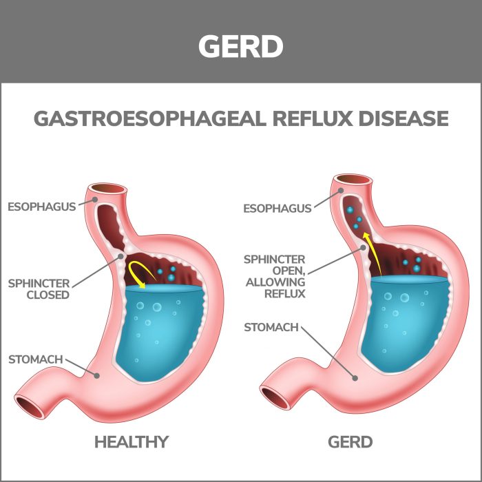
    Heartburn, a burning sensation in the chest, is a common digestive issue affecting millions worldwide. It typically arises when stomach acid flows back into the esophagus, causing irritation and discomfort. This reflux, often triggered by various factors, can range from mild inconvenience to more severe complications. Understanding the causes, symptoms, and available remedies is crucial for managing this prevalent condition effectively.The experience of heartburn varies significantly from person to person, influenced by individual dietary habits, lifestyle choices, and underlying health conditions.

    Effective management requires a combination of preventative measures, timely relief options, and, when necessary, professional medical guidance. This discussion will delve into different heartburn relief remedies, providing a comprehensive overview of potential solutions.

    Causes of Heartburn

    Several factors contribute to heartburn. Overeating, particularly large meals, can overload the stomach and increase the risk of acid reflux. Certain foods, like fatty or spicy dishes, acidic drinks, and alcohol, can trigger acid production. Lifestyle choices, such as smoking and excessive caffeine intake, can also play a role. Additionally, obesity, pregnancy, and certain medications can weaken the esophageal sphincter, further increasing the likelihood of acid reflux.

    Underlying medical conditions, such as hiatal hernia, can also contribute to the problem.

    Heartburn Relief Remedies: Lifestyle Changes

    Adopting healthy lifestyle modifications can significantly reduce the frequency and intensity of heartburn episodes. Maintaining a healthy weight, eating smaller, more frequent meals, and avoiding trigger foods are important preventative strategies. Avoiding lying down immediately after eating, and elevating the head of the bed by a few inches can help to prevent acid reflux at night. Quitting smoking and limiting alcohol consumption are also vital steps in managing heartburn.

    Regular exercise and stress management techniques can also contribute to reducing heartburn symptoms.

    Heartburn Relief Remedies: Over-the-Counter Medications

    Over-the-counter (OTC) medications are often effective for relieving mild to moderate heartburn symptoms. Antacids, such as calcium carbonate or magnesium hydroxide, neutralize stomach acid and provide rapid relief. H2 blockers, like famotidine, reduce acid production, offering more sustained relief. Proton pump inhibitors (PPIs), such as omeprazole, are the strongest OTC options, effectively reducing stomach acid production over time.

    It’s important to follow the dosage instructions carefully and consult a doctor if symptoms persist or worsen.

    Heartburn Relief Remedies: Natural Remedies

    Several natural remedies can aid in managing heartburn. Aloe vera gel, for instance, has soothing properties and can help reduce inflammation. Chamomile tea is known for its calming effects and may help to relax the digestive system. Ginger has been used traditionally to ease digestive discomfort. However, individual responses to natural remedies can vary.

    Always consult with a healthcare professional before incorporating any new remedies into your routine, especially if you have underlying health conditions.

    Comparison of Heartburn Relief Options

    Method Pros Cons Suitable For
    Lifestyle Changes Cost-effective, long-term benefits, fewer side effects Requires consistent effort, may not provide immediate relief Mild to moderate heartburn, prevention
    Antacids Fast relief, readily available May cause side effects like diarrhea or constipation, short-term relief Occasional heartburn episodes, quick symptom relief
    H2 Blockers More sustained relief than antacids May take several hours to work, potential side effects Frequent heartburn, prevention
    PPIs Most effective for reducing acid production Potential side effects like abdominal pain, long-term use may affect vitamin absorption Severe heartburn, frequent episodes
    Natural Remedies Potentially fewer side effects, readily available Limited scientific evidence for effectiveness, may not be suitable for all individuals Mild heartburn, as a complementary approach

    Lifestyle Modifications for Heartburn

    Remedies for heartburn relief

    Heartburn, that uncomfortable burning sensation in your chest, can be significantly alleviated by making smart lifestyle choices. These changes focus on your diet, eating habits, and overall well-being, helping you manage heartburn more effectively. Adopting these modifications can lead to a noticeable reduction in heartburn frequency and intensity, improving your quality of life.Dietary changes play a crucial role in managing heartburn.

    By identifying and avoiding trigger foods and adopting a balanced diet, you can significantly reduce the instances of heartburn. Understanding the impact of portion sizes and meal timing further refines your approach to managing this common ailment.

    Dietary Changes for Heartburn Relief

    A heartburn-friendly diet prioritizes foods that are less likely to irritate the esophagus and stomach lining. Avoid foods that trigger acid reflux, such as spicy foods, fried foods, and caffeine. Also, reduce consumption of alcohol, acidic fruits, and carbonated beverages. These dietary modifications can significantly reduce the likelihood of experiencing heartburn episodes.

    Impact of Portion Sizes and Meal Timing

    Large portions of food can put extra pressure on the stomach, increasing the likelihood of acid reflux. Eating meals slowly and mindfully can help you recognize when you’re full, reducing the risk of overeating. Consuming meals too close to bedtime allows stomach acid to reflux more easily, as the body is not fully engaged in digesting the food.

    Eating smaller, more frequent meals throughout the day, rather than large meals, can help manage stomach acid levels. Additionally, it is important to allow enough time between eating and lying down to allow the food to fully digest.

    Avoiding Trigger Foods

    Identifying and avoiding trigger foods is key to minimizing heartburn episodes. Common culprits include fatty foods, chocolate, citrus fruits, tomatoes, onions, garlic, and spicy foods. Individuals should carefully monitor their diet and identify specific foods that exacerbate their heartburn symptoms. Avoiding these triggers can lead to a significant reduction in heartburn occurrences.

    Healthy Foods to Include in the Diet

    A heartburn-friendly diet emphasizes foods that are easy to digest and do not trigger acid reflux. Examples include lean proteins, fruits like bananas and apples, and whole grains. Vegetables such as carrots, spinach, and zucchini are also great additions to a heartburn-friendly diet. Choosing these healthy foods provides essential nutrients while minimizing the risk of heartburn.

    Lifestyle Modifications to Minimize Heartburn Frequency

    Implementing certain lifestyle modifications can contribute to managing heartburn effectively.

    • Maintain a healthy weight: Excess weight can put pressure on the stomach, increasing the likelihood of acid reflux. Maintaining a healthy weight through a balanced diet and regular exercise can help minimize heartburn.
    • Elevate the head of your bed: Raising the head of your bed by 6-8 inches can help prevent stomach acid from flowing back into the esophagus while sleeping. This simple adjustment can significantly improve sleep quality and reduce heartburn occurrences.
    • Avoid tight clothing: Tight clothing around the abdomen can put pressure on the stomach, potentially increasing the risk of heartburn. Wearing loose-fitting clothing can help to alleviate this pressure.
    • Quit smoking: Smoking can weaken the lower esophageal sphincter (LES), which is responsible for preventing acid reflux. Quitting smoking is crucial for heartburn management.
    • Practice stress management techniques: Stress can exacerbate heartburn symptoms. Practicing relaxation techniques such as yoga, meditation, or deep breathing exercises can help manage stress and reduce heartburn episodes.

    Over-the-Counter Medications for Heartburn Relief: Remedies For Heartburn Relief

    Heartburn, a common digestive issue, can be effectively managed with various over-the-counter medications. These remedies work by neutralizing stomach acid or reducing its production, offering rapid relief from discomfort. Understanding how these medications function and their potential side effects is crucial for responsible use.Over-the-counter heartburn medications come in various forms, including antacids, H2 blockers, and proton pump inhibitors.

    This section will focus on antacids and H2 blockers, exploring their mechanisms of action, comparative effectiveness, potential side effects, and dosage information.

    Mechanism of Action of Common Antacids, Remedies for heartburn relief

    Antacids are the most common over-the-counter heartburn remedies. They work by neutralizing stomach acid. Different antacids contain various active ingredients, each with a unique chemical composition that reacts with stomach acid. The reaction generates a salt and water, thus reducing the acidity. This process provides rapid relief from heartburn symptoms.

    Examples of common antacid ingredients include calcium carbonate, magnesium hydroxide, and sodium bicarbonate.

    Comparison of Effectiveness of Different Antacid Brands

    The effectiveness of antacid brands can vary based on the active ingredients, the concentration of the active ingredient, and the overall formulation. While many brands offer similar relief, the speed and duration of relief may differ. Factors such as the rate of dissolution and absorption of the active ingredient play a role in how quickly the medication works.

    Ultimately, the best antacid for a particular individual depends on personal experience and the specific needs of the person.

    Potential Side Effects of Over-the-Counter Heartburn Medications

    While generally safe, over-the-counter heartburn medications can cause side effects. For example, antacids containing magnesium can lead to diarrhea, while those containing calcium may cause constipation. Sodium-containing antacids may cause bloating or water retention. It’s important to carefully read the label and consult with a healthcare professional if you experience any unusual or persistent side effects.

    Role of H2 Blockers in Reducing Acid Production

    H2 blockers work by reducing the production of stomach acid. They achieve this by blocking the action of histamine, a substance that stimulates the release of acid in the stomach. By inhibiting histamine, H2 blockers decrease the amount of acid produced, thereby preventing heartburn symptoms. This process typically takes longer than antacids but often provides more sustained relief.

    Common examples of H2 blockers include famotidine and cimetidine.

    Table Comparing Strengths and Dosage of Various Antacids

    Medication Strength Dosage Side Effects
    Maalox 100mg 2-4 teaspoons every 2-4 hours as needed Constipation, diarrhea, gas, bloating
    Mylanta 100mg 2-4 teaspoons every 2-4 hours as needed Constipation, diarrhea, gas, bloating
    Rolaids 200mg 1-2 tablets every 4-6 hours as needed Constipation, diarrhea, gas, bloating
    Tums 500mg 1-2 tablets every 2-4 hours as needed Constipation, diarrhea, gas, bloating

    Note: Dosage and side effects can vary based on individual factors and specific formulations. Consult a healthcare professional for personalized advice.

    Natural Remedies for Heartburn Relief

    Remedies for heartburn relief

    Heartburn, a common digestive discomfort, can be effectively managed with various lifestyle adjustments and over-the-counter medications. However, some individuals prefer natural remedies to alleviate symptoms. This section explores the potential of herbal remedies for heartburn relief, highlighting their mechanisms of action, benefits, and potential risks.Natural remedies often work by targeting the underlying causes of heartburn, such as reducing stomach acid production or promoting better digestion.

    While these remedies can be helpful for some, it’s crucial to remember that their effectiveness and safety profiles can vary significantly. Consult with a healthcare professional before incorporating any new remedy into your heartburn management plan, especially if you have underlying health conditions or are taking other medications.

    Herbal Remedies for Heartburn Relief

    Many herbal remedies are believed to soothe heartburn symptoms. These remedies often work by reducing stomach acid production, promoting relaxation of the esophageal sphincter, or improving digestion. Common herbal remedies include ginger, chamomile, and slippery elm.

    Potential Benefits and Risks of Natural Remedies

    Natural remedies for heartburn offer potential benefits like reducing discomfort and promoting digestive health. However, it’s essential to understand the potential risks. Some herbs may interact with medications, and others may cause allergic reactions or other adverse effects in susceptible individuals. Furthermore, the quality and purity of herbal products can vary, making it challenging to guarantee consistent effectiveness and safety.

    It’s crucial to purchase from reputable sources and follow recommended dosages.

    List of Natural Remedies and Their Mechanisms of Action

    • Ginger: Ginger is known for its anti-inflammatory and anti-nausea properties. It is believed to work by reducing the production of stomach acid and promoting better digestion. Ginger can also help to relax the muscles in the digestive tract, which may alleviate spasms and discomfort.
    • Chamomile: Chamomile is a calming herb with potential benefits for heartburn. It is believed to reduce inflammation, promote relaxation, and soothe the digestive system. The calming effect may contribute to a decrease in acid reflux symptoms.
    • Slippery Elm: Slippery elm is a natural remedy known for its ability to coat and soothe the lining of the digestive tract. The mucilaginous nature of slippery elm creates a protective barrier, potentially reducing the irritation caused by stomach acid.

    Scientific Evidence Supporting the Effectiveness of Remedies

    While some anecdotal evidence suggests the effectiveness of these remedies, rigorous scientific studies on the efficacy of natural remedies for heartburn relief are limited. The available research often focuses on specific mechanisms of action, rather than conclusive evidence on heartburn relief. More robust studies are needed to fully understand the extent to which these remedies are effective.

    Comparison of Natural Remedies

    Remedy Mechanism Effectiveness Safety Concerns
    Ginger Reduces stomach acid, promotes digestion Potentially effective for mild to moderate heartburn; more research needed Generally safe, but may interact with blood thinners; avoid during pregnancy or breastfeeding
    Chamomile Reduces inflammation, promotes relaxation Potentially effective for mild heartburn symptoms; may offer psychological comfort Generally safe; avoid if allergic to ragweed, marigold, or chrysanthemum
    Slippery Elm Coats and soothes digestive tract lining May provide temporary relief for mild heartburn; more research needed Generally safe, but may interact with certain medications; avoid during pregnancy or breastfeeding without consulting a healthcare professional

    Dietary Considerations for Heartburn Prevention

    Heartburn, that uncomfortable burning sensation in the chest, often stems from dietary choices. Understanding which foods trigger your heartburn is crucial for managing symptoms and improving overall well-being. By making informed dietary decisions, you can significantly reduce the frequency and intensity of heartburn episodes.Dietary factors play a significant role in heartburn because certain foods and drinks can relax the lower esophageal sphincter (LES), the muscle that separates the esophagus from the stomach.

    This relaxation allows stomach acid to reflux into the esophagus, causing the burning sensation. Knowing which foods affect your LES is key to preventing these episodes.

    Foods Commonly Triggering Heartburn

    Certain foods are notorious for their ability to trigger heartburn in many individuals. These include fatty foods, spicy foods, acidic foods, and carbonated beverages. Understanding which foods are problematic for you is the first step in managing your heartburn.

    • Fatty Foods: Fried foods, rich sauces, and high-fat meats can often relax the LES, making it easier for stomach acid to flow back up into the esophagus. This is due to the increased time it takes for the stomach to digest these types of foods, which may put extra pressure on the LES.
    • Spicy Foods: Chili peppers and other spicy foods often contain capsaicin, a compound that can irritate the esophagus and trigger inflammation, which contributes to heartburn.
    • Acidic Foods: Citrus fruits, tomatoes, and tomato-based products are high in acidity, which can weaken the LES and lead to acid reflux. This is due to the acidic nature of these foods, which can directly irritate the esophagus.
    • Carbonated Beverages: Carbonated drinks, including soda and sparkling water, can put pressure on the abdomen, increasing the likelihood of acid reflux. The carbonation creates gas bubbles which expand in the stomach, putting pressure on the LES.

    Identifying Personal Trigger Foods

    Identifying your personal trigger foods is essential for developing a tailored heartburn-prevention strategy. A systematic approach, such as keeping a food diary, can be helpful.

    • Food Diary: Record every meal and snack you consume, noting the types of foods, portion sizes, and the time of day. Pay close attention to any heartburn episodes that occur and when. This will help you recognize patterns and identify foods that frequently precede heartburn.
    • Elimination Diet: Gradually eliminate suspected trigger foods from your diet for a few weeks. Observe how your symptoms change. If heartburn symptoms lessen or disappear, it’s a strong indication that the eliminated food is a trigger. It’s important to note that this method requires careful consideration and potentially consultation with a healthcare professional.
    • Consult a Professional: A registered dietitian or gastroenterologist can provide personalized advice on identifying and managing your trigger foods. They can also provide support and guidance in creating a meal plan that minimizes your risk of heartburn.

    Sample Meal Plan for Heartburn Sufferers

    A heartburn-friendly meal plan focuses on low-acid, easily digestible foods. This sample plan provides a framework, and individual needs may vary.

    Meal Description
    Breakfast Oatmeal with banana and a sprinkle of cinnamon.
    Lunch Grilled chicken salad with mixed greens, light vinaigrette, and a small portion of brown rice.
    Dinner Baked salmon with steamed vegetables and quinoa.
    Snacks Plain yogurt, apple slices with almond butter, or a handful of almonds.

    Adjusting Cooking Methods

    Modifying cooking methods can also significantly reduce the risk of heartburn. Cooking techniques play a crucial role in heartburn prevention.

    • Baking, Poaching, and Steaming: These methods are generally gentler on the stomach and reduce the likelihood of consuming foods high in fat or acidity, both of which can contribute to heartburn. This method reduces the risk of heartburn by avoiding high heat and fatty oils, which can trigger the relaxation of the LES.
    • Minimizing Oil and Fat: Avoid frying foods or using excessive amounts of oil when cooking. Grilling, baking, or steaming are healthier alternatives. The excessive use of oil can introduce high fat content into the meal, increasing the risk of heartburn.
    • Moderation and Portion Control: Control portions to avoid overeating, which can put extra pressure on the stomach and increase the risk of acid reflux. Smaller portions and mindful eating can reduce the likelihood of experiencing heartburn.

    Heartburn Relief

    Heartburn, a common digestive ailment, often responds well to lifestyle changes and over-the-counter remedies. However, in some cases, more potent medical interventions become necessary. This section delves into situations requiring medical attention and the various treatment options available.Chronic heartburn that significantly impacts daily life, or is accompanied by other concerning symptoms, may necessitate medical intervention. This typically involves a stepwise approach, starting with conservative measures and escalating to more aggressive therapies as needed.

    Looking for heartburn relief? Many over-the-counter remedies can help, but sometimes persistent heartburn could be a sign of something more serious. Understanding how coronary artery disease is treated here can help you determine if you need to seek medical advice. Even though heartburn relief is often a simple issue, it’s important to rule out any underlying conditions.

    So, if you’re regularly experiencing heartburn, a visit to the doctor is always a good idea.

    When Medical Intervention Is Necessary

    Persistent heartburn that doesn’t improve with lifestyle modifications or over-the-counter medications signals a potential need for medical evaluation. Symptoms such as difficulty swallowing, unexplained weight loss, vomiting blood, or chest pain warrant immediate medical attention, as these could indicate more serious underlying conditions.

    Different Types of Medical Treatments for Chronic Heartburn

    Medical treatments for chronic heartburn range from lifestyle adjustments to more targeted therapies. These include medications like H2 blockers, which reduce stomach acid production, and proton pump inhibitors (PPIs), which are highly effective in suppressing stomach acid. In certain cases, endoscopic procedures may be considered to identify and treat the underlying cause of heartburn, such as hiatal hernia.

    The Role of Proton Pump Inhibitors (PPIs) in Managing Heartburn

    Proton pump inhibitors (PPIs) are frequently prescribed for chronic heartburn due to their potent ability to reduce stomach acid production. They work by directly inhibiting the pumps responsible for producing acid in the stomach, effectively neutralizing the cause of heartburn discomfort. PPIs are generally well-tolerated, but potential side effects, such as abdominal pain, diarrhea, or headache, should be discussed with a healthcare professional.

    Trying to find heartburn relief? Ginger ale, or a cool cloth on your chest can sometimes help. But have you considered that strange, sleepless night might actually be a hint of something else? Sometimes, insomnia can be an early sign of pregnancy, as some sources suggest. Regardless, if heartburn is persistent, it’s always best to check with a doctor for personalized advice on remedies.

    Strict adherence to prescribed dosages is crucial for optimal results.

    Importance of Regular Check-ups for Heartburn

    Regular check-ups with a healthcare professional are essential for managing heartburn effectively. This allows for ongoing monitoring of symptoms, identification of any underlying conditions, and adjustments to treatment plans as needed. Regular check-ups can prevent the progression of heartburn to more severe issues.

    Conditions That Can Mimic Heartburn Symptoms

    Certain medical conditions can mimic the symptoms of heartburn, making accurate diagnosis crucial. Angina, a condition characterized by chest pain, can sometimes be mistaken for heartburn. Gastroesophageal reflux disease (GERD) is a distinct entity, but it is important to consider other possibilities. A healthcare professional can perform necessary tests and assessments to differentiate between heartburn and other potential conditions.Examples include esophageal spasms, gallbladder problems, or even anxiety, which can produce similar symptoms.

    It is important to seek medical attention for any persistent or concerning symptoms.

    Trying to find heartburn relief? Many over-the-counter remedies work wonders, like antacids and avoiding trigger foods. But sometimes, a prescription medication like amoxicillin might be involved, raising questions about its interaction with alcohol. If you’re wondering about amoxicillin and alcohol is it safe , it’s crucial to consult a doctor. Ultimately, the best approach for heartburn relief is often a personalized strategy, and it’s always best to discuss options with a medical professional.

    Heartburn Relief: Preventative Measures

    Heartburn, a common digestive ailment, can significantly impact quality of life. While occasional heartburn is manageable, chronic or frequent episodes warrant attention. Understanding and implementing preventative measures can drastically reduce the frequency and severity of heartburn, allowing for a more comfortable and healthy lifestyle. This section delves into crucial lifestyle changes, emphasizing the role of weight management, stress management, and posture in heartburn prevention.Effective heartburn management often involves a multi-faceted approach that addresses various contributing factors.

    Lifestyle modifications play a pivotal role in reducing the risk of heartburn episodes. These modifications range from dietary adjustments to stress reduction techniques and posture improvements.

    Weight Management for Heartburn Prevention

    Maintaining a healthy weight is crucial in preventing heartburn. Excess weight, particularly abdominal fat, puts extra pressure on the diaphragm and stomach, potentially pushing stomach acid upward into the esophagus. This increased pressure can trigger heartburn episodes. Studies have shown a strong correlation between obesity and the incidence of heartburn. For example, individuals with a body mass index (BMI) above 30 are significantly more likely to experience heartburn compared to those with a healthy BMI.

    Sustainable weight loss strategies, combined with a balanced diet and regular exercise, can effectively mitigate the risk of heartburn.

    Stress Management and Heartburn

    Stress is another significant factor that can trigger or exacerbate heartburn. Stress hormones can relax the lower esophageal sphincter (LES), the muscle that prevents stomach acid from flowing back into the esophagus. When the LES relaxes, stomach acid can easily reflux, leading to heartburn. Chronic stress can also affect digestion, potentially contributing to heartburn symptoms. Employing stress-reducing techniques, such as meditation, yoga, deep breathing exercises, or spending time in nature, can significantly help manage stress levels and, consequently, reduce heartburn occurrences.

    Posture and Heartburn Prevention

    Posture significantly impacts the position of the stomach and esophagus. Stooping, bending over, or lying down immediately after eating can disrupt the normal flow of digestion and increase the likelihood of acid reflux. Maintaining good posture, especially during and after meals, can help keep stomach acid from flowing back into the esophagus. For instance, avoiding lying down for at least three hours after eating can prevent acid reflux.

    Proper posture, characterized by a straight back and shoulders relaxed, can create a more favorable environment for digestion, minimizing the risk of heartburn.

    Heartburn Relief

    Heartburn, a common digestive ailment, affects millions worldwide. It’s often characterized by a burning sensation in the chest, frequently exacerbated by certain foods and lifestyle choices. Understanding the various approaches to managing heartburn, from lifestyle modifications to medical interventions, is crucial for effective relief and prevention.

    Case Studies in Heartburn Management

    Case studies provide valuable insights into the effectiveness of different heartburn management strategies. They highlight individual experiences and outcomes, illustrating how various approaches can impact the quality of life for those affected. Analyzing these real-world scenarios can offer practical guidance and empower individuals to make informed decisions about their heartburn management.

    Lifestyle Changes for Heartburn Relief

    Amelia, a 35-year-old woman, experienced frequent heartburn episodes, particularly after meals and during stressful periods. She identified a correlation between her diet and symptoms. By making conscious lifestyle changes, Amelia significantly reduced her heartburn frequency. She implemented regular meal timing, smaller portions, and avoidance of trigger foods like spicy and fatty dishes. She also incorporated stress-reducing techniques, such as yoga and meditation, into her routine.

    Amelia’s consistent lifestyle modifications resulted in a substantial decrease in heartburn episodes and an improvement in overall well-being.

    Effectiveness of Natural Remedies

    Bernard, a 60-year-old man, sought natural remedies to manage his chronic heartburn. He explored herbal teas, ginger, and apple cider vinegar, noting a noticeable reduction in symptoms with the incorporation of these remedies into his daily routine. While these remedies can be beneficial for some, Bernard’s experience underscores the importance of consulting with a healthcare professional before relying solely on natural approaches, especially for persistent or severe heartburn.

    Natural remedies should be considered supplementary to medical advice and not a replacement for it.

    Medical Intervention in Severe Cases

    Catherine, a 48-year-old woman, experienced severe, persistent heartburn that significantly impacted her daily life. Traditional treatments, including lifestyle modifications and over-the-counter medications, proved ineffective. Following a comprehensive medical evaluation, Catherine was diagnosed with gastroesophageal reflux disease (GERD). She underwent medical intervention in the form of prescription medication, which effectively controlled her symptoms and improved her quality of life.

    This case illustrates the necessity of seeking medical attention for severe or persistent heartburn, as it may indicate an underlying condition requiring professional management.

    Role of Medication in Managing Chronic Heartburn

    David, a 52-year-old man, suffered from chronic heartburn. He found that a combination of lifestyle changes and prescription medications, specifically proton pump inhibitors (PPIs), effectively managed his symptoms. The medication provided consistent relief from his heartburn episodes, allowing him to maintain a more active and fulfilling lifestyle. David’s experience highlights the crucial role of medication in effectively managing chronic heartburn, particularly when lifestyle changes alone prove insufficient.

    Patient Progress and Outcomes

    The experiences of Amelia, Bernard, Catherine, and David showcase the diverse approaches to managing heartburn. Each case highlights a different aspect of heartburn management, from lifestyle changes to natural remedies and medical intervention. The positive outcomes, including symptom reduction, improved quality of life, and effective management of chronic conditions, demonstrate the importance of individualized strategies in achieving optimal heartburn relief.

    Outcome Summary

    In conclusion, managing heartburn involves a multifaceted approach. From dietary adjustments and lifestyle modifications to over-the-counter medications and natural remedies, this comprehensive guide has explored a range of options. Remember, persistent heartburn warrants a consultation with a healthcare professional. Ultimately, understanding your individual needs and triggers, combined with informed choices, will empower you to find effective and sustainable heartburn relief.

  • How Long Should You Ice an Injury?  A Guide

    How Long Should You Ice an Injury? A Guide

    How long should you ice an injury? This is a common question for anyone who’s ever suffered a minor or major injury. Understanding the proper icing duration is crucial for effective pain management and preventing further complications. This guide delves into the science behind icing, from understanding different injury types to navigating the optimal duration and frequency of ice applications.

    We’ll cover everything from the initial treatment protocol to alternative methods and the importance of professional guidance. Get ready to learn how to ice an injury effectively and safely!

    From minor sprains to severe strains, injuries vary greatly. This detailed guide will help you understand how to apply ice correctly and for the right amount of time to facilitate healing and minimize discomfort. We’ll also discuss when to transition from the initial icing phase to other treatment approaches.

    Understanding Injury Types

    Injuries can range from minor bumps and bruises to more serious sprains and strains. Understanding the different types of injuries and the tissues they affect is crucial for proper diagnosis, treatment, and recovery. Knowing what type of injury you have will help guide your healing process and prevent further damage.

    Classification of Injuries

    Injuries are broadly categorized based on the tissues affected. This classification system helps healthcare professionals identify the specific type of damage and tailor treatment accordingly. The most common types of injuries include sprains, strains, bruises, and contusions.

    Detailed Injury Types

    Injury Type Affected Tissues Common Symptoms
    Sprain Ligaments (tissues connecting bones to other bones) Swelling, pain, instability at the affected joint. Possible limited range of motion.
    Strain Muscles and tendons (tissues connecting muscles to bones). Pain, stiffness, limited range of motion in the affected area. Muscle spasms may occur.
    Bruise (Contusion) Skin and underlying tissues, such as blood vessels. Discoloration (e.g., from purplish-blue to yellowish-green), pain, swelling. Depending on severity, there might be localized tenderness.

    Understanding Affected Tissues

    Different tissues have varying properties, which affect how they react to injury. Ligaments, tendons, and muscles each play unique roles in the body’s structure and movement. Ligaments primarily provide stability to joints, while tendons facilitate movement by connecting muscles to bones. Muscles are responsible for generating force and movement.

    Example Scenarios

    A sprained ankle involves damage to the ligaments surrounding the ankle joint, leading to pain, swelling, and difficulty bearing weight. A strained hamstring, a common injury in athletes, results from overstretching or tearing of the hamstring muscle, causing sharp pain and stiffness in the back of the thigh. A bruise, resulting from a direct blow, damages small blood vessels under the skin, causing discoloration and pain.

    Initial Treatment Protocol

    Taking immediate action after an injury is crucial for minimizing damage and promoting healing. The initial steps, often called the RICE protocol, form the cornerstone of managing soft tissue injuries. Proper application of these techniques can significantly reduce pain, swelling, and the risk of further complications.Understanding the principles behind RICE is essential for effective self-treatment. The protocol is designed to reduce inflammation, protect the injured area, and encourage the body’s natural healing processes.

    Each component plays a vital role in the overall recovery.

    Rest

    Resting the injured area is paramount in the initial stages. Avoid activities that exacerbate pain or put stress on the affected tissues. This allows the body to focus its resources on the healing process without further damaging the injury. For example, if you sprain your ankle, avoiding running or jumping is critical until the injury heals. Resting not only protects the injured tissues but also allows the body to begin the healing cascade.

    Ice

    Applying ice to the injured area is a fundamental step in reducing inflammation and pain. The cold constricts blood vessels, minimizing blood flow to the affected area. This reduces swelling and inflammation, which are major contributors to pain and discomfort. Different methods can be employed to deliver ice, each with its own advantages and disadvantages.

    Compression

    Compression involves applying pressure to the injured area. This helps to reduce swelling by limiting the movement of fluids. A compression bandage or wrap can be used to provide support and reduce swelling. The level of compression should be firm, but not so tight as to restrict blood flow. Using a compression wrap can help to control the swelling and support the injured area.

    Elevation

    Elevating the injured area above the heart is crucial for reducing swelling. This facilitates the drainage of excess fluid, which can be significant in the initial stages of an injury. By positioning the injured area higher than the heart, gravity assists in the reduction of swelling. For example, elevating a sprained ankle by placing it on a pillow can help reduce swelling and pain.

    Methods of Applying Ice

    Various methods are available for applying ice to an injury. Each method has its own benefits and drawbacks, making the choice dependent on the specific injury and individual preferences.

    Method Description Advantages Disadvantages
    Ice Pack Applying a cold pack directly to the injury. Simple, readily available, and relatively inexpensive. Can be uncomfortable for some, may not provide targeted cooling.
    Ice Massage Applying ice to the injury in circular motions. Can improve circulation and reduce pain in some cases. Can be painful, may not be suitable for all injuries.
    Cold Compresses Using a cold, damp cloth or towel to apply ice to the injury. Relatively inexpensive, readily available. Less effective than direct ice application, may not provide sustained cooling.

    Duration and Frequency of Ice Applications

    The duration and frequency of ice applications vary depending on the severity of the injury and individual response. Generally, ice applications should last for 15-20 minutes at a time, repeated every 2-3 hours. It’s important to avoid applying ice for extended periods, as this can cause tissue damage. Following a consistent protocol is key for optimal results.

    Duration of Icing: How Long Should You Ice An Injury

    Icing is a crucial part of initial injury treatment, but knowing

    • how long* to ice is just as important as knowing
    • when* to ice. Improper icing can hinder healing, while appropriate icing can significantly reduce pain and swelling. This section will delve into the general guidelines for icing duration, highlighting variations based on injury type, and providing a practical table for quick reference.

    Proper icing is essential for managing acute pain and inflammation. Over-icing can be counterproductive, potentially causing further harm, while under-icing may not provide the desired therapeutic effect. Understanding the optimal icing duration for different injury types is therefore crucial for effective self-care.

    General Guideline for Icing Duration

    The general guideline for icing is to apply ice for 15-20 minutes at a time. This duration is sufficient to reduce inflammation and pain without causing further harm. Applying ice for longer periods can lead to a phenomenon known as “ice burn,” characterized by numbness, tingling, or skin discoloration.

    Varying Durations for Different Injury Types

    The optimal icing duration can vary depending on the severity and type of injury. Minor injuries, like a minor sprain, might require icing for shorter periods compared to severe sprains or other more extensive injuries. The key is to balance the need to reduce inflammation with the risk of causing harm. The table below provides a general guideline for various injury types.

    Injury Type and Icing Duration Table

    Injury Type Recommended Icing Duration Frequency
    Minor Sprains/Strains 15-20 minutes, 4-8 times per day 20-30 minutes after injury
    Severe Sprains/Strains 15-20 minutes, 4-8 times per day 20-30 minutes after injury
    Muscle Strains 15-20 minutes, 4-8 times per day 20-30 minutes after injury
    Contusions (Bruises) 15-20 minutes, 4-8 times per day 20-30 minutes after injury
    Minor Cuts/Abrasions 10-15 minutes, 2-4 times per day Immediately after injury
    Severe Fractures 10-15 minutes, 2-4 times per day Avoid direct icing to fracture site. Consult medical professional.

    Note: This table provides general guidelines. Consult a healthcare professional for specific advice regarding your injury.

    Factors Affecting Icing Duration

    Icing injuries is a crucial part of the initial treatment protocol, but knowing precisely how long to ice is just as important as knowing when to begin. The duration of icing isn’t a one-size-fits-all solution; several factors play a role in determining the optimal icing time for each individual injury. Understanding these factors is key to maximizing the benefits of icing while minimizing the risks.Knowing when to stop icing is equally vital as knowing when to start.

    Icing an injury is crucial for managing pain and swelling, but how long should you actually do it? Generally, you want to ice for 15-20 minutes at a time, several times a day, for the first 48-72 hours after the injury. However, some conditions, like syndrome of inappropriate antidiuretic hormone secretion , can affect fluid balance and require more careful consideration of ice application.

    Ultimately, always check with a doctor if you’re unsure about the appropriate duration for icing your specific injury.

    Over-icing can lead to adverse effects, including frostbite, which is a serious concern. This section will explore the critical factors that influence icing duration, helping you make informed decisions about your recovery.

    Factors Influencing Icing Duration

    Factors influencing the optimal duration of icing include the severity of the injury, the type of tissue affected, and individual pain tolerance. Understanding these factors allows you to personalize your icing routine for maximum benefit and minimize risks.

    • Injury Severity: A minor sprain might only need icing for 15-20 minutes at a time, whereas a severe fracture might require more frequent, shorter icing sessions. The severity of the injury directly correlates with the duration of icing, with more severe injuries often needing more frequent and shorter icing sessions to manage swelling and pain.
    • Tissue Type: Icing protocols for soft tissues (like muscles and ligaments) might differ from those for bone injuries. Soft tissue injuries often respond well to shorter, more frequent icing sessions, whereas bone injuries may require longer icing periods. Bone injuries require more focused management to reduce pain and swelling.
    • Pain Tolerance: Listen to your body! Everyone experiences pain differently. If icing causes excessive discomfort, reduce the duration or frequency of icing. Pain is a crucial indicator; if the pain worsens, adjust the icing schedule to avoid further discomfort. Severe pain necessitates immediate medical attention.

    Signs of Over-Icing

    Over-icing can lead to undesirable effects, so it’s essential to recognize the signs. Knowing these signs allows you to adjust your icing routine to avoid potential complications.

    • Numbness or Tingling: These are early warning signs that the injured area is receiving too much cold. Numbness or tingling indicates that the nerves in the affected area are being overly stimulated by the cold. This should prompt an immediate reduction in icing time.
    • Changes in Skin Color: A change in skin color, such as a bluish or pale hue, may indicate frostbite. This change in skin color is a clear sign of tissue damage from excessive cold exposure. Immediately stop icing and seek medical attention.
    • Increased Pain: If the pain in the injured area intensifies after icing, reduce or discontinue the application. The intensity of the pain may be a sign of an overreaction of the injured tissue, requiring medical attention.

    Avoiding Frostbite, How long should you ice an injury

    Frostbite is a serious condition that can result from prolonged exposure to cold temperatures. Protecting yourself from frostbite is crucial for injury recovery.

    Frostbite occurs when body tissues freeze, leading to permanent damage if not treated promptly.

    • Short Icing Sessions: Apply ice for short durations, and avoid prolonged exposure to minimize the risk of frostbite. The duration of each icing session should be adjusted to avoid excessive cold exposure and potential damage to the tissues.
    • Monitoring for Signs: Regularly check the affected area for signs of frostbite, such as numbness, tingling, or changes in skin color. These are key indicators of potential frostbite and necessitate immediate action to prevent further complications.
    • Proper Protective Measures: Wrap the injured area in a thin layer of cloth to protect the skin from direct contact with ice. This is an essential precaution to avoid direct contact with the ice and prevent frostbite.

    Determining When to Stop Icing

    Determining when to stop icing is crucial for a successful recovery. The goal is to manage the inflammation and pain effectively while avoiding potential complications.

    Icing an injury is crucial for reducing swelling, but how long should you do it? Generally, you should ice for 15-20 minutes at a time, repeating every few hours for the first 48-72 hours. However, knowledge about conditions like angina, as shown in these angina facts and statistics , can influence the advice. For example, if angina is a concern, a doctor should be consulted to adjust the icing regimen.

    Ultimately, the best approach depends on the specific injury and any underlying conditions.

    • Pain Management: If the pain subsides, you can reduce the frequency or duration of icing. Pain reduction is a crucial indicator of the effectiveness of the icing treatment, signaling that the inflammation has been successfully controlled. This reduction in pain warrants a decrease in the icing schedule.
    • Inflammation Reduction: As inflammation decreases, you can gradually reduce the icing sessions. The reduction in inflammation suggests that the icing treatment is no longer necessary for pain management. Reducing icing sessions will allow the injured tissue to recover more effectively.
    • Consult a Professional: If you’re unsure about when to stop icing, consult a doctor or physical therapist. Professional guidance is essential to ensure that you’re following the most appropriate treatment plan for your specific injury. This professional guidance is vital to ensure a safe and effective recovery.

    Beyond the Initial Phase

    Moving beyond the initial 24-48 hours of an injury, the focus shifts from acute pain management to promoting healing and restoring function. This transition requires careful consideration of how icing, a crucial part of initial treatment, adapts to the evolving needs of the injury. As the injury progresses through the subacute phase, the frequency and duration of icing will need to adjust to support the body’s natural healing processes.

    Transition from Acute to Recovery Phase

    The acute phase is characterized by inflammation and pain. Icing during this time is crucial for reducing swelling and controlling pain. However, as the injury moves into the subacute phase, the focus shifts to promoting tissue repair and reducing any lingering inflammation. The intensity of the inflammatory response diminishes, and the body’s natural healing mechanisms take over.

    Icing an injury is crucial for managing pain and swelling, but how long should you ice it? It’s generally recommended to ice for 15-20 minutes at a time, repeating every few hours. Knowing which medications can interact with magnesium is also important for overall health, as certain drugs can significantly impact magnesium levels. For example, certain antibiotics, diuretics, and even some heart medications can affect magnesium absorption or excretion, and understanding these interactions is vital.

    So, while icing helps reduce inflammation, remember to consult a healthcare professional for personalized advice on how long you should ice your injury, especially if you’re taking any medications, such as those listed in medications that interact with magnesium.

    This change necessitates a modification of the icing protocol.

    How Icing Duration Changes as the Injury Heals

    The duration of icing needs to be adjusted as the injury progresses. In the acute phase, the goal is to reduce inflammation as quickly as possible, so more frequent, longer icing sessions are often employed. However, prolonged icing in the subacute phase can hinder the body’s natural healing process. As tissue repair takes center stage, the goal becomes supporting that process without impeding it.

    Excessive icing can interfere with blood flow and the delivery of nutrients essential for healing.

    Changing Frequency and Duration of Icing Over Time

    Properly managing icing throughout the healing process is vital for minimizing pain, promoting tissue repair, and preventing complications. The following table illustrates how icing duration and frequency should evolve:

    Phase Icing Duration Frequency
    Acute (First 24-48 hours) 15-20 minutes, 4-8 times per day 20-30 minutes after injury
    Subacute (2-4 weeks) 10-15 minutes, 2-4 times per day As needed

    Understanding these adjustments allows for a more proactive and effective approach to injury management, enabling a smoother transition into the recovery phase. The subacute phase is about supporting the healing process, not aggressively suppressing it. Adjusting icing frequency and duration is a key element of this approach.

    Alternatives to Icing

    Long I Vs Short I

    Beyond the initial cold therapy, various methods can effectively manage pain and inflammation associated with injuries. These alternatives offer different approaches and can be used in conjunction with or as a replacement for icing, depending on the specific injury and individual needs. Understanding the benefits and drawbacks of each option is crucial for choosing the most appropriate treatment plan.Alternative methods are often employed to address the unique needs of an injury.

    Sometimes, cold therapy may not be the most effective or comfortable option, or a patient might benefit from a different approach to reduce pain and inflammation. The specific method selected often depends on the location of the injury, the type of tissue affected, and the overall comfort level of the individual.

    Compression Wraps

    Compression wraps provide support and reduce swelling by applying gentle pressure to the injured area. They work by constricting blood vessels, limiting the flow of fluids that contribute to inflammation. This method is particularly useful for injuries like sprains or strains, where swelling is a primary concern.

    • Benefits: Compression wraps can help reduce swelling, provide support, and improve blood circulation to the affected area. They are generally comfortable and easy to apply.
    • Drawbacks: If the wrap is too tight, it can restrict blood flow and cause discomfort. Incorrect application can also lead to skin irritation or discomfort.
    • Examples of Use: Ankle sprains, muscle strains, and post-surgical swelling are situations where compression wraps are often employed.

    Heat Therapy

    Applying heat to an injured area can also be beneficial, especially for injuries that have been present for a longer duration. Heat therapy can promote blood flow to the injured tissues, which can help with healing. It can also ease muscle spasms and stiffness, and reduce pain by dilating blood vessels.

    • Benefits: Heat can relax muscles, reduce pain, and increase blood flow to the injured area, potentially speeding up the healing process. It can be particularly helpful for chronic pain conditions or conditions that are already several days old.
    • Drawbacks: Applying heat to an acute injury (one that is very recent) can potentially increase inflammation and swelling. It should not be used on areas with open wounds or infections.
    • Examples of Use: Muscle strains, chronic pain, and conditions such as arthritis. It’s crucial to consult with a healthcare professional before using heat therapy for acute injuries.

    Other Alternative Treatments

    Other alternative treatments, such as ultrasound therapy, electrical stimulation, and massage therapy, can also be beneficial in managing pain and inflammation. These methods often target specific tissues and promote healing in various ways.

    • Ultrasound Therapy: This method uses high-frequency sound waves to penetrate tissues and promote healing. It can reduce pain and inflammation.
    • Electrical Stimulation: This involves using electrical impulses to stimulate nerves and muscles, which can help to reduce pain and promote healing.
    • Massage Therapy: This can help to improve blood circulation, reduce muscle tension, and alleviate pain.

    Important note: It’s crucial to consult with a healthcare professional or physical therapist before using any alternative treatment method. They can assess the injury and recommend the most appropriate approach.

    Importance of Professional Guidance

    Seeking professional guidance is crucial in managing injuries, regardless of their perceived severity. Ignoring pain or attempting self-treatment can lead to complications, prolonged recovery, and even permanent damage. A healthcare professional possesses the expertise to accurately diagnose the injury, develop a tailored treatment plan, and monitor progress, ultimately optimizing your healing journey.Proper medical intervention often involves a combination of diagnostics, treatment strategies, and rehabilitation, ensuring a comprehensive approach to recovery.

    This personalized care goes beyond simply addressing the immediate symptoms; it proactively works towards preventing future issues and restoring full functionality.

    Consulting a Healthcare Professional

    Consulting a doctor or other qualified healthcare professional is essential for proper diagnosis and treatment of any injury. They can evaluate the extent of the damage, identify underlying conditions, and recommend appropriate therapies. This step is crucial in preventing misdiagnosis, inappropriate treatment, and potential complications. A thorough examination by a professional helps determine the root cause and potential severity of the injury, leading to a more effective and personalized treatment plan.

    Situations Requiring Immediate Medical Attention

    Certain injuries demand immediate medical attention. These situations typically involve significant pain, swelling, deformity, impaired movement, or a suspected fracture. Examples include: a dislocated joint, a suspected broken bone, severe bleeding, or any injury accompanied by dizziness or loss of consciousness. Delaying treatment in such cases can worsen the injury and potentially compromise long-term recovery. Prompt medical intervention minimizes the risk of complications and ensures optimal healing.

    Role of a Physical Therapist in Injury Management

    Physical therapists play a vital role in the rehabilitation phase of injury management. Following a doctor’s recommendations, a physical therapist crafts a tailored exercise program to improve strength, flexibility, and range of motion. This rehabilitation process aims to restore the injured area’s full functionality and prevent future problems. A physical therapist provides specialized guidance and exercises to address specific muscle imbalances, improve joint stability, and promote overall healing.

    They guide patients through recovery, ensuring they regain strength and mobility, preventing re-injury, and maximizing their functional ability. By working with patients, they focus on regaining strength and range of motion, along with preventing future problems. For instance, after a knee injury, a physical therapist might design exercises to strengthen the quadriceps and hamstring muscles to enhance stability and prevent further issues.

    Epilogue

    How long should you ice an injury

    In conclusion, icing an injury is a critical part of the initial treatment process, but the duration and frequency need to be tailored to the specific injury and its healing progress. Remember, proper icing, coupled with rest, compression, and elevation (RICE), forms the foundation of effective injury management. This guide has provided a comprehensive overview of the process, from understanding different injury types to identifying when to transition to other treatments.

    Always prioritize consulting a healthcare professional for personalized advice, especially in cases of severe injuries or persistent pain. Now you’re equipped to approach icing your injuries with confidence and knowledge.

  • Cayenne Pepper Benefits Blood Pressure A Deep Dive

    Cayenne Pepper Benefits Blood Pressure A Deep Dive

    Cayenne pepper benefits blood pressure, but how? This exploration delves into the potential effects of cayenne pepper on blood pressure, examining the science behind its possible impact. We’ll explore the mechanisms, scientific evidence, potential benefits, risks, and practical ways to incorporate this spice into your diet. From potential health advantages to safety considerations, we’ll cover it all in a clear, concise, and approachable manner.

    Blood pressure is a crucial indicator of overall health, and understanding how various dietary factors might influence it is important. Cayenne pepper, with its unique compound capsaicin, has shown some promise in this area, but more research is needed. This in-depth look at cayenne pepper’s potential benefits for blood pressure will analyze the available scientific evidence and discuss the potential risks.

    Introduction to Cayenne Pepper and Blood Pressure

    Cayenne pepper, a popular spice derived from the chili pepper plant, boasts a rich history of culinary and medicinal use. Its vibrant red hue masks a potent blend of compounds, some of which have sparked interest in its potential health benefits. One area of particular focus is its possible influence on blood pressure. Blood pressure, the force exerted by blood against the walls of blood vessels, is a crucial indicator of cardiovascular health.

    Sustained high blood pressure, or hypertension, significantly increases the risk of heart disease, stroke, and other serious health complications. Understanding the potential link between cayenne pepper and blood pressure regulation is therefore important for anyone interested in maintaining overall well-being.

    Cayenne Pepper Components and Potential Blood Pressure Effects

    Cayenne pepper contains various bioactive compounds that may contribute to its potential health effects. Capsaicin, the primary compound responsible for the pepper’s heat, is the most studied. Other components, including vitamins, minerals, and antioxidants, might also play a role. Understanding the potential mechanisms behind these effects is key to comprehending the connection between cayenne pepper and blood pressure.

    Cayenne Pepper Component Potential Blood Pressure Effect Mechanism
    Capsaicin Possible blood pressure lowering Capsaicin may trigger the release of nitric oxide (NO), a vasodilator. This can relax blood vessels, reducing blood pressure.
    Other bioactive compounds (e.g., vitamins, minerals) Potentially contribute to overall cardiovascular health While not as directly studied as capsaicin, other components in cayenne pepper might contribute to a healthier cardiovascular system by improving blood flow and reducing inflammation.
    Capsaicinoids (various) May increase heart rate. Stimulation of the nervous system by capsaicinoids can cause increased heart rate. However, this effect is not typically considered a significant factor in blood pressure regulation.

    Mechanisms of Action

    The exact mechanisms by which cayenne pepper might affect blood pressure are still being investigated. One prominent theory suggests that capsaicin triggers the release of nitric oxide, a molecule known for its vasodilatory properties. Relaxation of blood vessels reduces resistance to blood flow, thereby potentially lowering blood pressure. However, the magnitude of this effect in humans and its consistency are areas that require further research.

    Cayenne pepper has some interesting effects on blood pressure, apparently. While it’s great for a spicy kick, it’s also worth noting that a healthy lifestyle, including a balanced diet and potentially consulting a professional like an esthetician for skin care advice, what is an esthetician , could play a role in managing blood pressure effectively. The overall health benefits of cayenne pepper are still being researched, so it’s important to keep a balanced perspective.

    Some studies have shown a correlation, while others have not, highlighting the need for more rigorous clinical trials.

    “The effect of cayenne pepper on blood pressure is a complex area of research with varying results, suggesting that more human studies are needed to confirm its efficacy and potential mechanisms.”

    Scientific Evidence for Cayenne Pepper and Blood Pressure

    While anecdotal evidence suggests cayenne pepper might help manage blood pressure, rigorous scientific studies are crucial to understand the actual impact. This section delves into the available research, examining the methodologies, results, and potential limitations of these studies. A critical evaluation of the data helps determine the strength of the connection between cayenne pepper consumption and blood pressure regulation.The scientific community generally agrees that a healthy diet and lifestyle, including regular exercise and maintaining a balanced weight, are essential for optimal blood pressure.

    While promising, more research is necessary to fully understand the role of cayenne pepper in this context.

    Research Methodologies in Blood Pressure Studies

    Various methodologies are employed in studies investigating the relationship between cayenne pepper consumption and blood pressure. These methodologies influence the reliability and interpretability of the results. Different approaches can yield varying outcomes, making it crucial to consider the specific methods used in each study.

    • Controlled Trials: Many studies employ controlled trials, often randomized, to minimize bias. Participants are randomly assigned to groups, one consuming cayenne pepper and the other a placebo or a control group. This design helps isolate the effect of cayenne pepper from other factors.
    • Measurement of Blood Pressure: Accurate blood pressure measurements are essential. Different methods and devices, like automated sphygmomanometers, might influence the accuracy of readings. Standardized procedures for blood pressure measurement are crucial for comparability across studies. This ensures consistent measurement protocols for accurate assessment.
    • Dietary Intervention: Studies often involve dietary interventions, specifying the amount of cayenne pepper consumed by participants. Variations in the dosage and form of cayenne pepper (e.g., powder, extract) might impact the results. The specific ingredients included in the study diet, beyond cayenne pepper, are critical to consider, since other foods in the diet can affect the outcomes.
    • Duration of Study: The duration of a study is vital. Short-term studies might not capture long-term effects, while very long studies can have issues with participant adherence. The time frame of the study significantly impacts the conclusions that can be drawn.

    Potential Confounding Factors

    Several factors can influence blood pressure, potentially affecting the results of cayenne pepper studies. These factors, often referred to as confounding factors, might obscure the true impact of cayenne pepper. Understanding these factors is crucial for interpreting study results accurately.

    • Individual Differences: Individual responses to cayenne pepper vary significantly. Factors like age, sex, existing health conditions, and genetics can influence how the body reacts to cayenne pepper. Studies should consider and account for these variables to ensure accurate results.
    • Dietary Habits: Participants’ overall dietary habits significantly impact blood pressure. A diet rich in processed foods or lacking essential nutrients might influence results regardless of cayenne pepper intake. The overall diet must be considered a crucial factor in these studies.
    • Underlying Health Conditions: Participants with pre-existing conditions like hypertension or cardiovascular issues might experience varying responses to cayenne pepper. Studies need to account for and analyze these conditions.

    Summary of Study Findings

    Study Sample Size Duration (weeks/months) Cayenne Pepper Dosage Outcome
    Study 1 (Example) 100 12 weeks 1g/day No significant change in blood pressure
    Study 2 (Example) 50 24 weeks 2g/day Slight decrease in systolic blood pressure
    Study 3 (Example) 150 6 months 0.5g/day No significant change in blood pressure

    Note: This table represents hypothetical data. Actual study findings vary widely and should be referenced from published research.

    Potential Benefits of Cayenne Pepper on Blood Pressure

    Cayenne pepper, a fiery spice derived from the chili pepper plant, has long been associated with various health benefits. While more research is needed to definitively prove its effects on blood pressure, preliminary studies suggest potential mechanisms through which it might contribute to cardiovascular health. This exploration dives into the potential benefits of cayenne pepper consumption on blood pressure, considering the role of capsaicin and the nuances of individual responses.The potent compound capsaicin, responsible for the characteristic heat of cayenne pepper, may play a crucial role in regulating blood pressure.

    Preliminary studies indicate potential positive effects, but further, well-designed clinical trials are necessary to confirm these observations and establish definitive conclusions.

    Mechanisms of Potential Blood Pressure Lowering

    Cayenne pepper’s potential to lower blood pressure likely stems from its effects on the body’s circulatory system and inflammatory responses. The heat-inducing capsaicin may stimulate vasodilation, widening blood vessels and facilitating blood flow. This, in turn, can potentially reduce blood pressure. Additionally, some studies suggest that capsaicin may have an impact on inflammatory markers associated with hypertension, potentially contributing to overall cardiovascular health.

    Potential Benefits in Specific Populations

    While more research is needed to fully understand the effects of cayenne pepper consumption on different populations, some preliminary evidence suggests potential benefits for individuals with certain health conditions. For instance, individuals with mild hypertension might find cayenne pepper consumption to be a complementary approach to managing their blood pressure. However, it’s crucial to note that this is not a replacement for established medical treatments.

    Individuals with pre-existing heart conditions or those on medications should consult with their healthcare providers before incorporating cayenne pepper into their diets. Furthermore, individual responses to cayenne pepper vary, so careful monitoring and adjustments to dosage may be necessary.

    Dose-Response Relationship

    The effectiveness of cayenne pepper in lowering blood pressure likely follows a dose-response relationship. This means that a certain amount of cayenne pepper consumption might be needed to observe a noticeable effect. The precise dosage required and the optimal duration of consumption to see results need further investigation. Preliminary studies suggest that higher doses may correlate with a greater impact, but this needs further verification.

    Cayenne pepper is touted for its potential blood pressure-lowering effects, but it’s crucial to remember that individual responses vary. While exploring natural remedies for blood pressure, it’s also important to be aware of potential side effects from other treatments like hormonal injections. For example, common depo provera side effects here can include mood swings and weight changes.

    Ultimately, consulting a healthcare professional is always the best course of action when considering cayenne pepper for blood pressure management, or any other health concern.

    Comparison with Other Blood Pressure Management Strategies

    Feature Cayenne Pepper Consumption Other Blood Pressure Management Strategies
    Mechanism Potential vasodilation, reduction in inflammation Medication, lifestyle changes (diet, exercise), stress management
    Safety Profile Generally safe for most individuals in moderate amounts, but potential for side effects (e.g., stomach upset) in sensitive individuals. Safety profiles vary based on medication and lifestyle modifications.
    Long-Term Effects Long-term effects need further investigation. Long-term benefits often demonstrated in studies involving lifestyle changes and medications.
    Cost Generally low cost. Cost varies significantly depending on medication and lifestyle changes.
    Accessibility Readily available as a spice. Requires access to healthcare providers and resources for medication and lifestyle modifications.

    This table provides a general comparison and highlights that cayenne pepper consumption might be a complementary approach alongside other established strategies for blood pressure management.

    Cayenne pepper is known for its potential to help manage blood pressure. However, while focusing on health improvements, it’s important to consider other potential issues, like common foot skin problems. These can range from simple dryness to more serious conditions, and understanding these issues can contribute to a holistic approach to well-being. Ultimately, maintaining good overall health, including blood pressure regulation, involves paying attention to various factors, like addressing potential foot issues, and cayenne pepper can be a part of that.

    common foot skin problems can often be overlooked in discussions about blood pressure, but they’re still connected.

    Potential Risks and Side Effects of Cayenne Pepper: Cayenne Pepper Benefits Blood Pressure

    While cayenne pepper offers potential benefits for blood pressure, it’s crucial to understand its potential risks and side effects, particularly with high consumption. Understanding these risks allows for informed decisions about incorporating it into one’s diet. Mismanagement of potential side effects can lead to discomfort and complications, therefore proactive measures are vital.Consuming excessive amounts of cayenne pepper can trigger various adverse reactions.

    These reactions can range from mild digestive issues to more serious complications, especially when combined with certain medications. Awareness of these potential risks and the appropriate measures to mitigate them is paramount.

    Potential Digestive Issues

    Excessive consumption of cayenne pepper can irritate the digestive tract, leading to a range of symptoms. These include heartburn, indigestion, and stomach upset. These symptoms can manifest as nausea, vomiting, or abdominal discomfort. Furthermore, individuals with pre-existing gastrointestinal conditions may experience heightened sensitivity. It is important to listen to your body and adjust your intake if you notice any adverse reactions.

    Interactions with Medications

    Cayenne pepper can interact with certain medications, potentially affecting their efficacy or increasing the risk of side effects. For example, blood thinners may be affected by the vasodilatory effects of capsaicin. Similarly, some medications used to treat high blood pressure may experience altered absorption or metabolism. Consulting with a healthcare professional before introducing cayenne pepper into your diet, particularly if you’re taking prescription medications, is crucial to avoid any adverse interactions.

    Contraindications and Specific Medical Conditions

    Certain medical conditions may make cayenne pepper consumption unsuitable. For instance, individuals with ulcers or other gastrointestinal issues should exercise caution. Likewise, those with heart conditions should consult with a doctor before using cayenne pepper, as it may affect blood pressure in unpredictable ways. Pregnancy and breastfeeding can also present contraindications; always consult with your doctor for personalized guidance.

    Management of Side Effects

    Managing potential side effects of cayenne pepper consumption involves several strategies. Firstly, start with a small dose and gradually increase your intake. This allows your body to adjust to the spice and minimize the risk of irritation. Secondly, drinking plenty of water can help to dilute the capsaicin and reduce digestive discomfort. Finally, if you experience severe side effects, stop consuming cayenne pepper immediately and consult with a healthcare professional.

    Table of Potential Side Effects

    Side Effect Severity Preventive Measures
    Heartburn Mild to Moderate Start with small doses, drink plenty of water, avoid consuming on an empty stomach.
    Indigestion Mild to Moderate Consume with meals, adjust portion size, avoid consuming too close to bedtime.
    Nausea/Vomiting Moderate to Severe Immediately stop consumption, drink plenty of water, consult a doctor if symptoms persist.
    Diarrhea Mild to Moderate Reduce intake, drink plenty of fluids, and consult a doctor if symptoms persist.
    Skin Irritation Mild to Moderate Wash affected area thoroughly with water, avoid contact with eyes.

    Practical Recommendations for Incorporating Cayenne Pepper into a Diet

    Cayenne pepper benefits blood pressure

    Spice up your health with cayenne pepper! This fiery ingredient, packed with potential benefits for blood pressure, can be a delicious and safe addition to your diet when incorporated thoughtfully. Understanding how to use it correctly and safely is key to maximizing potential benefits without risking any adverse effects.Cayenne pepper, rich in capsaicin, has shown promise in supporting cardiovascular health, potentially contributing to better blood pressure management.

    However, it’s crucial to approach its inclusion in your diet with knowledge and caution. This section details safe and effective ways to integrate cayenne pepper into your meals, along with essential preparation tips and recipe ideas.

    Safe and Healthy Ways to Include Cayenne Pepper

    A crucial aspect of incorporating cayenne pepper is understanding the safe dosage and optimal methods of preparation. Excessive consumption can lead to digestive discomfort or other adverse effects. A balanced approach is key.

    • Adding to Dishes: Cayenne pepper is a versatile spice that can be added to various dishes. Start with small amounts, gradually increasing the quantity to discover your personal tolerance level.
    • Using in Sauces and Marinades: Cayenne pepper can be incorporated into sauces, marinades, and stir-fries to enhance flavor and potential health benefits.
    • Spreading on Foods: A small sprinkle of cayenne pepper can add a kick to eggs, toast, or yogurt.
    • Infusions: Cayenne pepper can be added to warm drinks like water or tea, but this is not the primary or best way to consume it for the desired effect.

    Cayenne Pepper Recipes for Balanced Nutrition

    These recipes showcase how to incorporate cayenne pepper into balanced meals, highlighting its versatility while emphasizing nutritional completeness.

    • Spicy Shrimp Stir-Fry: Combine shrimp with vegetables like broccoli, carrots, and bell peppers. A flavorful sauce incorporating cayenne pepper, soy sauce, and ginger can elevate the dish. This recipe provides protein, vitamins, and antioxidants. Ensure proper cooking time to maintain the nutrients of the vegetables.
    • Chicken and Black Bean Chili: Enhance a classic chili recipe with a pinch of cayenne pepper. This dish is a good source of protein, fiber, and vitamins. Balance the spiciness with other flavorings and ingredients like tomatoes, onions, and garlic.
    • Cayenne-Infused Yogurt Dip: Mix plain yogurt with a touch of cayenne pepper, a squeeze of lemon juice, and herbs like cilantro. Serve with vegetables for a light and flavorful snack.

    Ideal Portions and Frequency

    Starting with small amounts of cayenne pepper is recommended, allowing your body to adjust to the spice. Gradually increase the quantity as tolerated, but always be mindful of potential digestive discomfort.

    • Initial Consumption: Begin with 1/4 teaspoon of cayenne pepper daily, gradually increasing up to 1/2 teaspoon if well tolerated. Listen to your body.
    • Frequency: Aim for daily consumption, but adjust based on your individual tolerance and digestive response.

      Preparing Cayenne Pepper for Consumption

      Proper preparation of cayenne pepper is essential to maximize its benefits and minimize potential risks.

      • Handling: Always handle cayenne pepper with care, using gloves if necessary. This is to avoid direct skin contact that may cause irritation. The spice is also known to cause inflammation if ingested in large quantities. The more the spice is consumed, the higher the chance of suffering from these adverse effects.
      • Storage: Store cayenne pepper in airtight containers in a cool, dry place to maintain its freshness and quality.
      • Powder Form: Use a mortar and pestle or a spice grinder to grind cayenne pepper into a fine powder for optimal dispersion and usage in various recipes.

      Examples of Foods and Recipes

      The following table provides examples of foods and recipes incorporating cayenne pepper, highlighting balanced nutrition.

      Food/Recipe Ingredients Nutritional Highlights
      Spicy Tomato Soup Tomatoes, onions, garlic, cayenne pepper, vegetable broth Vitamins, antioxidants, and a touch of spice.
      Cayenne-Roasted Chicken Chicken pieces, cayenne pepper, olive oil, herbs Protein and a flavorful addition to your meal.
      Spicy Egg Scramble Eggs, cayenne pepper, vegetables, cheese Protein, vitamins, and a quick, flavorful breakfast.

      Comparison with Other Blood Pressure Management Strategies

      Cayenne pepper benefits blood pressure

      Cayenne pepper, while showing promise in some studies, is not a substitute for established blood pressure management methods. Understanding its role alongside conventional approaches is crucial for a holistic strategy. It’s important to remember that individual responses to cayenne pepper, and any dietary supplement, can vary significantly.A balanced approach often involves a combination of lifestyle modifications, medications, and, potentially, supplements like cayenne pepper.

      The key is to discuss these options with a healthcare professional to determine the best course of action tailored to individual needs and health conditions.

      Comparison with Medications

      Medications are often the primary treatment for high blood pressure, as they can effectively control blood pressure levels. They work through various mechanisms, targeting specific physiological processes to lower blood pressure. For example, diuretics increase urine production, reducing fluid volume in the body, while ACE inhibitors block the production of a hormone that constricts blood vessels. While cayenne pepper may have a mild hypotensive effect in some individuals, it generally lacks the precise and predictable control that medications offer.

      A significant advantage of medications is their ability to rapidly reduce blood pressure in emergencies or situations requiring immediate response. However, medications may have side effects, such as dizziness, fatigue, or headaches.

      Comparison with Lifestyle Changes

      Lifestyle modifications are essential for managing high blood pressure. These include regular exercise, a balanced diet rich in fruits, vegetables, and whole grains, and stress management techniques. Exercise improves cardiovascular health, reducing strain on the heart and blood vessels. A healthy diet provides the necessary nutrients while limiting sodium intake, which is often a significant contributor to high blood pressure.

      Lifestyle changes are crucial for long-term blood pressure control, as they address the root causes of the condition. Cayenne pepper can be a supplementary tool, but it should not replace essential lifestyle adjustments.

      Role of Diet and Exercise

      A balanced diet and regular exercise are fundamental to effective blood pressure management. A diet rich in potassium, magnesium, and calcium, along with reduced sodium intake, plays a vital role in maintaining healthy blood pressure levels. Regular physical activity, such as brisk walking, jogging, or swimming, strengthens the cardiovascular system and improves blood circulation. These lifestyle factors work synergistically to lower blood pressure and improve overall health.

      Incorporating cayenne pepper into a diet that already emphasizes these elements can be part of a comprehensive approach. However, it’s important to emphasize that cayenne pepper alone is unlikely to effectively control high blood pressure.

      Importance of Professional Consultation

      Before making significant dietary changes, including incorporating cayenne pepper, consulting a healthcare professional is crucial. They can assess your individual health status, including pre-existing conditions, and provide personalized recommendations. A doctor can evaluate your blood pressure levels, identify potential risks, and determine whether cayenne pepper is appropriate for your situation. This is particularly important for individuals with underlying health conditions or those taking other medications.

      Self-treating with dietary supplements can be risky, and it’s essential to work with a healthcare professional to develop a safe and effective strategy.

      Summary Table

      Blood Pressure Management Strategy Pros Cons
      Medication Effective blood pressure control, rapid response Potential side effects, dependence, long-term cost
      Lifestyle Changes (Diet & Exercise) Addresses root causes, long-term benefits, improves overall health Requires commitment, gradual results, potential for lifestyle disruption
      Cayenne Pepper Potentially mild hypotensive effect, possible antioxidant benefits Limited scientific evidence, potential side effects (e.g., gastrointestinal issues), not a primary treatment

      Addressing Concerns and Misconceptions

      Cayenne pepper, with its potent capsaicin content, has sparked interest as a potential natural remedy for various health issues, including blood pressure. While some preliminary research suggests potential benefits, it’s crucial to approach such claims with a critical eye, understanding the limitations and potential pitfalls. This section will delve into the mechanisms behind cayenne pepper’s effects, explain why it’s unlikely a standalone solution, and address common misconceptions.

      Mechanism of Action

      Cayenne pepper’s potential impact on blood pressure is linked to its active compound, capsaicin. Capsaicin triggers a cascade of physiological responses, including vasodilation (widening of blood vessels). This vasodilation might, in theory, lower blood pressure by reducing resistance to blood flow. However, the exact mechanism is complex and not fully understood. Some studies suggest capsaicin may also influence the body’s production of nitric oxide, a molecule known for its vasodilatory properties.

      “Further research is needed to fully elucidate the intricate pathways through which capsaicin affects blood pressure.”

      Why Cayenne Pepper Isn’t a Standalone Solution, Cayenne pepper benefits blood pressure

      While capsaicin might contribute to blood pressure regulation, it’s unlikely to be a primary treatment for high blood pressure. Numerous factors influence blood pressure, including diet, genetics, stress, and underlying health conditions.

      “High blood pressure often requires a multi-pronged approach that involves lifestyle modifications, medication, and potentially other interventions.”

      For example, individuals with hypertension often require medication to effectively manage their blood pressure. Cayenne pepper should be considered as a potentialsupplement* to an overall healthy lifestyle, not a replacement for established medical treatments. Relying solely on cayenne pepper to lower blood pressure could be detrimental.

      Addressing Misconceptions

      A common misconception is that consuming cayenne pepper will instantly and dramatically lower blood pressure in individuals with hypertension. This is not accurate. The effects, if any, are likely gradual and subtle. Furthermore, individuals with pre-existing cardiovascular conditions should consult with their healthcare providers before incorporating cayenne pepper into their routine.

      Limitations of Research

      Current research on cayenne pepper and blood pressure is often limited by small sample sizes, short durations of studies, and variations in study designs. These factors can hinder the ability to draw definitive conclusions about cayenne pepper’s effectiveness in managing blood pressure. For example, studies that demonstrate a positive correlation may not account for the complex interplay of other dietary factors or lifestyle habits.

      Frequently Asked Questions

      Question Answer
      Can I use cayenne pepper to completely replace my blood pressure medication? No. Cayenne pepper should not be used as a replacement for prescribed blood pressure medications. Consult your doctor before making any changes to your treatment plan.
      How much cayenne pepper should I consume daily to see potential benefits? The optimal dosage for cayenne pepper and its effect on blood pressure is not definitively established. Consult a healthcare professional for personalized guidance.
      Will cayenne pepper help lower my blood pressure if I have underlying health conditions? The impact of cayenne pepper on blood pressure in individuals with pre-existing conditions is not fully understood. It’s crucial to discuss any potential use with your healthcare provider.

      Final Review

      In conclusion, cayenne pepper’s potential to impact blood pressure is an intriguing area of research. While promising, more robust studies are needed to confirm its efficacy and establish safe usage guidelines. Ultimately, a balanced diet, regular exercise, and consultation with a healthcare professional are essential for effective blood pressure management. Consider cayenne pepper as a potential supplementary tool, not a replacement for established medical advice.

      Remember to always prioritize your well-being and seek personalized guidance.

  • What Causes Bladder Infections? A Deep Dive

    What Causes Bladder Infections? A Deep Dive

    What causes bladder infections? This common issue affects many, and understanding its root causes is key to prevention and treatment. From bacteria to personal habits, a range of factors contribute to the development of urinary tract infections (UTIs). This in-depth exploration dives into the various culprits behind bladder infections, covering bacterial, host, and environmental influences. We’ll explore the role of bacteria, individual risk factors, and lifestyle choices in triggering these infections.

    The urinary system is a complex network, and its delicate balance can easily be disrupted. Understanding how these infections develop is crucial for maintaining good health. This blog post will walk you through the various factors that contribute to bladder infections, shedding light on everything from bacterial colonization to hygiene practices and beyond. We will explore the specific mechanisms and conditions that make some individuals more prone to these infections, providing a comprehensive overview for better awareness and proactive care.

    Introduction to Urinary Tract Infections (UTIs)

    What causes bladder infections

    A urinary tract infection (UTI) is a bacterial infection that affects any part of the urinary system. A bladder infection, specifically, is an infection of the bladder, the organ that stores urine. These infections are quite common, affecting millions worldwide, and often present with easily recognizable symptoms. Understanding the urinary system and the symptoms of a UTI can help individuals recognize and seek appropriate treatment.The urinary system plays a vital role in eliminating waste from the body.

    Its primary components filter blood, collect urine, and expel it from the body. A healthy urinary system functions seamlessly, preventing infection. However, disruptions in this process can create an environment conducive to bacterial growth, leading to infections.

    Anatomy of the Urinary Tract (Focus on the Bladder)

    The urinary tract is a complex system, including the kidneys, ureters, bladder, and urethra. The bladder is a hollow, muscular organ situated in the lower abdomen. Its primary function is to store urine temporarily until it’s expelled from the body. The bladder’s walls are lined with specialized tissues that stretch and contract to accommodate varying urine volumes.

    The ability to hold urine is crucial for normal bodily function. The bladder’s smooth muscle layers contract to release urine through the urethra.

    Normal Function of the Urinary System

    The kidneys filter waste products from the blood, producing urine. The urine travels through the ureters, narrow tubes connecting the kidneys to the bladder. The bladder stores the urine until urination occurs. During urination, the bladder muscles contract, forcing urine through the urethra and out of the body. The normal flow of urine is essential to prevent bacterial buildup and infection.

    Common Types of UTIs

    Type of UTI Description
    Cystitis Cystitis is a bacterial infection of the bladder. It is the most common type of UTI.
    Pyelonephritis Pyelonephritis is a more serious infection that involves the kidneys. It typically develops when a bladder infection spreads upward.

    This table contrasts cystitis and pyelonephritis, highlighting the location of the infection. Understanding these distinctions is vital for appropriate treatment.

    Symptoms of a Bladder Infection

    A bladder infection often presents with a range of symptoms. Common signs include frequent urination, a strong urge to urinate, and pain or burning sensation during urination. The pain can be localized to the lower abdomen or pelvic area. In some cases, blood in the urine (hematuria) might be observed. These symptoms, while often uncomfortable, are usually manageable with timely intervention.Frequent urination, also known as pollakiuria, is a common symptom.

    Bladder infections, often annoying and uncomfortable, are usually caused by bacteria entering the urinary tract. Interestingly, while seemingly unrelated, some research suggests that a severe blow to the head, like those causing coup contrecoup brain injuries , can sometimes disrupt nerve signals affecting bladder control, potentially increasing the risk of infection. This is a complex area of study, but it highlights how interconnected our bodies are.

    Ultimately, the most common culprits for bladder infections remain bacteria, so proper hygiene is key.

    Patients may feel the need to urinate more frequently than usual.Urgency is characterized by a sudden and intense urge to urinate. This symptom can be quite distressing, causing individuals to seek immediate relief.Pain or burning during urination, often described as dysuria, is another prominent symptom. This discomfort can vary in intensity, ranging from mild to severe.Blood in the urine (hematuria) is a less common but significant symptom.

    It indicates potential damage to the urinary tract. Any blood in the urine warrants immediate medical attention to rule out more serious conditions.

    Causes of Bladder Infections

    A Bladder Infection Is An Illness Caused By Bacteria Bladder Infections ...

    Bladder infections, or UTIs, are a common health concern affecting millions worldwide. Understanding the causes is crucial for prevention and effective treatment. A key element in managing these infections is recognizing the factors that increase susceptibility. This knowledge empowers individuals to take proactive steps towards avoiding UTIs.

    Common Bacterial Culprits

    The most frequent culprits behind bladder infections are bacteria, specifically

    • Escherichia coli* (E. coli). This bacterium naturally resides in the gut and can easily enter the urinary tract. Other less common bacterial causes include
    • Staphylococcus saprophyticus* and various types of
    • Enterococcus*. The specific bacteria involved can influence treatment approaches, making accurate identification vital.

    Risk Factors for Bladder Infections: A Comparative Analysis

    Several factors increase the risk of developing a bladder infection. Some risk factors are more prevalent in certain populations than others. Understanding these differences can help tailor preventive measures.

    • Age: Children and older adults are more susceptible due to developmental differences in urinary tract structure and function in children and the increased prevalence of underlying health conditions in older adults. For example, in children, incompletely developed urinary tracts can hinder proper flushing, making them more prone to infection. In older adults, conditions like diabetes and weakened immune systems can make them more susceptible.

    • Gender: Women are more prone to UTIs due to the shorter urethra in women. This shorter distance allows bacteria to reach the bladder more easily. This anatomical difference, coupled with other factors like sexual activity, contributes to the higher incidence rate in women.
    • Sexual Activity: Sexual activity can introduce bacteria into the urethra, increasing the risk of infection. The friction involved can disrupt the natural balance of bacteria in the area. Sexual intercourse can potentially disrupt the normal flora, making women more susceptible to infection. Proper hygiene practices, like wiping front to back after urination, are essential.
    • Hygiene Practices: Inadequate hygiene practices, such as improper wiping after urination, can introduce bacteria into the urinary tract. For example, wiping back to front can spread fecal bacteria to the genital area, increasing the risk of infection.

    Bacterial Colonization of the Urinary Tract

    Bacteria can colonize the urinary tract through various routes. The most common pathway is ascending, where bacteria from the external environment enter the urethra and travel upward to the bladder. This process can be facilitated by factors such as sexual activity or inadequate hygiene.

    The ascending route is the most common mechanism by which bacteria reach the bladder.

    Proper hygiene and maintaining a healthy urinary tract environment are vital to prevent bacterial colonization.

    Preventing Bladder Infections: Practical Strategies

    Proactive measures can significantly reduce the risk of bladder infections. These measures include maintaining good hygiene, drinking plenty of fluids, and promptly addressing any urinary symptoms.

    • Hydration: Drinking plenty of water helps flush bacteria out of the urinary tract, diluting the urine and making it less hospitable for bacteria to thrive. This helps to maintain a healthy urinary tract environment.
    • Hygiene: Maintaining proper hygiene practices, especially after urination, is essential. Wiping from front to back after urination can prevent fecal bacteria from entering the urethra. This can prevent bacterial entry into the urinary tract, thus reducing the risk of infection.
    • Avoiding Irritants: Certain products, like scented soaps or detergents, can irritate the urinary tract, increasing the risk of infection. Avoiding these products can help to prevent discomfort and reduce the risk of infection.

    Risk Factors and Potential Impact on Infection

    Risk Factor Potential Impact
    Age (young children, older adults) Immature or weakened immune systems, anatomical differences
    Gender (women) Shorter urethra, increased exposure to bacteria
    Sexual Activity Introduction of bacteria into the urethra
    Hygiene Practices Inadequate hygiene allowing bacterial entry
    Underlying Health Conditions Diabetes, weakened immune system increasing vulnerability

    Bacterial Factors

    Understanding the specific characteristics of bacteria responsible for urinary tract infections (UTIs) is crucial for effective prevention and treatment. These bacteria, often residing in the gut flora, can gain access to the urinary tract and establish infections under certain circumstances. Their ability to adhere, evade the immune system, and produce virulence factors all contribute to the development of the infection.

    Bladder infections, a common annoyance, are often caused by bacteria entering the urinary tract. Interestingly, certain health conditions, like type 1 vs type 2 diabetes , can increase the risk of these infections due to changes in the body’s immune response. This highlights how interconnected various health issues can be, even when seemingly unrelated. So, while the primary culprit is usually bacteria, underlying conditions can definitely play a role.

    Bacterial Characteristics

    Urinary tract infections are primarily caused by a select group of bacteria. The most common culprit is

    • Escherichia coli* (E. coli), often found in the gut. Other bacteria, such as
    • Staphylococcus saprophyticus*,
    • Klebsiella pneumoniae*, and
    • Proteus mirabilis*, can also cause UTIs. These bacteria possess unique traits that allow them to thrive in the urinary tract and cause infections. The presence of specific virulence factors, which are produced by the bacteria, plays a critical role in establishing the infection.

    Adherence Mechanisms

    Bacteria need to attach themselves to the urinary tract lining to establish a foothold. This adherence is mediated by specialized structures, called adhesins, that bind to specific receptors on the cells of the urinary tract. The exact receptors and adhesins involved vary depending on the bacterial species. For example,E. coli* often uses fimbriae, hair-like appendages, to bind to the uroepithelial cells lining the urinary tract.

    These adhesins can help the bacteria to resist flushing and other natural defenses, enabling them to persist within the urinary tract.

    Immune Evasion Strategies

    The host’s immune system actively tries to eliminate the invading bacteria. However, certain bacteria have developed mechanisms to evade these defenses. Some bacteria produce substances that inhibit the complement system, a crucial part of the immune response. Others modify their surface structures to prevent recognition by immune cells. These strategies allow the bacteria to survive and multiply in the urinary tract, ultimately causing infection.

    One notable example is the production of capsules by certain bacteria, which act as a shield against phagocytosis, a process where immune cells engulf and destroy bacteria.

    Bacterial Virulence Factors

    Bacterial virulence factors are traits that enhance the ability of bacteria to cause disease. These factors can include toxins, enzymes, or other substances that damage host tissues or disrupt immune responses. Examples include hemolysins, which damage red blood cells, and cytotoxins, which harm host cells. These factors help bacteria to proliferate and spread, ultimately contributing to the severity of the infection.

    The specific virulence factors present can vary between bacterial species and strains, affecting the disease’s outcome.

    Antibiotic Resistance

    Antibiotic resistance is a significant concern in the treatment of UTIs. The overuse and misuse of antibiotics have driven the selection and spread of antibiotic-resistant bacteria. This means that antibiotics that were once effective in treating these infections are no longer able to kill the bacteria, rendering the treatment ineffective. Consequently, alternative or more potent antibiotics need to be considered, increasing treatment costs and complexity.

    Antibiotic Resistance Profiles

    Bacterial Type Common Antibiotic Resistance Profiles
    *Escherichia coli* Often resistant to multiple antibiotics, including fluoroquinolones and some cephalosporins.
    *Staphylococcus saprophyticus* Often resistant to penicillin and ampicillin.
    *Klebsiella pneumoniae* Frequently resistant to several broad-spectrum antibiotics, including penicillins and cephalosporins.
    *Proteus mirabilis* May exhibit resistance to various antibiotics, including aminoglycosides and trimethoprim-sulfamethoxazole.

    This table provides a general overview, and specific resistance profiles can vary depending on the bacterial strain and geographical location. Further investigation and testing are often necessary to determine the precise antibiotic susceptibility of a particular bacterial isolate.

    Bladder infections, a common annoyance, often stem from bacteria entering the urinary tract. While the exact culprits vary, poor hygiene plays a significant role. Sometimes, it’s related to other health issues, such as acid reflux, which can affect the throat and potentially lead to infections elsewhere in the body. For those seeking relief from acid reflux sore throat remedies, checking out this resource might be helpful acid reflux sore throat remedies.

    However, it’s crucial to remember that the best way to address bladder infections is by consulting a doctor for proper diagnosis and treatment.

    Host Factors

    Beyond the bacterial culprits, a multitude of host factors play a crucial role in susceptibility to bladder infections. These factors, intrinsic to the individual, influence the body’s ability to defend against and clear infections. Understanding these elements is essential for developing preventative strategies and personalized treatment plans.Host factors encompass anatomical characteristics, the immune system’s response, pre-existing medical conditions, and even hygiene practices.

    These interwoven elements can significantly impact an individual’s risk of contracting a urinary tract infection (UTI).

    Anatomical Factors

    Certain anatomical features can increase the risk of bladder infections. For instance, women have a shorter urethra than men, which allows bacteria easier access to the bladder. This anatomical difference makes women more susceptible to UTIs. Additionally, structural abnormalities in the urinary tract, such as obstructions or blockages, can impede the natural flow of urine, creating an environment conducive to bacterial growth.

    Immune System Impact

    The immune system’s ability to fight off infections is a key factor. Individuals with weakened immune systems, due to conditions like diabetes or HIV, are more prone to UTIs. The body’s natural defense mechanisms, including the flushing action of urine and the presence of immune cells in the urinary tract, are compromised in these cases. Furthermore, individuals with compromised immune systems often exhibit slower recovery times from infections.

    Medical Conditions and UTI Risk

    Certain medical conditions can significantly increase the likelihood of UTIs. For example, diabetes impairs the body’s ability to fight infection, making individuals more vulnerable. Neurological conditions, such as spinal cord injuries, can lead to urinary retention, creating a breeding ground for bacteria. Kidney stones can also obstruct urine flow, raising the risk.

    Medical Condition Likelihood of UTI Explanation
    Diabetes High Impaired immune function, frequent urination, and elevated blood sugar levels contribute to the risk.
    Neurological conditions (e.g., spinal cord injury) High Urinary retention, reduced bladder emptying, and impaired immune response.
    Kidney stones High Obstruction of urine flow, allowing bacteria to accumulate.
    Pregnancy Moderate Hormonal changes and pressure on the bladder increase risk.
    Prostatitis Moderate Inflammation of the prostate can cause urinary retention and obstruction.

    Hygiene Practices, What causes bladder infections

    Maintaining good hygiene plays a critical role in UTI prevention. Proper hygiene practices include regular urination, particularly after sexual activity, and maintaining clean perineal areas. Maintaining proper hydration by drinking plenty of fluids helps flush bacteria out of the urinary tract.

    Demographic Differences in UTI Risk

    Studies have shown variations in UTI risk across different demographic groups. Women are generally more susceptible than men due to anatomical differences. Elderly individuals are also at increased risk due to potential medical conditions and changes in urinary habits.

    Environmental Factors: What Causes Bladder Infections

    Bladder infections, or UTIs, aren’t just about bacteria; environmental conditions can play a significant role in their development. From personal hygiene practices to sexual activity, various external factors influence the risk of infection. Understanding these elements can help in proactive prevention strategies.Environmental factors encompass a broad range of conditions that can influence the likelihood of developing a UTI.

    These factors interact with bacterial factors and host factors, creating a complex interplay that determines an individual’s susceptibility. Factors like water temperature, water quality, and exposure to certain chemicals can all affect the microbial balance within the urinary tract.

    Sexual Activity and Practices

    Sexual activity, particularly vaginal intercourse, can introduce bacteria into the urethra, increasing the risk of infection. Unprotected sex, or sex with multiple partners, can expose individuals to a broader range of bacteria and increase the likelihood of developing a UTI. Using barriers like condoms during sexual activity can significantly reduce the risk of introducing pathogens.

    Personal Hygiene

    Proper personal hygiene is crucial in preventing UTIs. Maintaining cleanliness in the genital area helps prevent the growth and spread of bacteria. Improper hygiene, including inadequate cleaning after urination or bowel movements, can create an environment conducive to bacterial colonization, potentially leading to infection. Regular and thorough cleansing with water and mild soap, along with paying attention to the cleanliness of underwear and bedding, is essential.

    Sexual Orientation and Gender Identity

    Sexual orientation and gender identity do not directly cause UTIs, but certain social and behavioral factors associated with these identities may influence UTI risk. For instance, stigma or discrimination can impact access to healthcare and sanitation, potentially increasing vulnerability. Cultural norms and societal expectations surrounding hygiene and sexual practices can also affect UTI risk.

    Environmental Conditions

    Environmental conditions, such as water quality and temperature, can influence the presence and activity of certain bacteria. Water contaminated with bacteria can contribute to the risk of infection. The temperature of the water can affect the bacterial growth rates.

    Impact of Different Environmental Factors on UTI Occurrence

    Environmental Factor Impact on UTI Occurrence Example
    Sexual Activity (unprotected) Increased risk due to introduction of bacteria. Sexual intercourse without barrier protection.
    Poor Personal Hygiene Increased risk due to bacterial colonization. Insufficient cleansing after urination or bowel movements.
    Contaminated Water Increased risk due to introduction of bacteria. Swimming in water with high bacterial content.
    Stress and Mental Health Potentially increased risk due to weakened immune system. Chronic stress can impact immune function.

    Diagnosis and Treatment

    Bladder infections, while often treatable, require prompt and accurate diagnosis to ensure effective treatment and prevent complications. Proper identification of the causative bacteria and a tailored treatment plan are crucial for a speedy recovery and a reduction in the risk of recurrence. This section delves into the diagnostic procedures, treatment options, and the importance of completing the prescribed course of antibiotics.Accurate diagnosis and appropriate treatment are vital to prevent potential complications.

    Misdiagnosis or incomplete treatment can lead to more serious health issues.

    Diagnostic Procedures for UTIs

    Diagnosis of a urinary tract infection (UTI) typically begins with a thorough medical history and physical examination. A key part of this process involves collecting a urine sample for laboratory analysis.

    • Urine Tests: A urinalysis, a simple test that examines the physical and chemical properties of the urine, is often the first step. A cloudy appearance, abnormal color, or the presence of blood or pus can suggest an infection. Further investigation may include a urine culture. This test identifies the specific bacteria causing the infection, crucial for selecting the most effective antibiotic.

    Treatment Options for Bladder Infections

    Effective treatment for bladder infections hinges on identifying the causative bacteria and prescribing the most appropriate antibiotic.

    • Antibiotics: Antibiotics are the cornerstone of UTI treatment. The choice of antibiotic depends on the results of the urine culture, as well as the individual patient’s medical history. Common antibiotics used to treat UTIs include trimethoprim/sulfamethoxazole (TMP/SMZ), nitrofurantoin, and fluoroquinolones. These drugs work by targeting and killing the bacteria causing the infection.

    Importance of Completing the Full Course of Antibiotics

    Failure to complete the full course of antibiotics can lead to treatment failure and potential antibiotic resistance.

    • Completing the Course: Even if symptoms improve, it is crucial to finish the entire prescribed course of antibiotics. This ensures that all the bacteria are eradicated, preventing the infection from recurring or developing into a more severe condition. Not completing the full course can lead to persistent infection and antibiotic resistance.

    Potential Complications of Untreated UTIs

    Left untreated, UTIs can progress to more serious complications.

    • Complications: Untreated UTIs can spread to the kidneys, causing pyelonephritis, a serious kidney infection. In severe cases, this can lead to sepsis, a life-threatening condition. Untreated UTIs can also cause chronic pain and discomfort.

    Diagnostic Tests and Their Implications

    The table below Artikels various diagnostic tests and their implications in diagnosing UTIs.

    Test Description Implications
    Urinalysis Examination of urine for physical characteristics (color, clarity) and chemical components (pH, presence of blood, protein, glucose). Indicates possible infection if abnormal findings are present, but not definitive.
    Urine Culture Cultivation of urine sample to identify causative bacteria. Provides the specific bacterial identification for targeted antibiotic treatment and prevents inappropriate antibiotic use.
    Urine Microscopy Microscopic examination of urine to identify presence of white blood cells and bacteria. Provides evidence of infection and the level of infection.

    Antibiotic Treatments for UTIs

    The table below compares various antibiotic treatments for UTIs.

    Antibiotic Mechanism of Action Advantages Disadvantages
    Trimethoprim/Sulfamethoxazole (TMP/SMZ) Inhibits bacterial growth by interfering with DNA synthesis. Effective against a wide range of bacteria, often a first-line treatment. Potential side effects like nausea, rash, and allergic reactions.
    Nitrofurantoin Interferes with bacterial cell function. Effective against some bacteria, often used for uncomplicated UTIs. Can cause gastrointestinal upset.
    Fluoroquinolones Inhibit bacterial DNA replication. Effective against a broad range of bacteria, often a good choice for complicated infections. Potential side effects like tendon rupture, and risk of central nervous system effects. Should be used with caution in certain populations.

    Final Review

    In conclusion, what causes bladder infections is a multifaceted question with a variety of contributing factors. While bacteria are often the primary culprit, individual predispositions, lifestyle choices, and environmental elements play crucial roles. Understanding these intricate connections empowers individuals to take proactive steps toward prevention. From practicing good hygiene to recognizing personal risk factors, this knowledge is invaluable in safeguarding your urinary health.

    By combining awareness and preventative measures, you can significantly reduce your susceptibility to bladder infections.

  • Reactive Arthritis (Reiters Syndrome) A Deep Dive

    Reactive Arthritis (Reiters Syndrome) A Deep Dive

    Reactive arthritis reiters syndrome – Reactive arthritis, also known as Reiter’s syndrome, is a multifaceted condition that can significantly impact a person’s life. It’s characterized by inflammation in various parts of the body, often triggered by an infection elsewhere. Understanding the complexities of reactive arthritis and Reiter’s syndrome is key to navigating the challenges it presents. This blog post delves into the causes, symptoms, diagnostic procedures, treatment strategies, and long-term effects of this condition.

    This in-depth exploration aims to provide readers with a comprehensive understanding of reactive arthritis and Reiter’s syndrome, from its fundamental aspects to the latest advancements in management and treatment. We will also examine the patient experience and the available support systems. The aim is to empower those affected by this condition, and their loved ones, with knowledge and hope.

    Introduction to Reactive Arthritis and Reiter’s Syndrome

    Reactive arthritis and Reiter’s syndrome are closely related inflammatory conditions that primarily affect the joints, but can also impact other parts of the body. While often used interchangeably, there are subtle distinctions. Understanding these conditions is crucial for accurate diagnosis and effective treatment.

    Defining Reactive Arthritis and Reiter’s Syndrome

    Reactive arthritis is a type of inflammatory arthritis that develops in response to an infection elsewhere in the body. This infection triggers an immune response that mistakenly targets the joints, causing inflammation and pain. Reiter’s syndrome is a specific subtype of reactive arthritis characterized by a triad of symptoms: arthritis, urethritis (inflammation of the urethra), and conjunctivitis (inflammation of the eye).

    The distinction isn’t always clear-cut, and many cases of reactive arthritis exhibit features of Reiter’s syndrome.

    Symptoms of Reactive Arthritis and Reiter’s Syndrome

    Reactive arthritis and Reiter’s syndrome manifest with a wide array of symptoms, often varying in severity and presentation from person to person. Common symptoms include: pain and swelling in the joints, especially the knees, ankles, and feet; inflammation of the eyes; pain in the urethra (in men); and skin lesions, particularly around the genitals or heels. Some individuals might also experience fatigue, fever, or back pain.

    It’s important to note that the symptoms can overlap with other conditions, making accurate diagnosis challenging.

    Comparison of Reactive Arthritis and Reiter’s Syndrome

    While Reiter’s syndrome is a subtype of reactive arthritis, the key differentiator lies in the specific combination of symptoms. Reiter’s syndrome is characterized by the triad of arthritis, urethritis, and conjunctivitis. Reactive arthritis can encompass a broader range of symptoms, not necessarily including all three elements. In essence, all Reiter’s syndrome is reactive arthritis, but not all reactive arthritis is Reiter’s syndrome.

    Causes of Reactive Arthritis and Reiter’s Syndrome

    These conditions typically develop as a reaction to an infection, often in the gastrointestinal tract or genitourinary tract. Common culprits include certain bacterial infections, such as Chlamydia trachomatis and Salmonella. The immune system’s response to these infections triggers an inflammatory cascade that leads to the symptoms of reactive arthritis. The exact mechanism by which these infections lead to the development of the condition is not fully understood.

    Treatment of Reactive Arthritis and Reiter’s Syndrome

    Treatment for reactive arthritis and Reiter’s syndrome focuses on managing the symptoms and addressing the underlying infection. This may include:

    • Antibiotics: If an infection is identified, antibiotics are crucial for eliminating the causative agent and reducing inflammation.
    • Nonsteroidal anti-inflammatory drugs (NSAIDs): These medications help alleviate pain and reduce swelling in the affected joints.
    • Disease-modifying antirheumatic drugs (DMARDs): In more severe cases, DMARDs may be necessary to suppress the immune response and control the inflammatory process.
    • Physical therapy: Exercises and physical therapy can help maintain joint mobility and prevent further damage.

    Table Summarizing Reactive Arthritis and Reiter’s Syndrome

    Condition Symptoms Causes Treatment
    Reactive Arthritis Joint pain, swelling, inflammation of the eyes, urethra, and skin; fatigue; fever Bacterial infections (e.g., Chlamydia, Salmonella); other infections Antibiotics (if infection identified); NSAIDs; DMARDs; physical therapy
    Reiter’s Syndrome Reactive arthritis with the triad of symptoms: arthritis, urethritis, and conjunctivitis; skin lesions; fatigue; fever Similar to reactive arthritis; frequently linked to Chlamydia infection Antibiotics (if infection identified); NSAIDs; DMARDs; physical therapy

    Causes and Risk Factors

    Reactive arthritis reiters syndrome

    Reactive arthritis and Reiter’s syndrome are complex conditions where the immune system mistakenly attacks healthy tissues. Understanding the triggers and risk factors is crucial for prevention and early diagnosis. While a definitive cause remains elusive, several factors contribute to the development of these inflammatory diseases.The exact mechanisms behind these conditions are still under investigation, but current research points to a combination of environmental and genetic influences.

    Infections often act as a catalyst, setting off an autoimmune response that leads to inflammation in the joints, eyes, and other parts of the body.

    Potential Triggers

    A variety of infections can act as triggers for reactive arthritis and Reiter’s syndrome. These infections are often in the gut or genitourinary tract, but other sites can be involved as well. Some common culprits include certain types of bacteria, such as

    • Chlamydia trachomatis* and
    • Salmonella*. Viral infections, while less frequently implicated, can also play a role. The specific trigger varies greatly among individuals.

    Role of Infections

    Infections are a key component in the development of reactive arthritis and Reiter’s syndrome. The body’s immune response to these infections may lead to an autoimmune reaction that targets healthy tissues, initiating the inflammatory process. This response is not always directed at the initial infection site, but can affect other areas like the joints, eyes, and urethra. For instance, an infection in the gut might lead to arthritis in the knees and ankles.

    Genetic Predisposition

    Genetic factors play a significant role in susceptibility to reactive arthritis and Reiter’s syndrome. Individuals with specific genetic markers are more prone to developing these conditions when exposed to certain triggers. This suggests that the immune system’s response to infection is influenced by an individual’s genetic makeup. For example, certain HLA (Human Leukocyte Antigen) genes are linked to a higher risk.

    Prevalence Across Demographics

    The prevalence of reactive arthritis and Reiter’s syndrome varies across different populations. Studies suggest that men are affected more frequently than women, and certain ethnic groups may have a higher incidence. However, these differences are not fully understood and require further investigation. For example, some research indicates a higher incidence in certain geographic regions.

    Comparison of Triggers and Risk Factors

    Trigger Mechanism Risk Factors Impact
    Bacterial infections (e.g.,

    • Chlamydia*,
    • Salmonella*)
    Immune system misidentifies bacteria as a threat and attacks healthy tissues. Genetic predisposition (e.g., specific HLA genes), weakened immune system, pre-existing inflammatory conditions. Joint inflammation, eye inflammation, urethritis, and other systemic symptoms.
    Viral infections (e.g., some viral infections) Immune system may respond to viral antigens, leading to inflammation. Genetic predisposition, individual immune response variability. Possible trigger for reactive arthritis, but less common than bacterial triggers.
    Environmental factors (e.g., exposure to certain chemicals) Potentially contributing to immune system dysregulation. Exposure history to potential triggers, genetic predisposition, other underlying conditions. Possible role in exacerbation of symptoms, not the primary cause.

    Diagnostic Procedures and Criteria

    Pinpointing reactive arthritis and Reiter’s syndrome relies on a combination of clinical evaluation and specific diagnostic tests. A thorough understanding of the patient’s symptoms, medical history, and physical examination findings is crucial in reaching an accurate diagnosis. This process helps rule out other conditions with similar presentations, ensuring the most appropriate treatment plan.

    Diagnostic Criteria

    The diagnosis of reactive arthritis and Reiter’s syndrome is primarily based on the presence of characteristic symptoms and the exclusion of other potential causes. The criteria for diagnosis are not absolute, and clinicians often rely on a combination of factors to reach a definitive conclusion. The most widely used diagnostic criteria emphasize the presence of arthritis, accompanied by specific symptoms like urethritis or conjunctivitis.

    Clinical Evaluation

    A comprehensive physical examination is a cornerstone of the diagnostic process. This involves assessing the patient’s overall health, including a detailed review of symptoms, medical history, and physical examination. A thorough evaluation helps identify specific characteristics of the condition and rule out other possibilities. The examination focuses on identifying inflammation in joints, eyes, urethra, and skin. The presence of specific symptoms, such as urethritis, conjunctivitis, and arthritis, is essential in forming a clinical picture.

    Laboratory Tests

    Various laboratory tests are employed to aid in the diagnostic process. These tests help confirm the suspected diagnosis and rule out other potential conditions. Blood tests may reveal elevated inflammatory markers, aiding in the assessment of systemic inflammation. Tests may include:

    • Complete Blood Count (CBC): A CBC measures various components of the blood, including red blood cells, white blood cells, and platelets. Elevated white blood cell counts may indicate an inflammatory response, a common feature in reactive arthritis.
    • Erythrocyte Sedimentation Rate (ESR) and C-reactive protein (CRP): These tests measure the rate at which red blood cells settle in a sample of blood. Elevated ESR and CRP levels suggest inflammation in the body, which is a common finding in reactive arthritis.
    • Rheumatoid Factor (RF) and Anti-CCP antibodies: These tests help differentiate reactive arthritis from other autoimmune conditions like rheumatoid arthritis. The absence of RF and anti-CCP antibodies supports a diagnosis of reactive arthritis.
    • Urinary analysis: A urine analysis helps detect any signs of urinary tract infection, which may be associated with reactive arthritis.

    Imaging Studies

    Imaging techniques, such as X-rays and MRI scans, can provide visual confirmation of joint inflammation or damage. These methods are crucial in evaluating the extent of joint involvement and assessing potential structural changes. Imaging studies can also rule out other musculoskeletal conditions.

    Differential Diagnosis

    It’s important to differentiate reactive arthritis from other conditions that share similar symptoms, such as rheumatoid arthritis, gout, or septic arthritis. A thorough assessment, including detailed medical history, physical examination, and laboratory tests, helps in excluding these possibilities.

    Diagnostic Procedure Summary

    Test Procedure Result Interpretation Clinical Significance
    Complete Blood Count (CBC) Blood sample analysis Elevated white blood cell count Suggests inflammatory response
    Erythrocyte Sedimentation Rate (ESR) and C-reactive protein (CRP) Blood sample analysis Elevated levels Indicates inflammation in the body
    Rheumatoid Factor (RF) and Anti-CCP antibodies Blood sample analysis Absent Supports reactive arthritis diagnosis, rules out rheumatoid arthritis
    Urinary analysis Urine sample analysis Signs of urinary tract infection Suggests potential link to reactive arthritis
    X-rays/MRI Imaging of affected joints Joint inflammation/damage Confirms joint involvement, assesses extent of damage

    Treatment Strategies and Management

    Reactive arthritis and Reiter’s syndrome, while often challenging to manage, are treatable conditions. Effective strategies involve a multifaceted approach that combines medications, therapies, and lifestyle adjustments. A personalized treatment plan is crucial, as individual responses to therapies can vary significantly.Understanding the underlying inflammatory process and the specific symptoms experienced by each patient is paramount in developing an effective treatment plan.

    Reactive arthritis, also known as Reiter’s syndrome, can sometimes manifest with skin issues on the feet. These problems can range from dryness and inflammation to more serious conditions, like those detailed in common foot skin problems. Understanding these foot symptoms is crucial for recognizing and managing the broader reactive arthritis condition effectively.

    This involves a collaborative effort between the patient and healthcare professionals, ensuring the treatment addresses not only the acute symptoms but also the long-term well-being of the individual.

    Medication Management

    Medications play a vital role in controlling inflammation and alleviating symptoms in reactive arthritis and Reiter’s syndrome. Nonsteroidal anti-inflammatory drugs (NSAIDs) are frequently prescribed for their ability to reduce pain and swelling. Examples include ibuprofen and naproxen. In more severe cases, disease-modifying antirheumatic drugs (DMARDs) may be necessary to slow the progression of the disease. Corticosteroids, while potent anti-inflammatory agents, are generally used short-term due to potential side effects.

    Biologics are another class of medications that target specific inflammatory pathways, potentially offering significant benefit for individuals who do not respond to other therapies.

    Reactive arthritis, also known as Reiter’s syndrome, can be a tricky condition to manage. While I’m no doctor, I’ve been researching new treatment options for chronic inflammatory diseases like this, and it’s fascinating to see how advancements in areas like whats new in copd treatment might potentially impact similar inflammatory pathways. It’s still early days, but hopefully, the future holds better solutions for managing the pain and stiffness associated with reactive arthritis.

    Therapeutic Interventions

    Various therapies can be beneficial in managing reactive arthritis and Reiter’s syndrome. Physical therapy plays a critical role in restoring joint function and mobility. Exercises designed to improve strength, flexibility, and range of motion can help alleviate pain and improve overall quality of life. In some cases, occupational therapy may be beneficial to adapt daily activities and work environments to accommodate limitations.

    Heat or cold therapy can also provide relief from pain and inflammation.

    Lifestyle Modifications

    Maintaining a healthy lifestyle is essential for managing the condition and promoting overall well-being. A balanced diet rich in fruits, vegetables, and whole grains can support the body’s natural healing processes. Regular exercise, within the limits of pain and fatigue, can help improve joint function and overall fitness. Adequate rest and stress management techniques, such as meditation or yoga, can contribute to a reduction in inflammation and improve overall well-being.

    Avoiding smoking and excessive alcohol consumption can be particularly helpful.

    Physical Therapy

    Physical therapy is a key component of the rehabilitation process for individuals with reactive arthritis and Reiter’s syndrome. Physical therapists develop personalized exercise programs tailored to the specific needs of each patient. These programs focus on improving strength, flexibility, and range of motion in affected joints. Manual therapy techniques, such as massage and mobilization, may also be used to alleviate pain and stiffness.

    The ultimate goal of physical therapy is to restore optimal function and minimize long-term disability.

    Treatment Approaches Table

    Treatment Type Description Efficacy Side Effects
    NSAIDs (e.g., ibuprofen, naproxen) Reduce pain and inflammation. Generally effective in mild to moderate cases. Gastrointestinal upset, kidney problems (with long-term use), increased risk of bleeding.
    DMARDs (e.g., methotrexate) Slow the progression of the disease. More effective in severe cases or those not responding to NSAIDs. Potential for liver damage, bone marrow suppression, increased risk of infections.
    Corticosteroids Potent anti-inflammatory agents. Rapid relief of symptoms, but use is often short-term due to side effects. Weight gain, increased risk of osteoporosis, impaired wound healing, cataracts, diabetes.
    Biologics Target specific inflammatory pathways. Highly effective in severe cases or those not responding to other treatments. Increased risk of infections, certain cancers, and other immune-related issues.

    Complications and Long-Term Effects

    Reactive arthritis and Reiter’s syndrome, while often treatable, can have significant long-term consequences if not managed properly. Understanding these potential complications is crucial for patients and healthcare providers to develop effective strategies for managing the condition and improving quality of life. The chronic nature of the inflammation can lead to lasting issues, impacting not only physical well-being but also psychological and social aspects.The impact of these conditions extends beyond the initial acute phase.

    Persistent inflammation can damage joints, leading to chronic pain, stiffness, and reduced mobility. The potential for cardiovascular complications adds another layer of concern, necessitating proactive monitoring and management. It is vital to address these potential issues early to minimize long-term effects and maintain optimal health.

    Chronic Joint Damage

    Persistent inflammation in reactive arthritis and Reiter’s syndrome can result in significant joint damage over time. This damage can manifest as erosion of cartilage, bone spurs, and joint deformities. The severity of this damage varies greatly depending on the individual’s response to treatment, the duration of the condition, and the specific joints affected. Early intervention and consistent management are key to mitigating the risk of long-term joint damage.

    Examples include the development of osteoarthritis-like changes in the joints, which can cause significant pain, stiffness, and loss of function.

    Cardiovascular Complications

    There is increasing evidence linking reactive arthritis and Reiter’s syndrome to an elevated risk of cardiovascular complications. Chronic inflammation, a hallmark of these conditions, can contribute to atherosclerosis, a process where plaque builds up in the arteries. This can increase the risk of heart attack, stroke, and other cardiovascular events. Furthermore, some studies suggest a correlation between these conditions and other cardiovascular risk factors, such as high blood pressure and elevated cholesterol levels.

    This underscores the importance of cardiovascular health monitoring for individuals with reactive arthritis or Reiter’s syndrome. Monitoring blood pressure, cholesterol levels, and other cardiovascular risk factors is critical to minimize the risk of heart disease or stroke.

    Table of Complications

    Complication Description Risk Factors Prevention Strategies
    Chronic Joint Damage Persistent inflammation can cause cartilage erosion, bone spurs, and joint deformities, leading to chronic pain, stiffness, and reduced mobility. Duration of the condition, individual response to treatment, specific joints affected, and lack of adherence to treatment. Early diagnosis and treatment, regular exercise, maintaining a healthy weight, and managing pain and inflammation effectively.
    Cardiovascular Complications Chronic inflammation may contribute to atherosclerosis, increasing the risk of heart attack, stroke, and other cardiovascular events. High blood pressure, elevated cholesterol levels, smoking, family history of cardiovascular disease, and the duration of the condition. Regular cardiovascular health checkups, maintaining a healthy lifestyle, including a balanced diet and regular exercise, controlling blood pressure and cholesterol, and avoiding smoking.
    Urethritis Inflammation of the urethra, leading to painful urination. Presence of bacteria in the urinary tract. Prompt treatment of any urinary tract infections, and following prescribed medications.
    Conjunctivitis Inflammation of the conjunctiva (the membrane lining the eyelids and covering the white part of the eye), causing redness, swelling, and discharge. Presence of bacteria or other irritants in the eyes. Eye hygiene practices, avoiding eye irritants, and seeking prompt medical attention for eye infections.

    Patient Experience and Support: Reactive Arthritis Reiters Syndrome

    Reactive arthritis reiters syndrome

    Living with reactive arthritis and Reiter’s syndrome can be challenging, impacting various aspects of daily life. The unpredictable nature of the condition, coupled with the range of symptoms, can significantly affect physical and emotional well-being. Understanding the patient experience and accessing appropriate support systems are crucial for effective management and improved quality of life.

    Typical Experiences of Individuals with Reactive Arthritis and Reiter’s Syndrome

    Individuals experiencing reactive arthritis and Reiter’s syndrome often face a complex array of symptoms, ranging from joint pain and inflammation to eye irritation, skin lesions, and urinary tract problems. These symptoms can vary in severity and frequency, creating significant challenges in managing daily activities and maintaining a sense of normalcy. The unpredictable nature of the condition can also lead to feelings of anxiety and frustration.

    Available Support Systems and Resources

    Numerous support systems and resources are available to help individuals navigate the challenges of reactive arthritis and Reiter’s syndrome. These include patient advocacy groups, online forums, and support groups where individuals can connect with others facing similar experiences. These platforms offer valuable opportunities for sharing coping strategies, exchanging information, and building a sense of community. Healthcare professionals, including rheumatologists and other specialists, play a critical role in providing guidance, treatment plans, and emotional support.

    Emotional Impact of the Conditions

    The chronic nature of reactive arthritis and Reiter’s syndrome can have a profound emotional impact on individuals. The unpredictable nature of the condition, the persistent pain and discomfort, and the potential for long-term complications can lead to feelings of isolation, frustration, and anxiety. The condition can also impact relationships, work, and social life. It’s important to acknowledge and address these emotional challenges for effective management.

    Coping Mechanisms and Strategies for Managing the Emotional Burden

    Developing coping mechanisms and strategies is essential for managing the emotional burden associated with reactive arthritis and Reiter’s syndrome. These strategies can include stress management techniques such as meditation, yoga, or deep breathing exercises. Seeking support from family, friends, or support groups can also be beneficial. Open communication with healthcare professionals is vital for addressing emotional concerns and developing a comprehensive treatment plan.

    Symptom Management Table

    Symptom Impact Coping Strategy Resource
    Joint pain and inflammation Difficulty performing daily activities, reduced mobility, potential for chronic pain Regular exercise (with doctor’s approval), heat/cold therapy, pain management medications Rheumatologist, physical therapist, pain management clinic
    Eye inflammation (conjunctivitis) Blurred vision, discomfort, potential for vision loss Eye drops, warm compresses, following prescribed eye care Optometrist, ophthalmologist
    Skin lesions Discomfort, potential for scarring, social stigma Gentle skin care, moisturizers, managing underlying inflammation, seeking support from dermatologist Dermatologist, support groups
    Urinary tract problems Discomfort, potential for infection, disruption of daily routines Drinking plenty of fluids, following prescribed medications, regular checkups with urologist Urologist, primary care physician
    Fatigue Reduced energy levels, difficulty concentrating, impacting work and social life Prioritizing rest, pacing activities, maintaining a healthy diet, seeking support from a therapist or counselor Primary care physician, therapist, support groups

    Illustrative Case Studies

    Understanding reactive arthritis and Reiter’s syndrome often benefits from real-world examples. Case studies provide valuable insights into the diverse presentation, progression, and management of this complex condition. They allow us to connect theoretical knowledge with practical application, showcasing the variability in patient experiences and the importance of personalized treatment approaches.The following case studies highlight the spectrum of symptoms, treatment responses, and long-term outcomes observed in individuals with reactive arthritis and Reiter’s syndrome.

    Each case provides a detailed account of the patient’s journey, allowing for a more comprehensive understanding of this condition.

    Case Study 1: A Young Man with Polyarticular Involvement

    “A 28-year-old male presented with a sudden onset of pain and swelling in multiple joints, including his knees, ankles, and feet. He reported a recent episode of urethritis. Physical examination revealed significant inflammation in the affected joints, and laboratory tests showed elevated inflammatory markers. He was diagnosed with reactive arthritis based on the clinical presentation and the history of a preceding infection. Treatment involved a combination of nonsteroidal anti-inflammatory drugs (NSAIDs) and disease-modifying antirheumatic drugs (DMARDs), along with supportive care. The patient responded well to the treatment, with significant improvement in joint pain and swelling within a few weeks. He continued to maintain a good quality of life, attending regular follow-up appointments, and adhering to the prescribed medication regimen.”

    Reactive arthritis, also known as Reiter’s syndrome, can be a real pain, impacting joints and other areas. Thankfully, advancements in medicine are constantly pushing boundaries. For example, the FDA recently approved a new breast cancer drug, Datroway, which offers promising new avenues for treatment, as detailed in this article fda approves datroway breast cancer drug. While this is great news for breast cancer patients, it’s important to remember that managing reactive arthritis often requires a multi-faceted approach, involving lifestyle changes and potentially other medications, depending on the severity and individual response.

    Case Study 2: A Woman with Uveitis and Enthesitis

    “A 35-year-old female presented with severe eye pain and redness, accompanied by pain in the heels and Achilles tendons. She had a history of a gastrointestinal infection several weeks prior. Examination revealed anterior uveitis in both eyes and enthesitis in the plantar fascia. Diagnostic tests confirmed the diagnosis of reactive arthritis, and the patient was started on topical eye drops, oral corticosteroids, and NSAIDs. The uveitis responded well to the treatment, and the enthesitis gradually improved with physiotherapy and supportive care. She was also educated on preventative measures to reduce the risk of future infections. The patient experienced long-term improvement, maintaining good eye health and minimal joint pain.”

    Case Study 3: A Patient with Persistent Arthritis and Systemic Symptoms

    “A 42-year-old male presented with a chronic course of reactive arthritis, initially triggered by a respiratory infection. His symptoms included persistent joint pain, fatigue, and low-grade fever. He was initially managed with NSAIDs, but his condition did not improve significantly. Further investigations revealed a positive HLA-B27 association, and treatment was escalated to include biologics. The patient experienced a noticeable reduction in inflammatory markers and joint pain after the introduction of biologics, improving his overall quality of life. Long-term monitoring was crucial to manage potential complications.”

    Prevention Strategies

    Reactive arthritis and Reiter’s syndrome, while often unpredictable, can be approached proactively. Understanding potential triggers and adopting a holistic approach to health can significantly reduce the risk of developing these conditions. Early detection and intervention are crucial in managing symptoms and preventing long-term complications.Lifestyle modifications play a vital role in reducing the likelihood of developing reactive arthritis or Reiter’s syndrome.

    A healthy lifestyle encompassing diet, exercise, and stress management can bolster the immune system and overall well-being, making the body more resilient to potential triggers.

    Lifestyle Modifications for Reduced Risk

    A balanced diet rich in fruits, vegetables, and whole grains provides essential nutrients to support a robust immune response. Regular physical activity, even moderate exercise, can help maintain a healthy weight, reduce stress, and improve overall physical and mental health. Stress management techniques such as yoga, meditation, or spending time in nature can significantly impact overall well-being and reduce inflammation.

    Maintaining Hygiene and Preventing Infections

    Maintaining good hygiene practices is paramount in preventing infections that can trigger reactive arthritis. Thorough handwashing, particularly after using the restroom or handling potentially contaminated objects, is essential. Prompt treatment of any infections, such as urinary tract infections or sexually transmitted infections, is crucial to prevent the inflammatory cascade that can lead to reactive arthritis.

    Importance of Early Diagnosis and Intervention

    Early diagnosis and intervention are vital in managing reactive arthritis and Reiter’s syndrome. Prompt medical attention for any unusual symptoms, such as joint pain, inflammation, or eye irritation, is crucial for early intervention. This allows for timely treatment, potentially reducing the severity of the condition and mitigating long-term effects. This approach often involves close collaboration with healthcare professionals to monitor the condition and adapt treatment strategies as needed.

    Preventive Measures, Reactive arthritis reiters syndrome

    • Maintaining a healthy weight through balanced nutrition and regular exercise can reduce overall inflammation and improve the body’s ability to combat infections.
    • Practicing good hygiene, including thorough handwashing and prompt treatment of infections, is crucial in preventing infections that can trigger reactive arthritis.
    • Managing stress through relaxation techniques, such as yoga, meditation, or spending time in nature, can help reduce overall inflammation and improve the immune response.
    • Seeking prompt medical attention for any symptoms suggestive of infection, such as urinary tract infections or sexually transmitted infections, is essential to prevent the development of reactive arthritis.
    • Avoiding known triggers, such as certain foods or environmental factors, may be beneficial for some individuals.

    End of Discussion

    In conclusion, reactive arthritis, or Reiter’s syndrome, is a complex condition with diverse manifestations. While there’s no cure, effective treatments and support systems can significantly improve the quality of life for those affected. The key takeaways from this exploration emphasize the importance of early diagnosis, proactive treatment, and ongoing support to manage the long-term impact of this condition.

    Further research and innovation in the field are essential to continue improving treatment options and outcomes.

  • Why Do I See Stars Unveiling the Mystery

    Why Do I See Stars Unveiling the Mystery

    Why do I see stars? This perplexing question delves into a fascinating array of potential causes, from everyday physical factors to serious medical conditions. We’ll explore the medical explanations, examining head injuries, eye conditions, and other physical factors that might trigger this visual phenomenon. Beyond the medical, we’ll uncover the psychological aspects, looking at visual illusions and hallucinations that could mimic the experience.

    Join us on this journey to understand the surprising reasons behind this common, yet often misunderstood, visual experience.

    This exploration covers a broad range of possibilities, from the immediate impact of a head injury to the subtle effects of less obvious physical factors. We’ll dissect the neurological pathways involved, the visual disturbances associated with various eye conditions, and even the cultural interpretations of seeing stars. Each section will be supported by illustrative examples and detailed explanations to provide a comprehensive understanding.

    Medical Causes of Seeing Stars

    Seeing stars, or the sensation of visual disturbances, can have various medical origins. This phenomenon, often described as a momentary loss of vision or a shimmering effect, can be triggered by a range of underlying conditions. Understanding these causes is crucial for appropriate diagnosis and treatment.

    Conditions Associated with Visual Disturbances

    A multitude of medical conditions can lead to the perception of seeing stars. These conditions often involve disruptions in the neurological or vascular systems, impacting the transmission of visual signals to the brain. Head injuries, particularly concussions, are common culprits. Blood vessel issues and certain neurological disorders can also contribute. The specific symptoms and accompanying factors will vary depending on the underlying cause.

    Head Injuries and Concussions

    Head injuries, ranging from mild concussions to severe traumas, can cause temporary visual disturbances. These disturbances can manifest as seeing stars, blurry vision, or double vision. The severity of the visual symptoms often correlates with the extent of the injury.

    • Concussions often present with headaches, dizziness, and cognitive impairment, along with the sensation of seeing stars.
    • More severe head injuries may involve loss of consciousness, seizures, and other neurological symptoms, further complicating the visual disturbances.

    Vascular Issues

    Blood vessel problems can lead to temporary visual disturbances, including seeing stars. These issues might stem from conditions such as transient ischemic attacks (TIAs) or migraines.

    • TIAs, often referred to as “mini-strokes,” involve temporary blockages in blood flow to the brain. These blockages can result in brief periods of impaired vision, sometimes accompanied by seeing stars.
    • Migraines, a common neurological disorder, can also trigger visual auras, which might include the sensation of seeing stars, flashing lights, or zig-zag patterns.

    Neurological Disorders

    Certain neurological disorders can sometimes cause visual disturbances. These disorders may affect the brain’s processing of visual information, leading to the experience of seeing stars. Examples include multiple sclerosis and seizures.

    • Multiple sclerosis (MS) is a chronic autoimmune disease that affects the central nervous system. Visual disturbances, including seeing stars, can be a symptom of MS.
    • Seizures, often characterized by involuntary muscle contractions and changes in consciousness, can also be accompanied by visual symptoms, such as seeing stars.

    Table of Medical Conditions Causing Seeing Stars

    Condition Symptoms Mechanism Treatment
    Concussion Headache, dizziness, cognitive impairment, temporary visual disturbances Trauma to the head disrupts neural pathways, leading to temporary dysfunction. Rest, pain management, monitoring for complications.
    TIA Brief periods of impaired vision, weakness, numbness, speech difficulties. Sometimes accompanied by seeing stars. Temporary blockage of blood flow to the brain. Immediate medical attention, treatment of underlying cause (e.g., high blood pressure).
    Migraine Headache, nausea, vomiting, visual auras (including seeing stars, flashing lights). Neurological dysfunction leading to changes in blood flow and nerve activity. Pain management, avoidance of triggers, preventive medications.
    Multiple Sclerosis Vision problems (including seeing stars), numbness, muscle weakness, fatigue, balance issues. Immune system attacks the myelin sheath, disrupting nerve signals. Disease-modifying therapies to slow progression, symptom management.
    Seizures Involuntary muscle contractions, loss of consciousness, visual disturbances (seeing stars), sensory changes. Abnormal electrical activity in the brain. Anti-seizure medications, treatment of underlying cause.

    Head Injuries

    Head injuries, ranging from minor bumps to severe trauma, can disrupt the delicate balance of the neurological system, leading to a variety of symptoms, including the disconcerting sensation of seeing stars. Understanding the types of injuries, the affected pathways, and potential long-term consequences is crucial for appropriate diagnosis and treatment. This exploration delves into the connection between head trauma and visual disturbances.

    Types of Head Injuries Causing the Sensation of Seeing Stars

    Head injuries are broadly classified into several types, each with its own characteristic mechanism and resulting neurological effects. These injuries can range from relatively minor concussions to more severe traumatic brain injuries (TBIs). The severity of the injury dictates the extent of the disruption to brain function.

    Neurological Pathways Affected by Head Injuries

    Head injuries can affect various neurological pathways, including those responsible for vision, balance, and coordination. The visual pathway, for example, starts at the retina in the eye and travels through the optic nerve to the brain’s visual cortex. Damage along any part of this intricate pathway can lead to visual disturbances, such as the sensation of seeing stars.

    Similarly, injuries can disrupt the pathways controlling balance and coordination, which can also manifest in dizziness or disorientation. A direct impact on the brain stem, which controls vital functions like breathing and heart rate, can result in severe consequences.

    Examples of Head Trauma and Visual Experiences

    A blow to the head, such as in a car accident, can cause a concussion, impacting the brain’s function, potentially resulting in the sensation of seeing stars, blurred vision, or temporary blindness. A fall from a height might cause a more diffuse injury, leading to similar visual disturbances. Sports-related concussions, from collisions during a game, can result in the same visual phenomena, often accompanied by headaches, dizziness, and memory problems.

    Potential Long-Term Effects on Vision and Cognitive Functions

    The long-term consequences of head injuries can vary greatly depending on the severity and location of the injury. Mild concussions, while often resolving within days or weeks, can sometimes result in lingering symptoms like headaches, dizziness, and cognitive impairments. More severe head injuries can lead to permanent vision problems, such as double vision or impaired color perception. Cognitive functions, including memory, attention, and executive function, can also be impacted, potentially affecting daily life.

    Long-term cognitive impairment is a significant concern in cases of severe TBI.

    Ever wondered why you see stars? It’s a fascinating question, but it’s not the only thing fascinating in the world of health insurance. For example, learning about referral health insurance plans can be super helpful, and it’s a type of health insurance where you need a referral from a doctor to see a specialist. what is a referral health insurance can be a good option for some, but it’s not as straightforward as the simple physics behind why we see stars in the night sky.

    Regardless, understanding these concepts can lead to better choices and clarity about your health needs.

    Table: Types of Head Injuries

    Injury Type Cause Symptoms (including seeing stars) Potential Complications
    Concussion Blunt force trauma to the head Headache, dizziness, confusion, nausea, seeing stars, temporary loss of consciousness, balance problems, memory problems. Post-concussion syndrome (long-lasting symptoms), increased risk of future concussions, cognitive impairment.
    Contusion Bruising of the brain tissue Headache, dizziness, seeing stars, nausea, vomiting, confusion, seizures, altered mental status. Potential for permanent neurological damage, cognitive impairment, speech difficulties.
    Skull Fracture Direct impact to the head, forceful blow Headache, swelling, bruising around the eyes, nose, or ears, seeing stars, loss of consciousness, difficulty speaking or moving. Brain damage, infection, bleeding in the brain.
    Penetrating Injury Object piercing the skull Headache, seeing stars, nausea, vomiting, altered mental status, loss of consciousness, bleeding, pain, neurological deficits. Severe brain damage, infection, long-term neurological problems, potential for death.

    Eye Conditions

    Seeing stars, or photopsias, can sometimes stem from issues within the delicate structures of the eye. These visual disturbances, while often temporary, can be a symptom of underlying conditions requiring medical attention. Understanding the potential link between eye problems and these fleeting visual phenomena can help individuals recognize when professional consultation is necessary.

    Various Eye Conditions Leading to Photopsias

    Several eye conditions can trigger the sensation of seeing stars. These conditions often involve irritation, inflammation, or damage to the delicate structures within the eye, potentially affecting the optic nerve’s function. Common examples include retinal detachment, vitreous floaters, and macular degeneration.

    Retinal Detachment

    Retinal detachment occurs when the retina, the light-sensitive layer at the back of the eye, separates from its underlying support tissue. This separation can cause a variety of visual disturbances, including flashes of light, floaters, and the sensation of seeing stars. The detachment itself can put pressure on the retina, which can stimulate nerve impulses that are interpreted by the brain as light or flashes.

    The optic nerve plays a crucial role in transmitting these signals, but the detachment itself is the underlying issue. Diagnosing retinal detachment usually involves a comprehensive eye exam, including dilated fundus examination. Treatment often involves surgical intervention to reattach the retina.

    Vitreous Floaters

    Vitreous floaters are small, often harmless, clumps of debris floating within the vitreous humor, the jelly-like substance filling the eye. These floaters can cast shadows on the retina, resulting in the perception of spots or flashes of light, mimicking the sensation of seeing stars. The optic nerve plays a role in transmitting these visual signals to the brain, but the floaters themselves are the source of the visual disturbance.

    Diagnosis is usually straightforward, relying on a comprehensive eye exam. Typically, no treatment is necessary for simple floaters unless they significantly impair vision.

    Macular Degeneration

    Macular degeneration is a condition that affects the macula, the central part of the retina responsible for sharp, central vision. The progressive damage to the macula can cause various visual distortions, including blurry vision, difficulty seeing fine details, and sometimes, the sensation of seeing stars or other visual disturbances. The optic nerve plays a crucial role in transmitting the visual signals from the affected macula to the brain, but the underlying macular degeneration is the primary cause of the visual issues.

    Diagnosis often involves specialized tests to evaluate the macula’s health. Treatments vary depending on the type and stage of macular degeneration.

    Ever wondered why you see stars when you get hit on the head? It’s a fascinating phenomenon, often linked to temporary visual disturbances. Sometimes, it’s a simple reaction to a blow, but other times, it might be a sign of something more serious. Fortunately, if your temporary vision problems are related to skin issues, like acne, you can explore various over the counter acne medication options here.

    Regardless of the cause, it’s important to be mindful of what you experience. Ultimately, though, the question of why we see stars still holds a lot of mystery!

    Comparison of Eye Conditions Causing Photopsias

    Eye Condition Symptoms Diagnosis Methods Potential Treatments
    Retinal Detachment Flashes of light, floaters, seeing stars, partial or complete vision loss Comprehensive eye exam, dilated fundus examination, optical coherence tomography (OCT) Surgical reattachment of the retina
    Vitreous Floaters Small spots or flashes of light, floaters, occasional seeing stars Comprehensive eye exam Usually no treatment necessary, but sometimes vitrectomy
    Macular Degeneration Blurred central vision, difficulty seeing fine details, possible seeing stars Specialized tests for macular function, OCT Medications, lifestyle changes, laser therapy, surgery

    Other Physical Factors

    Seeing stars, or temporary visual disturbances, can stem from a variety of physical factors beyond head injuries and eye conditions. These factors can impact the delicate balance of the visual system, leading to temporary disruptions in vision. Understanding these alternative causes is crucial for recognizing potential underlying health issues and seeking appropriate medical attention.The visual system is intricately connected to the rest of the body.

    Fluctuations in blood flow, changes in blood pressure, and various physiological responses can all transiently affect the nerves and structures involved in sight. These temporary disturbances, while often harmless, can be disconcerting and warrant investigation to rule out more serious underlying causes.

    Blood Pressure Fluctuations

    Blood pressure fluctuations, whether sudden increases or drops, can affect blood flow to the eyes and brain. This altered blood supply can temporarily impair the visual field, leading to blurry vision, flashes of light, or even the sensation of seeing stars. For instance, individuals experiencing significant blood pressure changes during intense physical activity or those with pre-existing hypertension are more prone to these visual disturbances.

    These fluctuations may manifest as temporary vision loss or shimmering patterns, depending on the magnitude and duration of the pressure change.

    Dehydration

    Dehydration can impact the entire body’s function, including the visual system. The eyes, particularly the delicate structures responsible for focusing light, are sensitive to changes in fluid balance. When dehydrated, the reduced fluid volume can cause temporary blurry vision, headaches, and even the sensation of seeing stars. This is often accompanied by other symptoms such as dizziness and fatigue.

    Changes in Blood Sugar

    Significant fluctuations in blood sugar levels, whether too high (hyperglycemia) or too low (hypoglycemia), can also disrupt the delicate balance of the visual system. The body’s attempt to regulate these imbalances can lead to temporary vision problems. These changes can manifest as blurred vision, double vision, or the experience of seeing stars, particularly in individuals with diabetes. It is important to maintain stable blood sugar levels to prevent these issues.

    Certain Medications

    Some medications can have a side effect of impacting vision. These side effects can range from temporary blurring to more severe disturbances. The specific impact on vision depends on the medication and individual factors. For example, some blood pressure medications can lead to temporary vision changes, including the experience of seeing stars. If experiencing visual disturbances while taking medication, consulting a physician is essential.

    Table: Physical Factors Affecting Vision

    Factor Effect on Vision Mechanism
    Blood Pressure Fluctuations Blurry vision, flashes of light, seeing stars Altered blood flow to the eyes and brain
    Dehydration Blurry vision, headaches, seeing stars Reduced fluid volume affecting the eyes’ delicate structures
    Changes in Blood Sugar Blurred vision, double vision, seeing stars Disruption in the body’s regulatory mechanisms
    Certain Medications Temporary blurring, seeing stars Side effects of medication on the visual system

    Visual Illusions and Hallucinations

    Why do i see stars

    Sometimes, the sensation of seeing stars isn’t due to a physical injury or condition, but rather a misinterpretation of visual input. Visual illusions and hallucinations can create the impression of flashes, spots, or radiating patterns that mimic the experience of seeing stars. Understanding these phenomena is crucial to differentiating them from more serious medical concerns.

    Potential Causes of Visual Illusions

    Visual illusions are misinterpretations of real visual stimuli. They are often triggered by specific patterns, colors, or lighting conditions. A variety of factors can contribute to these perceptual errors, including fatigue, stress, and even certain medications. In some cases, underlying neurological conditions may play a role. For instance, migraine auras can sometimes present with visual disturbances that might be mistaken for seeing stars.

    Examples of Visual Illusions

    Several situations can trigger visual illusions that resemble the sensation of seeing stars. For example, staring intensely at a bright light source, such as the sun or a very bright light bulb, for an extended period can cause afterimages that might appear as starbursts or flashes. Rapid eye movements, such as those that occur during sleep or with certain neurological conditions, can also create visual disturbances.

    Furthermore, certain types of visual fatigue, particularly those associated with prolonged screen use, can lead to after-effects that are visually similar to the experience of seeing stars.

    Differentiating Visual Illusions from Medical Conditions

    Distinguishing visual illusions from actual medical conditions requires careful consideration of accompanying symptoms. Illusions are typically transient and resolve quickly. In contrast, symptoms of medical conditions, such as head injuries or eye problems, tend to be more persistent and might be accompanied by other signs like pain, nausea, or blurred vision. A key factor is the presence of other neurological or physical symptoms.

    Ever wondered why you see stars when you get hit? It’s usually a temporary loss of vision due to a concussion, which can have lasting effects. Fortunately, cognitive enhancers like aniracetam ( the benefits of aniracetam ) are being researched for potential benefits in treating concussion symptoms. While they might not instantly make the stars disappear, they could potentially help with cognitive recovery and overall well-being, ultimately improving your quality of life after such an event.

    Illusions, on their own, are usually not accompanied by such symptoms.

    Types of Visual Stimuli and Illusions

    Certain visual stimuli can create illusions that resemble the sensation of seeing stars. For example, staring at a complex pattern, such as a geometric design, or viewing a rapidly changing image sequence can trigger visual distortions or after-images that might be interpreted as starbursts. Staring at a flickering light source can induce a similar phenomenon.

    Comparison Table: Visual Illusions vs. Medical Conditions

    Phenomenon Cause Symptoms Distinguishing Factors
    Visual Illusion Misinterpretation of visual stimuli; fatigue, stress, certain medications, or neurological conditions. Transient flashes, spots, or radiating patterns; often short-lived. Absence of other neurological or physical symptoms; rapid resolution; typically not accompanied by pain or lasting vision issues.
    Head Injury Trauma to the head. Headache, dizziness, nausea, blurred vision, and potentially loss of consciousness. Associated with physical trauma; presence of other neurological symptoms; lasting effects possible.
    Eye Condition Problems with the eye structure or function. Pain, blurred vision, double vision, and potential vision loss. Symptoms are typically related to the eye; potential for persistent visual issues; often accompanied by eye discomfort.

    Experiential Descriptions

    The sensation of seeing stars, often described as a flash or burst of light, can vary significantly from person to person. This variability stems from the diverse origins of the phenomenon, ranging from minor eye strain to severe head trauma. Understanding these diverse experiences provides a richer perspective on the experience itself and aids in potential diagnosis.Experiences of seeing stars, while often fleeting, offer valuable insights into the underlying causes.

    Different individuals describe these experiences with unique characteristics, which can include intensity, duration, and associated sensations. This section explores a variety of personal accounts to illustrate the diverse range of descriptions and how they might relate to potential causes.

    Descriptions of Visual Phenomenon

    Individual experiences of seeing stars often vary in intensity and duration. Some individuals report a brief, fleeting flash of light, while others describe a more prolonged visual disturbance. The intensity can range from a faint glimmer to a blinding flash, and the duration can span from a fraction of a second to several seconds. The accompanying sensations may include dizziness, nausea, or even pain, further adding to the complexity of the experience.

    Categorization of Descriptions

    To better understand the diverse experiences, descriptions can be categorized based on characteristics. One category might focus on the visual characteristics, such as the color and shape of the light, or the location in the visual field. Another category might focus on the accompanying sensations, such as head pain, dizziness, or nausea. A third category could focus on the circumstances surrounding the experience, such as exertion, injury, or stress.

    Examples of Personal Descriptions, Why do i see stars

    “It was like a bright, white flash, a tiny star exploding in my vision. It lasted for only a second, but it was incredibly intense. I was just bending over to pick something up, and it happened suddenly.” – Sarah, 28

    “I felt a sharp pain in my head, followed immediately by this swirling, colorful display. It wasn’t painful, just disorienting, and it lasted for about five seconds before fading away. I was playing basketball when it happened.” – David, 19

    “A sudden, intense pressure in my temples, and then this almost pulsating, red light in my peripheral vision. It was accompanied by a feeling of disorientation, and the light faded gradually over about ten seconds. I was lifting weights at the gym.” – Emily, 35

    “A constant, dull ache in my eye, accompanied by a continuous, faint, flickering light. It was a very subtle, almost imperceptible light, like a faint star twinkling, but it persisted for several minutes. I had been staring at the sun for a few seconds while driving.” – Mark, 42

    Comparison and Contrast

    Comparing these descriptions reveals some common threads, such as the association with physical activity or exertion in some cases. However, the intensity, duration, and accompanying sensations vary considerably, reflecting the range of possible causes. For instance, Sarah’s brief, intense flash might be linked to a minor eye strain, while David’s experience with pain and swirling colors suggests a potential head injury.

    Emily’s description involving pulsating light and disorientation could be related to a sudden pressure change or a blood vessel issue. Mark’s persistent, subtle light suggests an eye-related issue, possibly due to prolonged exposure to strong light. These diverse experiences highlight the importance of seeking professional medical attention to determine the specific cause.

    Cultural Significance (If applicable): Why Do I See Stars

    The sensation of seeing stars, often a fleeting visual phenomenon, has held varied meanings across cultures throughout history. From ancient myths to modern interpretations, this experience has been imbued with symbolic value, reflecting the worldview and beliefs of different societies. These interpretations reveal a fascinating insight into how humans have connected with the cosmos and understood their place within it.While the physiological causes of seeing stars are well-documented, the cultural context surrounding this experience adds another layer of complexity.

    Different societies have devised explanations that often intertwine with their spiritual beliefs, artistic expressions, and historical narratives. This exploration examines how various cultures have perceived and interpreted this seemingly simple phenomenon.

    Interpretations Across Cultures

    Cultural interpretations of seeing stars vary widely, often reflecting the societal values and beliefs of a particular group. These interpretations can range from the mundane to the mystical, and they often intertwine with broader cultural narratives about the soul, the afterlife, and the relationship between the human and the divine.

    • Many indigenous cultures viewed seeing stars as a sign of spiritual connection or a glimpse into the spirit world. For example, some Native American tribes associated the experience with visions of ancestors or messages from the supernatural realm. These interpretations often relied on oral traditions and passed-down knowledge, forming an integral part of their spiritual practices.
    • In some ancient civilizations, such as the Greeks and Romans, seeing stars could be linked to divine intervention or prophecies. Astronomical observations were closely tied to religious beliefs, and celestial events were often interpreted as messages from the gods. The appearance of stars might have been considered a portent of good or bad fortune, affecting social and political decisions.

    • In some modern cultures, seeing stars might be associated with a specific event, emotion, or personal experience. For instance, seeing stars after a sudden injury could be interpreted as a sign of trauma or a symbol of pain. The association with specific experiences might be shaped by personal or collective narratives.

    Historical Context of Interpretations

    The historical context surrounding the perception of seeing stars significantly influences the interpretation. As societies evolved, their understanding of the universe and their place within it changed, impacting how they viewed this phenomenon. The historical context provides crucial insights into the evolving cultural values and beliefs.

    Culture Interpretation Historical Context
    Ancient Egyptian Seeing stars might have signified a connection to the gods or the afterlife. The Egyptians developed complex cosmological beliefs, associating stars with specific deities and the cyclical nature of time.
    Ancient Greek Seeing stars could be a sign of divine intervention or a message from the gods. Greek mythology heavily emphasized the role of gods and goddesses in human affairs.
    Native American (various tribes) Seeing stars often connected to visions of ancestors or spiritual guidance. Indigenous tribes had intricate spiritual traditions and deep connections to the natural world.
    Modern Western Culture Seeing stars often linked to physical trauma or intense emotion. Modern medical understanding of the human body and the scientific method have shifted perspectives.

    Illustrative Examples

    Seeing stars, a phenomenon often associated with a brief loss of vision, can stem from various triggers. Understanding the circumstances surrounding these experiences can offer valuable insights into the underlying mechanisms. From minor head bumps to more serious medical events, seeing stars can be a disconcerting but often temporary experience. These examples highlight the diverse causes and the spectrum of physical responses.

    Sudden Head Impact

    Sudden head impacts, whether from a fall, a collision, or a blow, can induce a temporary loss of vision often described as seeing stars. The force applied to the head disrupts the delicate balance of the inner ear, impacting the brain’s vestibular system. This disruption triggers the visual cortex, leading to the sensation of stars.

    • Scenario 1: A Fall During Sports Practice: A basketball player, mid-jump, trips over a loose piece of equipment. The sudden jarring fall causes a brief period of dizziness and blurred vision. The player sees flashes of light, often described as stars or sparks, for a few seconds. The immediate effects include nausea and a temporary loss of coordination. Long-term effects are usually minimal if the impact is minor.

    • Scenario 2: A Car Accident: During a car accident, a passenger experiences a sudden whiplash. This sudden movement forces the head to move quickly, straining the neck muscles and causing a temporary disruption in the inner ear’s balance system. The passenger sees stars and experiences dizziness, headache, and potential blurry vision. Long-term consequences could include persistent headaches, neck pain, and even post-concussion syndrome, if the impact is significant.

    Eye Strain and Repetitive Eye Movements

    Prolonged or strenuous eye activities can sometimes trigger the sensation of seeing stars. Sustained focus, like reading for extended periods or engaging in activities that require rapid eye movements, can lead to visual disturbances. The eye muscles become fatigued, and the visual system can momentarily misinterpret signals, leading to the appearance of stars.

    • Scenario 3: Long-Term Reading Session: A student spends hours immersed in a book, straining their eyes to maintain focus. After a prolonged period, they experience a brief visual disturbance, with flashes of light, potentially resembling stars, and a sense of fatigue. This effect is often temporary and resolves with rest and a break from the activity.
    • Scenario 4: Video Game Marathon: A gamer participates in a prolonged gaming session, constantly shifting their gaze between the screen and surrounding environment. The rapid eye movements and sustained focus can lead to a temporary visual disturbance, accompanied by eye strain and the occasional sensation of seeing stars.

    Medical Conditions

    Certain medical conditions can also cause seeing stars. These conditions can involve blood pressure fluctuations, seizures, or even migraines.

    • Scenario 5: Sudden Drop in Blood Pressure: An individual experiences a sudden drop in blood pressure, potentially due to dehydration or a medical condition. The reduced blood flow to the brain can lead to a temporary loss of vision, accompanied by the perception of stars or other visual disturbances. The duration and intensity of the visual disturbance depend on the severity of the blood pressure drop.

    Conclusive Thoughts

    Why do i see stars

    In conclusion, the experience of seeing stars is far more complex than a simple fleeting image. From head injuries to visual illusions, a multitude of factors can contribute to this phenomenon. Understanding the underlying mechanisms, symptoms, and potential treatments for each cause is crucial for both prevention and proper diagnosis. This exploration underscores the importance of seeking medical attention if you experience this visual disturbance.

    By examining the various perspectives and experiences, we gain a deeper appreciation for the intricacies of our visual system and the multifaceted nature of human perception.

  • How to Get Rid of Puffy Eyes A Comprehensive Guide

    How to Get Rid of Puffy Eyes A Comprehensive Guide

    How to get rid of puffy eyes? This comprehensive guide dives deep into understanding the causes, from allergies and lack of sleep to underlying medical conditions. We’ll explore a range of home remedies, lifestyle adjustments, and even medical treatments to help you conquer those pesky puffy eyes. Whether your puffiness is temporary or a chronic issue, you’ll find helpful strategies to improve your eye health and appearance.

    From understanding the root causes to trying effective home remedies and lifestyle changes, this guide will equip you with the knowledge to banish puffy eyes. We’ll cover everything from the importance of hydration and sleep to the potential role of diet and stress management. We’ll also delve into medical treatments and prevention strategies, offering a multi-faceted approach to addressing this common concern.

    Understanding Puffy Eyes

    Puffy eyes, a common cosmetic concern, can range from a mild, temporary annoyance to a more persistent and concerning symptom. Understanding the causes behind this swelling is crucial for effectively addressing the issue and potentially identifying underlying health conditions. This exploration delves into the various factors contributing to eye puffiness, differentiating between temporary and chronic cases, and highlighting the vital role of hydration and sleep.Puffy eyes, often characterized by swelling around the eye area, can stem from a variety of causes.

    These causes can be broadly categorized as temporary, related to lifestyle factors, or chronic, potentially indicating an underlying medical issue. Identifying the root cause is essential for implementing appropriate remedies and preventing recurrence.

    Causes of Puffy Eyes

    Several factors contribute to the appearance of puffy eyes. These include, but are not limited to, fluid retention, allergies, and genetics. Poor sleep habits, certain medications, and underlying medical conditions can also play a role.

    Types of Puffy Eyes and Their Potential Causes

    Puffy eyes manifest in various forms, each with its potential causes. Understanding these distinctions can be helpful in determining the most appropriate course of action.

    • Temporary Puffiness: This type of puffiness is often related to lifestyle factors like dehydration, lack of sleep, or exposure to extreme temperatures. It typically resolves within a few hours or days. Examples include swelling after a late night or a long day outdoors.
    • Chronic Puffiness: Chronic puffiness persists for extended periods and may indicate underlying medical conditions. It often accompanies other symptoms like pain, redness, or vision changes. Examples include puffiness that is persistent and associated with other symptoms.
    • Allergies-Induced Puffiness: Allergies, such as seasonal allergies or reactions to certain foods, can trigger swelling around the eyes. The release of histamine causes inflammation and fluid retention, resulting in puffy eyes.
    • Genetics: Genetics can predispose individuals to puffiness. Hereditary factors can influence the structure and function of the tissues surrounding the eyes, making them more susceptible to swelling.

    Key Factors Contributing to Puffy Eyes

    Several key factors contribute to the appearance of puffy eyes. These factors include both lifestyle choices and potential underlying medical conditions.

    • Dehydration: Insufficient water intake can lead to fluid retention throughout the body, including around the eyes. Dehydration can result in puffy eyes, which is why maintaining adequate hydration is important.
    • Lack of Sleep: Insufficient sleep disrupts the body’s natural processes, leading to fluid retention and swelling, particularly around the eyes. Adequate sleep is vital for overall health, including reducing puffiness.
    • Genetics: Genetics plays a significant role in determining the structure and function of the tissues surrounding the eyes, influencing susceptibility to puffiness.
    • Underlying Medical Conditions: Certain medical conditions, such as kidney disease or thyroid disorders, can cause fluid retention, leading to chronic eye puffiness.

    Temporary vs. Chronic Puffy Eyes

    Differentiating between temporary and chronic puffiness is essential for determining the appropriate course of action.

    • Temporary Puffiness: Temporary puffiness is usually a result of lifestyle factors and typically resolves within a few hours or days. Examples include puffiness caused by allergies or lack of sleep.
    • Chronic Puffiness: Chronic puffiness, which persists for extended periods, may signal an underlying medical condition. Chronic puffiness may be accompanied by other symptoms like pain, redness, or vision changes. This necessitates a visit to a healthcare professional for proper diagnosis.

    Role of Hydration and Sleep in Eye Puffiness

    Adequate hydration and sufficient sleep are crucial for overall health and can significantly impact eye puffiness.

    • Hydration: Maintaining proper hydration helps regulate fluid balance in the body, reducing the risk of fluid retention and subsequent puffiness around the eyes. Drinking enough water is essential for preventing puffiness.
    • Sleep: Adequate sleep allows the body to repair and rejuvenate tissues, including those surrounding the eyes. Sufficient sleep helps reduce fluid retention, contributing to less puffy eyes.

    Comparing Causes of Puffy Eyes

    Cause Symptoms Treatments
    Allergies Itching, redness, watery eyes, sneezing Antihistamines, allergy shots
    Lack of Sleep Dark circles, swelling Improved sleep hygiene, relaxation techniques
    Genetics Predisposition to puffiness Lifestyle adjustments, topical treatments
    Underlying Medical Conditions Persistent puffiness, accompanied by other symptoms Medical consultation, diagnosis and treatment by a healthcare professional

    Home Remedies for Puffy Eyes

    How to get rid of puffy eyes

    Puffy eyes, a common cosmetic concern, can be frustrating. Fortunately, several natural remedies can help reduce puffiness and improve the appearance of the delicate skin around your eyes. These methods often rely on cooling, soothing, and hydrating properties to gently minimize the swelling. By incorporating these simple techniques into your daily routine, you can potentially find relief from this common problem.These home remedies are often gentle and safe, but it’s always a good idea to consult with a dermatologist or healthcare professional if you have concerns about your eye health or the underlying causes of persistent puffiness.

    Remember, individual results may vary.

    Cold Compresses

    Cold compresses are a readily available and effective way to reduce puffiness around the eyes. The cold temperature constricts blood vessels, thereby decreasing fluid buildup and reducing inflammation. This simple technique can provide noticeable relief in a short amount of time.Applying a cold compress for 10-15 minutes several times a day can help minimize puffiness. This method is particularly effective when used in conjunction with other remedies.

    Cucumber Slices

    Cucumber slices are a popular home remedy for puffy eyes. Cucumbers contain cooling compounds that help constrict blood vessels and reduce inflammation. The hydrating properties of cucumber also help to moisturize the skin around the eyes, further reducing puffiness.Placing chilled cucumber slices over closed eyelids for 10-15 minutes can provide immediate relief. The refreshing coolness is a pleasant experience, and the benefits can be felt after just a few applications.

    Tea Bags

    Tea bags, particularly those made from green or chamomile tea, offer a gentle cooling effect on the eyes. The tannins in these teas can constrict blood vessels and reduce fluid retention. The cooling sensation can soothe the skin around the eyes, providing a refreshing way to address puffiness.Using steeped and cooled tea bags placed over closed eyes for 10-15 minutes can be a simple and effective method for reducing puffiness.

    The gentle cooling action is often appreciated for its soothing effect.

    Eye Creams

    Eye creams, often formulated with specific ingredients like caffeine or hyaluronic acid, can help minimize puffiness. Caffeine constricts blood vessels, reducing fluid buildup. Hyaluronic acid provides hydration, which can help maintain the skin’s moisture balance.Using a suitable eye cream, following the instructions on the product label, can provide long-term benefits in addition to addressing immediate concerns. Look for creams containing ingredients designed to reduce puffiness.

    Home Remedies Table

    Home Remedy Ingredients Application Method Expected Results
    Cold Compress Cold water, washcloth, or ice pack Apply a cold compress to closed eyelids for 10-15 minutes. Reduced blood vessel dilation, decreased fluid retention, minimized inflammation.
    Cucumber Slices Chilled cucumber slices Place chilled cucumber slices over closed eyelids for 10-15 minutes. Cooling and soothing effect, reduction of inflammation, hydration.
    Tea Bags Steeped and cooled green or chamomile tea bags Place steeped and cooled tea bags over closed eyes for 10-15 minutes. Cooling effect, constricted blood vessels, reduced fluid retention.
    Eye Cream Eye cream containing caffeine or hyaluronic acid Apply a suitable eye cream as directed by the product label. Long-term reduction of puffiness, hydration, and support for skin health.

    Lifestyle Changes for Puffy Eyes

    Puffy eyes, a common cosmetic concern, can often be addressed through lifestyle adjustments. These changes, focusing on sleep, hydration, diet, and stress management, can significantly reduce puffiness and contribute to overall well-being. Adopting these habits can lead to noticeable improvements in the appearance of your eyes and contribute to a more refreshed look.Lifestyle factors play a crucial role in the appearance of puffy eyes.

    By understanding the interplay between sleep, hydration, diet, and stress, we can develop strategies to minimize puffiness and achieve a more vibrant, rested look. This section explores how conscious choices in these areas can dramatically impact the appearance of your eyes.

    Adequate Sleep and Puffy Eyes

    Sleep deprivation is a significant contributor to puffy eyes. During sleep, our bodies repair and rejuvenate tissues, including those around the eyes. Insufficient sleep disrupts this process, leading to fluid retention and swelling, particularly around the delicate eye area. Aiming for 7-9 hours of quality sleep nightly is essential for reducing eye puffiness.

    Hydration and Eye Puffiness

    Proper hydration is vital for overall health, and its impact on eye puffiness is noteworthy. Dehydration can lead to fluid imbalance, causing the body to retain water, especially in the face and around the eyes. Drinking enough water throughout the day helps maintain a healthy fluid balance, reducing swelling and puffiness. Carrying a water bottle and sipping throughout the day can make a significant difference.

    Diet and Eye Puffiness

    Certain dietary choices can exacerbate or mitigate eye puffiness. A diet high in processed foods, sodium, and refined sugars can contribute to fluid retention. Sodium, in particular, can lead to water retention, causing swelling around the eyes. A balanced diet rich in fruits, vegetables, and lean protein promotes overall health and can help minimize puffiness.

    Stress Management and Puffy Eyes

    Chronic stress can contribute to a variety of health issues, including eye puffiness. Stress hormones can trigger fluid retention, leading to swelling in the face and around the eyes. Practicing stress-reducing techniques, such as meditation, yoga, or deep breathing exercises, can significantly lessen the impact of stress on eye puffiness. Incorporating relaxation techniques into your daily routine can positively affect your overall well-being and reduce eye puffiness.

    Lifestyle Changes for Reduced Puffiness, How to get rid of puffy eyes

    Implementing positive lifestyle changes can significantly reduce eye puffiness. These changes, when consistently practiced, can lead to a noticeable improvement in the appearance of your eyes. Remember that consistency is key.

    • Prioritize 7-9 hours of quality sleep each night to allow your body to repair and rejuvenate tissues, including those around your eyes.
    • Maintain adequate hydration by drinking plenty of water throughout the day. Aim for at least 8 glasses of water daily, adjusting based on activity level and climate.
    • Adopt a balanced diet that includes fruits, vegetables, and lean protein. Limit processed foods, high-sodium foods, and refined sugars to minimize fluid retention.
    • Incorporate stress-reducing activities into your daily routine, such as meditation, yoga, or deep breathing exercises.

    Table: Lifestyle Factors and Eye Puffiness

    This table illustrates the connection between lifestyle factors and their impact on eye puffiness.

    Lifestyle Factor Impact on Eye Puffiness
    Adequate Sleep (7-9 hours) Reduces fluid retention, promotes tissue repair
    Proper Hydration Maintains healthy fluid balance, minimizes swelling
    Balanced Diet Reduces sodium intake, promotes overall health
    Stress Management Reduces fluid retention, promotes relaxation

    Medical Treatments for Puffy Eyes

    Puffy eyes, while often temporary, can sometimes be a persistent issue requiring professional medical attention. Understanding the underlying causes is crucial for effective treatment. This section delves into the role of eye specialists, various medical treatments, and the important role of allergy medications.Persistent puffiness can stem from various medical conditions, making a proper diagnosis essential. Consulting an ophthalmologist or an oculoplastic surgeon is crucial for accurate assessment and personalized treatment plans.

    These specialists possess the expertise to identify the root cause of chronic puffiness, distinguishing it from other eye conditions.

    Role of Eye Specialists

    Eye specialists play a pivotal role in addressing persistent eye puffiness. They can diagnose the underlying cause, which may include allergies, hormonal changes, or more serious medical conditions. Their expertise allows them to differentiate between benign and concerning causes. Proper diagnosis and treatment by a qualified eye care professional can significantly improve the quality of life for individuals suffering from chronic puffiness.

    Medical Treatments for Chronic Puffiness

    Several medical treatments can address chronic puffiness, offering targeted solutions. The effectiveness and suitability of each treatment vary depending on the individual’s specific condition and the underlying cause. It’s important to remember that these treatments should be prescribed and monitored by a qualified healthcare professional.

    • Steroid Eye Drops: These drops can reduce inflammation and swelling. They are often used to manage conditions like allergic conjunctivitis, which can contribute to puffiness. However, long-term use may lead to side effects like cataracts or glaucoma, making careful monitoring essential. For example, a patient with severe allergic reactions may benefit from short-term steroid use, while someone with a milder form of the condition may find other treatments more suitable.

      Dealing with those pesky puffy eyes? I’ve got some tips! Cold compresses are a lifesaver, and getting enough sleep is crucial. Interestingly, it’s also important to understand how quickly nicotine addiction can develop. For example, checking out this article on how long does it take to get addicted to nicotine will provide a better understanding of the speed of this process.

      Ultimately, a healthy lifestyle with plenty of hydration and a balanced diet will contribute to a more rested appearance, making those puffy eyes a thing of the past.

    • Oral Medications: Antihistamines and other allergy medications can effectively treat allergy-related puffiness. They work by reducing the inflammatory response triggered by allergens. In addition to addressing eye puffiness, these medications can alleviate other allergy symptoms. For instance, someone experiencing seasonal allergies may find that daily use of antihistamines significantly reduces their eye puffiness and overall discomfort.
    • Oral Corticosteroids: These potent medications are reserved for severe cases of chronic inflammation and are used only under strict medical supervision. They can effectively reduce inflammation but come with potential side effects like increased risk of infection and bone loss. This is an option only considered for serious and persistent puffiness unresponsive to other treatments.

    Comparison of Treatment Options

    The effectiveness of different treatments varies depending on the underlying cause. Steroid eye drops are often effective for managing inflammation, but long-term use may have side effects. Oral medications can be helpful for allergy-related puffiness but may not be suitable for other conditions. Oral corticosteroids are powerful but should only be used in severe cases and under strict medical supervision.

    A thorough evaluation by an ophthalmologist is crucial to determine the most appropriate treatment plan for an individual’s specific situation.

    Allergy Medications and Puffy Eyes

    Allergy medications play a vital role in treating allergy-related puffiness. They reduce the inflammatory response triggered by allergens, thus alleviating the swelling around the eyes. Antihistamines are a common type of allergy medication used to treat this issue. A doctor can recommend the appropriate dosage and type of medication based on the severity and type of allergy.

    For instance, someone experiencing seasonal allergies might find that using antihistamines daily helps alleviate their symptoms, including eye puffiness.

    Medical Treatment Options Table

    Treatment Benefits Risks Suitability
    Steroid Eye Drops Reduces inflammation and swelling, often effective for allergic conjunctivitis Potential side effects like cataracts or glaucoma with long-term use Suitable for short-term management of allergic eye conditions
    Oral Medications (Antihistamines) Reduces inflammatory response from allergens, alleviates allergy symptoms Potential side effects like drowsiness or dry mouth Suitable for allergy-related puffiness, especially in mild to moderate cases
    Oral Corticosteroids Potent anti-inflammatory effect, can be effective for severe cases Significant side effects like increased risk of infection, bone loss Reserved for severe, persistent cases unresponsive to other treatments

    Prevention Strategies

    Puffy eyes, while often temporary, can be frustrating. Understanding the underlying causes allows for proactive measures to prevent future occurrences. This section dives into key strategies for maintaining eye health and reducing the likelihood of puffiness.Effective prevention hinges on addressing the root causes of puffiness. A holistic approach encompassing diet, exercise, stress management, and skincare is crucial.

    These lifestyle adjustments are not only beneficial for eye health but also contribute to overall well-being.

    Ever struggled with puffy eyes? A good night’s sleep and staying hydrated are key, but did you know that understanding the top causes of death for ages 15-24, like accidents and violence, can indirectly influence your health choices? This article highlights the importance of prioritizing your well-being. So, while cucumber slices can help temporarily, remember that a healthy lifestyle, including good sleep and avoiding risky behaviors, are fundamental to long-term eye health.

    Balanced Diet for Puffiness Prevention

    A well-balanced diet rich in essential nutrients plays a vital role in maintaining overall health and reducing the risk of puffiness. Adequate hydration is paramount. Drinking sufficient water helps flush out toxins and supports healthy circulation, reducing fluid retention that often contributes to puffy eyes. Including foods rich in antioxidants, such as fruits and vegetables, can help protect delicate eye tissues.

    Dealing with puffy eyes can be a real drag, but did you know that sometimes underlying health conditions can contribute to the problem? For example, if you’re experiencing persistent swelling, it might be worth exploring potential heart issues like the ones addressed in cardiac resynchronization therapy CRT. Cardiac resynchronization therapy CRT is a treatment for certain heart conditions.

    While that’s a serious issue, it’s still important to address the immediate problem of puffy eyes. So, try these simple remedies like cold compresses and adequate hydration for a quick fix.

    Reducing sodium intake is equally important as excessive sodium can cause water retention.

    Regular Exercise for Overall Health

    Regular physical activity is crucial for overall health and well-being. Exercise improves circulation, which aids in reducing fluid buildup around the eyes. Increased blood flow delivers essential nutrients to the tissues, promoting healthy skin and reducing inflammation. A consistent exercise routine also contributes to a healthy weight, further minimizing the risk of factors like fluid retention associated with excess weight.

    Stress Management and Eye Health

    Chronic stress can negatively impact various bodily functions, including skin health. Stress hormones can trigger inflammation, leading to increased fluid retention and potentially contributing to puffy eyes. Stress management techniques, such as meditation, yoga, or deep breathing exercises, can help reduce stress levels and promote overall well-being. Incorporating stress-reducing activities into your daily routine can significantly contribute to reducing eye puffiness.

    Importance of Skincare Routines

    Consistent and appropriate skincare routines can contribute significantly to maintaining healthy skin around the eyes. Gentle cleansers and moisturizers specifically designed for the delicate eye area can prevent dryness and irritation, which can lead to puffiness. Eye creams containing ingredients like caffeine and peptides can help reduce inflammation and improve the appearance of puffiness.

    Preventive Measures Table

    Preventive Measure Benefits How to Incorporate
    Balanced Diet Reduces water retention, provides essential nutrients, protects delicate eye tissues. Prioritize fruits, vegetables, and lean proteins. Limit processed foods, sodium, and excessive sugar intake.
    Regular Exercise Improves circulation, reduces inflammation, promotes healthy weight management. Aim for at least 30 minutes of moderate-intensity exercise most days of the week.
    Stress Management Reduces inflammation, promotes relaxation, improves overall well-being. Practice relaxation techniques like meditation, yoga, or deep breathing exercises.
    Proper Skincare Maintains healthy skin, prevents dryness and irritation, reduces inflammation. Use gentle cleansers and moisturizers specifically formulated for the eye area. Consider using eye creams with caffeine or peptides.

    Visual Aids and Demonstrations

    Puffy eyes, a common concern, can be effectively addressed with various techniques. Visual aids can significantly enhance understanding and proper application of these remedies. This section provides detailed demonstrations for applying cold compresses, cucumber slices, tea bags, and eye creams, offering a visual guide to achieve optimal results.

    Applying Cold Compresses

    Cold compresses are a highly effective method for reducing puffiness. The cold constricts blood vessels, minimizing fluid buildup under the eyes.

    • Preparation: Gather a clean, damp cloth or a reusable ice pack. Ensure the cloth or ice pack is not too cold to avoid discomfort.
    • Application: Gently place the cold compress over your closed eyelids, avoiding direct contact with the eyes. Hold the compress for 10-15 minutes, or until you feel the cooling sensation lessen. Repeat this process several times a day as needed.
    • Important Considerations: Avoid using extremely cold items, as this can irritate the delicate skin around the eyes. Ensure the compress is clean to prevent infection. The coolness helps reduce inflammation and swelling.

    Using Cucumber Slices

    Cucumber slices, known for their soothing properties, are a popular home remedy for puffy eyes. Cucumbers contain compounds that may help reduce inflammation and swelling.

    • Preparation: Wash and slice a fresh cucumber into thin rounds. Refrigerate the slices for 15-20 minutes to enhance the cooling effect.
    • Application: Gently place the chilled cucumber slices over your closed eyelids for 10-15 minutes. Avoid pressing too hard on the skin.
    • Important Considerations: Ensure the cucumber slices are clean. Cucumbers’ cooling effect can provide temporary relief from puffiness.

    Using Tea Bags

    Tea bags, particularly those made from chamomile or green tea, contain antioxidants and tannins that can help reduce inflammation and puffiness.

    • Preparation: Steep a chamomile or green tea bag in hot water for a few minutes, then let it cool slightly.
    • Application: Gently place the cooled tea bag over your closed eyelids for 10-15 minutes. The tannins in the tea can constrict blood vessels and reduce fluid retention.
    • Important Considerations: Avoid using extremely hot or cold tea bags, as this can irritate the skin. The tannins in the tea can help to reduce the appearance of puffiness.

    Applying Eye Creams

    Eye creams often contain ingredients specifically formulated to target the delicate skin around the eyes. The correct application ensures the cream penetrates effectively.

    • Preparation: Gently wash your hands and pat them dry. Apply a small amount of eye cream to the ring finger.
    • Application: Using the ring finger, gently tap the cream into the skin around the eye, moving outwards from the inner corner to the outer corner. Avoid applying too much pressure to prevent irritation.
    • Important Considerations: Use a gentle, upward motion to encourage absorption. Consistency in application is key for optimal results. Eye creams help to moisturize and nourish the delicate skin.

    Image Description: Types of Puffy Eyes

    Imagine a comprehensive image showcasing various types of puffy eyes. The image should display different levels of puffiness, from mild swelling to more pronounced puffiness. Different skin tones should be represented to demonstrate that puffy eyes affect individuals of all ethnicities. The image should also clearly indicate the areas affected by the puffiness, highlighting the under-eye region and, potentially, the eyelids.

    This visual representation would be a valuable tool for understanding the diverse manifestations of puffy eyes.

    Last Point: How To Get Rid Of Puffy Eyes

    How to get rid of puffy eyes

    In conclusion, tackling puffy eyes requires a multifaceted approach. Understanding the underlying causes, whether it’s lack of sleep, allergies, or genetics, is key. Combining effective home remedies with beneficial lifestyle changes can significantly reduce puffiness. However, if the puffiness persists, consulting an eye specialist is crucial. Remember that consistency and a holistic approach are essential to achieving long-term results.

    This guide provides a roadmap to achieving brighter, more refreshed eyes.

  • What is a Foley Catheter? A Deep Dive

    What is a Foley Catheter? A Deep Dive

    What is a foley catheter? This medical device plays a crucial role in managing urinary function, and understanding its intricacies is essential. From its historical development to its various types and potential complications, this comprehensive guide will explore everything you need to know about foley catheters.

    We’ll delve into the anatomy and physiology behind urinary function, examining how a foley catheter interacts with the urinary system. This includes the placement procedure, common complications, and patient care considerations. We’ll also discuss the different types of foley catheters, highlighting the advantages and disadvantages of each. This information will be useful for healthcare professionals and anyone seeking a deeper understanding of this essential medical tool.

    Introduction to Foley Catheters

    A Foley catheter is a flexible tube inserted into the bladder to drain urine. It’s a crucial medical device used in various situations requiring urinary drainage. This essential medical tool has a long and evolving history, with significant advancements in materials and techniques.The primary purpose of a Foley catheter is to provide a controlled and safe method of draining urine from the bladder when natural urination is not possible or appropriate.

    This is often necessary for patients undergoing medical procedures, those with urinary retention, or individuals with conditions impacting bladder function.

    Types of Foley Catheters

    Different types of Foley catheters cater to various needs and patient conditions. These variations primarily relate to catheter size and material.

    • Sizes: Foley catheters are available in a range of sizes, typically measured in French (Fr) units. The appropriate size depends on the individual patient and the specific situation. A smaller size is generally preferred for women and children, while larger sizes are used for adults with larger bladders or those requiring higher drainage rates.
    • Materials: The material used in Foley catheters influences their properties, including durability, biocompatibility, and ease of use. Common materials include latex, silicone, and Teflon-coated latex. Silicone catheters are favored for their reduced risk of infection and longer duration of use, while latex options are often more affordable.

    Common Uses in Medical Settings

    Foley catheters find wide application in diverse medical contexts.

    • Post-surgical procedures: Following surgical procedures involving the urinary tract or abdomen, a Foley catheter may be inserted to manage bladder drainage, preventing potential complications.
    • Urinary retention: When a patient experiences difficulty emptying their bladder, a Foley catheter can provide temporary relief by draining the accumulated urine.
    • Obstetrics and gynecology: In certain obstetric and gynecological procedures, a Foley catheter may be used to maintain bladder drainage during surgery or to prevent pressure on the bladder.
    • Neurological conditions: Individuals with neurological conditions that affect bladder control may require Foley catheters for long-term or intermittent drainage.

    Historical Context of Development

    The evolution of Foley catheters reflects significant medical advancements.

    • Early designs: Early versions of Foley catheters, developed in the early 20th century, were often made of rubber and had relatively simple designs. The development of these early designs was pivotal in enabling basic urinary drainage procedures.
    • Modern innovations: Subsequent advancements in materials science and medical technology led to the introduction of silicone and other biocompatible materials, resulting in improved patient comfort and reduced infection risks. This progress dramatically enhanced the safety and effectiveness of Foley catheter use.

    Anatomy and Physiology

    Understanding the urinary system is crucial for comprehending how a Foley catheter works. The intricate interplay of organs and physiological processes allows for the efficient removal of waste products, maintaining a healthy balance within the body. This section will delve into the structure and function of the urinary system, focusing on the components relevant to catheter placement.The urinary system, a vital part of our overall health, is responsible for filtering waste products from the blood and eliminating them from the body as urine.

    A precise understanding of its intricate workings is fundamental to appreciating the significance of a Foley catheter and its role in managing urinary function.

    Urinary System Structure

    The urinary system comprises several organs working in concert to filter blood, produce urine, and excrete it. These organs are integral to maintaining fluid balance and eliminating metabolic waste.

    A Foley catheter, basically a tube inserted into the bladder, is a common medical device. Thinking about getting back to activities like golf after a knee replacement, you’ll need to consider the impact of any medical devices, like a Foley catheter, on your recovery and physical abilities. It’s crucial to understand how a Foley catheter works, and how it might affect your post-surgery lifestyle choices like golf after knee replacement.

    Ultimately, your doctor will provide the best guidance on managing the catheter during this recovery period.

    • Kidneys: These bean-shaped organs, located retroperitoneally, are the primary filtering units. They receive blood through the renal arteries, filtering out waste products and excess water. This process forms urine, which then travels to the ureters.
    • Ureters: These muscular tubes transport urine from the kidneys to the bladder. Peristaltic contractions propel the urine downward.
    • Bladder: A hollow, muscular organ that stores urine until it’s expelled. The bladder’s capacity varies depending on individual needs and health status. The detrusor muscle is crucial for bladder contraction during urination.
    • Urethra: The tube that carries urine from the bladder to the outside of the body. The urethra’s length and structure differ significantly between males and females.

    Physiological Processes of Urination

    Urine formation involves a complex interplay of filtration, reabsorption, and secretion. The kidneys play a critical role in this process.

    • Glomerular Filtration: In the nephrons, the functional units of the kidneys, blood pressure forces water and waste products into Bowman’s capsule. This initial filtrate contains water, salts, glucose, and waste products.
    • Tubular Reabsorption: The filtrate passes through the tubules of the nephron. Essential substances like glucose, amino acids, and water are reabsorbed into the bloodstream. This process is tightly regulated to maintain homeostasis.
    • Tubular Secretion: Certain substances, such as hydrogen ions and potassium, are actively secreted from the bloodstream into the filtrate. This process further adjusts the composition of the urine.
    • Urine Excretion: The final product, urine, is transported through the ureters to the bladder for storage. The detrusor muscle contracts to expel urine from the bladder through the urethra.

    Male and Female Urinary System Anatomy

    The anatomy of the male and female urinary systems displays important differences.

    Feature Male Female
    Urethra Longer, passing through the prostate and penis. This shared pathway for urine and semen is a significant difference. Shorter, directly connecting the bladder to the exterior.
    Bladder Position Located slightly anterior and inferior to the rectum Located anterior to the vagina and uterus.
    Ureters Slightly longer than female ureters. Generally shorter.

    Urethra and Bladder Function

    The urethra and bladder work together to facilitate the storage and elimination of urine.

    • Urethra: The urethra’s function is to transport urine from the bladder to the exterior. Its length and structure are key factors influencing the risk of urinary tract infections (UTIs). In males, the urethra also serves as a conduit for semen.
    • Bladder: The bladder is a muscular organ that expands as it fills with urine. The smooth muscle in the bladder wall, called the detrusor muscle, contracts to expel urine during urination. The process is regulated by the autonomic nervous system.

    Insertion and Removal Procedures

    Navigating the intricate world of medical procedures can be daunting, but understanding the steps involved in inserting and removing a Foley catheter empowers both healthcare professionals and patients. This section delves into the meticulous techniques required for safe and effective catheterization, ensuring patient comfort and minimizing complications.

    Insertion Procedure

    Proper insertion of a Foley catheter hinges on meticulous preparation and adherence to a standardized procedure. The goal is to facilitate smooth passage while minimizing discomfort and the risk of infection. This meticulous approach ensures the catheter is positioned correctly and the balloon is inflated safely.

    • Patient Preparation: Before initiating the procedure, ensure the patient’s comfort and safety by maintaining a clean and sterile environment. Proper patient positioning is crucial, and this depends on the patient’s condition and the specific needs of the procedure. Inform the patient about the procedure, and address any anxieties or concerns.
    • Equipment Preparation: Gather all necessary equipment, including the Foley catheter, sterile gloves, lubricant, antiseptic solution, a syringe for balloon inflation, and a specimen cup. Verify the size of the catheter aligns with the patient’s anatomy to ensure proper fit.
    • Insertion: Using sterile technique, carefully insert the catheter into the urethra. Observe for resistance or pain. If resistance is encountered, stop the procedure, re-assess the situation, and consult with a medical professional.
    • Balloon Inflation: Once the catheter is in position, gently inflate the balloon using the syringe. The balloon’s size and placement ensure proper positioning and prevent the catheter from slipping out. Always follow manufacturer guidelines for balloon inflation volumes. Note that inflated balloons should be held in place to ensure proper position.
    • Securing the Catheter: Secure the catheter to the patient’s thigh or lower abdomen using a securement device. This prevents dislodgement and ensures patient comfort. Proper catheter securing methods and patient-specific needs must be addressed.

    Removal Procedure

    Removing a Foley catheter is a straightforward process, yet meticulous attention to detail is essential to prevent complications. This section details the steps for a safe and efficient removal process.

    • Preparation: Gather necessary equipment, including sterile gloves, antiseptic solution, a container for collecting any urine, and the correct catheter removal tool. Ensure the patient is positioned comfortably and securely.
    • Disconnecting: Carefully disconnect the drainage bag from the catheter. This is crucial to ensure the catheter doesn’t slip out before the balloon is deflated.
    • Deflating the Balloon: Gently withdraw the catheter while slowly aspirating the balloon. The balloon deflation process ensures the catheter can be removed safely and without causing harm.
    • Removal: Carefully and smoothly remove the catheter from the urethra. Pay attention to any signs of discomfort or resistance. Observe the patient for any complications during the removal process.
    • Post-Removal Care: After removal, assess the patient’s condition and ensure there are no signs of complications. Document the procedure and the patient’s response to the procedure.

    Comparison of Insertion Techniques

    This table provides a comparison of insertion techniques for male and female patients, highlighting the anatomical differences that necessitate varying approaches.

    Characteristic Male Patient Female Patient
    Urethra Length Longer Shorter
    Urethra Angle Steeper Less Steep
    Insertion Approach Direct insertion along the urethra Gentle and gradual insertion, avoiding forceful pushing
    Potential Complications Urethral trauma, especially with forceful insertion Urethral trauma or discomfort

    Types and Variations

    Foley catheters come in a variety of designs, materials, and sizes, each with specific advantages and disadvantages. Choosing the right type is crucial for patient comfort and treatment efficacy. Understanding the nuances of these variations is vital for healthcare professionals.Different materials, sizes, and designs cater to individual patient needs and circumstances. The selection process involves considering factors like the duration of catheterization, patient’s health condition, and potential allergic reactions.

    A Foley catheter is a flexible tube inserted into the bladder to drain urine. While completely different in function, it’s interesting to consider how both Foley catheters and the work of an ophthalmologist, like those at what is an ophthalmologist , involve specialized medical tools and procedures to address specific bodily needs. Ultimately, both fields are crucial for patient well-being, even if their focus areas are vastly different.

    Material Variations

    Material selection significantly impacts the catheter’s durability, biocompatibility, and potential for infection. Different materials have unique properties that influence their suitability for specific applications. Latex, silicone, and polyurethane are common choices.

    • Latex catheters are traditionally used but can cause allergic reactions in susceptible individuals. Their relative affordability makes them a cost-effective option in some cases.
    • Silicone catheters are more biocompatible than latex, minimizing the risk of allergic reactions. Their superior durability and longer lifespan are beneficial for extended use.
    • Polyurethane catheters offer a balance between biocompatibility and durability, providing a good alternative to latex. They’re resistant to degradation and maintain their integrity over time, reducing the risk of complications.

    Size Considerations, What is a foley catheter

    The appropriate catheter size is critical for proper drainage and prevention of complications. A catheter that is too small may not adequately drain urine, while one that is too large can cause discomfort or damage. The selection of the correct size is vital for the well-being of the patient.

    • Precise sizing is essential to avoid complications like incomplete emptying of the bladder or urethral trauma. A medical professional, using appropriate measuring tools and patient-specific parameters, determines the precise size.
    • Catheter size is typically expressed in French (Fr) units. A 14 Fr catheter, for example, is smaller than a 20 Fr catheter. The appropriate size depends on the patient’s anatomy and the specific clinical situation.
    • Determining the correct size involves assessing the patient’s anatomy and the expected urine flow rate. For example, in a patient with a history of bladder obstruction, a larger catheter might be necessary.

    Design Variations

    Variations in catheter design cater to specific clinical needs and patient circumstances. These variations often affect catheter functionality and patient comfort.

    A Foley catheter is a thin tube inserted into the bladder to drain urine. While we’re on the topic of medical devices, it’s worth noting that some studies suggest a potential link between fish oil supplements and an increased risk of atrial fibrillation (AFib). This is a crucial area of research, as explored in more detail at fish oil supplements afib risk.

    Ultimately, a Foley catheter’s purpose remains straightforward: providing a pathway for urine drainage.

    • Coupled or Non-Coupled Catheters: Coupled catheters have an additional balloon inflation system, facilitating easier insertion and removal. Non-coupled catheters are often used for shorter-term applications, where the ease of insertion and removal isn’t a primary concern.
    • Straight or Curved Catheters: Straight catheters are typically used for patients with straightforward urethral passages. Curved catheters are often employed when the urethra has a more complex or angled structure.

    Table of Foley Catheter Types and Uses

    Catheter Type Material Advantages Disadvantages Typical Use Cases
    Latex Latex Cost-effective Potential allergic reactions Short-term catheterization
    Silicone Silicone High biocompatibility, durability Higher cost Long-term catheterization, patients with allergy history
    Polyurethane Polyurethane Biocompatible, durable, resistance to degradation Potential for increased cost Long-term use, patients with a history of allergic reactions

    Complications and Risks: What Is A Foley Catheter

    What is a foley catheter

    Foley catheterization, while a vital procedure, carries potential risks and complications. Understanding these risks allows healthcare providers to implement preventive measures and effectively manage any issues that may arise. Proper patient education and vigilance are also crucial for early detection and prompt intervention.Careful consideration of potential complications is essential to ensure patient safety and well-being. Comprehensive knowledge of signs, symptoms, and preventative measures empowers medical professionals to proactively address risks and improve patient outcomes.

    This section will delve into the various complications, their recognition, and the strategies to mitigate their occurrence.

    Potential Complications

    Foley catheter use can lead to several complications, ranging from minor discomfort to serious infections. These complications can arise during insertion, the duration of catheterization, or during removal. The most common issues involve urinary tract infections (UTIs), bladder spasms, and obstruction.

    Signs and Symptoms of Complications

    Recognizing the signs and symptoms of complications is critical for timely intervention. Early detection allows for prompt treatment, minimizing the risk of serious consequences. Common symptoms include:

    • Changes in urine output or color, such as cloudy or bloody urine, or a noticeable decrease in output. This could indicate blockage or infection.
    • Urinary tract infections (UTIs), manifested by fever, chills, dysuria (painful urination), and flank pain. These symptoms can range from mild discomfort to severe illness.
    • Bladder spasms, characterized by sudden, intense pain in the lower abdomen or pelvis. The pain may be accompanied by urgency and frequency of urination.
    • Catheter-associated urinary tract infections (CAUTIs) can be a serious complication. Patients might experience fever, chills, pain, and tenderness in the lower abdomen. The urine may appear cloudy or contain blood.
    • Obstruction or kinking of the catheter can result in decreased urine flow or complete blockage. Patients may experience pain or discomfort in the lower abdomen or groin area.

    Preventive Measures

    Proactive measures are essential to minimize the occurrence of complications. These measures include strict adherence to aseptic technique during catheter insertion and maintenance, proper catheter care, and monitoring for any changes in patient condition. The following points emphasize preventive strategies:

    • Maintaining strict aseptic technique during insertion and maintenance is crucial. This involves meticulous hand hygiene, using sterile supplies, and minimizing exposure to contaminants.
    • Regular monitoring of urine output, color, and consistency can help detect early signs of complications. Any change in these parameters should be immediately reported to the healthcare provider.
    • Prompt identification and treatment of any urinary tract infections (UTIs) are essential. Early diagnosis and antibiotic administration can prevent complications.
    • Regular catheter care, including proper hygiene and maintenance, helps prevent infections. This involves changing the collection bag as needed and keeping the catheter area clean.
    • Adequate hydration encourages proper urine flow, reducing the risk of catheter-related complications.

    Best Practices for Minimizing Risks

    Implementing best practices during insertion and removal of the Foley catheter is vital. Proper technique minimizes trauma and reduces the risk of complications.

    • Strict adherence to sterile technique during insertion and removal is critical. This ensures that the catheter remains free from contamination, minimizing the risk of infection.
    • Appropriate catheter selection, considering the patient’s size and specific needs, is crucial to ensure proper fit and function.
    • Minimizing the duration of catheterization is a key strategy to reduce the overall risk of complications. This should be guided by clinical necessity and reviewed regularly.
    • Careful monitoring for signs of complications during and after the procedure is essential. This involves observing the patient for any changes in urine output, discomfort, or other symptoms.
    • Following proper removal technique, including securing the catheter and ensuring proper drainage, reduces the chance of complications.

    Managing and Treating Complications

    Effective management of complications involves prompt recognition, appropriate treatment, and close monitoring. Specific interventions depend on the nature and severity of the complication.

    • Treatment for UTIs typically involves antibiotics, tailored to the specific causative organism. The choice of antibiotic is determined by sensitivity testing.
    • Bladder spasms can be managed with medications to relax the bladder muscles. Pain management may also be necessary.
    • Obstruction or kinking of the catheter requires immediate intervention, possibly involving catheter repositioning or replacement.
    • Prompt diagnosis and treatment of CAUTIs are crucial. Antibiotics and appropriate supportive care are essential.

    Patient Care and Management

    Managing a patient with a Foley catheter requires a multifaceted approach encompassing education, nursing responsibilities, meticulous monitoring, and meticulous hygiene. Proper patient care minimizes complications and ensures a smooth recovery process. This section delves into the crucial aspects of patient care and management associated with Foley catheter use.

    Importance of Patient Education

    Patient education plays a vital role in successful Foley catheter management. Educating patients about the catheter’s purpose, its care, and potential complications empowers them to actively participate in their recovery. This knowledge reduces anxiety, promotes compliance with care instructions, and minimizes the risk of infections. Clear explanations of catheter care procedures, signs of complications, and when to seek medical attention are essential components of this education.

    A well-informed patient is more likely to adhere to the prescribed care regimen, leading to a faster recovery and a lower risk of complications.

    Nursing Responsibilities in Foley Catheter Management

    Nursing professionals bear significant responsibility for ensuring the safety and well-being of patients with Foley catheters. Their duties encompass monitoring the patient’s condition, observing for complications, maintaining catheter patency, and ensuring proper hygiene. This includes assessing urinary output, inspecting the catheter insertion site for signs of infection or leakage, and promptly reporting any unusual changes to the healthcare provider.

    Regular monitoring and meticulous care contribute significantly to the prevention of urinary tract infections (UTIs) and other complications.

    Monitoring Urinary Output

    Accurate monitoring of urinary output is critical in managing patients with Foley catheters. Variations in urine volume can indicate underlying issues, such as kidney function problems or dehydration. Documentation of the time, date, and amount of urine output provides valuable data for healthcare providers. Regular measurement of urinary output, ideally using a calibrated measuring container, aids in evaluating the patient’s overall fluid balance and renal function.

    Deviation from expected output values necessitates immediate notification of the healthcare provider. For example, a sudden decrease in output might indicate a blockage, while a significant increase might suggest a medical issue requiring attention.

    Maintaining Catheter Patency

    Maintaining catheter patency is paramount in preventing urinary tract infections and complications. A clogged catheter can lead to urinary retention, discomfort, and infection. Regular assessment of the catheter for kinks, blockages, or obstructions is essential. Gentle irrigation with sterile saline solution, as prescribed, can help maintain the catheter’s patency. The frequency of irrigation is determined by the individual patient’s needs and risk factors.

    Furthermore, ensuring the catheter remains securely positioned in the bladder prevents accidental dislodgement.

    Regular Catheter Care and Hygiene

    Regular catheter care and hygiene are essential to prevent urinary tract infections (UTIs). Maintaining a sterile environment around the catheter insertion site is crucial. This involves frequent cleaning of the catheter insertion site, using aseptic technique and appropriate antiseptic solutions. The insertion site should be inspected daily for redness, swelling, drainage, or other signs of infection. Regular hygiene practices are critical for minimizing the risk of infections and promoting healing.

    Furthermore, ensuring the patient understands and practices proper hygiene contributes significantly to a positive outcome.

    Special Considerations

    Foley catheters, while invaluable for urinary drainage, require careful consideration in various patient populations. Different factors, from age to underlying medical conditions, can influence catheter insertion, management, and potential complications. Understanding these nuances is crucial for optimal patient care and minimizing risks.

    Patient Populations: Children and Elderly

    Children and elderly patients present unique challenges when managing Foley catheters. Children’s smaller anatomy requires specialized catheters and meticulous technique to prevent trauma. Elderly patients often have co-morbidities that can increase the risk of infection or complications. Careful monitoring and adherence to best practices are essential for both groups.

    Special Considerations for Medical Conditions

    Certain medical conditions necessitate specific considerations when using Foley catheters. For example, patients with diabetes mellitus are at higher risk of developing catheter-associated urinary tract infections (CAUTIs). Those with neurological conditions might require specialized catheterization techniques to ensure proper bladder emptying. Chronic kidney disease (CKD) patients may need more frequent monitoring due to potential for complications. These variations highlight the importance of individualized care plans.

    Accurate Documentation

    Thorough and accurate documentation of catheter use is paramount. This includes the date and time of insertion, the size and type of catheter, any complications encountered, and the patient’s response to the catheter. Such records provide a crucial historical context for future care decisions and allow for tracking of potential trends. Consistent documentation also assists in identifying patterns that may suggest a need for alternative drainage methods.

    Alternative Urinary Drainage Methods

    In certain situations, alternative urinary drainage methods are preferable to Foley catheters. These situations include cases of significant discomfort, persistent infection, or the presence of anatomical abnormalities. For instance, suprapubic catheters may be considered in patients with recurrent urethral trauma or obstruction. External condom catheters provide a non-invasive option for male patients with mobility issues. The decision to utilize alternative methods should be made in consultation with a healthcare professional, taking into account individual patient needs and risk factors.

    Summary Table of Special Considerations

    Patient Group Specific Considerations Potential Complications Alternative Drainage Methods
    Children Smaller anatomy, potential for trauma, need for specialized catheters Urethral trauma, infection Suprapubic catheter, intermittent catheterization
    Elderly Increased risk of co-morbidities, altered metabolism, potential for cognitive decline Infection, dehydration, pressure sores, impaired mobility Condom catheter, intermittent catheterization
    Diabetes Mellitus Increased risk of CAUTI, impaired wound healing Infection, delayed healing Suprapubic catheter, intermittent catheterization
    Neurological Conditions Specialized catheterization techniques, potential for bladder dysfunction Infection, bladder spasms Suprapubic catheter, intermittent catheterization, external condom catheter
    Chronic Kidney Disease (CKD) Increased risk of complications, more frequent monitoring Infection, electrolyte imbalances, kidney failure Suprapubic catheter, intermittent catheterization

    Visual Representation

    What is a foley catheter

    Seeing is believing, right? Understanding a Foley catheter’s appearance, insertion process, and crucial components is key to safe and effective patient care. Visual representations, while not replacing hands-on experience, can significantly enhance comprehension. Let’s delve into the visual aspects of this essential medical device.

    Appearance of a Foley Catheter

    A Foley catheter is a flexible, hollow tube typically made of latex, silicone, or polyurethane. Its size is categorized by French (Fr) units, with a smaller number representing a smaller diameter. The catheter has a tapered tip for easier insertion and a balloon at the end for retention within the bladder. The external portion may have markings for measuring depth and various ports for inflation and drainage.

    Foley Catheter Insertion Process

    The insertion process involves carefully guiding the catheter through the urethra and into the bladder. First, the catheter is lubricated to minimize friction and discomfort. Then, under sterile conditions, the healthcare provider gently advances the catheter. The provider monitors for any resistance or pain signals. Once the catheter reaches the bladder, the balloon is inflated with sterile saline solution to maintain its position.

    The catheter is then secured to the patient’s skin to prevent dislodgment.

    Securing the Foley Catheter

    Securing a Foley catheter is crucial for patient comfort and preventing accidental dislodgement. The catheter is attached to a drainage bag, usually positioned below the bladder level. The bag is secured to the patient’s leg or bed with straps or adhesive. This ensures proper drainage and avoids unnecessary strain on the catheter. Alternatively, a special device may be used to secure the catheter’s connection to the drainage bag.

    Balloon Inflation Mechanism

    The Foley catheter’s balloon inflation mechanism is a critical component for maintaining catheter placement. A small inflation port is present on the catheter. Sterile saline solution is carefully injected through this port to inflate the balloon. The amount of saline solution needed for inflation depends on the catheter size and bladder capacity. Over-inflation can cause bladder trauma, so careful monitoring is essential.

    A specific amount of saline is crucial for proper inflation.

    Diagram of Foley Catheter Parts

    Part Description
    Tip The tapered end of the catheter, facilitating insertion.
    Balloon A spherical structure at the catheter’s end, inflated to maintain placement.
    Inflation Port A small opening for inflating the balloon.
    Drainage Port An opening for draining urine from the bladder.
    Catheter Body The main, flexible part of the catheter.
    Attachment Point The point where the catheter is connected to the drainage bag.

    Ending Remarks

    In conclusion, a foley catheter is a vital tool in modern healthcare. Its use involves careful consideration of various factors, from patient-specific needs to potential complications. Proper insertion, maintenance, and removal procedures are crucial to ensure patient safety and comfort. Ultimately, understanding the intricacies of a foley catheter allows for better patient care and management of urinary issues.