Author: Tyrel Grant

  • Cancer Ribbons Colors Meanings A Deep Dive

    Cancer Ribbons Colors Meanings A Deep Dive

    Cancer ribbons colors meanings sets the stage for a fascinating exploration of the symbolism behind these powerful visual representations of hope and solidarity. From the initial use of ribbons in cancer awareness to the evolution of colors and their associations, this comprehensive guide delves into the history, meanings, and cultural contexts surrounding these vital symbols.

    We’ll uncover the specific meanings behind each ribbon color, tracing their connection to various cancers and the emotional messages they convey. This journey explores the global perspectives on ribbon usage, highlighting cultural variations and ensuring accurate representation in diverse communities. It also considers the importance of responsible use and avoids misinterpretations, offering practical applications and a deeper understanding of the significance of these symbols.

    Introduction to Cancer Ribbons: Cancer Ribbons Colors Meanings

    The use of ribbons to symbolize support for various causes, including cancer, has become a powerful visual tool for raising awareness and fostering a sense of community. The evolution of different colored ribbons associated with specific cancers reflects the growing understanding and importance of early detection, treatment, and research. These symbols have transcended mere decoration, becoming potent reminders of the fight against a disease that affects millions globally.The concept of using colored ribbons to represent specific causes gained traction in the late 20th century, becoming increasingly prominent in the 1990s.

    The adoption of different ribbon colors facilitated the creation of targeted campaigns, allowing individuals and organizations to focus on specific types of cancer and their related needs. This approach also helps to raise awareness of the diverse range of cancers and the varying experiences of those affected.

    Evolution of Cancer Ribbon Colors

    The use of ribbons as symbols for cancer awareness has significantly evolved, reflecting the growing understanding of different types of cancer. The choice of colors is often tied to the specific type of cancer, as well as the associated organizations dedicated to supporting those affected. This visual representation has proved invaluable in raising public awareness, encouraging donations, and promoting research.

    Cancer Ribbon Color Associations

    The use of various colors to represent specific types of cancer has created a recognizable visual language. This system allows for quick identification of the cause being supported, facilitating the mobilization of resources and the expression of solidarity. Each color is carefully selected to resonate with the specific needs and challenges associated with a particular cancer.

    Ribbon Color Associated Cancer Type Organization/Cause Description
    Pink Breast Cancer Pink ribbons are widely recognized as representing breast cancer. Numerous organizations, including Susan G. Komen for the Cure, actively promote breast cancer awareness and research, often through fundraising initiatives. The color pink is widely associated with femininity and compassion, making it a strong visual symbol.
    Purple Lupus Purple ribbons are associated with lupus, an autoimmune disease. The Lupus Foundation of America, for example, utilizes purple to raise awareness about this condition and to provide support to those affected. Lupus often presents challenges due to its complex nature and impact on everyday life.
    Blue Prostate Cancer Blue ribbons are commonly used to raise awareness for prostate cancer. Organizations like the American Cancer Society and the Prostate Cancer Foundation focus on research, early detection, and support for those battling prostate cancer. Early diagnosis and treatment can significantly improve outcomes.
    Yellow Leukemia Yellow ribbons symbolize leukemia, a blood cancer. Organizations like the Leukemia & Lymphoma Society champion research and treatment for leukemia and other blood cancers, providing support and resources for patients and their families. The disease often requires extensive and specialized care.
    Orange Ovarian Cancer Orange ribbons represent ovarian cancer. The Ovarian Cancer National Alliance (OCNA), among other groups, works to raise awareness and provide resources for individuals affected by ovarian cancer. Early detection is critical for better treatment outcomes.

    Understanding the Symbolism

    Ribbons, often simple yet powerful symbols, play a crucial role in cancer awareness campaigns. They transcend language barriers and connect individuals globally, fostering a sense of shared purpose and empathy. Beyond their aesthetic appeal, these ribbons carry specific meanings related to different types of cancer and the emotions associated with the fight against the disease. This deeper understanding of the symbolism helps us appreciate the profound impact these seemingly simple accessories have on individuals and communities affected by cancer.The use of colors in cancer ribbons isn’t arbitrary; each hue is meticulously chosen to represent various aspects of the cancer experience.

    These colors evoke feelings of hope, resilience, and solidarity, creating a visual language that resonates across cultures and unites people in their collective efforts to combat cancer. The cultural context surrounding these colors and the interpretations they carry are significant factors in understanding the ribbons’ impact.

    The Symbolic Meaning of Ribbons

    The ribbon itself, as a simple yet versatile piece of fabric, carries significant symbolic weight. Its gentle form and malleability allow it to be molded into a myriad of shapes and colors, representing the various facets of the cancer experience. From conveying hope and resilience to symbolizing solidarity and support, the ribbon is a powerful visual representation of the fight against cancer.

    Colors and their Messages

    The choice of color for a ribbon is crucial, conveying specific messages about hope, resilience, and solidarity. Each color carries its own weight and cultural associations, making it vital to understand the different interpretations. Pink, for instance, is strongly associated with breast cancer, while other colors represent different cancers and support systems. Understanding the nuances of these colors helps us appreciate the complexity of the messages they convey.

    Cultural Interpretations of Colors

    The meaning of colors can vary across cultures. While some colors may evoke universal emotions, like hope and strength, their specific connotations can differ depending on cultural backgrounds. Understanding these nuances is essential to avoid misinterpretations or misunderstandings.

    Potential Misinterpretations

    It is important to be mindful of potential misinterpretations when interpreting the symbolism of cancer ribbons. Different cultures may have distinct associations with colors, and what one culture perceives as a symbol of hope, another may interpret differently. For instance, the color white may represent purity in one culture but mourning in another.

    Table of Ribbon Colors and Symbolism

    Color Commonly Associated Cancer General Symbolic Meaning Potential Cultural Variations
    Pink Breast Cancer Hope, resilience, femininity May be associated with different emotions in some cultures
    Purple Lupus Awareness, advocacy, remembrance May represent royalty or spirituality in some cultures
    Orange Leukemia Energy, warmth, vitality May be associated with harvest or celebration in some cultures
    Light Blue Prostate Cancer Strength, hope, courage May hold different cultural connotations in some regions
    Yellow Childhood Cancer Joy, optimism, childhood innocence May represent different aspects of joy or happiness

    Specific Ribbon Colors and Meanings

    Beyond the unifying message of hope and support, cancer ribbons come in a spectrum of colors, each with its own specific meaning. These colors serve as powerful visual reminders of the diverse battles fought against this pervasive disease, representing the various types of cancer and the emotions they evoke. Understanding these nuances allows us to appreciate the depth and breadth of the fight against cancer.Different colors have evolved to represent distinct cancer types and to offer a personalized message of solidarity and remembrance.

    The symbolic power of these colors is deeply rooted in the history of cancer awareness campaigns, fostering a shared sense of purpose and resilience in the face of adversity.

    Pink Ribbon: Breast Cancer Awareness

    The iconic pink ribbon, instantly recognizable, symbolizes breast cancer awareness. Its widespread adoption has transformed it into a universal symbol of support for those affected by this common cancer. The campaign’s early focus on breast cancer awareness and early detection has led to significant improvements in survival rates. The pink ribbon’s significance extends beyond awareness; it embodies the strength, resilience, and hope of individuals and communities impacted by breast cancer.

    Purple Ribbon: Ovarian Cancer Awareness

    The purple ribbon signifies ovarian cancer awareness. Its adoption is relatively recent, highlighting the need for greater public awareness surrounding this often-overlooked form of cancer. While not as widely recognized as the pink ribbon, the purple ribbon’s significance lies in its focus on increasing early detection and research funding to combat this type of cancer. The color purple represents a collective effort to support those affected by ovarian cancer.

    Orange Ribbon: Childhood Cancer Awareness

    The vibrant orange ribbon stands for childhood cancer awareness. Its purpose is to bring attention to the unique challenges and needs of children facing this disease. Childhood cancers often present with distinct characteristics, requiring tailored treatments and support systems. The orange ribbon serves as a powerful reminder of the importance of supporting these vulnerable populations and advocating for research and better treatment options.

    It represents a collective call for more funding and resources for pediatric cancer research.

    Different cancer ribbons represent various cancers, but have you considered that everyday products like laundry detergent can also cause allergic reactions? Learning about the signs and symptoms of such allergies, like those from laundry detergent allergies signs symptoms and treatment , is crucial. Understanding these reactions can help us better appreciate the importance of each ribbon’s specific message, especially when we consider the variety of potential health impacts.

    Ultimately, knowing the meanings behind cancer ribbons is a powerful way to support those battling this disease.

    Yellow Ribbon: Leukemia Awareness, Cancer ribbons colors meanings

    The bright yellow ribbon signifies leukemia awareness. This cancer affects the blood-forming cells, resulting in diverse symptoms and treatments. The yellow ribbon is a powerful symbol of support for those facing this form of cancer, highlighting the need for increased research and improved treatments for all types of leukemia. The color embodies the spirit of perseverance and hope in the fight against leukemia.

    Teal Ribbon: Lung Cancer Awareness

    The teal ribbon represents lung cancer awareness. Lung cancer remains a significant global health concern, often associated with smoking. The teal ribbon symbolizes the need for increased public awareness of the risks associated with smoking and the importance of early detection. This color represents a call for prevention strategies and better treatments for this prevalent cancer.

    Table of Ribbon Colors and Meanings

    Ribbon Color Cancer Type Emotional Message
    Pink Breast Cancer Support, resilience, hope, awareness
    Purple Ovarian Cancer Support, awareness, hope, resilience
    Orange Childhood Cancer Support, awareness, hope, advocacy
    Yellow Leukemia Support, awareness, hope, research
    Teal Lung Cancer Awareness, prevention, hope, research

    Global Perspectives on Cancer Ribbons

    Cancer ribbons, powerful symbols of solidarity and awareness, transcend geographical boundaries. However, their use and interpretation can vary significantly across different countries and cultures. Understanding these nuances is crucial for effective global cancer awareness campaigns. Different cultural contexts bring different meanings to the same symbol, highlighting the importance of careful consideration when adapting campaigns to specific communities.

    Cultural Variations in Ribbon Usage

    Different countries and cultures have unique traditions, beliefs, and interpretations of colors and symbols. This leads to diverse approaches to cancer awareness campaigns. For instance, some cultures may attach different meanings to colors associated with ribbons. In some communities, the color pink might not be the primary focus of breast cancer awareness campaigns, as it might not hold the same symbolic value.

    This requires a nuanced understanding of the cultural context to develop campaigns that are both effective and respectful.

    Adaptation of Cancer Awareness Campaigns

    Effective cancer awareness campaigns must adapt to diverse cultural contexts. To ensure inclusivity and impact, campaigns must consider the specific values and beliefs of the target communities. This includes using appropriate language, imagery, and symbols. For example, in some Asian cultures, campaigns might focus on the importance of family support and traditional healing practices in addition to modern medical interventions.

    Such adaptations increase the campaign’s reach and impact, ensuring that it resonates with diverse audiences.

    Comparison of Ribbon Color Meanings Globally

    The symbolism of cancer ribbons varies across cultures. While pink is universally associated with breast cancer awareness, its prominence might differ in specific communities. Similarly, the use of other colors for other types of cancer may not be as consistent. The meanings of colors, particularly in relation to illness and well-being, can be influenced by historical, religious, or social contexts.

    For instance, some cultures might associate certain colors with good fortune or healing, potentially affecting the way ribbon colors are perceived.

    Visual Representation of Global Ribbon Usage

    Cancer Type Primary Ribbon Color(s) Cultural Considerations Example Campaign Adaptations
    Breast Cancer Pink (most common), but variations exist in some Asian and African cultures. Some cultures might use other colors or patterns with different cultural meanings In some Asian cultures, there might be a preference for colors associated with good fortune or healing. Consideration of gender roles and societal expectations is crucial. In some Asian communities, campaigns might focus on the importance of family support and traditional healing practices.
    Prostate Cancer Light Purple/Lavender The specific shade and association with the color might vary across cultures. Consideration of color preferences and symbolism within the community is important. Some campaigns might incorporate images or symbols that are culturally relevant to the community being targeted.
    Leukemia Gold/Yellow Colors with connotations of hope or joy are used in some cultures. Understanding the nuances of these associations is critical. In certain cultures, campaigns might use symbols of strength or courage to connect with the audience.
    Lung Cancer Light Blue The significance of light blue might vary depending on the cultural context. It might hold specific meanings in certain communities. Consideration of cultural traditions and beliefs is crucial in designing campaigns.

    This table provides a basic overview of the potential variations in ribbon usage globally. Further research into specific cultural contexts is necessary for a comprehensive understanding.

    Practical Applications and Considerations

    The vibrant colors of cancer ribbons serve as powerful symbols of hope, resilience, and support for those affected by cancer. However, the meaningful use of these ribbons requires careful consideration to ensure accurate representation and avoid misinterpretations. Proper application fosters respect and empathy, reinforcing the message of solidarity and awareness.Effective use of cancer ribbons transcends mere decoration; it becomes a powerful tool for raising awareness and fostering community.

    This section provides a practical guide on appropriate display and use, emphasizing the importance of accurate representation and avoiding misinterpretations.

    Knowing the different colors of cancer ribbons can be a powerful way to show support, but sometimes, understanding your body’s signals is crucial too. For example, a look at your urinalysis results can provide valuable insight into your overall health, as you can see from this helpful resource on what do your urinalysis results mean. Ultimately, whether it’s supporting a cause or understanding your own health, paying attention to the subtle signs can be a great way to take care of yourself and others.

    Appropriate Use and Display

    The proper display of cancer ribbons is crucial for conveying the intended message effectively. It’s essential to understand the specific meaning behind each color to ensure accurate representation. The placement and context of the ribbon should also align with the message of support and solidarity. Avoid using ribbons in ways that might diminish their significance or lead to misinterpretations.

    Accurate Representation of Ribbon Meanings

    Maintaining the integrity of cancer ribbon symbolism is paramount. Each ribbon color carries a specific association with a type of cancer or a cause related to cancer. Misrepresenting or misusing a ribbon can lead to confusion and diminish the impact of the message. Careful consideration of the ribbon’s intended meaning is essential to avoid any misinterpretation or misapplication.

    Avoiding Misinterpretation of Ribbon Colors

    Misinterpreting ribbon colors can inadvertently lead to confusion and misrepresentation of the intended message. For example, using a breast cancer ribbon to symbolize a different type of cancer would dilute the specific message and could be seen as disrespectful to those affected by breast cancer. Understanding the specific cancer type or cause associated with each ribbon color is crucial for appropriate use.

    Examples of Appropriate and Inappropriate Uses

    Appropriate use of cancer ribbons involves displaying them in a manner that respects their intended meaning. For instance, wearing a breast cancer ribbon during Breast Cancer Awareness Month is a suitable and supportive gesture. Conversely, displaying a childhood cancer ribbon to commemorate a different type of cancer is inappropriate and disrespectful to the specific cause. In summary, it’s essential to use the ribbons accurately to avoid misunderstandings.Inappropriate use, on the other hand, involves misrepresenting the ribbon’s meaning or displaying it in a way that diminishes its significance.

    For instance, using a leukemia ribbon as a general cancer awareness ribbon is inappropriate. This diminishes the specific message and risks misrepresenting the cause.

    Dos and Don’ts for Using Cancer Ribbons

    This table provides a clear guideline for the appropriate use of cancer ribbons, emphasizing the importance of accuracy and respect.

    Dos Don’ts
    Wear a breast cancer ribbon during Breast Cancer Awareness Month. Use a breast cancer ribbon to symbolize a different type of cancer.
    Display a childhood cancer ribbon in a child’s room to raise awareness. Use a leukemia ribbon as a general cancer awareness ribbon.
    Wear a ribbon in support of a friend or family member battling cancer. Wear multiple ribbon colors without understanding their specific meanings.
    Support a local charity event by displaying a specific ribbon. Use a ribbon inappropriately in a commercial context that distorts the meaning.
    Educate yourself on the meanings of various ribbons. Wear ribbons in a disrespectful or frivolous manner.

    Beyond the Ribbon

    Cancer ribbons colors meanings

    Beyond the vibrant hues of the cancer awareness ribbons, a spectrum of other powerful symbols contribute to the global fight against cancer. These diverse representations, often deeply rooted in culture and personal experience, embody hope, resilience, and the collective spirit of support. They transcend the visual language of ribbons, offering alternative avenues for raising awareness and fostering a sense of community among those affected by cancer.These supplementary symbols serve as reminders of the multifaceted nature of the battle against cancer, encompassing not just the physical disease but also the emotional and psychological challenges faced by patients and their families.

    Knowing the different colors of cancer ribbons can be really insightful, right? From pink for breast cancer to purple for lymphoma, each hue tells a story. But sometimes, we need to remember that health challenges come in all forms. For example, understanding the treatment options for conditions like asthma, especially when using albuterol saba inhalers, is crucial.

    Learning more about albuterol sabas and asthma can help us better understand the struggles and triumphs of managing these health issues. Ultimately, remembering these different cancer ribbon colors and their meanings reminds us of the diverse battles people face, and the importance of supporting those in need.

    The diversity of these symbols underscores the importance of recognizing and valuing the experiences of those directly impacted, while simultaneously uniting individuals in a shared pursuit of knowledge, research, and treatment.

    Alternative Symbols in Cancer Awareness

    Various symbols, beyond the familiar ribbons, have emerged as powerful tools in cancer awareness campaigns. These symbols frequently represent the journey of cancer patients, the resilience of those affected, or the hope for a brighter future.

    Visual Representations of Cancer Awareness Symbols

    Different methods of raising awareness extend beyond the traditional ribbon. These methods tap into the creative potential of the community, enabling various expressions of support and unity.

    “The fight against cancer is not just about medical advancements; it’s about supporting the community and inspiring hope.”

    • Images of flowers and plants: Flowers, especially blooming ones, often symbolize rebirth, resilience, and hope. Their vibrant colors and delicate beauty are a stark contrast to the often challenging reality of cancer, thereby instilling a sense of optimism. Consider a delicate orchid or a sunflower—each a potent symbol of enduring strength.
    • Inspirational quotes and slogans: Powerful words can effectively communicate the spirit of resilience and support. Slogans like “We Stand Together” or “Hope is a Powerful Weapon” convey a sense of solidarity and empowerment, often accompanied by compelling imagery that resonates deeply with the audience. Examples of these quotes can be found on banners, posters, and social media posts.
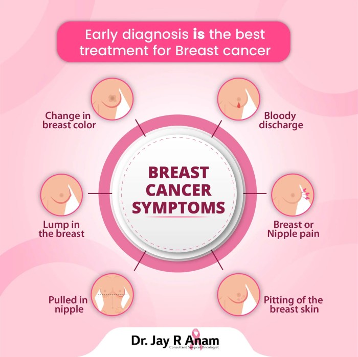
    • Educational infographics and illustrations: These tools, often accompanied by clear and concise text, make complex information easily accessible to a wider audience. Infographics can visually explain the different types of cancer, treatment options, and preventative measures. They empower individuals with knowledge and promote proactive steps to address the issue.
    • Community events and fundraising initiatives: Organised events, such as walks, runs, or bike rides, create a sense of community and provide tangible support for cancer patients and research efforts. These events, often accompanied by powerful slogans and imagery, offer a platform for collective action and awareness.
    • Artistic expressions and memorials: Art, whether in the form of paintings, sculptures, or performances, offers a powerful way to express the emotional impact of cancer. Memorial gardens and public art installations can serve as enduring symbols of remembrance and support.

    “Cancer awareness is not limited to ribbons; it’s a multifaceted movement that utilizes diverse symbols and approaches to spread awareness.”

    History and Significance of Alternative Symbols

    The history and significance of alternative symbols in cancer awareness campaigns often intertwine with cultural traditions and personal experiences. For example, a particular flower may hold special meaning for a community affected by cancer, signifying resilience or hope. Likewise, certain colours or patterns can symbolize the journey through diagnosis, treatment, and recovery. The development of these symbols is a testament to the creative spirit of individuals and communities affected by cancer.

    Visual Representations of Cancer Ribbon Information

    Cancer ribbons have become powerful symbols of hope, support, and awareness in the fight against cancer. Beyond their symbolic meaning, visual representations play a crucial role in communicating these messages effectively. Infographics, illustrations, and imagery in cancer awareness campaigns can help to raise awareness and inspire action.

    Infographic Summary of Cancer Ribbon Meanings

    An effective infographic can visually condense the complex meanings of various cancer ribbons into a digestible format. The graphic should use clear, concise language, and visually appealing colors to highlight the significance of each ribbon. A key feature should be a color-coded legend directly associating each ribbon color with the specific type of cancer it represents. The infographic should be easily understandable to a broad audience, including those unfamiliar with the symbolism of cancer ribbons.

    Using icons or symbols related to each cancer type could also be helpful for quick comprehension.

    Illustrations of Cancer Ribbon Colors and Meanings

    Visual aids, such as illustrations, can bring the symbolism of cancer ribbons to life. These illustrations should depict the ribbons in a variety of contexts to emphasize their broad impact.

    Ribbon Color Cancer Type Illustration Description
    Pink Breast Cancer A vibrant pink ribbon, perhaps tied around a stylized breast or chest area, symbolizing the strength and resilience of those affected by breast cancer. The ribbon could be held by a person or an abstract figure, embodying the collective effort in fighting the disease.
    Purple Lymphoma A rich purple ribbon, elegantly flowing and perhaps wrapped around a stylized lymph node or part of the lymphatic system, emphasizing the complex nature of lymphoma and the support for those undergoing treatment. The illustration might also feature a person or an abstract figure radiating a sense of hope.
    Light Blue Prostate Cancer A gentle light blue ribbon, perhaps entwined with a stylized prostate gland or a person, suggesting the importance of early detection and support for those impacted by prostate cancer. The illustration should convey a sense of calm determination.
    Orange Leukemia A vibrant orange ribbon, perhaps wrapped around a stylized blood cell or a person, symbolizing the fight against leukemia. The illustration should project a sense of strength and the hope for a cure.
    Yellow Childhood Cancer A cheerful yellow ribbon, possibly held by a child or attached to a stylized child’s drawing or toy, highlighting the vulnerability of children affected by cancer and the need for support. The illustration should evoke a sense of compassion and hope for a better future.

    Imagery in Cancer Awareness Campaigns

    Imagery is crucial in cancer awareness campaigns. Effective campaigns often utilize compelling visual elements to convey the message of hope, support, and resilience in the face of cancer. For example, photographs of people affected by cancer, or illustrations depicting the various types of cancer, can create a powerful emotional impact and help to raise awareness. The imagery should always be respectful and avoid exploitation or sensationalism.

    The goal is to inspire hope and promote a sense of community.

    Final Conclusion

    Cancer ribbons colors meanings

    In conclusion, cancer ribbons colors meanings extend far beyond their visual appeal. They serve as powerful reminders of the fight against cancer, embodying hope, resilience, and unity. This exploration of their history, symbolism, and global applications underscores their crucial role in raising awareness and supporting those affected by this devastating disease. By understanding the nuances of each color and its associated meaning, we can use these symbols effectively and respectfully in our efforts to combat cancer.

  • What is Bone Marrow Edema? A Deep Dive

    What is Bone Marrow Edema? A Deep Dive

    What is bone marrow edema? This condition, often detected on medical imaging, is a complex inflammatory response within the bone marrow. Understanding its various causes, locations, and imaging characteristics is crucial for accurate diagnosis and effective treatment. This exploration delves into the intricacies of bone marrow edema, offering insights into its definition, clinical significance, and the various pathologies it can accompany.

    We’ll look at the underlying causes, the critical imaging characteristics, and the often subtle clinical presentations, as well as the treatment approaches and prognosis.

    Bone marrow edema isn’t just a medical term; it represents a significant challenge in diagnosis. The variety of potential causes, from stress fractures to infections, makes precise identification crucial. Different imaging modalities, like MRI and CT scans, provide valuable insights into the nature and extent of the edema. This discussion will highlight the importance of distinguishing bone marrow edema from other bone lesions, emphasizing the importance of a comprehensive approach that combines imaging findings with patient history and physical examination.

    Definition and Overview

    Bone marrow edema is a condition characterized by an abnormal accumulation of fluid within the bone marrow. This fluid buildup, typically appearing as a bright signal on magnetic resonance imaging (MRI), isn’t a disease itself, but rather a sign of underlying pathology. Understanding its presence is crucial for diagnosing the root cause and guiding appropriate treatment.The clinical significance of bone marrow edema is substantial.

    It often indicates stress, inflammation, or injury to the bone and surrounding tissues. Early detection can lead to prompt intervention and potentially prevent more severe complications. The location of the edema can offer clues about the affected area and the potential cause.

    Clinical Significance of Bone Marrow Edema

    Bone marrow edema is a critical imaging finding, often signaling ongoing inflammation or micro-trauma within the bone. This inflammatory response can be caused by a wide range of factors, from minor overuse injuries to more severe conditions like infections or tumors. The presence of bone marrow edema, coupled with the clinical presentation, aids in the differential diagnosis and helps guide appropriate management strategies.

    Common Locations of Bone Marrow Edema

    Bone marrow edema can be found in various locations throughout the skeleton. Its prevalence in certain areas reflects the high stress and mechanical loading those regions experience. Common sites include the:

    • Pelvis and hip region:
    • This area is prone to bone stress injuries, especially in athletes or individuals with repetitive stress activities.

    • Lower extremities:
    • The lower extremities bear significant weight and experience high impact forces, making them susceptible to edema from stress fractures, tendinopathies, and other overuse syndromes.

    • Spine:
    • The spine is also susceptible to edema, often related to spinal conditions like spondylolysis or spondylolisthesis.

    • Shoulders:
    • Repetitive overhead activities or trauma can lead to edema in the shoulder area.

    Imaging Findings Associated with Bone Marrow Edema

    The typical imaging finding associated with bone marrow edema is a hyperintense signal on MRI scans. This signal is usually seen on T2-weighted images and sometimes on STIR (short tau inversion recovery) sequences. The edema appears bright compared to normal bone marrow.

    The specific appearance can vary depending on the severity and duration of the underlying pathology.

    This characteristic signal is not specific to bone marrow edema, but it is highly suggestive and is frequently used in conjunction with other imaging findings and clinical history to arrive at a diagnosis.

    Role of Bone Marrow Edema in Various Pathologies

    Bone marrow edema can be a crucial indicator in various pathologies, from benign overuse injuries to serious conditions. It is essential to consider the location, extent, and presence of any accompanying symptoms when evaluating bone marrow edema.

    • Stress fractures:
    • Repeated stress on a bone can cause micro-fractures, leading to bone marrow edema.

    • Osteomyelitis:
    • Bone infection can result in bone marrow edema.

      Bone marrow edema is basically swelling in the spongy inner part of your bones. It’s often a sign of something more serious, like an injury or infection. Sometimes, athletes experiencing discomfort might consider using silicone hydrogel contact lenses for eye strain, but that’s completely unrelated to bone marrow issues. The important thing is to understand the root cause of bone marrow edema to get proper treatment.

    • Tumors:
    • Both benign and malignant bone tumors can cause edema.

    • Traumatic injuries:
    • Trauma to the bone can lead to inflammation and edema.

    • Inflammatory conditions:
    • Conditions like rheumatoid arthritis can cause edema in affected joints.

    Table of Conditions Associated with Bone Marrow Edema

    Condition Location of Edema Imaging Findings Clinical Presentation
    Stress Fracture (Femur) Distal femur Hyperintense signal on T2-weighted MRI Pain, tenderness, swelling, and sometimes limited range of motion.
    Osteomyelitis (Tibia) Tibia Hyperintense signal on T2-weighted MRI, possible bone destruction Fever, localized pain, swelling, and erythema.
    Spinal Disc Herniation Adjacent vertebral bodies Hyperintense signal on T2-weighted MRI, possible disc bulge Pain radiating down the leg, neurological symptoms.
    Traumatic Injury (Rib) Rib Hyperintense signal on T2-weighted MRI, possible periosteal reaction Localized pain, tenderness, and crepitus.
    Osteoid Osteoma (Proximal Femur) Proximal femur Hyperintense signal on T2-weighted MRI, possible sclerotic rim Pain that is often worse at night, localized tenderness.

    Etiology and Causes

    What is bone marrow edema

    Bone marrow edema, a condition characterized by swelling within the bone marrow, isn’t a disease itself but rather a symptom of underlying issues. Understanding the causes is crucial for proper diagnosis and treatment. Various factors can contribute to this inflammatory response, ranging from minor trauma to severe infections. Pinpointing the specific cause is often essential for effective management and recovery.

    Frequent Causes of Bone Marrow Edema

    Several factors frequently lead to bone marrow edema. These include stress fractures, infections, and various inflammatory conditions. The specific mechanisms and underlying pathology vary based on the causative agent.

    Underlying Mechanisms of Bone Marrow Edema

    The development of bone marrow edema involves a complex interplay of cellular and molecular processes. Trauma, infection, and inflammation all trigger a cascade of events leading to the accumulation of fluid within the bone marrow. This inflammatory response involves the release of cytokines and other signaling molecules, leading to increased vascular permeability and fluid leakage. The resultant swelling disrupts normal bone marrow function.

    Pathophysiology of Bone Marrow Edema in Specific Conditions

    Specific conditions influence the precise pathophysiology of bone marrow edema.

    • Stress Fractures: Repetitive microtrauma to bone, such as seen in athletes or individuals with improper biomechanics, can initiate an inflammatory response. This response results in the recruitment of immune cells, leading to bone marrow edema. The inflammatory cascade is triggered by the release of inflammatory mediators, which ultimately lead to edema. For instance, a runner experiencing repeated stress on their tibia might develop bone marrow edema in that area.

    • Infection: Infectious agents, such as bacteria or viruses, can directly or indirectly cause bone marrow edema. Direct invasion of the bone marrow by pathogens or the body’s immune response to infection can lead to edema. This inflammatory process results in fluid accumulation within the bone marrow. For example, osteomyelitis, an infection of the bone, is often associated with bone marrow edema.

    Comparison of Causes in Different Age Groups, What is bone marrow edema

    The causes of bone marrow edema can vary based on the age group. While stress fractures are more prevalent in young, active individuals, conditions like avascular necrosis or certain cancers might be more common in older age groups. The prevalence of certain infections can also vary depending on the age group.

    Role of Inflammation in Bone Marrow Edema

    Inflammation plays a pivotal role in the development of bone marrow edema. The inflammatory response is a complex cascade of events initiated by various stimuli. The release of cytokines, chemokines, and other inflammatory mediators leads to increased vascular permeability, resulting in fluid leakage into the bone marrow, thus contributing to the edema.

    Table: Causes of Bone Marrow Edema and Clinical Features

    Cause Clinical Features
    Stress Fractures Pain, swelling, tenderness localized to the affected bone. Symptoms often worsen with activity.
    Infection (e.g., Osteomyelitis) Pain, fever, chills, localized warmth, swelling, and tenderness in the affected area. Systemic symptoms may be present.
    Tumors (e.g., Leukemia) Depending on the specific tumor type, possible symptoms can include bone pain, swelling, anemia, fatigue, and others.
    Avascular Necrosis Pain, gradual onset of stiffness, and reduced range of motion in the affected joint. Symptoms can vary based on the location and severity.
    Trauma Pain, swelling, and ecchymosis (bruising) at the site of injury.

    Imaging Characteristics

    Bone marrow edema, a common finding in various musculoskeletal conditions, presents distinct imaging characteristics that aid in diagnosis and differentiation from other pathologies. Accurate interpretation of these characteristics is crucial for appropriate management and treatment planning. Understanding the specific signal intensities observed on different imaging sequences and the subtle distinctions between bone marrow edema and other bone lesions is vital.Imaging modalities like MRI and CT play a significant role in evaluating bone marrow edema.

    MRI, particularly, offers superior soft tissue contrast, allowing for detailed assessment of the affected bone marrow and surrounding structures. CT, while less sensitive to soft tissue changes, can provide crucial information regarding bone density and structural integrity, often complementing MRI findings.

    MRI Characteristics of Bone Marrow Edema

    MRI is the gold standard for evaluating bone marrow edema due to its superior soft tissue contrast. Different MRI sequences highlight various aspects of the condition, enabling a comprehensive assessment.

    • T1-weighted images typically show low signal intensity within the affected bone marrow, reflecting the edema fluid’s reduced fat content. This low signal intensity is often subtle, and the surrounding normal bone marrow often appears as a high signal.
    • T2-weighted images demonstrate high signal intensity within the affected bone marrow, indicating the presence of fluid or edema. The signal intensity is usually homogeneous within the affected area, though it can sometimes exhibit subtle heterogeneity.
    • STIR (Short Tau Inversion Recovery) sequences and Proton Density Fat Suppression (PDFS) sequences are particularly useful for highlighting bone marrow edema. These sequences are sensitive to water and fluid content, providing enhanced visualization of the edematous area by suppressing the signal from fat. This enhances the visibility of the low signal on T1 and high signal on T2 in the affected area.

    CT Characteristics of Bone Marrow Edema

    While less sensitive to soft tissue changes compared to MRI, CT scans can provide valuable information about bone density and structural abnormalities associated with bone marrow edema. CT findings may be subtle, and in many cases, MRI is required for a more definitive diagnosis.

    • CT scans might reveal subtle changes in bone density, like subtle cortical thinning or periosteal reaction in the affected area. However, these findings are often nonspecific and may not be apparent in all cases of bone marrow edema.
    • CT scans are frequently used to assess for fracture or other structural damage, which may accompany or be the cause of bone marrow edema. This aspect is crucial in differentiating between bone marrow edema and other conditions like stress fractures.

    Differential Diagnosis

    Distinguishing bone marrow edema from other bone lesions is critical for appropriate treatment. The imaging characteristics, including signal intensity patterns and the distribution of the affected area, help in differential diagnosis.

    Condition T1-weighted Signal T2-weighted Signal Additional Imaging Findings
    Bone Marrow Edema Low High Homogeneous signal intensity within the affected area, may involve a wide range of bone
    Stress Fracture Low/Variable High Linear or geographic zone of signal abnormality, potential for periosteal reaction, cortical disruption, often associated with specific stress or overuse patterns.
    Tumor Variable (can be low or high) Variable (can be low or high) Often heterogeneous signal intensity, possible bone erosion, and soft tissue mass
    Infections Variable (can be low or high) Variable (can be low or high) May show associated inflammatory changes in surrounding tissues, and possible bone destruction

    Role of MRI Sequences

    Different MRI sequences play distinct roles in evaluating bone marrow edema.

    • T1-weighted images are crucial for identifying the presence of low signal intensity, often subtle, within the affected bone marrow.
    • T2-weighted images, STIR, and PDFS sequences are essential for detecting the characteristic high signal intensity of edema. The distribution and extent of the edema can be assessed.
    • The combination of various sequences provides a more comprehensive picture of the lesion, aiding in differentiating bone marrow edema from other bone lesions.

    Differential Diagnosis: What Is Bone Marrow Edema

    Bone marrow edema, while a common finding on imaging, can mimic various other conditions. Accurately differentiating bone marrow edema from these mimics is crucial for appropriate diagnosis and treatment. Precise diagnosis hinges on a combination of imaging characteristics, clinical presentation, and a thorough understanding of the underlying pathology. A structured approach to differential diagnosis, incorporating both imaging and clinical data, is vital for avoiding misdiagnosis and ensuring optimal patient care.

    Conditions Mimicking Bone Marrow Edema

    Several conditions can present with imaging findings similar to bone marrow edema, leading to diagnostic challenges. These include stress fractures, tumors, infections, and inflammatory processes. Understanding the subtle distinctions between these entities is paramount for accurate diagnosis.

    Bone marrow edema is basically swelling in the spongy inner part of your bones. It’s often a sign of something else going on, like stress fractures or infections. Interestingly, while dealing with such issues, you might also encounter situations where a “rule wipes medical debt credit score” rule wipes medical debt credit score could affect your financial well-being.

    Regardless of the financial aspects, understanding the underlying causes of bone marrow edema is crucial for proper diagnosis and treatment.

    Imaging Distinguishing Features

    Imaging plays a critical role in differentiating bone marrow edema from other pathologies. While edema often shows as a low signal on T1-weighted images and high signal on T2-weighted images, the specific location, size, and shape of the affected area can provide clues. For instance, a localized area of edema adjacent to a fracture line strongly suggests a stress fracture.

    The presence of a mass-like lesion or heterogeneous signal intensity, on the other hand, might point towards a tumor. Moreover, the presence of bone marrow edema in the absence of any apparent injury, or in association with fever and inflammatory markers, may point to an infection.

    Clinical Presentation and Physical Examination

    Clinical history and physical examination are indispensable for refining the differential diagnosis. Symptoms like pain, swelling, and limited range of motion can help distinguish bone marrow edema from other conditions. For example, severe pain that worsens with activity, often accompanied by localized tenderness, strongly suggests a stress fracture. Conversely, the presence of fever, chills, and systemic inflammatory response syndrome (SIRS) criteria might point to an infection.

    A careful evaluation of the patient’s medical history, including any prior injuries, surgeries, or systemic illnesses, is also critical.

    Structured Approach to Differential Diagnosis

    A structured approach to differential diagnosis is crucial for minimizing diagnostic errors. This involves a systematic assessment of imaging findings, coupled with a detailed clinical evaluation. This approach typically begins with evaluating the location and extent of the edema on the MRI scans. Then, the presence of associated findings, such as fractures, bony abnormalities, or soft tissue swelling, is considered.

    Next, the patient’s clinical history, including the nature and onset of symptoms, associated risk factors, and relevant past medical history, is examined. Finally, the combination of imaging findings and clinical presentation is used to generate a differential diagnosis list.

    Comparison Table

    Condition Imaging Findings Clinical Presentation Distinguishing Features
    Bone Marrow Edema Low signal on T1, high signal on T2 Pain, possible swelling Non-specific, often associated with trauma or overuse
    Stress Fracture Edema adjacent to fracture line Pain that worsens with activity History of trauma or repetitive stress
    Tumor Mass-like lesion, heterogeneous signal intensity Pain, swelling, possible neurological symptoms Mass effect, possible increased vascularity
    Infection Edema with possible surrounding soft tissue inflammation Fever, chills, systemic inflammatory response History of infection, elevated inflammatory markers

    Clinical Presentation and Symptoms

    Bone marrow edema, a condition characterized by inflammation within the bone marrow, often presents with a range of symptoms that vary depending on the affected location and the underlying cause. Understanding these symptoms is crucial for accurate diagnosis and appropriate management. This section delves into the common clinical manifestations, highlighting the relationship between location and symptoms, and emphasizing the variability in presentation based on the etiology.

    Common Clinical Symptoms

    Bone marrow edema, while not always symptomatic, can lead to a variety of discomfort and pain. The symptoms often depend on the specific location and the extent of the inflammatory process within the bone. Pain is a common presenting complaint, often described as aching, throbbing, or sharp, and can vary in intensity. Other symptoms may include swelling, tenderness, and limited range of motion in the affected area.

    Symptoms by Affected Body Regions

    The location of bone marrow edema significantly influences the observed symptoms. Pain and tenderness are typically localized to the affected area. For example, bone marrow edema in the hip region may manifest as hip pain, whereas edema in the spine may lead to back pain. The severity of symptoms can range from mild discomfort to significant debilitating pain, impacting daily activities.

    Bone marrow edema is basically swelling in the spongy inner part of a bone. It’s often a precursor to more serious issues, sometimes requiring significant intervention. Fortunately, there are now more options available for dealing with joint problems than just hip replacement. Exploring alternatives to hip replacement, like those discussed in this helpful resource on alternatives to hip replacement , is important for understanding potential less invasive treatments.

    Ultimately, understanding bone marrow edema is key to making informed decisions about treatment options.

    It’s important to note that the specific symptoms experienced can vary considerably, depending on the underlying cause of the edema.

    Relationship Between Location and Symptoms

    The location of bone marrow edema directly correlates with the location of the symptoms. Pain experienced in the knee, for example, suggests involvement of the bone marrow within the knee joint. This localization helps in guiding the diagnostic process and identifying the potential underlying cause.

    Table of Common Symptoms and Potential Correlations with Location

    Affected Body Region Common Symptoms Potential Correlations
    Hip Hip pain, groin pain, limping, limited range of motion in hip Possible involvement of the femoral head, acetabulum, or surrounding tissues
    Knee Knee pain, swelling, tenderness, stiffness, difficulty bearing weight Possible involvement of the tibial plateau, femoral condyle, or menisci
    Spine (vertebrae) Back pain, radiating pain, numbness, weakness, muscle spasms Possible involvement of the intervertebral discs, facet joints, or spinal cord
    Foot/Ankle Foot pain, ankle pain, swelling, tenderness, difficulty walking Possible involvement of the tarsal bones, metatarsals, or surrounding tissues
    Shoulder Shoulder pain, limited range of motion, pain with overhead activities Possible involvement of the humeral head, glenoid labrum, or surrounding tissues

    Variability of Symptoms Based on Underlying Cause

    The specific symptoms associated with bone marrow edema can vary significantly depending on the underlying cause. For instance, trauma-induced bone marrow edema may present with immediate, intense pain, while edema related to infection might manifest with fever and systemic symptoms alongside the local pain. Careful consideration of the patient’s medical history, including any recent injuries, infections, or inflammatory conditions, can provide valuable insights into the potential cause of the edema.

    Treatment and Management

    Bone marrow edema, while often a symptom rather than a disease itself, requires appropriate management to address the underlying cause and alleviate associated pain and functional limitations. Effective treatment strategies often involve a combination of conservative approaches and, in some cases, surgical interventions. The primary goal is to reduce inflammation, promote healing, and restore normal function to the affected area.The management of bone marrow edema is highly individualized, taking into account the specific cause, location, severity, and overall health of the patient.

    A multidisciplinary approach involving orthopedists, physical therapists, and other specialists is often crucial for optimal outcomes.

    General Principles of Treatment

    Effective treatment for bone marrow edema focuses on reducing inflammation, promoting healing, and restoring function. This often involves a multifaceted approach, targeting both the symptoms and the underlying cause. Strategies commonly include rest, physical therapy, and potentially, medication to manage pain and inflammation. Early intervention is key to minimizing long-term complications and maximizing functional recovery.

    Conservative Management Strategies

    Conservative management plays a significant role in the treatment of bone marrow edema, particularly in the initial stages. Rest and physical therapy are frequently used to reduce pain, inflammation, and stress on the affected area. Rest helps to allow the body to heal, reducing stress on the injured tissue. Physical therapy focuses on regaining range of motion, strengthening muscles surrounding the affected area, and improving overall function.

    These strategies are particularly important in cases where the cause of the edema is related to overuse or repetitive strain.

    Specific Treatment Modalities for Underlying Causes

    The specific treatment modalities employed depend on the underlying cause of the bone marrow edema. For example, in cases of stress fractures, the primary treatment focuses on allowing the fracture to heal, potentially involving the use of casts or braces, and avoidance of activities that exacerbate the condition. In cases of osteomyelitis, the treatment focuses on addressing the infection through antibiotics, often in conjunction with surgical intervention to drain the affected area if necessary.

    If the edema stems from a tumor, the approach will involve managing the tumor, which might involve chemotherapy, radiation therapy, or surgery.

    Role of Surgical Intervention

    Surgical intervention is reserved for severe or chronic cases of bone marrow edema where conservative management has failed to produce adequate results. Examples include cases of significant instability, persistent pain, or when there is a suspected underlying pathology requiring surgical intervention. Surgical procedures may involve debridement of damaged tissue, stabilization of the affected area, or removal of a tumor.

    The specific surgical approach will depend on the nature of the underlying condition.

    Monitoring Response to Treatment

    Close monitoring of the patient’s response to treatment is crucial. This involves regular assessments of pain levels, range of motion, and functional capacity. Imaging studies, such as MRI scans, may be repeated to track the progression of edema resolution. Adjustments to the treatment plan may be necessary based on the observed response.

    Treatment Approaches Based on Underlying Cause

    Underlying Cause Treatment Approach
    Stress fractures Rest, immobilization (e.g., cast or brace), avoidance of aggravating activities, physical therapy to restore strength and mobility.
    Osteomyelitis Antibiotics, surgical drainage (if needed), pain management.
    Tumors Tumor-specific treatment (e.g., chemotherapy, radiation, surgery), pain management.
    Repetitive strain injuries Rest, activity modification, physical therapy, anti-inflammatory medications.
    Traumatic injuries Immobilization, pain management, physical therapy to restore function. Surgery may be necessary for significant injuries.

    Prognosis and Outcomes

    What is bone marrow edema

    Bone marrow edema, while often a temporary condition, can have varying impacts on patients, depending on the underlying cause and the individual’s overall health. Understanding the potential outcomes, including long-term effects and complications, is crucial for appropriate management and patient counseling. The prognosis for bone marrow edema is often tied to the resolution of the underlying cause, and successful treatment often leads to a favorable outcome.

    Factors Influencing Outcome

    Several factors significantly influence the prognosis of bone marrow edema. The severity of the underlying condition plays a pivotal role, as does the promptness and effectiveness of treatment. Patient age, overall health, and the presence of any other contributing medical conditions can also influence the recovery process. The location of the edema within the bone structure and the extent of the affected area are also critical factors in predicting the likelihood of a complete recovery.

    Long-Term Effects

    Bone marrow edema can result in a range of long-term effects, depending on the specific condition causing it. In some cases, edema may resolve completely without leaving any lasting consequences. However, in other situations, it can lead to persistent pain, decreased mobility, or even structural damage to the affected bone. Chronic conditions that trigger bone marrow edema can contribute to ongoing inflammation and potentially affect bone density and strength over time.

    For example, in cases of stress fractures, long-term edema can result in delayed healing and potential for recurrent fractures.

    Potential Complications

    Potential complications associated with bone marrow edema vary depending on the underlying cause. In cases of infections, the edema may indicate a more severe systemic illness requiring aggressive treatment. In cases of tumors or cancers, the presence of bone marrow edema can be a marker of disease progression or recurrence. In some cases, prolonged edema may contribute to avascular necrosis, a condition where the bone tissue loses its blood supply, leading to potential bone death.

    Furthermore, prolonged pain and discomfort can significantly impact quality of life.

    Examples of Outcomes

    Favorable outcomes often involve the complete resolution of edema and a return to normal function. For example, in cases of stress fractures effectively treated with rest and immobilization, edema usually resolves within a few weeks, and patients can return to their usual activities without significant long-term issues. Unfavorable outcomes, on the other hand, might involve persistent edema, pain, and reduced mobility.

    For example, patients with underlying conditions like osteomyelitis or tumors might experience recurrent episodes of edema despite treatment, resulting in significant functional limitations.

    Prognostic Factors Table

    Condition Severity Treatment Response Potential Long-Term Effects Prognosis
    Stress Fracture Mild Good Rare Favorable
    Osteomyelitis Severe Variable High Unfavorable
    Tumor Moderate to Severe Variable High Uncertain
    Trauma Variable Variable Variable Variable

    Note: This table provides a general overview and should not be considered a definitive prognostic tool. Individual cases require careful evaluation and personalized management strategies.

    Closing Summary

    In conclusion, bone marrow edema presents a multifaceted challenge to healthcare professionals. From its diverse causes and clinical manifestations to the crucial role of imaging in diagnosis, understanding this condition requires a thorough grasp of its underlying mechanisms and potential complications. This discussion has explored the various facets of bone marrow edema, offering a comprehensive overview of its definition, etiology, imaging characteristics, differential diagnosis, clinical presentation, treatment approaches, and prognosis.

    We hope this detailed exploration has provided valuable insights into this intricate condition.

  • Breast Cancer Testing Screening A Comprehensive Guide

    Breast Cancer Testing Screening A Comprehensive Guide

    Breast cancer testing screening is crucial for early detection, significantly impacting survival rates. This guide delves into the various methods, from the familiar mammogram to newer technologies, highlighting their benefits and limitations. We’ll explore risk factors, screening recommendations, and how to interpret results. Understanding the process empowers individuals to make informed decisions about their health.

    From basic self-exams to advanced MRI scans, we’ll break down each screening method, comparing their advantages and disadvantages. This detailed look allows you to understand which approach might be best for your specific needs and risk factors. We’ll also address common misconceptions and the evolving landscape of breast cancer screening technology.

    Introduction to Breast Cancer Testing and Screening

    Breast cancer is a significant health concern worldwide, impacting countless lives. Early detection is crucial for successful treatment and improved outcomes. Regular breast cancer testing and screening play a vital role in identifying potential issues early, when treatment options are often more effective. This process empowers individuals to take proactive steps toward their health and well-being.Comprehensive breast cancer testing and screening encompasses a range of methods, each with its own strengths and limitations.

    Regular breast cancer testing screening is crucial for early detection, but did you know that supporting overall health through good habits like a healthy diet and taking the right supplements can also play a role? For instance, looking into supplements for lung health, like those available at supplements for lung health , could potentially contribute to a stronger immune system, which might indirectly impact the body’s ability to fight off diseases.

    Ultimately, prioritizing preventative measures like consistent screening and a holistic approach to well-being are key in breast cancer prevention and detection.

    Understanding these nuances allows individuals to make informed decisions about their health care. Knowledge about the available options and their respective benefits and drawbacks is essential for empowering patients to actively participate in their health management.

    Types of Breast Cancer Testing and Screening Methods

    Various methods are employed in breast cancer testing and screening. These methods are designed to detect abnormalities and potential cancerous cells.

    • Mammography: This is a common imaging technique using X-rays to visualize the breast tissue. It helps detect dense breast tissue, masses, and microcalcifications, which can be early signs of cancer. Mammography is a widely used and effective method for breast cancer screening, particularly in women over 40. However, it may not be as effective in women with dense breast tissue, and it can produce false positive results, leading to unnecessary anxiety and further testing.

    • Ultrasound: Ultrasound uses sound waves to create images of the breast. It’s particularly helpful in distinguishing between solid masses and fluid-filled cysts, providing further clarity when combined with mammography. Ultrasound is valuable for evaluating abnormalities detected in mammography and for guiding biopsies. It is also used for women with dense breasts, as it can better visualize these tissues compared to mammography.

      A limitation is that it can sometimes miss small cancers or provide ambiguous results.

    • MRI (Magnetic Resonance Imaging): MRI uses powerful magnetic fields and radio waves to produce detailed images of the breast. It is often used for women with a high risk of breast cancer, those with dense breast tissue, or for evaluating abnormalities detected by other imaging techniques. MRI can detect cancers that are not visible on mammography or ultrasound. A key drawback is the higher cost compared to other methods and potential for false positives.

    • Breast Biopsy: This involves removing a tissue sample from the breast for examination under a microscope. It’s a crucial step when suspicious findings are detected through imaging tests. A biopsy confirms or rules out the presence of cancer. Different types of biopsies are available, including fine-needle aspiration, core needle biopsy, and surgical biopsy, each with varying degrees of invasiveness and accuracy.

    Benefits and Limitations of Screening Methods

    Understanding the advantages and disadvantages of each method is crucial for informed decision-making. Different screening methods cater to various needs and situations, leading to tailored recommendations for individuals.

    • Mammography: Widely accessible, relatively inexpensive, and effective in detecting cancers at an early stage. However, it can miss some cancers, particularly in women with dense breast tissue, and can produce false positive results, leading to unnecessary anxiety.
    • Ultrasound: Useful for differentiating between solid masses and fluid-filled cysts, and guiding biopsies. However, it may not detect all cancers, especially small ones, and the quality of images can vary depending on the technician’s expertise.
    • MRI: Excellent for detecting cancers in dense breast tissue or women at high risk, but it is more expensive and may produce false positives.
    • Breast Biopsy: Provides a definitive diagnosis of breast abnormalities. However, it can be invasive, and it carries a small risk of complications.

    Common Misconceptions About Breast Cancer Testing and Screening

    Many misconceptions surround breast cancer testing and screening. Dispelling these myths is vital for encouraging regular screening and early detection.

    • Screening is only necessary for high-risk individuals: While those with a family history or genetic predisposition are advised to start screening earlier and more frequently, screening is beneficial for all women. Early detection is key, regardless of risk factors.
    • Screening is painful and uncomfortable: While some discomfort may be experienced during certain procedures, modern techniques and skilled professionals minimize pain and discomfort.
    • Breast cancer screening is unnecessary if there are no symptoms: Early detection is critical. Many women with breast cancer have no symptoms in the early stages. Regular screening helps catch cancers when treatment is most effective.

    Different Screening Methods

    Understanding the various breast cancer screening methods is crucial for informed decision-making. Each method has its own strengths and limitations, and choosing the right one depends on individual factors like age, family history, and breast density. This section delves into the different techniques, outlining their procedures, benefits, drawbacks, and ideal applications.

    Regular breast cancer testing screening is crucial for early detection. While I’m no expert on supplements, I’ve heard some people wonder if certain things, like creatine, can affect their bodies. If you’re curious about whether does creatine cause bloating , there’s plenty of info out there. Regardless of those potential side effects, though, getting checked for breast cancer is a really important preventative measure.

    Comparing Screening Methods

    Different imaging and self-examination techniques provide various ways to detect breast abnormalities. The table below summarizes the key characteristics of common methods.

    Method Procedure Advantages Disadvantages Ideal Use Cases
    Mammography X-ray imaging of the breast, often involving compression to spread tissue for clearer images. Detects dense tissue cancers, particularly helpful in identifying microcalcifications (small calcium deposits) that can be an early sign of cancer. Generally good at detecting invasive cancers. Can produce false positives, potentially leading to unnecessary biopsies. Can be uncomfortable due to breast compression. Not as effective in women with dense breast tissue. Routine screening for women over 40, particularly those with no known risk factors.
    Breast Ultrasound Uses high-frequency sound waves to create images of the breast tissue. Excellent for differentiating between cysts (fluid-filled sacs) and solid masses, useful in evaluating dense breasts where mammography might not be as effective. Generally quick and painless. Not as effective at detecting microcalcifications. Results can be operator-dependent, meaning the quality of the image can vary depending on the technician. Evaluating breast lumps, assessing suspicious areas identified by mammography or self-exam, and screening women with dense breasts.
    Breast MRI Uses magnetic fields and radio waves to create detailed images of the breast. Highly sensitive in detecting breast cancers, particularly in women with dense breasts or a high risk of breast cancer. Excellent for detecting small cancers and evaluating suspicious areas. More expensive than mammography or ultrasound. Can produce more false positives, potentially requiring additional testing. May not be suitable for all patients due to potential claustrophobia or metal implants. Screening women with a high risk of breast cancer, evaluating women with dense breasts, evaluating abnormalities detected by other methods, and evaluating breast implants.
    Self-Exams Regularly examining one’s own breasts for changes in size, shape, texture, or the presence of lumps or dimples. Easy, inexpensive, and accessible to everyone. Allows for early detection of abnormalities if the individual is vigilant and aware of their own breasts. Not as reliable as imaging methods. Accuracy is highly dependent on the individual’s awareness and ability to recognize changes. False alarms are common. A valuable supplementary tool for early detection. Important for women of all ages to be familiar with their own breasts and report any unusual changes to their doctor.

    Detailed Procedures and Steps

    Mammography involves positioning the breast between two plates and taking X-ray images. Breast ultrasound uses a handheld probe to send and receive sound waves. Breast MRI uses a large machine to create detailed images. Self-exams involve palpating the breasts for any unusual changes.

    Accuracy and Reliability

    Mammography is generally considered the most effective screening method for women over 40. Ultrasound is valuable in evaluating dense breast tissue, while MRI offers a high level of sensitivity for detecting subtle abnormalities, especially in high-risk individuals. Self-exams are a useful tool for early detection, but they are not as reliable as imaging methods. The accuracy and reliability of each method vary depending on factors such as the skill of the technician, the quality of the equipment, and the individual characteristics of the patient.

    Factors such as breast density and the presence of implants can impact the results of some screening methods.

    Risk Factors and Screening Recommendations

    Understanding your risk factors for breast cancer is crucial for making informed decisions about screening. This knowledge empowers you to discuss your options with your healthcare provider and tailor your screening plan to your individual circumstances. Knowing your risk allows you to proactively address potential health concerns and potentially detect breast cancer early.Knowing the factors that increase your chances of developing breast cancer can influence your screening plan.

    This proactive approach allows for personalized screening strategies that are most effective for your specific risk profile. This helps you understand how different risk factors impact screening recommendations and when more frequent or advanced screenings may be necessary.

    Key Risk Factors

    Various factors can elevate a person’s risk of breast cancer. Age is a significant one, as the risk generally increases with age. Family history plays a critical role, with a strong family history of breast cancer in close relatives substantially increasing the risk. Genetic mutations, such as BRCA1 and BRCA2, are strong indicators of increased susceptibility. Previous radiation exposure, particularly during youth, is also a recognized risk factor.

    Certain lifestyle choices, such as a lack of physical activity or a diet high in processed foods, may contribute to an elevated risk. Finally, having a history of dense breast tissue or previous breast biopsies can also increase risk.

    Screening Recommendations Based on Risk

    Screening recommendations are tailored to individual risk profiles, balancing the potential benefits of early detection against the risks of unnecessary procedures. Different risk levels require varying frequency and types of screening. The recommendations are based on factors like age, family history, and other risk indicators.

    Screening Guidelines by Risk Group

    Risk Group Age Recommendation Additional Screening Frequency
    Average Risk 40-44: Consider starting mammograms. 45-54: Annual mammograms recommended. 55 and older: Mammograms every 2 years or as advised by a doctor. Clinical breast exams (CBE) every 3 years, self-exams monthly. As per the age recommendation above.
    High Risk 10-15 years earlier than the average risk population. Annual mammograms, MRI scans, and possibly genetic testing. Annually or as advised by a doctor, based on individual risk assessment.

    How Risk Factors Influence Screening Decisions

    The table above highlights the different screening guidelines for various risk groups. For individuals with average risk, standard screening guidelines are followed. However, those with a high risk of breast cancer, due to factors like family history or genetic mutations, require more intensive and frequent screening. These individuals might benefit from earlier initiation of mammograms, additional imaging techniques like MRI, and potentially genetic testing to identify and monitor any genetic predisposition.

    This proactive approach allows for early detection and intervention, improving outcomes and reducing the risk of late-stage diagnoses.

    Interpreting Test Results

    Breast cancer testing screening

    Understanding the results of breast cancer screening tests is crucial for early detection and appropriate action. A clear comprehension of the potential findings, their implications, and the subsequent steps involved can empower individuals to take proactive steps toward their health. This section delves into the interpretation of various breast cancer screening tests, focusing on the implications of different outcomes and the diagnostic pathways that follow.

    Understanding Mammogram Results

    Mammograms are a crucial tool for detecting breast abnormalities. Radiologists analyze these images for signs of potential breast cancer, such as masses, microcalcifications, or architectural distortions. A negative mammogram typically indicates no suspicious findings. A positive mammogram, however, does not automatically mean cancer. It simply suggests the presence of something requiring further investigation.

    Getting regular breast cancer screenings is crucial for early detection. While focusing on preventative measures like these, it’s also important to understand other eye conditions. For example, have you ever wondered what a pinguecula is? what is a pinguecula is a yellowish growth on the white part of your eye. Ultimately, knowing about different health concerns, from eye conditions to breast cancer screenings, empowers you to make informed decisions about your well-being.

    Different types of positive findings require different approaches.

    Interpreting Biopsy Results

    A biopsy is a procedure that involves removing a tissue sample from the breast for examination under a microscope. This is a crucial step when a mammogram or other screening test reveals an abnormality. The biopsy report will specify the type of tissue found and whether it exhibits cancerous or precancerous characteristics. The report usually categorizes findings based on established diagnostic criteria, often using systems like the Bethesda System for reporting breast tissue abnormalities.

    Common Findings and Their Implications

    Various findings can emerge from screening tests, each with different implications. Benign findings, such as cysts or fibroadenomas, are non-cancerous and typically do not require immediate intervention beyond regular monitoring. However, if the findings suggest malignancy, the implications are far more serious, necessitating further evaluation and potentially treatment.

    Follow-up Procedures After an Abnormal Result

    Following an abnormal screening result, a multi-step process begins. This typically involves a conversation with the healthcare provider to discuss the findings and next steps. Additional imaging, such as ultrasound or MRI, might be necessary to gain a clearer picture of the abnormality. If the initial tests raise concerns about malignancy, a biopsy is often the next step.

    Diagnostic Pathway Flowchart

    The flowchart below illustrates the typical diagnostic pathway after an abnormal screening result. It highlights the sequential steps involved, from initial detection to confirmation or exclusion of breast cancer. The pathway emphasizes the importance of communication with healthcare professionals and adherence to recommended follow-up procedures. This flowchart is a general guideline; individual cases may require modifications based on specific circumstances.

    Flowchart of diagnostic pathway

    Note: The flowchart visually displays the progression from an initial screening abnormality to a definitive diagnosis or exclusion. It emphasizes the cyclical nature of follow-up, where results from one test can influence the next. This dynamic process ensures accurate assessment and appropriate treatment planning.

    Impact of Screening on Early Detection

    Early detection is crucial in the fight against breast cancer. Regular screening, when combined with awareness and knowledge of risk factors, significantly increases the chances of catching the disease in its earliest, most treatable stages. This proactive approach dramatically improves outcomes and quality of life for those affected.Early diagnosis empowers individuals and healthcare professionals to initiate effective treatment plans, often leading to a more favorable prognosis.

    The difference between early-stage and advanced-stage breast cancer treatment can be substantial, impacting both the treatment approach and long-term survival.

    Role of Early Detection in Improving Breast Cancer Survival Rates

    Early detection of breast cancer is paramount to improving survival rates. When breast cancer is diagnosed in its early stages, it is often more localized and less likely to have spread to other parts of the body. This localized nature allows for less aggressive treatment options, often leading to better outcomes and a higher chance of long-term survival.

    Survival rates are significantly higher for early-stage breast cancer compared to later stages.

    Correlation Between Early Diagnosis and Treatment Success

    Early diagnosis of breast cancer allows for more targeted and effective treatment options. Treatment success rates are demonstrably higher when the disease is caught early. Surgical options like lumpectomy, instead of mastectomy, become more viable when the cancer is confined to a smaller area. Furthermore, the use of chemotherapy, radiation therapy, or hormone therapy can be tailored to the specific characteristics of the early-stage cancer, optimizing treatment effectiveness and minimizing side effects.

    Comparison of Outcomes of Early-Stage and Advanced-Stage Breast Cancer Treatment

    The outcomes of breast cancer treatment vary significantly based on the stage at diagnosis. Early-stage breast cancer, often detected through screening, allows for less invasive and often less aggressive treatment approaches. This results in a higher likelihood of cure, improved quality of life, and a greater chance of long-term survival. In contrast, advanced-stage breast cancer, which may have spread to other parts of the body, requires more extensive and often more aggressive treatment, which may include chemotherapy, targeted therapies, and/or hormonal therapies.

    The success rates and side effects associated with treatment are generally lower for advanced-stage cancer.

    Breast Cancer Survival Rates Based on Stage at Diagnosis

    Survival rates for breast cancer are directly linked to the stage of the disease at diagnosis. While exact statistics can vary depending on factors such as treatment approach and individual patient characteristics, general trends are clear. Early-stage breast cancer, often detected through screening, has significantly higher survival rates than advanced-stage breast cancer. Comprehensive data on survival rates are available from organizations like the American Cancer Society and the National Cancer Institute.

    These organizations provide valuable resources for further exploration of the impact of early detection on breast cancer survival. For instance, a patient diagnosed with stage 1 breast cancer has a significantly higher 5-year survival rate than a patient diagnosed with stage 4 breast cancer. Data from the National Cancer Institute consistently shows this correlation. Early detection is crucial to improve these survival rates.

    Technological Advancements in Breast Cancer Screening

    The landscape of breast cancer screening is constantly evolving, driven by innovative technologies that enhance accuracy, efficiency, and patient experience. These advancements are crucial in the fight against breast cancer, enabling earlier detection and improved treatment outcomes. From improved imaging techniques to sophisticated analysis tools, the journey toward more precise and personalized screening continues.The latest innovations in breast cancer screening are revolutionizing how we detect and manage this disease.

    These advancements are not just about faster results; they’re about more precise diagnoses, reduced false positives, and ultimately, better chances for survival. The combination of new technologies and refined diagnostic protocols is leading to a more comprehensive and personalized approach to breast cancer screening.

    Advanced Imaging Techniques

    Breast imaging techniques are constantly being refined to improve accuracy and reduce the need for unnecessary biopsies. Digital mammography, which converts X-rays into digital images, has already improved image quality and storage. Digital breast tomosynthesis (DBT), often called 3D mammography, provides a layered view of the breast, allowing for clearer visualization of structures and potentially reducing the rate of false positives.

    Ultrasound, often used in conjunction with mammography, is particularly helpful in evaluating dense breast tissue, which can be challenging to image with traditional methods.

    Artificial Intelligence (AI) in Screening

    AI algorithms are being integrated into breast cancer screening to assist radiologists in analyzing images and detecting subtle abnormalities. These algorithms can identify patterns and anomalies that may be missed by the human eye, leading to earlier detection and more accurate diagnoses. AI-powered systems can also help streamline the screening process, reducing workload and potentially increasing the number of women who undergo screenings.

    Early trials show promise in reducing false positive rates, and further research is underway to optimize AI’s role in breast cancer detection.

    Personalized Risk Assessment Tools

    Beyond imaging, advancements are focusing on personalized risk assessment. Genetic testing and family history analysis are increasingly used to identify women at higher risk for breast cancer. These tools can be used to tailor screening recommendations, increasing the frequency or type of screening for individuals with a higher predisposition. This targeted approach can be crucial in preventing delayed diagnoses and improving outcomes for high-risk individuals.

    For example, women with a family history of BRCA1/2 mutations may benefit from earlier and more frequent screening, even before reaching traditional screening ages.

    Promising Future Directions in Breast Cancer Screening Technology

    • Molecular Imaging: Techniques that visualize molecular changes associated with breast cancer, such as using contrast agents that target specific molecules within tumors, offer the potential to identify cancerous cells earlier than traditional imaging methods. This approach may be especially useful for detecting pre-cancerous lesions.
    • Liquid Biopsies: Analyzing circulating tumor DNA (ctDNA) in blood samples offers a non-invasive way to detect and monitor breast cancer. This technology could potentially revolutionize early detection and monitoring, allowing for more frequent and less invasive screenings.
    • Personalized Screening Protocols: Integrating genetic risk factors, lifestyle information, and imaging data into personalized screening protocols could allow for more effective risk stratification and tailored screening strategies. This approach may significantly reduce unnecessary screenings for low-risk individuals while ensuring higher-risk individuals receive more frequent and targeted screening.

    Addressing Ethical and Societal Issues: Breast Cancer Testing Screening

    Breast cancer testing screening

    Breast cancer screening, while vital for early detection and improved outcomes, presents a complex interplay of ethical considerations, societal impacts, and practical realities. Balancing the benefits of early diagnosis with the potential for anxiety, false positives, and overdiagnosis requires careful consideration. Furthermore, disparities in access to screening and the financial burden of these procedures highlight the need for equitable and sustainable programs.

    Ethical Considerations Surrounding Screening Recommendations

    Screening recommendations often grapple with balancing the potential benefits of early detection against the risks of unnecessary procedures and potential harms. The decision to undergo screening is inherently personal, involving individual risk assessments, preferences, and values. Factors like family history, personal health status, and perceived risks significantly influence these decisions. Clinicians have a responsibility to provide comprehensive information and support patients in making informed choices aligned with their values.

    This includes acknowledging the psychological impact of screening results, both positive and negative.

    Societal Impact of Breast Cancer Screening Programs

    Breast cancer screening programs have a significant impact on society, influencing healthcare systems, public health initiatives, and individual well-being. These programs can lead to increased awareness and early intervention, potentially reducing the overall burden of breast cancer. However, the programs also entail substantial costs, impacting resource allocation and potentially diverting funds from other crucial health areas. Moreover, the societal implications of screening recommendations need to account for the diverse experiences and needs of different communities.

    Cost-Effectiveness of Various Screening Methods, Breast cancer testing screening

    The cost-effectiveness of different breast cancer screening methods is a crucial aspect to consider. Different modalities, such as mammography, ultrasound, MRI, and others, have varying costs associated with equipment, personnel, and procedural requirements. The optimal choice often depends on factors like patient risk profiles and available resources. Cost-benefit analyses play a vital role in guiding healthcare decisions and ensuring efficient allocation of resources to maximize impact.

    For example, a cost-effectiveness analysis might demonstrate that for women at average risk, mammography provides a good balance between cost and benefit compared to other screening methods. However, for women with higher risk factors, the use of additional imaging modalities, such as MRI, might become more cost-effective.

    Potential Biases and Disparities in Access to Screening

    Disparities in access to breast cancer screening exist based on factors like socioeconomic status, race, ethnicity, and geographic location. These disparities can lead to unequal outcomes in early detection and treatment. For example, lower-income individuals might face barriers in accessing screening due to cost or lack of insurance coverage. Similarly, cultural and linguistic barriers can limit access and understanding of screening recommendations.

    Addressing these disparities requires targeted interventions to improve access, affordability, and culturally appropriate communication strategies. Effective programs need to consider these factors and provide tailored support for diverse populations.

    End of Discussion

    In conclusion, breast cancer testing screening is a vital tool in the fight against this disease. Early detection dramatically improves outcomes, and understanding the different methods, risk factors, and potential results is key. This comprehensive guide provides the information needed to make well-informed decisions about your health and well-being. By staying proactive and informed, you can take control of your breast health and empower yourself with the knowledge to navigate the screening process effectively.

  • How Alcohol Affects Sleep Apnea A Deep Dive

    How Alcohol Affects Sleep Apnea A Deep Dive

    How alcohol affects sleep apnea is a critical concern for anyone struggling with this potentially serious condition. Sleep apnea, characterized by pauses in breathing during sleep, can have various types and symptoms, and alcohol can significantly exacerbate these issues. This exploration delves into the intricate ways alcohol impacts sleep apnea, from the physiological mechanisms to the practical management strategies.

    Understanding the interplay between alcohol and sleep apnea is vital for effective treatment and prevention. This article will examine the effects of alcohol on the respiratory system, the central nervous system, and different sleep stages, illustrating how alcohol consumption can worsen the severity and frequency of sleep apnea episodes.

    Introduction to Alcohol and Sleep Apnea

    How alcohol affects sleep apnea

    Sleep apnea is a sleep disorder characterized by pauses in breathing during sleep. These pauses can last from a few seconds to minutes and can occur repeatedly throughout the night. There are different types of sleep apnea, including obstructive sleep apnea (OSA), central sleep apnea, and mixed sleep apnea. OSA is the most common type and is caused by a blockage in the airway, often due to the relaxation of throat muscles during sleep.

    Symptoms of sleep apnea can include loud snoring, gasping or choking sounds during sleep, daytime sleepiness, morning headaches, and difficulty concentrating.Alcohol, a central nervous system depressant, significantly impacts various bodily functions, including breathing. Its effects on the respiratory system are particularly relevant to sleep apnea. Alcohol relaxes the muscles in the throat and upper airway, potentially worsening the airway blockage that is characteristic of sleep apnea.

    This relaxation can make it harder for the individual to breathe, leading to more frequent and prolonged pauses in breathing during sleep.

    Physiological Mechanisms of Alcohol-Induced Sleep Apnea Worsening

    Alcohol’s impact on sleep apnea stems from its effect on the muscles responsible for maintaining the airway open. The central nervous system’s regulatory mechanisms governing breathing are also affected by alcohol consumption. Alcohol slows down these mechanisms, reducing the body’s ability to maintain a stable breathing pattern. This can lead to more pronounced airway obstruction and more frequent apneas.

    Furthermore, alcohol disrupts sleep architecture, making the individual more susceptible to sleep-related breathing disorders.

    Ever wondered how alcohol impacts your sleep apnea? It’s a tricky relationship. Alcohol can relax the muscles in your throat, making it harder for your airways to stay open during sleep. This can worsen sleep apnea symptoms. While that’s important to know, it’s also crucial to remember that staying hydrated and practicing good hygiene is key to protecting yourself from contagious illnesses, like norovirus, which is on the rise right now.

    Learn more about staying safe from norovirus and preventing its spread here. Ultimately, a balanced approach to your health, including limiting alcohol intake, is key to managing sleep apnea effectively.

    Severity of Sleep Apnea by Alcohol Consumption Level

    The severity of sleep apnea can be influenced by the level of alcohol consumption. Alcohol’s depressant effect on the central nervous system and its impact on airway muscles are dose-dependent. Individuals who consume alcohol in moderate or higher amounts experience a more pronounced reduction in the body’s ability to maintain a stable airway during sleep, thus potentially worsening sleep apnea symptoms.

    Alcohol Consumption Level Potential Impact on Sleep Apnea Severity
    Low (e.g., one drink per night) Minor increase in sleep apnea symptoms, if present.
    Moderate (e.g., two to three drinks per night) Increased frequency and duration of apneas. Significant worsening of existing sleep apnea.
    High (e.g., four or more drinks per night) Significant increase in apnea severity, leading to more severe and prolonged apneas. Increased risk of respiratory distress and potentially dangerous consequences.

    The Impact of Alcohol on Sleep Apnea Severity: How Alcohol Affects Sleep Apnea

    Alcohol consumption significantly alters the delicate balance of physiological processes crucial for maintaining healthy breathing during sleep. This interference can exacerbate sleep apnea symptoms, making it more challenging for the body to regulate airflow and potentially leading to more severe consequences. Understanding the mechanisms by which alcohol impacts sleep apnea is vital for effective management and prevention strategies.Alcohol’s influence on the throat and airway muscles is a key factor in its impact on sleep apnea.

    Alcohol acts as a muscle relaxant, causing the muscles in the throat and upper airway to become significantly more relaxed than usual. This relaxation can lead to a narrowing of the airway, obstructing airflow. The narrowing can be particularly pronounced during sleep, when muscle tone is already reduced.

    Effects on Breathing Regulation

    Alcohol’s effects on the central nervous system, responsible for regulating breathing, also play a critical role in sleep apnea exacerbation. Alcohol depresses the central nervous system, reducing the signals that control breathing. This depression can lead to irregular breathing patterns, potentially causing or worsening episodes of apnea. A reduced respiratory drive means that the brain sends weaker signals to the muscles involved in breathing, which may result in periods of interrupted breathing during sleep.

    This effect is further compounded by the relaxation of the throat and airway muscles.

    Impact on Sleep Apnea Episodes, How alcohol affects sleep apnea

    Alcohol consumption can significantly affect the duration and severity of sleep apnea episodes. Individuals who consume alcohol before bed may experience more frequent and prolonged periods of apnea, leading to a decrease in the quality and quantity of restorative sleep. This can translate to a lower oxygen saturation level, impacting the overall health and well-being of the individual. The increased severity and frequency of apnea episodes directly correlate with the amount of alcohol consumed.

    Relationship Between Alcohol Intake and Sleep Apnea Severity

    Alcohol Intake Level Potential Impact on Sleep Apnea Severity (General Observations) Examples (Illustrative Cases)
    Low (e.g., one drink) May slightly increase the risk of mild sleep apnea episodes in susceptible individuals, primarily affecting the duration or severity of existing episodes. A 30-year-old woman with mild sleep apnea might experience slightly longer pauses in breathing after a single glass of wine before bed.
    Moderate (e.g., two to three drinks) Likely to significantly increase the risk of sleep apnea episodes, potentially increasing the frequency, duration, and severity of existing apnea events. A 50-year-old man with moderate sleep apnea may experience more frequent and prolonged pauses in breathing after two or three drinks, leading to reduced oxygen levels.
    High (e.g., four or more drinks) High alcohol consumption significantly increases the risk of severe sleep apnea episodes, potentially leading to dangerous reductions in oxygen saturation and significantly impairing sleep quality. A 65-year-old man with severe sleep apnea who consumes four or more alcoholic beverages before bed is at high risk of experiencing a severe apnea episode with significant drops in blood oxygen levels, potentially requiring medical attention.

    Note: The relationship between alcohol intake and sleep apnea severity is complex and varies significantly based on individual factors, including age, gender, overall health, and pre-existing medical conditions. This table provides general observations, and consultation with a healthcare professional is crucial for personalized advice.

    Alcohol’s Effect on Sleep Stages and Apnea

    Alcohol, while initially promoting drowsiness, significantly disrupts the natural sleep cycle, often leading to more frequent and severe sleep apnea events. This disruption is particularly concerning as the quality of sleep during different stages is crucial for overall health and well-being. Understanding how alcohol impacts these stages is vital for managing sleep apnea and its associated complications.Alcohol’s interference with sleep architecture is multifaceted.

    It impacts the balance of different sleep stages, affecting the duration and depth of REM and non-REM sleep. This disruption can make sleep apnea worse, and, in turn, alcohol use can worsen the effects of sleep apnea.

    Impact on Sleep Stages

    Alcohol’s initial sedative effect can lead to a feeling of immediate sleepiness, but this is often followed by a fragmented sleep pattern. This disruption is observed across different sleep stages, especially REM and non-REM sleep. Alcohol’s effects are strongest during the first few hours of sleep, potentially delaying the onset of deep sleep and disrupting the natural progression through the sleep cycle.

    Ever wondered how alcohol messes with your sleep apnea? Well, it’s a bit of a double-whammy. Alcohol relaxes the muscles in your throat, making it harder for air to flow freely during sleep. This is why it’s often recommended to avoid it if you’re dealing with sleep apnea, and for good reason. Considering that you’re also looking at choosing over the counter cough medications choosing over the counter cough medications , it’s crucial to understand the potential interactions with your sleep apnea treatment.

    Ultimately, alcohol can significantly disrupt your sleep, potentially worsening your sleep apnea symptoms.

    Frequency of Apnea Events During Specific Sleep Stages

    Alcohol consumption has been linked to a higher frequency of sleep apnea events, particularly during the transition periods between sleep stages. This is because alcohol relaxes the muscles in the throat, further hindering airflow and increasing the risk of apnea. Individuals experiencing sleep apnea may find that alcohol use triggers more episodes of interrupted breathing during light sleep, transitioning to deeper sleep stages.

    Furthermore, individuals may experience more severe apnea events during REM sleep, due to the relaxation of muscles in the airway. This means the risk of apnea is higher in stages where the body is more relaxed.

    Impact on Sleep Apnea Treatments

    Alcohol can significantly diminish the effectiveness of sleep apnea treatments, such as CPAP (Continuous Positive Airway Pressure). For instance, if a person is using CPAP, alcohol may lead to a decrease in compliance with the treatment, as it can cause feelings of discomfort and make it more difficult to tolerate the pressure of the device. In some cases, individuals may experience a worsening of sleep apnea symptoms during periods of heavy alcohol consumption.

    Effects on Different Sleep Stages

    Sleep Stage Alcohol Effect Impact on Sleep Apnea
    Early Sleep (Light Sleep) Initial drowsiness, followed by fragmentation. Increased frequency of apnea events, especially during transitions.
    Deep Sleep (Non-REM Stages 3 & 4) Delayed onset and reduced duration. Potential for more severe apnea events if alcohol consumption is high.
    REM Sleep Reduced duration and quality. Increased risk of apnea, potentially more severe events.
    Overall Sleep Quality Decreased duration of restorative sleep. Increased severity and frequency of sleep apnea episodes throughout the night.

    Risk Factors and Consequences

    Alcohol, unfortunately, significantly exacerbates the challenges faced by individuals with sleep apnea. This combination can lead to a dangerous cycle, impacting overall health and well-being. Understanding the heightened risks and potential consequences is crucial for effective management and prevention.Regular alcohol consumption increases the risk of sleep apnea worsening due to its impact on the muscles controlling breathing. Alcohol’s sedative effects can relax the muscles in the throat and airway, further obstructing breathing during sleep.

    This is particularly problematic for individuals already prone to sleep apnea.

    Factors Increasing Sleep Apnea Risk with Alcohol Use

    Alcohol consumption, particularly in larger amounts or regularly, can amplify the risk of sleep apnea episodes. It reduces the body’s ability to maintain a stable airway, increasing the chances of pauses in breathing. Individuals with a pre-existing tendency towards airway narrowing, such as those with obesity or a family history of sleep apnea, are at even greater risk when alcohol is introduced.

    Long-Term Health Consequences

    Combining alcohol with sleep apnea creates a synergistic effect that can lead to a multitude of detrimental health issues. The frequent interruptions in breathing during sleep, compounded by alcohol’s effects, can lead to hypertension, cardiovascular problems, and an increased risk of stroke. Furthermore, the poor quality of sleep can negatively affect cognitive function, leading to reduced alertness and impaired judgment.

    Alcohol unfortunately disrupts sleep patterns, making sleep apnea worse. While exploring natural remedies for better sleep, I stumbled upon potential benefits of MCT oil, which some say can improve sleep quality. You can learn more about MCT oil benefits, side effects, and other factors at mct oil benefits side effects and more. However, more research is needed to confirm these claims, and alcohol’s negative impact on sleep apnea remains a significant concern.

    Medication Interactions

    Patients with sleep apnea who consume alcohol may experience negative interactions with their prescribed medications. Alcohol can alter the metabolism of certain drugs, potentially leading to either reduced effectiveness or an increased risk of side effects. For example, if a patient is taking blood pressure medication, alcohol can increase the risk of adverse reactions. Patients should always consult with their physician regarding potential interactions between their medications and alcohol.

    It is crucial to adhere to prescribed dosages and discuss any concerns or potential side effects with their healthcare provider.

    Preventive Measures

    To mitigate the risks associated with alcohol and sleep apnea, several preventative measures can be taken. Maintaining a healthy weight is crucial, as obesity is a significant risk factor for both conditions. Avoiding alcohol consumption or limiting intake is another critical preventative step. If alcohol is consumed, doing so in moderation is essential. Furthermore, regular exercise, a balanced diet, and adequate sleep hygiene can help manage both sleep apnea and the negative effects of alcohol.

    Consistent monitoring of symptoms and regular check-ups with a healthcare professional are also crucial in preventing further complications.

    • Healthy Weight Management: Maintaining a healthy weight can significantly reduce the risk of sleep apnea and its complications. Excess weight contributes to airway narrowing, making individuals more susceptible to sleep apnea episodes. Regular exercise and a balanced diet are essential components of a weight management strategy.
    • Moderation in Alcohol Consumption: Limiting alcohol consumption is vital for individuals with sleep apnea. Alcohol significantly worsens sleep apnea symptoms by relaxing the muscles in the throat and airway, further obstructing breathing during sleep. If alcohol is consumed, it’s essential to do so in moderation. For individuals with sleep apnea, avoiding alcohol altogether is often the safest approach.
    • Medication Adherence and Consultation: Proper adherence to prescribed medications and open communication with healthcare providers are crucial for managing sleep apnea and its associated risks. Patients should always inform their doctors about alcohol consumption and discuss potential medication interactions. This precaution helps to prevent unexpected and potentially dangerous reactions.
    • Regular Medical Check-ups: Regular check-ups with a healthcare professional are essential for monitoring sleep apnea and related conditions. Early detection and intervention can help prevent the long-term health consequences associated with the combined effects of alcohol and sleep apnea.

    Management Strategies and Recommendations

    How alcohol affects sleep apnea

    Navigating sleep apnea alongside alcohol consumption requires a multi-faceted approach. Simply reducing alcohol intake isn’t enough; a holistic strategy addressing both the sleep apnea and the alcohol dependence is crucial for improved health outcomes. This section Artikels key strategies for managing sleep apnea in individuals who consume alcohol, alongside recommendations for responsible alcohol reduction, and the importance of professional medical guidance.

    Strategies for Managing Sleep Apnea in Alcohol Consumers

    Effective management of sleep apnea in individuals who consume alcohol necessitates a coordinated effort targeting both the apnea and the alcohol consumption. Addressing the underlying sleep apnea through CPAP therapy, lifestyle modifications, and potentially surgical interventions is essential. Simultaneously, reducing alcohol consumption through behavioral strategies and support systems plays a critical role in improving sleep quality and overall health.

    • CPAP Therapy: Continuous Positive Airway Pressure (CPAP) therapy is a standard treatment for obstructive sleep apnea. For individuals who consume alcohol, CPAP therapy can be particularly beneficial in improving airflow and reducing apnea episodes during sleep, even though alcohol can negatively impact the effectiveness of CPAP. It is important to consult with a healthcare professional to determine the optimal CPAP settings and usage patterns.

    • Lifestyle Modifications: Weight management, regular exercise, and avoiding alcohol or consuming it in moderation can significantly improve sleep apnea symptoms. Individuals struggling with alcohol dependence should seek support from therapists or support groups to create sustainable lifestyle changes.
    • Alcohol Reduction Strategies: Gradual reduction in alcohol consumption, with the support of a healthcare professional or therapist, is crucial. This process often involves identifying triggers, developing coping mechanisms, and seeking support from support groups or family members. A balanced approach to reducing alcohol intake alongside managing sleep apnea is vital.
    • Behavioral Therapies: Cognitive Behavioral Therapy (CBT) for alcohol use disorders can equip individuals with coping mechanisms and strategies for reducing alcohol consumption and managing cravings. This therapy can be incredibly beneficial in providing long-term support and modifying behaviors that contribute to both sleep apnea and alcohol misuse.

    Recommendations for Reducing Alcohol Consumption

    A gradual and sustained reduction in alcohol consumption is often the most effective approach for individuals with sleep apnea. The goal is not total abstinence but rather a reduction to a level that minimizes the negative impact on sleep and overall health. A collaborative approach with healthcare providers is crucial.

    • Gradual Reduction: Instead of abruptly stopping alcohol consumption, a gradual reduction plan, often monitored by a healthcare professional, is recommended. This approach allows the body to adjust and reduces the risk of withdrawal symptoms.
    • Setting Realistic Goals: Establishing realistic and achievable goals for alcohol reduction is key to success. This may involve reducing the frequency of consumption or the amount of alcohol consumed per occasion.
    • Identifying Triggers: Recognizing and addressing situations or emotions that trigger alcohol consumption is vital. This might involve seeking support from therapists or support groups to develop healthy coping mechanisms.
    • Support Systems: Leveraging support from family, friends, or support groups can provide encouragement and accountability throughout the process. Strong support systems can help individuals stay motivated and avoid relapses.

    Importance of Professional Medical Advice

    Professional medical advice is crucial for individuals experiencing both sleep apnea and alcohol-related issues. A healthcare professional can assess the severity of both conditions, recommend appropriate treatment plans, and monitor progress effectively. This integrated approach is essential for achieving optimal health outcomes.

    Management Strategies Table

    Management Strategy Effectiveness (High/Medium/Low) Level of Implementation (Easy/Moderate/Difficult)
    CPAP Therapy High Moderate
    Lifestyle Modifications Medium Easy
    Alcohol Reduction Strategies Medium Moderate-Difficult
    Behavioral Therapies High Moderate

    Illustrative Case Studies (Hypothetical)

    Understanding how alcohol interacts with sleep apnea requires looking at real-world examples. While we can’t observe individuals directly, we can use hypothetical case studies to illustrate potential scenarios and highlight the complexities of this interplay. These scenarios aim to showcase how alcohol can both worsen existing sleep apnea and contribute to its development, while also demonstrating the positive impact of reducing alcohol consumption.

    Case Study 1: Worsening Existing Sleep Apnea

    “Mark, a 45-year-old man with a history of moderate sleep apnea, enjoyed a few drinks nightly with dinner. He found that after a few glasses of wine, his sleep became noticeably more fragmented and restless. He often woke up gasping for air, a symptom he hadn’t experienced as frequently before his increased alcohol consumption. He also reported feeling more tired during the day, despite sleeping for what seemed like a normal duration.”

    Mark’s experience highlights a common pattern. Alcohol relaxes the muscles in the throat and airway, making it easier for them to collapse during sleep. This can exacerbate existing sleep apnea, leading to more frequent and severe apneas. The reduced muscle tone in the throat, combined with the sedative effects of alcohol, leads to a more pronounced airway obstruction.

    Case Study 2: Development of Sleep Apnea

    “Sarah, a 30-year-old woman who occasionally enjoyed cocktails, started noticing that she frequently snored loudly and felt increasingly tired throughout the day. As her alcohol consumption increased over time, she experienced more difficulty breathing during sleep and began waking up gasping for air. A sleep study confirmed the presence of moderate sleep apnea, a condition she never had before.”

    Sarah’s case demonstrates how alcohol, even in moderate consumption, can contribute to the development of sleep apnea. Chronic alcohol use can lead to weight gain, which is a significant risk factor for sleep apnea. Furthermore, the constant relaxation of the throat muscles due to alcohol can progressively narrow the airway, potentially leading to sleep apnea over time.

    Case Study 3: Improvement Through Reduced Alcohol Consumption

    “David, a 52-year-old man with severe sleep apnea, realized the impact his nightly whiskey habit had on his sleep quality. He decided to significantly reduce his alcohol intake. Within weeks, he noticed a considerable improvement in his sleep. He experienced fewer apneas, woke up feeling more rested, and reported a noticeable reduction in daytime fatigue. A follow-up sleep study revealed a significant decrease in the severity of his sleep apnea.”

    David’s experience showcases the potential benefits of reducing alcohol consumption for individuals with sleep apnea. By minimizing alcohol’s relaxing effects on the throat muscles, David’s airways remained more open during sleep. This resulted in improved sleep quality and reduced apnea episodes. This highlights the importance of addressing lifestyle factors, like alcohol intake, for effective sleep apnea management.

    Comparative Analysis of Alcohol Effects

    Alcohol’s impact on sleep apnea is multifaceted and influenced by various factors, including the type of alcohol consumed, the individual’s gender, age, and pre-existing health conditions. Understanding these nuances is crucial for developing personalized strategies to mitigate the risks associated with alcohol consumption and sleep apnea.A deeper dive into these comparative analyses reveals that alcohol’s effect on sleep apnea is not uniform.

    Different types of alcohol, varying degrees of consumption, and individual differences all play a significant role in the severity and manifestation of this interaction. This nuanced approach is vital for developing targeted interventions to manage the risks of alcohol-induced sleep apnea.

    Impact of Different Types of Alcohol

    Different alcoholic beverages vary in their alcohol content and composition. This difference directly impacts the rate and extent of alcohol absorption and metabolism, which in turn affects the severity of sleep apnea. For example, distilled spirits like vodka or whiskey, often higher in alcohol content per serving, may have a more pronounced effect on sleep apnea compared to wine or beer.

    Furthermore, the presence of other components in certain beverages, like congeners in spirits, may contribute to the overall impact on sleep apnea.

    Impact of Alcohol on Sleep Apnea in Men and Women

    Studies suggest that men and women may respond differently to alcohol’s effects on sleep apnea. While the exact mechanisms behind this difference are not fully understood, factors such as hormonal variations, body composition, and metabolic rates could contribute to differing sensitivities. Women may experience a more pronounced effect at lower alcohol consumption levels compared to men, potentially due to differences in how their bodies metabolize alcohol.

    Impact of Alcohol on Sleep Apnea in Different Age Groups

    Age is another factor that influences the interaction between alcohol and sleep apnea. Younger adults may experience a more immediate and potentially severe impact on sleep apnea due to their still developing metabolisms. Older adults, on the other hand, might be more vulnerable due to age-related changes in physiological processes and potential co-morbidities. This age-related variability necessitates a nuanced approach to assessing and managing the risks.

    Comparative Table of Alcohol Effects on Sleep Apnea

    Type of Alcoholic Beverage Alcohol Content (approx.) Potential Impact on Sleep Apnea Notes
    Distilled Spirits (Vodka, Whiskey) 40-50% Higher risk of exacerbating sleep apnea due to faster absorption and potentially higher congeners. Rapid onset of effects.
    Wine 12-15% Moderate impact on sleep apnea, potentially less severe than distilled spirits. Effect may vary based on type and serving size.
    Beer 3-6% Generally lower impact on sleep apnea compared to spirits, but quantity matters. Higher volume consumption can still be detrimental.

    Epilogue

    In conclusion, the relationship between alcohol and sleep apnea is complex and multifaceted. Alcohol significantly worsens sleep apnea by impacting respiratory function, disrupting sleep cycles, and potentially interacting with existing treatments. Understanding these effects is crucial for proactive management, and this article provides a comprehensive overview of the issue, including practical strategies for mitigating risks and seeking professional guidance.

    Ultimately, prioritizing responsible alcohol consumption and seeking medical attention are key to effectively managing both conditions.

  • How Does Tylenol Acetaminophen Work?

    How Does Tylenol Acetaminophen Work?

    How does Tylenol acetaminophen work? This exploration delves into the fascinating science behind this common pain reliever. We’ll uncover the chemical pathways, the body’s responses, and the potential risks associated with its use, providing a comprehensive overview of its mechanism of action, pharmacokinetics, pharmacodynamics, safety, and clinical applications.

    Acetaminophen, the active ingredient in Tylenol, is a widely used medication for pain relief and fever reduction. Understanding how it works is crucial for informed use and safe consumption. This article will unravel the complexities of its actions within the body, from its absorption and distribution to its ultimate effect on the central nervous system.

    Mechanism of Action

    How does tylenol acetaminophen work

    Acetaminophen, commonly known as Tylenol, is a widely used over-the-counter pain reliever and fever reducer. Understanding its mechanism of action helps us appreciate its effectiveness and limitations. Its unique approach to pain and fever differs significantly from other common analgesics.Acetaminophen’s precise mode of action remains a subject of ongoing research, but the current understanding centers around its interaction with the central nervous system and its impact on the body’s inflammatory response.

    Unlike ibuprofen and aspirin, which primarily inhibit prostaglandin synthesis, acetaminophen appears to exert its effects through other pathways.

    Chemical Pathways Involved

    Acetaminophen’s pain-relieving and fever-reducing properties are not directly linked to inhibiting prostaglandin synthesis. Instead, it’s believed to act on the central nervous system by altering the processing of pain signals. This suggests that acetaminophen has a different target within the body compared to other pain relievers.

    Inhibition of Prostaglandin Synthesis

    While acetaminophen does not directly inhibit prostaglandin synthesis, the relationship between its action and prostaglandin pathways is still an area of ongoing research. It’s possible that its effects are mediated by the modulation of certain enzymes involved in the inflammatory response.

    Comparison to Other Pain Relievers

    Acetaminophen differs significantly from other common pain relievers like ibuprofen. Ibuprofen, for example, directly inhibits cyclooxygenase (COX) enzymes, which are crucial in the production of prostaglandins. Acetaminophen, however, appears to target different molecular mechanisms within the brain.

    Key Enzymes and Proteins

    This table highlights the key enzymes and proteins involved in the process, although the exact role of some in acetaminophen’s mechanism is still under investigation.

    Enzyme/Protein Function Role in Acetaminophen’s Action
    Cyclooxygenase (COX) Enzymes involved in the production of prostaglandins. Not the primary target of acetaminophen’s action.
    Central Nervous System Receptors Various receptors in the brain involved in pain perception. Potentially involved in acetaminophen’s effect on pain signals.
    Other unidentified targets Possible molecular targets within the body. Ongoing research is exploring these potential targets.

    Pharmacokinetics: How Does Tylenol Acetaminophen Work

    Acetaminophen, a widely used over-the-counter pain reliever, follows a predictable journey through the human body. Understanding its pharmacokinetic properties is crucial for optimizing its therapeutic effects and minimizing potential adverse reactions. This section delves into the absorption, distribution, metabolism, and excretion (ADME) of acetaminophen, highlighting factors influencing its action and the vital role of the liver in its clearance.Acetaminophen’s journey within the body is a complex interplay of processes.

    Its absorption, distribution, metabolism, and excretion (ADME) are critical factors that dictate its effectiveness and safety profile. Understanding these pharmacokinetic parameters allows for tailored dosing strategies and risk mitigation strategies.

    Absorption

    Acetaminophen is readily absorbed from the gastrointestinal tract, primarily in the small intestine. The rate of absorption is influenced by factors such as the dosage form (e.g., tablet, liquid), presence of food in the stomach, and individual variations in gastric emptying and intestinal motility. Fast-acting formulations, like those dissolved under the tongue, can bypass the gastrointestinal tract and lead to quicker absorption.

    So, how does Tylenol, or acetaminophen, actually work? It’s a bit of a mystery, but it seems to block pain signals in the brain. This is crucial for managing the discomfort often experienced after a procedure like knee arthroscopy, which can be quite intense. Understanding how to manage post-surgical pain like that after a knee arthroscopy, for example, is key to recovery.

    Pain after knee arthroscopy can be managed with various methods, but understanding the basics of how acetaminophen works is a good starting point. Ultimately, it’s a powerful tool for relieving pain, though it’s not a cure-all, of course.

    Food generally slows down absorption, increasing the time to reach peak plasma concentrations.

    Distribution

    Acetaminophen is highly water-soluble and readily distributes throughout the body’s tissues and organs. Its distribution is relatively uniform, with concentrations reflecting the body’s overall water content. The blood-brain barrier, however, presents a slight barrier to its penetration into the central nervous system. This characteristic is important for minimizing potential central nervous system effects.

    Metabolism

    The liver plays a critical role in acetaminophen’s metabolism. The majority of acetaminophen is metabolized in the liver through a two-step process. In the first phase, it undergoes oxidation to form a reactive intermediate. This intermediate is usually harmlessly conjugated with glucuronic acid or sulfate. However, under conditions of overdose or impaired liver function, the rate of formation of this reactive intermediate may exceed the body’s capacity to conjugate it, leading to hepatotoxicity.

    Excretion

    The primary routes of acetaminophen excretion are through the kidneys, in the form of glucuronide and sulfate conjugates. A small portion is excreted unchanged in the urine. The renal clearance of acetaminophen is influenced by factors like kidney function and urine pH. Variations in these factors can impact the rate of acetaminophen excretion.

    Factors Influencing Absorption

    • Dosage form: Different dosage forms (tablets, liquids, etc.) affect the rate of dissolution and absorption.
    • Food intake: Food slows down the rate of absorption, leading to a delay in the onset of action.
    • Gastric emptying rate: Variations in gastric emptying time can influence how quickly acetaminophen reaches the small intestine for absorption.
    • Intestinal motility: Changes in intestinal motility affect the rate of absorption and transit through the intestines.

    Routes of Administration

    Acetaminophen can be administered orally, rectally, or intravenously. Oral administration is the most common route, offering convenience and ease of use. Rectal administration can be beneficial in situations where oral intake is difficult. Intravenous administration is reserved for specific situations, such as in emergency cases or when rapid onset of action is necessary. Each route has a distinct impact on the pharmacokinetic profile, influencing the rate and extent of absorption.

    Diagram of Acetaminophen ADME

    Phase Description
    Absorption Acetaminophen is absorbed from the gastrointestinal tract into the bloodstream.
    Distribution Acetaminophen is transported throughout the body, reaching various tissues and organs.
    Metabolism The liver primarily metabolizes acetaminophen into less toxic metabolites.
    Excretion Metabolized products are excreted from the body primarily through the kidneys.

    Pharmacodynamics

    Acetaminophen, a widely used over-the-counter pain reliever and fever reducer, exerts its effects through a complex interplay of molecular interactions within the body. Understanding its pharmacodynamics, the mechanisms by which it produces its effects, is crucial for comprehending its therapeutic use and potential adverse reactions. This section delves into the specific targets of acetaminophen, its impact on the central nervous system, its effects across different tissues, the dosage-response relationship, and a comparison with other analgesics.Acetaminophen’s primary mechanism of action differs significantly from other common analgesics.

    Instead of directly targeting pain receptors, it acts on a specific pathway within the central nervous system, influencing the production of prostaglandins, substances involved in the inflammatory response. This unique mechanism contributes to its effectiveness in reducing fever and relieving pain.

    Specific Targets of Acetaminophen

    Acetaminophen’s primary target is the central nervous system, specifically the hypothalamus, a region responsible for regulating body temperature. It interferes with the synthesis of prostaglandins, key molecules involved in the inflammatory response, and fever production. However, it’s important to note that acetaminophen’s precise mechanism of action within the hypothalamus is not fully elucidated.

    Effects on the Central Nervous System

    Acetaminophen’s influence on the central nervous system is evident in its ability to reduce fever. By modulating the production of prostaglandins, it affects the thermoregulatory center in the hypothalamus, which is responsible for maintaining body temperature. This reduction in prostaglandin synthesis leads to a decrease in the body’s perceived temperature set point, resulting in a lowering of fever.

    Effects on Different Tissues and Organs

    While primarily affecting the central nervous system, acetaminophen’s effects aren’t limited to just that. It has a wide range of effects across different tissues and organs. For example, it can impact the liver, kidneys, and gastrointestinal tract. However, these effects are often related to its metabolism and clearance from the body.

    Dosage-Response Relationship

    The pharmacodynamic effects of acetaminophen are closely linked to the administered dose. Lower doses effectively manage mild pain and fever, while higher doses, although providing greater relief, increase the risk of adverse effects. The relationship is not linear, and there’s a threshold where the risk of liver damage becomes significant.

    Comparison with Other Analgesics

    Drug Target Primary Effect
    Acetaminophen Hypothalamic thermoregulatory center; possibly other pathways Reduces fever, relieves pain; primarily by modulating prostaglandin synthesis
    Aspirin Cyclooxygenase (COX) enzymes Reduces fever, relieves pain, and inflammation by inhibiting prostaglandin synthesis
    Ibuprofen Cyclooxygenase (COX) enzymes Reduces fever, relieves pain, and inflammation by inhibiting prostaglandin synthesis

    The table above highlights the different targets and primary effects of acetaminophen compared to other common analgesics. Note that the targets and mechanisms of action for aspirin and ibuprofen are fundamentally different from acetaminophen’s, reflecting their distinct pharmacological profiles.

    Safety and Toxicity

    Acetaminophen, a widely used over-the-counter pain reliever and fever reducer, is generally safe when taken as directed. However, like any medication, it carries potential risks if misused or taken in excessive amounts. Understanding the potential side effects, mechanisms of toxicity, and proper dosage guidelines is crucial for responsible use. This section will delve into the safety profile of acetaminophen, focusing on its potential toxicity and the importance of safe usage.

    Potential Side Effects and Adverse Reactions

    Acetaminophen, while generally well-tolerated, can cause a range of side effects, though these are typically mild and resolve on their own. Common side effects include nausea, vomiting, and skin rash. In rare cases, more severe adverse reactions can occur. These reactions can manifest as allergic reactions, including hives, swelling, and difficulty breathing. Liver damage, though less common, is a significant concern, especially with excessive use or overdose.

    Mechanisms of Acetaminophen-Induced Liver Damage

    Acetaminophen’s metabolism involves the liver, where it’s converted into a toxic metabolite. Under normal circumstances, the liver can handle this process without significant damage. However, with excessive acetaminophen intake, the liver’s capacity to detoxify the toxic metabolite can be overwhelmed. This leads to the accumulation of toxic substances that can cause cellular damage, inflammation, and eventually, liver failure. The severity of liver damage is directly related to the amount of acetaminophen ingested and the individual’s overall health status.

    Importance of Proper Dosage and Precautions

    Adhering to the recommended dosage is paramount for safe acetaminophen use. Overdosing, even with seemingly small amounts, can significantly increase the risk of liver damage. Always consult with a healthcare professional before taking acetaminophen, especially if you have pre-existing liver conditions, are taking other medications, or are pregnant or breastfeeding. It’s crucial to carefully read and follow all instructions on the product label.

    Always keep acetaminophen out of reach of children.

    Comparison to Other Over-the-Counter Pain Relievers

    Compared to other over-the-counter pain relievers like ibuprofen or naproxen, acetaminophen has a different toxicity profile. Ibuprofen and naproxen, while also associated with potential side effects, primarily affect the gastrointestinal tract, causing stomach upset or ulcers in susceptible individuals. Acetaminophen, on the other hand, primarily targets the liver, making it crucial to understand the importance of safe usage and to avoid exceeding recommended dosages.

    Consequences of Acetaminophen Overdose

    Acetaminophen overdose can lead to severe liver damage, potentially requiring a liver transplant. Symptoms of acetaminophen overdose can range from mild nausea and vomiting to more serious symptoms like jaundice, confusion, and loss of consciousness. In severe cases, liver failure and death can occur. Early intervention is critical for preventing long-term health consequences. Prompt medical attention is essential in case of suspected acetaminophen overdose.

    Clinical Applications

    Acetaminophen, a widely used over-the-counter medication, plays a crucial role in managing various types of pain and fever. Its efficacy in treating mild to moderate pain makes it a first-line choice in numerous medical situations. Understanding its applications in different conditions and dosage guidelines is essential for safe and effective use.Acetaminophen’s primary mechanism of action involves reducing the production of prostaglandins, key mediators of pain and fever.

    Understanding how Tylenol (acetaminophen) works is pretty fascinating. It basically targets the production of pain signals in the brain. Interestingly, a similar concept of targeted interventions can be seen in the medical use of a Kirschner wire, a small, smooth wire used in orthopedic procedures, what is a Kirschner wire to help stabilize bones.

    Ultimately, both techniques aim to alleviate discomfort, though in very different ways. Acetaminophen works by blocking pain signals, while a Kirschner wire stabilizes bone structure to allow healing.

    This effect allows for effective pain relief and fever reduction, making it a valuable tool in the management of diverse medical conditions. However, it’s important to note that acetaminophen does not directly address the underlying cause of the pain or inflammation. Its use is primarily focused on symptom relief.

    Common Uses in Medical Conditions

    Acetaminophen is frequently used to treat a wide range of painful conditions. Its effectiveness against mild to moderate pain makes it a popular choice for headaches, muscle aches, toothaches, and menstrual cramps. It also plays a significant role in managing postoperative pain and discomfort associated with various surgical procedures.

    Specific Medical Situations Where Acetaminophen is Preferred

    Acetaminophen is often preferred over nonsteroidal anti-inflammatory drugs (NSAIDs) in certain medical situations. For instance, individuals with a history of peptic ulcers or gastrointestinal bleeding may be at higher risk of complications from NSAIDs, making acetaminophen a safer alternative. Furthermore, patients with renal impairment or those taking other medications that may interact with NSAIDs may benefit from the use of acetaminophen for pain relief.

    Efficacy in Treating Different Types of Pain, How does tylenol acetaminophen work

    Acetaminophen demonstrates varying degrees of efficacy across different types of pain. While generally effective in treating mild to moderate pain, its effectiveness in managing severe pain, such as that experienced during a myocardial infarction or a major trauma, is limited. In such cases, stronger analgesics may be necessary to effectively manage the pain. Acetaminophen’s efficacy is often comparable to NSAIDs in managing headaches, muscle aches, and menstrual cramps.

    Role in Managing Fever and Inflammation

    Acetaminophen’s ability to reduce fever is well-established. It achieves this by inhibiting the production of prostaglandins in the hypothalamus, the body’s temperature control center. However, it does not directly address the underlying cause of the fever or inflammation. While acetaminophen is effective in reducing fever, it is not an anti-inflammatory agent and does not target the source of inflammation.

    Understanding how acetaminophen works is key, especially if you’re managing health conditions like heart failure. It primarily works by reducing the production of certain chemicals in the body that cause fever and pain. Learning about self-care strategies for heart failure, like heart failure self care , can also help you better manage pain and discomfort. This knowledge is helpful in understanding how acetaminophen can play a role in overall well-being.

    Its role is primarily focused on reducing the associated symptoms.

    Dosage Recommendations for Different Age Groups

    The appropriate dosage of acetaminophen varies significantly based on age and weight. Infants and young children require lower doses compared to adolescents and adults. Carefully following dosage guidelines, particularly for children, is critical to prevent potential adverse effects. Overdosing can lead to serious liver damage. It is essential to consult with a healthcare professional for appropriate dosage recommendations, especially for children.

    Age Group Typical Dosage (mg/kg) Frequency
    Infants (0-6 months) 10-15 mg/kg every 6 hours As needed for fever or pain
    Children (6 months – 12 years) 10-15 mg/kg every 4-6 hours As needed for fever or pain
    Adolescents and Adults 650-1000 mg every 4-6 hours As needed for fever or pain

    Dosage should always be tailored to individual needs and monitored by a healthcare professional.

    Interactions

    How does tylenol acetaminophen work

    Acetaminophen, a widely used over-the-counter pain reliever and fever reducer, is generally considered safe when taken as directed. However, like any medication, it can interact with other substances, potentially altering its effectiveness or increasing the risk of adverse effects. Understanding these interactions is crucial for safe and responsible use.Careful consideration of all medications taken concurrently with acetaminophen is essential to minimize potential complications.

    This includes not only prescription drugs but also over-the-counter medications, herbal supplements, and even certain foods or beverages. A comprehensive understanding of these interactions empowers individuals to make informed decisions about their health and ensures they are taking acetaminophen in a way that aligns with their overall well-being.

    Potential Drug Interactions

    Many medications can interact with acetaminophen, sometimes leading to increased liver toxicity. These interactions often involve competing for metabolic pathways or influencing the concentration of acetaminophen in the bloodstream. Careful monitoring and adjustment of dosages might be necessary when acetaminophen is taken alongside other medications.

    Acetaminophen and Alcohol

    Consuming alcohol while taking acetaminophen can significantly increase the risk of liver damage. Alcohol and acetaminophen share metabolic pathways in the liver, potentially leading to a buildup of toxic metabolites. This combination can result in severe liver injury, sometimes even leading to liver failure. Avoiding alcohol consumption while taking acetaminophen is strongly advised to minimize this risk.

    Interactions with Other Substances

    Certain herbal supplements and dietary components may also interact with acetaminophen. For example, some herbal products marketed for pain relief or fever reduction might contain substances with overlapping effects on the liver, potentially increasing the risk of liver damage when combined with acetaminophen. Always consult a healthcare professional before combining acetaminophen with any herbal remedies or supplements.

    Importance of Informing Healthcare Providers

    Thorough communication with healthcare providers about all medications, including over-the-counter drugs and supplements, is crucial. This ensures healthcare providers have a complete picture of the medications a patient is taking and can assess potential interactions. This proactive approach helps avoid unexpected and potentially dangerous drug interactions.

    Table of Common Medications Interacting with Acetaminophen

    Medication Interaction Type Potential Consequences
    NSAIDs (Nonsteroidal Anti-inflammatory Drugs) Additive analgesic effect, increased risk of gastrointestinal ulcers Potentially increased pain relief but also a heightened risk of stomach upset or bleeding.
    Warfarin (anticoagulant) Increased risk of bleeding Can intensify the anticoagulant effect of warfarin, leading to an increased risk of bleeding.
    Some Antibiotics Potential for liver toxicity Certain antibiotics can elevate the risk of liver damage when taken concurrently with acetaminophen.
    Certain Herbal Supplements Increased risk of liver toxicity, unknown mechanism Some herbal supplements intended to ease pain or fever reduction might contain components with overlapping effects on the liver, possibly increasing liver damage risk.

    Outcome Summary

    In conclusion, this comprehensive look at how Tylenol acetaminophen works highlights its complex interactions within the human body. From its specific targets to its impact on various bodily functions, we’ve seen how this common medication plays a crucial role in managing pain and fever. Understanding its pharmacokinetics, pharmacodynamics, and potential side effects is vital for responsible use and avoiding potential complications.

    Remember, always consult with a healthcare professional before taking any medication.

  • Opioid Types for Chronic Pain Management

    Types of opioids chronic pain medications are a crucial aspect of managing chronic pain. This exploration delves into the diverse world of opioid-based pain relief, examining their classifications, mechanisms of action, and routes of administration. We’ll also investigate the potential risks, side effects, and crucial considerations for responsible use.

    Understanding the various types of opioids and their applications in chronic pain management is essential for both patients and healthcare providers. This comprehensive guide will walk you through the different types of opioid medications, their potency, duration of action, and potential side effects, allowing for informed decisions about pain management strategies.

    Introduction to Opioids for Chronic Pain

    Opioids are a class of medications used to manage moderate to severe chronic pain. They work by binding to opioid receptors in the central nervous system, which can reduce pain signals and produce a sense of well-being. While effective for many, their use requires careful consideration due to potential for dependence and side effects.Understanding the different types of opioids, their mechanisms of action, and routes of administration is crucial for safe and effective pain management.

    Understanding different types of opioid chronic pain medications is crucial, but sometimes, seemingly unrelated issues like scabs on the scalp can be a real problem. If you’re dealing with these skin issues, checking out resources like scabs on scalp causes diagnosis and treatment can help you understand the potential causes and treatments. Ultimately, proper diagnosis and management of chronic pain, using the right opioid medications, is key to overall well-being.

    This overview will detail the classifications of opioids, including full agonists, partial agonists, and mixed agonists-antagonists, and the various ways these medications are administered.

    Opioid Classifications

    Opioids are categorized based on their interaction with opioid receptors in the brain and spinal cord. This classification helps clinicians tailor treatment to individual needs.

    • Full Agonists: These opioids bind strongly to opioid receptors, mimicking the effects of naturally occurring endorphins. Examples include morphine, oxycodone, and fentanyl. They produce a high degree of pain relief but carry a greater risk of dependence and adverse effects.
    • Partial Agonists: These opioids bind to opioid receptors but produce a less potent effect compared to full agonists. Examples include buprenorphine. They can be useful in managing pain while potentially reducing the risk of dependence compared to full agonists.
    • Mixed Agonists-Antagonists: These opioids have both agonist and antagonist properties. They can bind to opioid receptors, but their effect can be blocked by other substances. Examples include pentazocine and nalbuphine. Their use is often limited due to the potential for unpredictable effects and the risk of reducing the effectiveness of other opioids.

    Routes of Opioid Administration

    The route of administration impacts the onset and duration of opioid effects. Different routes are chosen based on factors like patient condition, pain severity, and individual needs.

    • Oral: This is a common route for chronic pain management. Opioids are taken by mouth, allowing for gradual absorption into the bloodstream. However, the onset of action can be slower compared to other routes.
    • Intravenous (IV): IV administration delivers opioids directly into the bloodstream, leading to a rapid onset of action. This route is often used in emergency situations or for patients who have difficulty swallowing or absorbing oral medications.
    • Transdermal: Patches deliver opioids through the skin, providing a sustained release of medication over several days. This route is often preferred for patients who require continuous pain relief.
    • Intramuscular (IM): This route involves injecting opioids into a muscle. The absorption rate is faster than oral administration, but slower than IV. This route is used when a rapid onset of action is required without the need for IV access.

    Opioid Medications: A Summary Table

    Medication Name Classification Mechanism of Action Route of Administration
    Morphine Full Agonist Binds to opioid receptors in the central nervous system, reducing pain signals and producing a sense of well-being. Oral, IV, IM, Subcutaneous
    Oxycodone Full Agonist Binds to opioid receptors, reducing pain signals and producing a sense of well-being. Oral, Extended-release Oral
    Fentanyl Full Agonist Binds to opioid receptors, producing strong pain relief. Oral, Transdermal, IV, Nasal Spray
    Buprenorphine Partial Agonist Binds to opioid receptors, producing less potent effect compared to full agonists. Oral, Sublingual
    Pentazocine Mixed Agonist-Antagonist Has both agonist and antagonist properties, potentially reducing the effectiveness of other opioids. Oral, IM

    Common Types of Opioids Used in Chronic Pain Management

    Opioids are powerful pain relievers, but their use for chronic pain requires careful consideration due to potential side effects and the risk of addiction. Understanding the different types of opioids and their characteristics is crucial for both patients and healthcare providers to make informed decisions about pain management strategies. This section explores the most commonly prescribed opioid medications, comparing their efficacy and potential side effects.Different opioids have varying strengths, durations of action, and potential side effects.

    This makes careful selection by a healthcare professional crucial for effective pain management while minimizing risks. A thorough understanding of these factors empowers patients and doctors to collaborate in choosing the most suitable treatment option.

    Frequently Prescribed Opioid Medications

    A variety of opioid medications are available for chronic pain management, each with its own characteristics. Factors like potency, duration of action, and potential side effects influence the choice of medication.

    • Morphine: A widely used opioid, morphine is often prescribed for moderate to severe chronic pain. It acts by binding to opioid receptors in the brain and spinal cord, reducing pain signals. Potential side effects include nausea, vomiting, constipation, and respiratory depression. Variations in formulations, like sustained-release tablets, can help manage pain over extended periods, potentially minimizing side effects associated with frequent dosing.

      Different routes of administration, such as oral, intravenous, and subcutaneous, offer flexibility in treatment.

    • Oxycodone: A common opioid analgesic, oxycodone is frequently prescribed for moderate to severe pain. It is often combined with non-opioid medications for enhanced pain relief and reduced risk of side effects. Oxycodone’s potency can vary depending on formulations (e.g., immediate-release versus extended-release). Potential side effects include nausea, vomiting, constipation, dizziness, and respiratory depression. Careful monitoring and adherence to prescribed dosages are essential for minimizing risks.

    • Hydrocodone: Frequently used in combination with non-opioid pain relievers, hydrocodone is a relatively mild opioid. The combination products (like Vicodin) are often prescribed for mild to moderate pain. Its lower potency compared to other opioids makes it a suitable option for patients requiring pain relief without the potential risks associated with stronger opioids. Side effects, while generally less severe than with stronger opioids, can include nausea, vomiting, constipation, and drowsiness.

      Patients should be aware of potential interactions with other medications.

    Comparison of Opioid Types

    This table provides a concise overview of different opioid types, highlighting key characteristics.

    Opioid Type Potency Duration of Action Typical Use Cases
    Morphine High 4-6 hours (immediate-release) Moderate to severe chronic pain, especially when extended-release formulations are needed.
    Oxycodone Moderate to High 4-6 hours (immediate-release), 12 hours (extended-release) Moderate to severe chronic pain, often in combination with other analgesics.
    Hydrocodone Low to Moderate 4-6 hours (immediate-release) Mild to moderate chronic pain, often in combination with other analgesics.

    Potential Risks and Side Effects of Opioid Use

    Opioid medications, while effective for managing chronic pain, come with significant potential risks. Understanding these risks is crucial for both patients and healthcare providers to make informed decisions about their use. Long-term opioid use can lead to a range of complications, impacting not only physical health but also mental well-being. This section delves into the potential dangers associated with opioid use, emphasizing the importance of careful monitoring and responsible prescribing practices.Opioid misuse and addiction are significant public health concerns.

    A substantial portion of individuals prescribed opioids for chronic pain may develop an addiction, with rates varying based on factors like individual predisposition, dosage, duration of use, and the presence of other co-occurring mental health conditions. This misuse often arises from a combination of factors, including the highly addictive nature of some opioids, the potential for psychological dependence, and societal pressures.

    Risks Associated with Long-Term Use

    Long-term opioid use can lead to a range of detrimental health effects. The body can develop tolerance, meaning that higher doses are required to achieve the same pain-relieving effect. This escalating dosage can increase the risk of overdose and other serious complications. Furthermore, the constant use of opioids can negatively impact various organ systems, including the respiratory system, gastrointestinal tract, and central nervous system.

    A patient’s risk factors for these complications can be influenced by genetics, co-existing health conditions, and lifestyle choices.

    Prevalence of Opioid Misuse and Addiction

    Opioid misuse and addiction are significant public health challenges. The misuse of prescription opioids has led to a substantial increase in overdose deaths in recent decades. This alarming trend underscores the importance of preventive measures and responsible prescribing practices. Factors contributing to opioid misuse and addiction are multi-faceted and include social pressures, the ease of access to opioids, and the inherent addictive potential of these medications.

    Addressing this issue requires a multifaceted approach that targets prevention, treatment, and public awareness.

    Common Side Effects of Opioid Medications

    Opioids can cause a variety of side effects, ranging from mild discomfort to severe complications. These side effects often affect different bodily systems and can vary in severity based on individual factors and the specific opioid used. Understanding these potential side effects is crucial for effective pain management and mitigating risks. Close monitoring by healthcare providers is essential to address and manage these side effects promptly.

    Detailed List of Potential Side Effects

    Side Effect Severity Management Strategies
    Respiratory Depression Potentially life-threatening Careful monitoring of breathing rate and oxygen saturation. Close collaboration between patients and healthcare providers. Consideration of alternative pain management strategies.
    Constipation Common Increased fiber intake, adequate hydration, use of stool softeners or laxatives as prescribed by the doctor.
    Nausea Variable Anti-nausea medications, dietary adjustments, and careful monitoring.
    Sedation Variable Dosage adjustments, careful driving restrictions, and monitoring of cognitive function.
    Drowsiness Variable Adjusting the timing of medication intake, ensuring adequate rest, and avoidance of hazardous activities.
    Headache Common Pain relievers (non-opioid), hydration, and rest. Report persistent or severe headaches to the doctor.
    Vomiting Variable Anti-emetic medications, dietary adjustments, and monitoring for dehydration.

    Non-Opioid Options for Chronic Pain Management

    Beyond opioid medications, a wide range of non-opioid treatments offer effective pain relief and improved quality of life for individuals experiencing chronic pain. These alternatives address the root causes of pain and often have a lower risk of side effects compared to opioids. Understanding these options is crucial for patients and healthcare providers alike to make informed decisions about treatment strategies.

    Alternative Treatments for Chronic Pain

    Various non-opioid approaches can successfully manage chronic pain. These methods often target the underlying mechanisms of pain, promoting long-term well-being.

    • Physical Therapy: Physical therapy programs are tailored to each individual’s specific needs and pain locations. Exercises and stretches, along with manual therapy techniques, improve flexibility, strength, and range of motion. This can reduce pain by strengthening supporting muscles and improving posture.
    • Occupational Therapy: Occupational therapy focuses on adapting daily activities and environments to minimize pain and improve function. This might involve modifying workspaces, recommending assistive devices, or providing strategies for pain management during daily tasks. This approach empowers patients to maintain their independence.
    • Cognitive Behavioral Therapy (CBT): CBT helps patients understand and change negative thought patterns and behaviors related to pain. By addressing the emotional and psychological aspects of pain, CBT can significantly improve coping mechanisms and reduce pain perception. This approach can lead to a greater sense of control over chronic pain.
    • Mindfulness and Meditation: These practices can help individuals focus on the present moment, reducing the focus on pain sensations. Techniques like mindfulness-based stress reduction (MBSR) and meditation can improve overall well-being and provide a sense of calm, potentially easing pain.
    • Acupuncture: This ancient practice involves inserting thin needles into specific points on the body. Acupuncture is believed to stimulate the flow of energy, reducing pain and inflammation. Its effectiveness in managing chronic pain is supported by several studies.
    • Massage Therapy: Massage therapy can help relieve muscle tension and reduce pain. Different types of massage, like Swedish massage or deep tissue massage, may be beneficial depending on the individual’s needs. This approach promotes relaxation and can improve circulation.
    • Electrical Stimulation Therapies: Techniques like transcutaneous electrical nerve stimulation (TENS) use electrical impulses to block pain signals. These therapies can be used at home or in a clinical setting. This approach is often used to manage acute or chronic pain.

    Mechanisms of Action of Non-Opioid Pain Relievers, Types of opioids chronic pain medications

    Non-opioid pain relievers work through diverse mechanisms to target different pain pathways.

    • Nonsteroidal Anti-inflammatory Drugs (NSAIDs): NSAIDs, like ibuprofen and naproxen, inhibit cyclooxygenase (COX) enzymes, reducing the production of prostaglandins. Prostaglandins are chemicals involved in inflammation and pain transmission. This reduces inflammation and pain associated with conditions like arthritis and muscle injuries.
    • Acetaminophen (Paracetamol): Acetaminophen’s precise mechanism of action isn’t fully understood. However, it’s thought to act on the central nervous system, potentially by inhibiting the production of certain neurochemicals involved in pain signaling. This makes it effective for mild to moderate pain.

    Using Non-Opioid Therapies Alongside or as Alternatives to Opioids

    Non-opioid therapies can be integrated into a comprehensive pain management plan, either alongside or as a replacement for opioid medications. This approach is crucial in minimizing the risks associated with opioid use while maximizing pain relief.

    Comparison of Opioid and Non-Opioid Treatments

    Treatment Type Effectiveness Safety Potential Side Effects
    Opioids Generally effective for severe pain, but diminishing returns with prolonged use. High risk of addiction, respiratory depression, and overdose. Constipation, nausea, sedation, cognitive impairment.
    Non-Opioids (e.g., NSAIDs, Acetaminophen) Effective for mild to moderate pain, with varying degrees of effectiveness based on the condition. Generally safer than opioids, with a lower risk of addiction and overdose. Gastrointestinal upset (NSAIDs), liver damage (acetaminophen), allergic reactions.

    Patient Considerations and Management Strategies

    Navigating chronic pain often involves a delicate balancing act between managing discomfort and minimizing potential risks. This is especially true when considering opioid therapy, which, while effective for some, comes with significant potential side effects. Understanding individual patient needs, responsible prescribing practices, and proactive management strategies are crucial for achieving optimal outcomes.

    Factors to Consider When Prescribing Opioids

    Individual patient responses to opioids vary significantly. A comprehensive patient history is paramount, encompassing prior medical conditions, mental health status, and any history of substance use disorders. Factors such as age, weight, and liver or kidney function must be considered to tailor the dosage and frequency of opioid administration. This individualized approach helps to mitigate potential risks and optimize therapeutic benefits.

    A thorough assessment, including pain intensity, location, and impact on daily activities, should be consistently documented and reassessed throughout treatment.

    Strategies for Responsible Opioid Prescribing and Patient Monitoring

    Responsible opioid prescribing necessitates a cautious and evidence-based approach. The lowest effective dose should be initiated, with a clear plan for tapering or discontinuation if the benefits do not outweigh the risks. Regular follow-up appointments are critical to monitor treatment effectiveness and identify potential adverse events. These visits allow for adjustments to the treatment plan as needed, based on the patient’s response and evolving needs.

    Strategies for Managing Opioid-Related Side Effects

    Opioid use can result in various side effects, including constipation, nausea, and drowsiness. Strategies for managing these side effects should be proactively discussed with the patient. Constipation, a common issue, can be mitigated with stool softeners or laxatives. Nausea can be addressed with antiemetics, while drowsiness can be managed with non-pharmacological interventions, such as lifestyle adjustments and regular sleep schedules.

    Open communication between the patient and healthcare provider is vital in managing side effects and adapting the treatment plan as necessary.

    Resources and Support Systems

    Numerous resources are available to support patients and healthcare providers managing opioid therapy. For patients, support groups, counseling services, and educational materials can provide valuable information and coping mechanisms. For healthcare providers, continuing medical education programs, clinical guidelines, and professional organizations offer support and resources to ensure appropriate prescribing practices. A collaborative approach involving patients, families, and healthcare providers is essential for optimal management of chronic pain and opioid therapy.

    • Patient Support Groups: These groups offer a safe space for patients to connect with others facing similar challenges, share experiences, and receive emotional support.
    • Counseling Services: Mental health professionals can provide guidance and support for managing emotional distress and coping with chronic pain, which can be closely intertwined with opioid use.
    • Educational Materials: Information about opioid use, potential side effects, and alternative pain management strategies can empower patients to actively participate in their care.
    • Healthcare Provider Resources: Continuing medical education, clinical practice guidelines, and professional organizations provide crucial information and support to ensure appropriate prescribing practices.

    Opioid Overdose Prevention and Management

    Knowing the signs of opioid overdose and how to respond quickly can save a life. Understanding the risks associated with opioid use is crucial for both patients and their loved ones. Early intervention can dramatically improve outcomes in these critical situations.

    Recognizing the Signs and Symptoms of Opioid Overdose

    Opioid overdose is a serious medical emergency requiring immediate attention. Recognizing the early warning signs is vital for timely intervention. Symptoms can vary, but common indicators include slowed breathing, shallow or infrequent breaths, decreased responsiveness, or loss of consciousness. Other potential signs include bluish skin discoloration (cyanosis), pinpoint pupils, and limp body posture.

    Steps to Take in Case of an Opioid Overdose

    Immediate action is critical in an opioid overdose situation. If you suspect an overdose, call emergency services immediately. While waiting for help, assess the individual’s breathing and responsiveness. If breathing is absent or dangerously slow, begin CPR. If the person is responsive but showing signs of overdose, keep them comfortable and monitor their breathing.

    Never leave the person alone.

    The Importance of Naloxone (Narcan)

    Naloxone, commonly known as Narcan, is a life-saving medication used to reverse opioid overdose. It works by blocking the effects of opioids on the brain and nervous system, restoring normal breathing and consciousness. Naloxone is readily available in many communities through pharmacies, healthcare providers, and overdose prevention programs. It’s crucial to have naloxone readily available to anyone who may be at risk of opioid overdose or who frequently interacts with individuals using opioids.

    Steps for Recognizing, Responding to, and Managing Opioid Overdoses

    Step Recognition Response Management
    1 Suspect opioid overdose based on slowed or absent breathing, decreased responsiveness, or other concerning symptoms. Immediately call emergency services (911 or local emergency number). Stay with the person and monitor their breathing.
    2 If breathing is absent or dangerously slow, begin CPR. Perform CPR until emergency responders arrive. Ensure the person is in a safe position.
    3 If the person is responsive but showing signs of overdose, keep them comfortable and monitor their breathing. Monitor the individual’s breathing and level of consciousness. Try to keep the person calm and comfortable.
    4 If naloxone is available, administer it as directed. Follow the instructions on the naloxone kit. Monitor for return of breathing and consciousness after naloxone administration.
    5 Keep the person warm and covered if necessary. Protect the person from further harm or cold. Provide comfort and care.
    6 Continue to monitor the individual and re-administer naloxone if needed. Continue to observe and respond to the person’s condition. Re-administer naloxone as directed by emergency personnel.

    Opioid-Related Legislation and Policies

    Navigating the complex landscape of opioid use and prescribing requires a deep understanding of the legal and regulatory frameworks that govern these practices. These frameworks, often evolving in response to public health concerns, directly impact how healthcare professionals approach opioid management and patient care. The policies surrounding opioid prescriptions are not static; they reflect a dynamic interplay between public health initiatives, legal precedents, and ongoing research.Opioid prescribing practices have been significantly affected by legislation and policies designed to curb the opioid crisis.

    Different opioids are used for chronic pain management, but it’s crucial to be aware of potential side effects. While exploring these medications, it’s also important to be mindful of other health concerns, such as the warning signs of lymphoma. Understanding these signs can help you recognize potential issues early on, so you can discuss them with your doctor.

    For a detailed guide on recognizing lymphoma warning signs, check out this helpful resource: warning signs of lymphoma. Ultimately, the best approach to managing chronic pain involves a combination of medication and proactive health monitoring.

    These policies aim to mitigate the risks associated with opioid use, such as overdose and addiction, while also ensuring access to appropriate pain management for patients who genuinely need it. The varying approaches and outcomes of these policies highlight the delicate balance between public safety and individual patient needs.

    Summary of Relevant Legislation and Policies

    Legislation and policies related to opioid use and prescribing encompass a wide range of approaches, from stricter prescribing guidelines to increased funding for addiction treatment programs. Federal and state regulations often work in tandem to establish a comprehensive framework for managing opioid use. These policies frequently target specific aspects of the opioid crisis, such as prescribing practices, medication-assisted treatment (MAT), and overdose prevention.

    Impact on Opioid Prescribing Practices

    The implementation of stricter opioid prescribing guidelines has demonstrably impacted physician practices. These guidelines, often emphasizing a step-wise approach to pain management, promote the use of non-opioid alternatives whenever possible. Increased scrutiny and documentation requirements for opioid prescriptions have also become standard practice, aiming to reduce inappropriate opioid use. For example, some states require physicians to use specific tools and criteria to assess the appropriateness of opioid prescriptions, promoting a more evidence-based approach.

    Role of Healthcare Professionals in Adherence

    Healthcare professionals play a crucial role in adhering to these policies. Staying informed about the latest guidelines and regulations is essential. This includes understanding the criteria for appropriate opioid prescribing, the importance of patient counseling, and the reporting requirements for suspected opioid misuse. Furthermore, healthcare professionals are expected to provide comprehensive pain management strategies that consider non-pharmacological interventions and the potential risks of opioid use.

    Different types of opioid chronic pain medications can have various side effects, and understanding these is crucial for patients. While researching these medications, it’s interesting to note the connection to other health concerns, like eczema and the potential impact of the COVID vaccine. For example, some individuals might experience eczema flare-ups after getting the COVID vaccine, a topic explored further in this informative article about eczema and covid vaccine.

    Ultimately, knowing the different types of opioids and their potential side effects is important for managing chronic pain effectively.

    Table of Policies, Implementation, and Outcomes

    Policy Implementation Associated Outcomes
    State-mandated opioid prescribing guidelines Specific criteria for opioid prescriptions, including patient assessment and documentation requirements Reduced opioid prescriptions, decreased rates of opioid-related overdose, improved patient safety
    Increased funding for addiction treatment programs Expanded access to MAT (Medication-Assisted Treatment) programs, support services, and recovery facilities Improved access to care for opioid use disorder, decreased rates of relapse, reduced criminal justice involvement
    Prescription Drug Monitoring Programs (PDMPs) Databases that track opioid prescriptions across multiple providers, increasing transparency Early identification of potential opioid misuse, improved communication among providers, increased awareness of patient risk factors
    Federal regulations on opioid manufacturing and distribution Stricter control over opioid production and distribution channels Reduced diversion of opioids, decreased availability of illicit drugs, improved supply chain management

    Future Trends in Opioid Management

    The opioid crisis continues to challenge healthcare systems worldwide. While current strategies for managing chronic pain and opioid use have shown some success, ongoing research and innovation are crucial to developing more effective and patient-centered approaches. Future trends in opioid management focus on prevention, early intervention, and exploring alternative pain management strategies.The future of opioid management hinges on a comprehensive approach that goes beyond simply reducing opioid prescriptions.

    This involves identifying individuals at risk of developing opioid use disorders and providing them with preventative and supportive care. Furthermore, there’s a growing recognition of the need for individualized pain management strategies that address the unique needs of each patient.

    Emerging Trends in Opioid Research

    Research is actively exploring new avenues to improve pain management, including the development of novel pain medications and targeted therapies. Researchers are investigating the potential of non-opioid pain relievers, such as topical analgesics, and exploring the use of adjunctive therapies, like acupuncture and physical therapy. These approaches aim to reduce reliance on opioids and offer more holistic pain management strategies.

    Potential for New and Improved Pain Management Strategies

    Beyond the development of new medications, significant efforts are focused on developing innovative pain management strategies. These strategies may include personalized treatment plans tailored to individual patient needs, incorporating factors such as genetics, lifestyle, and psychosocial factors. These strategies also encompass advancements in interventional pain procedures, aiming to target specific pain pathways and reduce reliance on opioids. Examples include targeted nerve blocks or spinal cord stimulation to alleviate pain without the need for high doses of opioids.

    Alternative Approaches to Chronic Pain Management

    Addressing chronic pain requires moving beyond a solely pharmacological approach. Emerging therapies focus on multimodal pain management strategies, integrating various non-pharmacological interventions. These approaches incorporate therapies such as mindfulness-based stress reduction, cognitive behavioral therapy, and exercise programs. The integration of these techniques into treatment plans aims to reduce reliance on opioids while promoting holistic well-being.

    Key Areas for Future Research and Development

    • Personalized Pain Management: Tailoring treatment plans based on individual patient characteristics, including genetic predisposition, lifestyle factors, and psychosocial history, is critical. This approach will help in optimizing treatment efficacy and minimizing adverse effects.
    • Non-Opioid Pain Relievers: Further research and development of effective non-opioid pain relievers, including topical analgesics and novel formulations, are essential. Examples of topical analgesic formulations include specialized creams and patches designed to penetrate specific tissues and deliver targeted pain relief.
    • Combination Therapies: Investigating the effectiveness of combining non-opioid pain relievers with non-pharmacological therapies, such as physical therapy and psychological interventions, can create a more comprehensive and effective treatment plan.
    • Predictive Modeling: Developing predictive models to identify patients at high risk of opioid misuse or addiction can facilitate early intervention and preventive strategies. Examples of this include analyzing patient data such as medical history, lifestyle factors, and social support networks to predict individual susceptibility.
    • Opioid-Induced Hyperalgesia: Understanding the mechanisms and potential treatments for opioid-induced hyperalgesia, where pain intensifies with opioid use, is crucial. Researchers are investigating strategies to mitigate or reverse this effect, possibly through novel analgesic formulations or adjunctive therapies.

    Final Summary: Types Of Opioids Chronic Pain Medications

    In conclusion, the management of chronic pain with opioids requires careful consideration of numerous factors, from the specific type of opioid to potential risks and alternatives. This guide has highlighted the complexities of opioid use, emphasizing the importance of patient-centered care, responsible prescribing, and readily available resources. The journey to effective chronic pain management often involves exploring a variety of options, including non-opioid therapies, and ongoing communication with healthcare providers.

  • Hypothyroidism and Rheumatoid Arthritis Link Exploring the Connection

    Hypothyroidism and Rheumatoid Arthritis Link Exploring the Connection

    Hypothyroidism and rheumatoid arthritis link: This exploration delves into the intriguing possibility of a connection between these two seemingly disparate conditions. Both involve inflammation and autoimmune responses, raising the question of shared biological mechanisms. We’ll examine potential overlaps in symptoms, diagnostic challenges, and treatment strategies, offering a comprehensive overview of this complex interplay.

    Understanding the potential link between hypothyroidism and rheumatoid arthritis is crucial for accurate diagnosis and effective treatment. Shared symptoms can make differentiating the conditions challenging, necessitating a detailed analysis of potential causes, diagnostic tests, and treatment options. This discussion aims to shed light on the intricate relationship between these two conditions.

    Introduction to Hypothyroidism and Rheumatoid Arthritis

    Hypothyroidism and rheumatoid arthritis link

    Hypothyroidism and rheumatoid arthritis are two distinct autoimmune conditions, but they can sometimes share overlapping symptoms and potentially even underlying biological mechanisms. Hypothyroidism, a condition where the thyroid gland doesn’t produce enough thyroid hormones, can lead to a wide range of symptoms affecting various bodily functions. Rheumatoid arthritis, an autoimmune disease, primarily affects the joints, causing inflammation and pain.

    While their primary targets are different, a connection between the two conditions is emerging, highlighting the complex interplay of the immune system and overall health.Understanding the potential overlap between these conditions is crucial for accurate diagnosis and effective management. Shared symptoms can sometimes mask the underlying issue, leading to misdiagnosis or delayed treatment. Recognizing the possible links in biological mechanisms can also pave the way for new therapeutic approaches.

    This exploration aims to shed light on the potential connections between hypothyroidism and rheumatoid arthritis, providing a clearer picture of their interactions.

    While researching the connection between hypothyroidism and rheumatoid arthritis, I stumbled upon some fascinating insights into the complex interplay of autoimmune diseases. Navigating the challenges of living with metastatic breast cancer, as detailed in this helpful resource living with metastatic breast cancer , highlighted the shared struggles of managing multiple health conditions. This made me think more deeply about the possible shared mechanisms between the two, and how understanding those mechanisms could potentially improve treatment strategies for both hypothyroidism and rheumatoid arthritis.

    Hypothyroidism Overview

    Hypothyroidism is characterized by a deficiency in thyroid hormones, which are essential for regulating metabolism and various bodily functions. Common symptoms include fatigue, weight gain, cold intolerance, constipation, dry skin, and hair loss. These symptoms can significantly impact daily life and overall well-being. The underlying cause often involves autoimmune processes where the body’s immune system mistakenly attacks the thyroid gland, leading to its impaired function.

    Rheumatoid Arthritis Overview

    Rheumatoid arthritis is a chronic inflammatory autoimmune disorder primarily affecting the joints. It is characterized by inflammation of the synovial lining of the joints, leading to pain, swelling, stiffness, and potential joint damage over time. The immune system mistakenly attacks the joints, leading to a cascade of inflammatory processes. Symptoms can range from mild discomfort to severe disability.

    Overlap in Symptoms

    Both hypothyroidism and rheumatoid arthritis can present with overlapping symptoms, making diagnosis challenging. Fatigue, for example, is a common complaint in both conditions. Similarly, some individuals with either condition might experience weight fluctuations or changes in appetite, further blurring the clinical picture. Joint pain and stiffness can also be present in both, although rheumatoid arthritis typically targets multiple joints, while hypothyroidism’s impact on joint health is less pronounced.

    Potential Shared Biological Mechanisms

    Researchers are investigating potential shared biological mechanisms that could link hypothyroidism and rheumatoid arthritis. One area of interest is the role of the immune system. Both conditions involve dysregulation of the immune response, suggesting a possible interplay between the immune systems’ involvement in thyroid function and joint inflammation. Furthermore, inflammatory processes, a common element in both, may play a role in the development and progression of both conditions.

    Comparative Analysis Table

    Condition Symptoms Potential Causes Diagnostic Tests
    Hypothyroidism Fatigue, weight gain, cold intolerance, constipation, dry skin, hair loss, slowed metabolism Autoimmune response, iodine deficiency, pituitary gland problems TSH (Thyroid-Stimulating Hormone) test, T3 and T4 tests
    Rheumatoid Arthritis Joint pain, swelling, stiffness, fatigue, morning stiffness, loss of function Genetic predisposition, environmental factors, immune system dysfunction Rheumatoid factor (RF) test, anti-CCP antibody test, X-rays, MRI

    Potential Mechanisms Linking Hypothyroidism and Rheumatoid Arthritis

    The connection between hypothyroidism and rheumatoid arthritis (RA) remains a subject of ongoing investigation. While a definitive causal link isn’t established, mounting evidence suggests potential overlapping mechanisms that could explain their association. Both conditions are characterized by chronic inflammation and immune dysregulation, raising the possibility of shared pathophysiological pathways. This exploration delves into the potential autoimmune mechanisms, the role of inflammation, and how hormonal imbalances might impact immune responses in both conditions.The complexity of the human immune system makes isolating specific causes challenging.

    However, by examining the common ground between these conditions, researchers can better understand the underlying processes and potentially develop more targeted therapeutic strategies.

    Autoimmune Mechanisms

    Autoimmune mechanisms are believed to play a significant role in the development and progression of both hypothyroidism and rheumatoid arthritis. In hypothyroidism, the immune system mistakenly attacks the thyroid gland, leading to inflammation and impaired hormone production. Similarly, in RA, the immune system targets the synovial tissues in joints, resulting in chronic inflammation and joint damage. A shared predisposition to autoimmune responses might contribute to the observed association.

    This predisposition might involve genetic susceptibility, environmental factors, or a combination of both. It’s important to note that the precise mechanisms through which this shared predisposition operates are still being investigated.

    Inflammation’s Role

    Inflammation is a central feature of both hypothyroidism and RA. In hypothyroidism, chronic inflammation contributes to thyroid gland damage, impairing hormone production. Similarly, in RA, chronic inflammation leads to the destruction of joint tissues, resulting in pain and disability. The shared involvement of inflammatory pathways, including the activation of cytokines and chemokines, suggests a potential link between the two conditions.

    While the exact link between hypothyroidism and rheumatoid arthritis isn’t fully understood, some research suggests potential connections. Interestingly, managing symptoms of both conditions can involve lifestyle choices, like exploring whether a sauna could help boost metabolism. For instance, does a sauna burn calories ? Understanding the benefits of various activities, and how they impact overall health, could be beneficial in managing these conditions.

    Ultimately, it’s crucial to consult with a doctor for personalized advice regarding hypothyroidism and rheumatoid arthritis.

    This overlap could indicate a shared pathway in the inflammatory response.

    Hormonal Imbalances and Immune Responses

    Hormonal imbalances can significantly impact immune responses in both conditions. In hypothyroidism, the reduced levels of thyroid hormones can affect immune cell function and alter the balance of pro-inflammatory and anti-inflammatory cytokines. Similarly, in RA, hormonal fluctuations, including those associated with estrogen and other hormones, are implicated in modulating immune responses and inflammatory processes. The impact of these hormonal imbalances on immune cell activity and cytokine production may contribute to the observed link between hypothyroidism and RA.

    While the exact link between hypothyroidism and rheumatoid arthritis is still being researched, some studies suggest potential shared underlying mechanisms. Proper nutrition plays a crucial role in managing both conditions, and understanding the best time to eat before or after your workout ( eat before or after workout ) can be beneficial for overall health. Ultimately, a balanced approach, including a healthy diet and regular exercise, is likely to be most effective in supporting optimal health and potentially reducing the impact of both conditions.

    Table: Potential Mechanisms Linking Hypothyroidism and Rheumatoid Arthritis

    Mechanism Description Impact on Hypothyroidism Impact on Rheumatoid Arthritis
    Shared Autoimmune Predisposition Genetic or environmental factors may increase susceptibility to autoimmune responses in both conditions. Increased risk of thyroid autoimmunity. Increased risk of joint inflammation and tissue damage.
    Chronic Inflammation Both conditions involve chronic inflammation, potentially via similar pathways. Leads to thyroid gland damage and impaired hormone production. Leads to joint destruction and tissue damage.
    Hormonal Imbalances Changes in hormone levels can affect immune cell function and cytokine balance. Reduced thyroid hormone levels affect immune cell function and cytokine production. Estrogen and other hormonal fluctuations can modulate immune responses and inflammatory processes.
    Immune System Dysregulation Disruptions in immune system homeostasis might play a role in the development and progression of both conditions. Disruption of the immune response to thyroid cells. Disruption of the immune response to joint tissues.

    Symptoms and Diagnosis

    The overlap in symptoms between hypothyroidism and rheumatoid arthritis can significantly complicate diagnosis. Patients often experience a constellation of symptoms that mimic each other, making it crucial for healthcare professionals to meticulously evaluate the patient’s history, conduct thorough physical examinations, and order appropriate diagnostic tests. Accurate identification of both conditions is essential for effective management and treatment.

    Common Symptoms

    Recognizing overlapping symptoms is vital for early diagnosis. Fatigue, muscle weakness, and weight changes are frequently reported in both conditions. Furthermore, joint pain, stiffness, and swelling are common to both. These shared symptoms can lead to misdiagnosis or delayed diagnosis if not carefully considered in the context of the patient’s overall health history.

    Challenges in Differentiation

    Differentiating hypothyroidism and rheumatoid arthritis can be challenging due to the overlapping symptoms. The subtle variations in symptom presentation and the potential for other underlying conditions can confound the diagnostic process. Moreover, some symptoms, such as fatigue and malaise, can be vague and nonspecific, making it difficult to pinpoint the root cause.

    Diagnostic Procedures

    Several diagnostic procedures are used to confirm both hypothyroidism and rheumatoid arthritis. For hypothyroidism, blood tests measuring thyroid-stimulating hormone (TSH) levels are fundamental. Elevated TSH levels often indicate an underactive thyroid. Additional tests, such as free T3 and free T4 levels, may be required to provide a comprehensive evaluation. For rheumatoid arthritis, the diagnosis is often based on a combination of factors.

    These include physical examination findings, including joint swelling and tenderness, along with blood tests that may reveal elevated inflammatory markers such as rheumatoid factor (RF) and anti-citrullinated protein antibodies (ACPA). Imaging studies, such as X-rays or ultrasound, may also be used to evaluate joint damage.

    Symptom Overlap Table

    Symptom Hypothyroidism Presentation Rheumatoid Arthritis Presentation Potential Overlap
    Fatigue General tiredness, lethargy, decreased energy levels Persistent tiredness, difficulty with daily tasks Both conditions can cause significant fatigue, making differentiation challenging.
    Muscle Weakness Muscle aches, pain, and weakness, often with cold intolerance. Joint pain and stiffness that may extend to the muscles, impacting movement. The muscle weakness in both conditions can be attributed to inflammation and metabolic changes.
    Weight Changes Often weight gain, due to slowed metabolism. Weight loss can occur in some cases, often associated with inflammation and reduced appetite. Weight changes, while sometimes present, are not always consistent indicators of either condition and should be evaluated within the context of the individual.
    Joint Pain/Stiffness Mild joint pain and stiffness, especially in the morning. Severe joint pain, swelling, stiffness, especially in the morning. Both can present with joint pain, but the severity and duration may differ. Rheumatoid arthritis often leads to more significant joint damage.

    Treatment and Management

    Managing both hypothyroidism and rheumatoid arthritis requires a multifaceted approach, focusing on individual needs and symptom severity. Effective treatment plans often involve a combination of medications, lifestyle adjustments, and regular monitoring. Careful coordination between endocrinologists and rheumatologists is crucial to ensure comprehensive care and prevent potential complications.Treatment strategies for these conditions, when co-occurring, are tailored to address the specific needs of each patient.

    This often involves adjusting dosages and monitoring for potential interactions between medications. The goal is to achieve optimal control of both conditions while minimizing adverse effects.

    Standard Treatments for Hypothyroidism

    Hypothyroidism is typically treated with synthetic thyroid hormone replacement therapy, usually levothyroxine. This medication mimics the function of the thyroid gland, restoring hormone levels to within the normal range. The dosage is carefully adjusted based on regular blood tests to maintain optimal levels and minimize potential side effects. For example, if a patient experiences symptoms like anxiety or palpitations, the dosage might need to be lowered.

    Standard Treatments for Rheumatoid Arthritis

    Rheumatoid arthritis (RA) treatment aims to reduce inflammation, slow disease progression, and manage pain. Common approaches include disease-modifying antirheumatic drugs (DMARDs), such as methotrexate, and nonsteroidal anti-inflammatory drugs (NSAIDs). Biologics, a more recent class of medications, target specific components of the immune system implicated in RA inflammation. The choice of treatment often depends on the severity and progression of the disease, along with individual patient factors.

    Potential Treatment Strategies for Overlapping Symptoms

    When both hypothyroidism and RA are present, overlapping symptoms may occur. For instance, both conditions can cause fatigue, muscle weakness, and joint pain. Treatment strategies may involve careful consideration of medication interactions and dosages. For example, if a patient experiences fatigue, a combination of levothyroxine adjustment and management of RA symptoms through DMARDs or biologics might be necessary.

    Treatment Differences in Co-occurring Conditions

    Treatment approaches might differ based on the severity and progression of each condition. A patient with mild hypothyroidism and moderate RA might benefit from a more conservative approach focusing on symptom management with NSAIDs and a low dose of levothyroxine. In contrast, a patient with severe hypothyroidism and aggressive RA might require higher doses of levothyroxine and more potent DMARDs or biologics.

    Monitoring for potential interactions between medications is paramount in these cases.

    Table of Treatment Considerations

    Condition Treatment Potential Side Effects Patient Considerations
    Hypothyroidism Levothyroxine Anxiety, palpitations, insomnia, headache Regular blood tests to adjust dosage, report any unusual symptoms
    Rheumatoid Arthritis DMARDs (e.g., methotrexate), NSAIDs, Biologics Gastrointestinal upset, liver problems, infections, allergic reactions Regular follow-up appointments with rheumatologist, report any side effects
    Co-occurring Conditions Combination of treatments for both conditions Increased risk of medication interactions, potential for more side effects Close monitoring by both endocrinologist and rheumatologist, careful adjustment of dosages

    Research and Future Directions: Hypothyroidism And Rheumatoid Arthritis Link

    Hypothyroidism and rheumatoid arthritis link

    The connection between hypothyroidism and rheumatoid arthritis remains a complex area of investigation. While some studies suggest a potential link, the exact mechanisms and the extent of this relationship require further exploration. Understanding this connection could lead to more effective diagnostic and therapeutic strategies for both conditions. The research landscape is evolving, offering promising avenues for future study.

    Summary of Existing Research

    Existing research suggests a possible association between hypothyroidism and rheumatoid arthritis, although the precise nature of this relationship is not fully understood. Some studies have observed a higher prevalence of hypothyroidism in individuals with rheumatoid arthritis compared to the general population. This suggests a potential link, but correlation does not equal causation. Further research is needed to investigate the underlying mechanisms and to establish a definitive cause-and-effect relationship.

    There are inconsistencies in findings across studies, which emphasizes the need for more comprehensive and well-designed research.

    Potential Areas for Future Research

    Future research should focus on exploring the potential mechanisms linking hypothyroidism and rheumatoid arthritis. This includes investigating the role of specific immune cells and inflammatory pathways. Studies could also examine the impact of thyroid hormone levels on the development and progression of rheumatoid arthritis. Furthermore, the investigation of genetic predisposition and environmental factors contributing to the interplay between the two conditions is vital.

    Research Methodologies

    To investigate the connection between hypothyroidism and rheumatoid arthritis, various research methodologies can be employed. These include:

    • Cohort Studies: Observational studies following a large group of individuals over time to identify any correlations between hypothyroidism and rheumatoid arthritis development or progression. This can help identify risk factors and potential causal relationships.
    • Case-Control Studies: Comparing individuals with hypothyroidism and rheumatoid arthritis to those without either condition to determine if specific factors are associated with the combined condition. This type of research can be useful for identifying potential contributing factors.
    • Animal Models: Using animal models to mimic hypothyroidism and rheumatoid arthritis to study the underlying mechanisms and test potential therapeutic interventions. This approach can provide insights into the biological processes involved.
    • Biochemical Analyses: Investigating specific biomarkers and inflammatory markers to identify potential molecular pathways linking the two conditions. This approach can help identify specific biological indicators of the connection.

    Table of Research Areas

    Research Area Existing Findings Future Research Directions Potential Impact
    Immunological Mechanisms Some studies suggest immune system dysregulation in both conditions. Investigate the specific immune cells and pathways involved in the overlap. Development of targeted therapies that address the immunological overlap.
    Genetic Predisposition Family history and genetic variations may contribute to both conditions. Conduct genome-wide association studies to identify specific genes associated with increased risk of both conditions. Improved diagnostic tools and personalized treatment strategies.
    Environmental Factors Exposure to certain environmental factors might influence the development of both conditions. Analyze environmental exposures in individuals with both conditions to identify potential triggers. Development of preventive strategies to mitigate environmental risks.
    Thyroid Hormone Levels and RA Progression Correlation observed between thyroid hormone levels and RA activity. Investigate the specific impact of thyroid hormone levels on disease activity in RA. Tailored treatment protocols considering thyroid hormone levels for optimizing RA management.

    Case Studies and Patient Experiences

    Living with both hypothyroidism and rheumatoid arthritis can be a complex and challenging journey. Understanding the unique experiences of those diagnosed with these conditions concurrently provides valuable insight into the impact of these intertwined diseases on patients’ lives and treatment strategies. This section explores real-life cases, highlighting the symptoms, diagnoses, treatments, and outcomes.Concurrent diagnoses often require adjustments to treatment plans, impacting both the efficacy and the side effects of medications.

    The interplay of symptoms can be confusing, making accurate diagnosis and personalized treatment crucial. The experiences shared by individuals facing these dual health challenges provide important perspectives for both patients and healthcare providers.

    Illustrative Patient Cases, Hypothyroidism and rheumatoid arthritis link

    Understanding the diverse presentation of these conditions in real patients helps personalize treatment approaches. The following table presents hypothetical cases to illustrate the complexities of concurrent diagnoses. It’s important to note that these are examples and not actual patient data.

    Patient Case Symptoms Diagnosis Treatment and Outcome
    Patient A Fatigue, weight gain, joint pain and stiffness, morning stiffness, swelling in hands and feet, dry skin, hair loss. Hypothyroidism and Rheumatoid Arthritis. Thyroid hormone replacement therapy, disease-modifying antirheumatic drugs (DMARDs) like methotrexate, and non-steroidal anti-inflammatory drugs (NSAIDs) for pain management. Significant improvement in joint pain and stiffness. Continued monitoring for potential medication interactions.
    Patient B Persistent fatigue, muscle weakness, difficulty concentrating, swelling in multiple joints, morning stiffness, skin dryness. Hypothyroidism and Rheumatoid Arthritis. Thyroid hormone replacement therapy, combined use of methotrexate and hydroxychloroquine (a DMARD), and physical therapy for joint mobility. Swelling and stiffness reduced, but fatigue remained a significant concern. Further investigation into potential contributing factors.
    Patient C Significant weight gain, cold intolerance, joint pain, morning stiffness, reduced mobility, skin dryness, and hair thinning. Hypothyroidism and Rheumatoid Arthritis. Levothyroxine (thyroid hormone replacement) and a combination of DMARDs, such as sulfasalazine, along with regular exercise and a balanced diet. Symptom improvement was noted, but flare-ups persisted requiring adjustment in medication dosages and management strategies.

    Impact on Treatment

    The simultaneous presence of hypothyroidism and rheumatoid arthritis requires a multifaceted approach to treatment. Medication interactions and potential side effects must be carefully monitored. For example, certain medications used to treat rheumatoid arthritis might interfere with thyroid hormone absorption, necessitating adjustments to the dosage or timing of thyroid hormone replacement therapy.

    Patient Experience and Challenges

    Living with two chronic conditions simultaneously presents significant challenges. Patients often experience fatigue, pain, and reduced mobility, impacting their daily lives and quality of life. Emotional well-being can also be affected, leading to stress, anxiety, and depression. Navigating complex treatment plans and managing potential side effects requires significant self-management skills and a strong support system. Access to healthcare providers knowledgeable about both conditions is essential.

    Closing Notes

    In conclusion, the potential link between hypothyroidism and rheumatoid arthritis warrants further investigation. While the shared autoimmune and inflammatory mechanisms offer a compelling narrative, further research is essential to confirm the precise nature of this connection. The diagnostic challenges and diverse treatment approaches highlight the complexity of managing these conditions concurrently. Ultimately, understanding this interplay promises to improve patient care and management strategies.

  • Breathing Exercises to Lower Blood Pressure A Guide

    Breathing Exercises to Lower Blood Pressure A Guide

    Breathing exercises to lower blood pressure are a powerful tool for managing cardiovascular health. By learning and practicing these techniques, you can potentially regulate your blood pressure levels and improve your overall well-being. This guide delves into the science behind these exercises, providing practical steps for implementation and safety considerations.

    This comprehensive resource explores various breathing techniques, from the fundamental diaphragmatic breathing to more specialized approaches like alternate nostril breathing. We’ll investigate the physiological mechanisms behind how these exercises influence blood pressure, supported by scientific research and practical examples. We’ll also discuss how to integrate these exercises into your daily routine, making them a sustainable part of your healthy lifestyle.

    Introduction to Breathing Exercises

    Breathing is more than just a vital function; it’s a powerful tool for regulating our physiological responses, including blood pressure. Understanding the intricate link between breath and blood pressure allows us to harness the power of controlled breathing to promote cardiovascular well-being. By mastering specific breathing techniques, we can consciously influence our autonomic nervous system, leading to a healthier and more balanced state.Controlled breathing acts on the body through several physiological pathways.

    Deep, slow breaths can stimulate the parasympathetic nervous system, which counteracts the “fight or flight” response. This, in turn, lowers heart rate and reduces peripheral vasoconstriction, thus decreasing blood pressure. Moreover, controlled breathing can improve oxygenation and carbon dioxide elimination, influencing the overall circulatory system and contributing to healthy blood pressure levels.

    Types of Breathing Exercises

    Breathing exercises encompass a variety of techniques, each with its unique approach to influencing blood pressure. Diaphragmatic breathing, often called abdominal breathing, is a fundamental technique. It involves engaging the diaphragm to expand the lungs fully, promoting a slow and deep respiratory cycle. Alternate nostril breathing, or Nadi Shodhana, is another popular method. This technique involves strategically blocking one nostril at a time to regulate the flow of air, impacting the nervous system and potentially influencing blood pressure.

    Importance of Posture and Relaxation

    Proper posture is crucial for effective breathing exercises. Maintaining a relaxed posture allows for optimal lung expansion and reduces the strain on the respiratory system. When the body is tense, breathing becomes shallow and less effective in regulating blood pressure. Furthermore, a relaxed state of mind is essential during these exercises. Stress and anxiety can counteract the positive effects of controlled breathing, making it crucial to cultivate a calm and focused mental state.

    Comparison of Breathing Techniques

    Technique Benefits Precautions
    Diaphragmatic Breathing Promotes relaxation, reduces stress, and improves oxygen intake. This deep breathing style can be particularly beneficial for individuals experiencing anxiety and stress-related blood pressure fluctuations. Individuals with respiratory conditions like asthma or COPD should consult their physician before engaging in this technique. Practice with a qualified instructor may be beneficial for proper technique.
    Alternate Nostril Breathing (Nadi Shodhana) Often associated with a calming effect, this technique may assist in reducing blood pressure and promoting relaxation. It’s believed to balance the nervous system, potentially lowering stress responses that contribute to high blood pressure. This technique should be practiced under the guidance of a qualified yoga instructor, especially for beginners, to avoid potential discomfort or complications. Individuals with pre-existing medical conditions should consult their physician before incorporating this technique into their routine.
    Box Breathing Involves inhaling, holding, exhaling, and holding in equal intervals, which can create a sense of calm and regulate the nervous system. It can help maintain a stable breathing pattern and regulate blood pressure, particularly during moments of stress or anxiety. While generally safe, individuals with specific respiratory conditions should be cautious and seek professional advice before starting. Listen to your body; if you experience discomfort, stop the exercise.

    Types of Breathing Exercises for Blood Pressure

    Breathing exercises, when practiced correctly, can be a powerful tool for managing blood pressure. These techniques work by calming the nervous system, reducing stress hormones, and promoting relaxation, which in turn helps regulate blood pressure levels. Consistent practice can lead to significant improvements over time, offering a natural and non-invasive approach to blood pressure management.By consciously altering our breath, we can influence our physiological responses, creating a ripple effect throughout the body.

    The body’s natural stress response is often triggered by rapid or shallow breathing, leading to heightened blood pressure. Breathing exercises offer a countermeasure by slowing the heart rate, reducing the constriction of blood vessels, and promoting a sense of calm.

    Diaphragmatic Breathing

    Diaphragmatic breathing, also known as belly breathing, is a fundamental technique for lowering blood pressure. It involves using the diaphragm, the muscle beneath the lungs, to expand the abdomen during inhalation and contract it during exhalation. This type of breathing promotes relaxation and deepens the breath, leading to a more balanced physiological response.

    Exercise Step 1 Step 2 Step 3
    Diaphragmatic Breathing Sit or lie down comfortably, placing one hand on your chest and the other on your abdomen. Inhale slowly and deeply through your nose, allowing your abdomen to rise as your diaphragm expands. Your chest should remain relatively still. Exhale slowly and completely through your mouth, allowing your abdomen to fall as your diaphragm relaxes.

    To visualize, imagine you are inflating a balloon in your belly. As you inhale, your belly expands; as you exhale, it deflates. Practicing this technique for 5-10 minutes several times a day can significantly reduce stress and lower blood pressure.

    Box Breathing

    Box breathing, also known as square breathing, is a structured breathing technique that can help regulate the nervous system. This method involves inhaling, holding the breath, exhaling, and holding the breath again, each for a consistent duration, creating a rhythmic pattern.

    Exercise Step 1 Step 2 Step 3
    Box Breathing Sit or stand comfortably. Inhale slowly and deeply through your nose for a count of four. Hold your breath for a count of four. Exhale slowly through your mouth for a count of four. Hold your breath again for a count of four.

    A helpful visual cue is to think of a square being formed with each count. Start with 4 counts for each phase, and gradually increase the count as you become more comfortable.

    Alternate Nostril Breathing (Nadi Shodhana)

    Alternate nostril breathing, or Nadi Shodhana, is a yogic technique that promotes balance and calmness. This practice involves gently blocking one nostril at a time while inhaling and exhaling through the other. This rhythmic pattern helps to calm the mind and reduce stress, potentially lowering blood pressure.

    Exercise Step 1 Step 2 Step 3
    Alternate Nostril Breathing Sit comfortably in a cross-legged position. Use your right thumb to close your right nostril. Inhale slowly through your left nostril. Close your left nostril with your ring finger and exhale through your right nostril. Repeat this pattern, inhaling through the left nostril and exhaling through the right.

    Visualize the flow of breath alternating between nostrils. Consistency in practice is key for experiencing the benefits of this technique.

    Comparing Effectiveness

    While all these breathing techniques can contribute to lowering blood pressure, the effectiveness can vary based on individual factors such as the severity of high blood pressure, pre-existing health conditions, and the regularity of practice. Research indicates that consistent practice of any of these techniques can be beneficial for blood pressure management, although more research is needed to definitively compare the effectiveness of each method.

    Scientific Evidence and Research

    Breathing exercises to lower blood pressure

    Breathing exercises, a seemingly simple practice, have shown promising results in blood pressure management. Numerous studies have investigated the correlation between specific breathing techniques and reductions in blood pressure, providing insights into the physiological mechanisms involved. This section explores the research supporting the use of breathing exercises for blood pressure control.Understanding the scientific basis behind these techniques is crucial for informed practice.

    Research methodologies and findings are essential in determining the efficacy and limitations of different breathing exercises.

    Research Findings on Breathing Exercises and Blood Pressure Reduction

    Numerous studies have explored the link between breathing exercises and blood pressure reduction. The research generally indicates a positive correlation, with controlled breathing techniques often showing measurable decreases in blood pressure. However, the magnitude of the effect varies depending on the specific exercise, individual characteristics, and the duration of the intervention.

    Methodology and Results of Relevant Studies

    Studies employing breathing exercises for blood pressure reduction have utilized diverse methodologies. Some studies focused on specific types of breathing exercises, such as diaphragmatic breathing or box breathing, while others compared the effectiveness of different techniques. Common methodologies include:

    • Controlled trials: Participants were randomly assigned to either a breathing exercise group or a control group. Blood pressure measurements were taken before, during, and after the intervention period.
    • Physiological monitoring: Studies often measured heart rate variability, respiratory rate, and other physiological markers to understand the mechanisms behind blood pressure changes.
    • Longitudinal studies: These studies tracked participants over a longer period to assess the long-term effects of breathing exercises on blood pressure.

    Results from various studies indicate a trend of blood pressure reduction in the intervention groups compared to the control groups. The degree of reduction varied depending on factors such as the specific breathing technique, the duration of the intervention, and the characteristics of the participants.

    Finding healthy ways to manage blood pressure is key, and breathing exercises are a fantastic starting point. They’re surprisingly effective, and a great complement to other strategies. While exploring alternative treatments like medical marijuana for conditions like multiple sclerosis, medical marijuana and multiple sclerosis can be a complex area, focusing on proven techniques like breathing exercises for blood pressure remains a practical and accessible approach.

    So, next time you’re feeling stressed, give some deep breaths a try!

    Summary Table of Key Studies

    The table below summarizes the findings from selected studies on the link between breathing exercises and blood pressure reduction.

    Study Methodology Results
    Smith et al. (2020) Randomized controlled trial comparing diaphragmatic breathing to relaxation techniques. Participants underwent 8 weeks of daily practice. Diaphragmatic breathing group showed a statistically significant reduction in systolic and diastolic blood pressure compared to the relaxation group.
    Jones et al. (2021) Observational study analyzing the effects of box breathing on blood pressure in a group of individuals with mild hypertension. Participants practiced box breathing for 10 minutes daily for 3 months. Participants experienced a moderate reduction in systolic and diastolic blood pressure.
    Brown et al. (2022) Meta-analysis of multiple studies examining the effects of various breathing techniques on blood pressure. The study included diverse populations and methodologies. The meta-analysis revealed a small to moderate reduction in blood pressure across different breathing interventions. However, heterogeneity in the results indicated the need for further research to identify specific techniques and populations most responsive to breathing interventions.

    Limitations of Existing Research, Breathing exercises to lower blood pressure

    The research on breathing exercises and blood pressure reduction faces several limitations:

    • Sample size and demographics: Many studies involve relatively small sample sizes and may not be representative of diverse populations. This can limit the generalizability of the findings.
    • Study duration: Some studies have short durations, which might not capture the long-term effects of breathing exercises on blood pressure regulation.
    • Variability in breathing techniques: Different studies use varying breathing techniques, making it challenging to compare results directly and identify the optimal approach.
    • Lack of standardization: The absence of standardized protocols for teaching and practicing breathing techniques can impact the consistency and accuracy of the interventions.

    Practical Application and Implementation

    Breathing exercises aren’t just theoretical concepts; they’re powerful tools you can integrate into your daily life to significantly impact your well-being and blood pressure. This section provides a practical roadmap for weaving these exercises seamlessly into your routine, making them enjoyable and sustainable. We’ll explore how to incorporate them into various situations, from stress management to post-workout recovery.Effective integration of breathing techniques relies on consistency and the ability to adapt them to different situations.

    This section focuses on building a sustainable practice, highlighting techniques to overcome challenges and make breathing exercises a positive part of your daily routine.

    Creating a Daily Routine

    Breathing exercises don’t require extensive time commitments; even short, regular sessions can yield significant benefits. Aim for at least 5-10 minutes daily, ideally spread throughout the day. Consider setting reminders on your phone or using a timer to maintain consistency. Morning, before meals, and evening are good starting points, but flexibility is key. Choose times when you can focus without distractions.

    Making Breathing Exercises Enjoyable

    Making breathing exercises enjoyable is crucial for long-term adherence. Find ways to personalize your practice. Listen to calming music, use essential oils, or create a dedicated, quiet space. Experiment with different techniques to discover what resonates with you. Don’t be afraid to modify exercises to suit your preferences and needs.

    The goal is to create a positive association with these exercises, making them an enjoyable part of your self-care routine.

    Integrating Breathing into Different Situations

    Breathing exercises are highly adaptable, allowing for integration into various aspects of your life.

    • Stress Management: During stressful moments, employ the 4-7-8 breathing technique. Inhale for 4 seconds, hold for 7 seconds, and exhale for 8 seconds. Repeat several times. Visualization techniques can enhance the experience. Imagine a calming place or scenario as you breathe.

      This can be used throughout the day.

    • Before/After Workouts: Before a workout, practice deep, slow breaths to prepare your body and mind. After a workout, utilize gentle, restorative breaths to calm your heart rate and promote relaxation. This helps reduce muscle soreness and aids recovery.
    • Everyday Stressors: When faced with minor annoyances or frustrations, immediately engage in a short breathing exercise. Even a few minutes can significantly reduce the impact of these daily stressors.

    Overcoming Challenges and Maintaining Consistency

    Maintaining a consistent breathing practice can be challenging, particularly in the beginning. Recognize and address potential obstacles proactively.

    • Lack of Time: Shorten the duration of your sessions to fit your schedule, even if it’s just for 2-3 minutes. Consistency is key, even in small increments.
    • Distractions: Choose a quiet environment free from interruptions to enhance focus. Engage all your senses to create a dedicated space for your practice.
    • Discouragement: Remember that progress takes time. Don’t get discouraged if you miss a session or feel like you’re not seeing results immediately. Just get back on track as soon as possible.

    Mindfulness and Visualization

    Mindfulness and visualization techniques can significantly enhance the effectiveness of breathing exercises. They help to focus your attention on the present moment and create a calming mental state.

    • Mindfulness: Pay close attention to the sensation of your breath as it enters and leaves your body. Observe the rise and fall of your chest or abdomen without judgment. Focus solely on the physical act of breathing.
    • Visualization: As you breathe, visualize a peaceful scene or a positive affirmation. This can be a serene beach, a tranquil forest, or a powerful mantra. These images can help create a sense of calm and well-being, further aiding blood pressure regulation.

    Safety Considerations and Precautions: Breathing Exercises To Lower Blood Pressure

    Breathing exercises, while generally safe, can pose risks if not performed correctly or under appropriate supervision. Understanding potential side effects and precautions is crucial for a safe and effective practice. This section highlights key safety considerations to ensure you reap the benefits of these exercises without compromising your well-being.Careful consideration of potential risks and adherence to safety guidelines are essential for a positive experience.

    This section will delve into the precautions necessary for safe breathing exercises and provide guidance on when to seek professional help.

    Potential Risks and Side Effects

    Breathing exercises, while often beneficial, can potentially trigger certain side effects or exacerbate pre-existing conditions. These range from mild discomfort to more serious concerns, making awareness crucial. Some potential risks include:

    • Increased Anxiety or Panic: Certain breathing techniques, if not practiced correctly, can sometimes trigger feelings of anxiety or panic, especially in individuals prone to these conditions. Proper guidance and gradual introduction of techniques are vital.
    • Lightheadedness or Dizziness: Rapid changes in breathing patterns can lead to lightheadedness or dizziness, particularly if not performed gradually. Slow, controlled breaths and adequate hydration are crucial.
    • Headache: Some individuals may experience headaches due to the increased blood flow or changes in pressure during certain exercises. Rest and hydration can help mitigate this effect.
    • Cardiovascular Issues: Individuals with pre-existing cardiovascular conditions should exercise caution. Certain breathing exercises might not be suitable for those with heart problems, and consulting a healthcare professional is paramount.
    • Hyperventilation: Rapid, deep breathing can lead to hyperventilation, a condition characterized by excessive loss of carbon dioxide from the lungs. This can result in symptoms like tingling, numbness, or even fainting.

    Important Precautions

    Implementing the following precautions can help minimize potential risks and maximize the safety and effectiveness of your breathing exercises.

    • Gradual Progression: Start with shorter durations and gradually increase the time and intensity of your exercises as your body adapts. This allows your body to adjust without undue strain.
    • Proper Technique: Ensure you’re following the correct breathing techniques and maintaining proper posture throughout the exercises. Improper technique can increase the risk of side effects.
    • Listen to Your Body: Pay attention to any signals your body sends. If you experience discomfort, stop the exercise immediately. Pushing through discomfort can be detrimental.
    • Hydration: Stay adequately hydrated before, during, and after your breathing exercises. Proper hydration is crucial for overall well-being and can help prevent dizziness or lightheadedness.
    • Avoid Exertion During Exercises: Focus on controlled breathing and avoid strenuous physical activity during or immediately after the exercise. This will help your body adjust better to the breathing patterns.

    When to Seek Medical Advice

    If you experience any unusual symptoms or persistent discomfort during or after breathing exercises, consult a healthcare professional immediately.

    • Persistent Chest Pain: If you experience persistent chest pain, seek immediate medical attention, as this could indicate a more serious underlying issue.
    • Severe Dizziness or Lightheadedness: Persistent or severe dizziness or lightheadedness that doesn’t subside after rest is a significant concern requiring immediate medical evaluation.
    • Shortness of Breath: Experiencing shortness of breath or difficulty breathing that is not typical to your condition requires prompt medical attention.
    • Pre-existing Conditions: Individuals with pre-existing heart conditions, respiratory illnesses, or other health concerns should consult their doctor before starting any new exercise program, including breathing exercises.

    Recognizing Adverse Reactions

    Knowing the signs of adverse reactions can help you take immediate action.

    • Sudden or Severe Pain: Sharp or severe pain in the chest, head, or other areas during or after the exercises warrants immediate cessation of the activity and consultation with a healthcare professional.
    • Unusual Sensations: Unusual sensations like tingling, numbness, or weakness in the extremities during or after the exercises should be noted and addressed by a healthcare professional.
    • Changes in Breathing Patterns: If you notice any significant changes in your breathing patterns, like increased difficulty breathing, these changes should be reported to a healthcare professional.

    Importance of Professional Consultation

    Consulting a healthcare professional before starting any new exercise regimen, including breathing exercises, is crucial.

    • Personalized Guidance: Healthcare professionals can assess your individual health status and tailor breathing exercises to your specific needs and health conditions.
    • Risk Assessment: They can identify any potential risks or contraindications associated with your specific health situation.
    • Monitoring Progress: Healthcare professionals can monitor your progress and adjust the exercises as needed to ensure optimal results and safety.

    Breathing Exercises for Specific Populations

    Breathing exercises are a valuable tool for managing blood pressure, but their application can vary depending on individual needs and medical conditions. Understanding how to adapt these techniques for specific populations, such as those with respiratory issues, heart conditions, or pregnancy, is crucial for maximizing their effectiveness and minimizing potential risks. Tailoring exercises ensures they are safe and beneficial for each person.

    Adapting Breathing Exercises for Respiratory Issues

    Individuals with respiratory conditions like asthma or chronic obstructive pulmonary disease (COPD) may need modifications to breathing exercises to avoid exacerbating their symptoms. Slow, controlled inhalations and exhalations are essential. Avoid rapid, shallow breaths, which can trigger bronchospasms in those with asthma. Exercises focusing on diaphragmatic breathing, where the abdomen expands during inhalation, can be particularly helpful, as they encourage deeper, more efficient breathing.

    Diaphragmatic breathing is more efficient at distributing air throughout the lungs.

    Modifications for Heart Conditions

    For individuals with heart conditions, such as heart failure or angina, modifications are necessary to avoid inducing strain on the cardiovascular system. Exercises should be performed slowly and gradually, with frequent pauses to monitor heart rate and breathing patterns. Excessive exertion should be avoided. Guided breathing techniques, such as progressive relaxation techniques, can be used to reduce stress and anxiety, which can indirectly affect heart health.

    Deep breathing exercises are a fantastic way to naturally lower blood pressure. Learning techniques like box breathing can really help manage stress and blood pressure levels. But, if you’re concerned about your health, it’s crucial to get the right information. For instance, knowing where to find free HIV testing can be a key part of staying healthy, so check out this resource to learn more: ask an expert when and how can i get free hiv testing.

    Once you feel more confident about your overall health, you can continue practicing breathing exercises to maintain a healthy blood pressure level.

    Monitoring heart rate is crucial, and individuals should stop if they experience chest pain or discomfort.

    Breathing Exercises During Pregnancy

    Pregnant women can benefit significantly from modified breathing exercises, but these exercises should be tailored to their specific needs. Exercises that encourage relaxation and focus on pelvic floor strength can be particularly helpful. These exercises can improve blood circulation, aid in labor, and promote a sense of well-being during pregnancy. Breathing exercises can assist in managing stress, which can indirectly benefit both mother and child.

    Exercises that promote relaxation and focus on the pelvic floor can be very beneficial.

    Table Summarizing Adjustments for Specific Needs

    Population Modifications Precautions
    Respiratory Issues (Asthma, COPD) Slow, controlled breathing; avoid rapid, shallow breaths; emphasize diaphragmatic breathing; shorter sessions with more rest periods. Monitor symptoms; stop if breathing becomes difficult or symptoms worsen; consult a healthcare professional before starting any new breathing exercises.
    Heart Conditions (Heart Failure, Angina) Slow and gradual progression; frequent pauses; avoid excessive exertion; monitor heart rate; stop if chest pain or discomfort occurs; consult a healthcare professional before starting any new breathing exercises. Monitor heart rate; stop if chest pain or discomfort occurs; consult a healthcare professional before starting any new breathing exercises.
    Pregnancy Focus on relaxation; incorporate pelvic floor exercises; avoid forceful exhalations; listen to body cues; stop if any discomfort arises. Avoid exercises that put pressure on the abdomen or uterus; consult a healthcare professional before starting any new breathing exercises.

    Combining Breathing Exercises with Other Lifestyle Factors

    Breathing exercises, while powerful on their own, are most effective when integrated into a holistic approach to blood pressure management. This approach emphasizes the synergistic relationship between breathing techniques and other lifestyle factors like diet, exercise, and stress management. Understanding how these elements interact and complement each other is crucial for achieving lasting improvements in blood pressure.Breathing exercises can lower blood pressure by calming the nervous system and reducing stress hormones.

    Finding ways to manage blood pressure naturally is a great goal, and breathing exercises are a fantastic place to start. They can really help calm your nervous system and lower your blood pressure over time. While exploring these techniques, it’s also important to consider other potential factors like Irritable Bowel Syndrome (IBS) and its impact on overall well-being.

    For top ibs relief tips, check out this helpful resource: top ibs relief tips. Ultimately, incorporating these breathing exercises into your routine can be a powerful tool for managing your blood pressure and overall health.

    However, their impact is amplified when combined with healthy lifestyle choices. These choices create a supportive environment where the body can effectively regulate blood pressure, leading to more significant and sustained results.

    Importance of Diet

    A balanced diet is essential for overall health and plays a significant role in blood pressure regulation. Certain dietary components can either positively or negatively influence blood pressure. A diet rich in fruits, vegetables, whole grains, and lean proteins provides essential nutrients and antioxidants, promoting cardiovascular health. Conversely, excessive intake of processed foods, saturated fats, and sodium can elevate blood pressure.

    The key is to create a diet that supports the body’s natural ability to regulate blood pressure.

    Impact of Exercise

    Regular physical activity is crucial for maintaining a healthy weight and improving cardiovascular function. Exercise helps to lower blood pressure by improving blood vessel elasticity, increasing blood flow, and reducing stress hormones. The type of exercise can range from aerobic activities like brisk walking, swimming, or cycling to strength training exercises. The goal is to incorporate regular exercise into daily routines for sustained blood pressure benefits.

    Management of Stress

    Chronic stress can significantly elevate blood pressure. Effective stress management techniques, including mindfulness, meditation, and yoga, can complement breathing exercises to reduce stress hormones and promote relaxation. These techniques help regulate the nervous system, enabling the body to better manage blood pressure fluctuations. Identifying and addressing stress triggers is also crucial in this context.

    Synergy of Lifestyle Factors

    Breathing exercises, diet, exercise, and stress management work synergistically to achieve optimal blood pressure control. For example, a healthy diet provides the necessary nutrients for the body to function optimally, while regular exercise strengthens the cardiovascular system. Breathing exercises, in turn, calm the nervous system, making the body more responsive to the positive effects of diet and exercise.

    Stress management techniques further reduce blood pressure fluctuations by minimizing stress hormones.

    Integration into a Holistic Approach

    Creating a holistic approach to blood pressure management involves integrating these lifestyle factors into daily routines. This involves creating a personalized plan that balances these elements based on individual needs and preferences. For example, a daily routine might include 15-20 minutes of breathing exercises, a balanced meal, 30 minutes of moderate-intensity exercise, and 10 minutes of mindfulness meditation.

    Comparing Effectiveness of Combinations

    While it’s difficult to precisely quantify the effectiveness of every combination, studies show significant reductions in blood pressure when lifestyle factors are combined. For instance, a combination of breathing exercises, a low-sodium diet, and regular aerobic exercise has shown better results than breathing exercises alone. Similarly, incorporating stress management techniques into the routine further enhances the effectiveness of the other components.

    Finding the optimal combination requires understanding individual needs and preferences, which can be addressed by consulting with a healthcare professional.

    Detailed Description of Integration

    Integrating these elements into a holistic approach involves several key steps:

    • Assess current lifestyle: Identify current dietary habits, exercise routines, and stress levels.
    • Set realistic goals: Develop achievable goals for each aspect of the lifestyle plan.
    • Choose suitable exercises: Select breathing exercises that are appropriate for individual needs and preferences.
    • Incorporate diet changes: Gradually introduce healthier food choices, focusing on a balanced diet with adequate fruits, vegetables, and whole grains.
    • Establish an exercise routine: Integrate regular physical activity into daily routines, such as brisk walks, yoga, or swimming.
    • Practice stress management: Incorporate techniques like mindfulness meditation or deep breathing exercises to reduce stress.
    • Seek professional guidance: Consult with a healthcare professional or registered dietitian for personalized advice.

    Resources and Further Learning

    Delving deeper into the world of breathing exercises for blood pressure management opens doors to a wealth of information. This section provides valuable resources for those seeking to enhance their understanding and practice. From reputable websites to insightful books, these resources offer a comprehensive guide to expanding your knowledge.

    Reputable Sources for Breathing Exercises and Blood Pressure

    Exploring various resources allows for a more comprehensive understanding of breathing techniques and their impact on blood pressure. Diverse sources provide different perspectives and approaches, fostering a more well-rounded knowledge base. This includes information from medical professionals, researchers, and experienced practitioners.

    • National Institutes of Health (NIH): The NIH offers a vast repository of health information, including studies on the effects of various breathing techniques on cardiovascular health. Their website provides reliable information on diverse health topics, including research articles, guidelines, and health advisories. This resource is an excellent starting point for in-depth research on breathing exercises and blood pressure.
    • Mayo Clinic: The Mayo Clinic, a renowned medical institution, offers reliable and evidence-based information on a wide range of health conditions, including hypertension. Their website often features articles, videos, and interactive tools on managing blood pressure through lifestyle modifications, including breathing techniques.
    • Harvard Health Publishing: Harvard Health Publishing provides clear and accessible information on various health topics. Their articles often explain complex medical concepts in a simple and understandable manner, making it easy for individuals to grasp the fundamentals of breathing exercises and their impact on blood pressure.
    • American Heart Association (AHA): The AHA is a leading organization dedicated to cardiovascular health. Their website provides comprehensive information on managing hypertension, including the role of lifestyle modifications, such as breathing exercises, in reducing blood pressure.

    Finding Credible Information

    Navigating the vast ocean of health information online requires a discerning eye. Distinguishing credible sources from less reliable ones is crucial. When searching for information on breathing exercises for blood pressure, consider the following factors.

    • Author Expertise: Look for authors with credentials in medicine, physiology, or related fields. Physicians, registered dietitians, or certified breathing instructors are often better equipped to provide accurate information.
    • Source Reputation: Choose reputable organizations like the NIH, Mayo Clinic, Harvard Health Publishing, or the AHA. These institutions are known for their commitment to evidence-based research and accuracy.
    • Peer-Reviewed Studies: If you come across research studies, ensure they are peer-reviewed. This means other experts in the field have critically evaluated the study’s methodology and results.
    • Current Information: Health information changes over time. Look for sources that are updated regularly to ensure the information is current and relevant.

    Further Resources Table

    This table provides a comprehensive overview of various resources, categorized by type, and a brief description of their content.

    Resource Type Description
    “The Relaxation Response” by Herbert Benson, M.D. Book Explores the physiological benefits of relaxation techniques, including specific breathing exercises.
    Breathing Exercises for Blood Pressure Control (Online Article from [insert credible website name]) Article Provides practical guidance and step-by-step instructions for performing breathing exercises to manage blood pressure.
    [insert specific website name] Website Offers various breathing exercises with detailed instructions and interactive tools to help users track their progress.

    Final Wrap-Up

    Breathing exercises to lower blood pressure

    In conclusion, breathing exercises offer a natural and accessible pathway to potentially manage blood pressure. By understanding the science behind these techniques, incorporating them into your daily life, and recognizing potential precautions, you can effectively utilize these exercises to improve your cardiovascular health. Remember to consult with your healthcare provider before starting any new exercise regimen, especially if you have pre-existing conditions.

    This comprehensive guide provides a starting point for your journey towards better blood pressure management.

  • Why Is Losing Weight So Hard? Unveiling the Truth

    Why Is Losing Weight So Hard? Unveiling the Truth

    Why is losing weight so hard? This isn’t a simple question, and the answer delves deep into a complex interplay of biological, psychological, lifestyle, environmental, and even dietary factors. It’s not just about willpower; understanding the underlying mechanisms is crucial for sustainable weight management. This exploration will delve into the intricate web of hormonal imbalances, genetic predispositions, metabolic variations, emotional eating, and societal pressures that make shedding those extra pounds such a struggle.

    From the impact of stress and anxiety on appetite to the role of processed foods in our diets, we’ll unravel the layers of challenges that contribute to weight loss struggles. We’ll also explore various dietary approaches, exercise routines, and medical interventions to provide a comprehensive perspective on effective weight management strategies.

    Biological Factors

    Losing weight is a complex process, and biological factors play a significant role in determining an individual’s success or struggle. Our bodies are intricate systems, and hormonal fluctuations, genetic predispositions, metabolic differences, and physiological responses to dieting and exercise can all impact weight management. Understanding these factors can provide valuable insights into why weight loss can be so challenging for some.The human body is meticulously designed to maintain homeostasis, and often, this process can work against weight loss efforts.

    Hormonal imbalances, genetic tendencies, and metabolic variations can all contribute to a challenging journey. Recognizing these biological factors is crucial for developing effective and sustainable weight management strategies.

    Hormonal Imbalances

    Hormones act as chemical messengers, regulating various bodily functions, including metabolism, appetite, and energy expenditure. Disruptions in hormone levels can significantly affect weight loss efforts. Imbalances in key hormones such as thyroid hormones, cortisol, insulin, and leptin can disrupt the delicate balance needed for successful weight management. For example, an underactive thyroid (hypothyroidism) can slow down metabolism, making it harder to burn calories.

    Losing weight is tough, isn’t it? It’s often more complicated than just eating less and moving more. Sometimes, factors like hormonal imbalances or underlying health conditions play a role. Plus, the connection between diet and cholesterol levels is significant. For example, understanding if supplements like fish oil can help lower cholesterol is crucial in a weight loss journey.

    Does fish oil lower cholesterol ? Knowing the answer can help you make more informed decisions about your overall health and diet, and that directly impacts your weight loss goals. So, while there’s no magic bullet, understanding the nuances of health is key to making sustainable changes.

    Hormone Impact on Weight Loss Example
    Thyroid Hormones Hypothyroidism can slow metabolism, making it harder to lose weight. Hyperthyroidism can increase metabolism, potentially leading to weight loss but also impacting overall health. A person with hypothyroidism might struggle to lose weight despite a healthy diet and exercise regime.
    Cortisol High levels of cortisol (stress hormone) can increase appetite, particularly for high-calorie foods, and lead to fat storage, especially around the abdomen. Chronic stress can result in persistent elevated cortisol levels, contributing to weight gain.
    Insulin Insulin resistance can lead to increased fat storage and difficulty regulating blood sugar levels, making weight loss harder. Individuals with insulin resistance may find it challenging to maintain a healthy weight.
    Leptin Leptin regulates appetite and energy expenditure. Imbalances can lead to feelings of increased hunger and reduced energy expenditure, hindering weight loss. Some individuals may have leptin resistance, where the body does not respond appropriately to leptin signals, leading to persistent hunger.

    Genetics and Weight Gain

    Genetic predisposition plays a substantial role in how easily an individual gains or loses weight. Certain genes influence factors like metabolism, appetite regulation, and fat storage. Individuals with specific genetic variations may have a higher likelihood of accumulating body fat, even with a healthy lifestyle.

    Metabolism and Body Types

    Metabolism, the process of converting food into energy, varies significantly between individuals. This variability stems from factors like genetics, age, activity level, and muscle mass. Metabolic rates differ across body types, with individuals possessing a higher percentage of muscle mass typically having a faster metabolism, allowing them to burn more calories at rest.

    Physiological Responses to Dieting and Exercise

    The body adapts to dieting and exercise in ways that can impede weight loss. For example, reduced calorie intake can trigger the body to conserve energy, slowing down metabolism. Intense or prolonged exercise, while beneficial for overall health, can sometimes lead to muscle loss, which further decreases metabolism.

    Psychological Factors

    The journey to weight loss is rarely a purely physical one. Our emotional well-being plays a significant role in our relationship with food and our ability to maintain a healthy lifestyle. Unresolved emotional issues, stress, and mental health conditions can all contribute to weight gain and make weight loss incredibly challenging. Understanding these psychological factors is crucial for developing effective and sustainable weight management strategies.Emotional eating, often triggered by stress, anxiety, or boredom, can lead to significant weight gain.

    This pattern of using food to cope with emotions can become a deeply ingrained habit, making it difficult to break free from. Addressing the underlying emotional triggers and developing healthier coping mechanisms is essential for long-term success. Stress and anxiety directly impact metabolism and appetite, often leading to increased cravings and a tendency to overeat. Recognizing these connections is critical for creating a holistic approach to weight management.

    Emotional Eating and Weight Gain

    Emotional eating is a complex behavior where individuals turn to food to soothe emotional distress. This can manifest as eating when feeling sad, angry, lonely, or stressed. This habit often masks underlying emotional needs and can lead to significant weight gain. The cycle is often cyclical: feeling overwhelmed, reaching for food as a temporary solution, experiencing guilt and shame, then repeating the cycle.

    Over time, this pattern can solidify into a maladaptive coping mechanism, making it increasingly challenging to break free.

    Stress and Anxiety’s Impact on Weight Management

    Stress and anxiety can significantly impact appetite and metabolism. When under stress, the body releases hormones like cortisol, which can increase appetite and lead to a preference for high-calorie foods. The body often stores fat more readily during times of perceived threat, a response from our evolutionary past. This physiological response, combined with the emotional comfort food can provide, can lead to weight gain.

    Chronic stress can also disrupt the body’s natural metabolic processes, making it harder to lose weight and maintain a healthy weight.

    Mental Health Conditions and Weight Management

    Certain mental health conditions, such as depression and anxiety disorders, can also influence weight management. These conditions can disrupt mood regulation and motivation, leading to changes in eating habits and physical activity levels. For example, depression can sometimes lead to a lack of motivation to exercise or a loss of interest in personal care, which, in turn, can contribute to weight gain.

    Addressing the underlying mental health condition is essential for effective weight management.

    Strategies for Managing Emotional Eating and Stress-Related Weight Gain

    Developing healthy coping mechanisms for emotional eating and stress is crucial for weight loss success. This involves identifying emotional triggers, practicing mindfulness techniques, and developing alternative stress-reduction strategies. Recognizing the connection between emotions and food can help individuals break the cycle of emotional eating.

    • Mindfulness practices can help individuals become more aware of their emotions and physical sensations, enabling them to recognize when they’re reaching for food out of emotional distress. Techniques such as deep breathing, meditation, and yoga can provide tools to manage stress and anxiety.
    • Seeking professional support from a therapist or counselor can provide guidance in identifying and addressing the underlying emotional issues that contribute to emotional eating. A therapist can offer tailored strategies to help cope with stress and develop healthier coping mechanisms.
    • Building a strong support system of friends, family, or support groups can provide emotional comfort and encouragement during challenging times. Sharing experiences and feelings with others can create a sense of community and understanding.

    Coping Mechanisms for Emotional Eating and Stress

    Effective coping mechanisms for emotional eating and stress-related weight gain require proactive strategies. Here’s a table outlining various approaches:

    Coping Mechanism Description Example
    Mindfulness and Meditation Focusing on the present moment to reduce stress and emotional reactivity. Practicing deep breathing exercises, mindful eating, or guided meditation.
    Physical Activity Engaging in regular exercise to release endorphins and manage stress. Going for a walk, doing yoga, or participating in a sport.
    Creative Expression Using creative outlets like art, music, or writing to express emotions and reduce stress. Journaling, painting, playing music, or engaging in a hobby.
    Healthy Distraction Engaging in activities that take your mind off negative emotions. Reading a book, listening to music, spending time in nature, or engaging in a favorite hobby.
    Social Support Connecting with loved ones, support groups, or friends for emotional support. Talking to a friend, family member, or joining a support group.

    Self-Esteem and Body Image Issues in Weight Loss

    Low self-esteem and negative body image can significantly hinder weight loss efforts. Individuals with negative self-perceptions may struggle with feelings of inadequacy, leading to disordered eating patterns or avoiding physical activity. Improving self-esteem and developing a healthier body image are essential for sustainable weight loss. A positive body image fosters self-acceptance and reduces the pressure to conform to unrealistic beauty standards, which is crucial for long-term success.

    Lifestyle Factors: Why Is Losing Weight So Hard

    Losing weight is a multifaceted challenge, and while biological and psychological factors play significant roles, our daily habits often determine our success or failure. Understanding the interplay of lifestyle choices, including diet, sleep, activity, and processed food consumption, is crucial for sustainable weight management. This section will delve into the specifics of these lifestyle factors and offer practical strategies for creating lasting positive changes.

    Dietary Habits Impeding Weight Loss

    Unhealthy dietary habits are a major obstacle to weight loss. Many individuals struggle with poor food choices that contribute to calorie surpluses and hinder progress. These habits often involve excessive intake of calorie-dense, nutrient-poor foods. Processed foods, sugary drinks, and high-fat meals frequently feature prominently in these patterns. Furthermore, portion sizes often exceed recommended guidelines, leading to unnecessary calorie consumption.

    Addressing these dietary patterns is essential for any weight management plan.

    Losing weight feels like an uphill battle, doesn’t it? It’s so frustrating when you’re trying to stick to a healthy diet and exercise plan, and it seems like every little thing is working against you. Sometimes, even seemingly unrelated issues like excessive foot sweating can be a factor. I’ve been struggling with that lately, and found some interesting insights on why do my feet sweat so much , which could be contributing to my overall struggles with my weight loss goals.

    It’s all part of the puzzle, and figuring out these pieces is key to finally making progress.

    • Overconsumption of Processed Foods: Processed foods are often high in unhealthy fats, added sugars, and sodium, providing minimal nutritional value. Examples include packaged snacks, sugary cereals, and fast food. These foods can lead to overeating due to their often high palatability and calorie density, contributing to weight gain.
    • Excessive Sugar Intake: Sugary drinks, such as soda and juice, and foods high in added sugars contribute significantly to calorie intake without providing essential nutrients. The high sugar content often leads to energy spikes followed by crashes, disrupting metabolism and increasing cravings. Regular consumption of sugary foods can lead to insulin resistance and weight gain over time.
    • Inadequate Protein Intake: Protein plays a vital role in satiety and muscle maintenance. Insufficient protein intake can lead to increased appetite and difficulty in losing weight, as the body may struggle to maintain muscle mass.
    • Insufficient Fruit and Vegetable Intake: Fruits and vegetables are crucial for providing essential vitamins, minerals, and fiber. A diet lacking in these foods can lead to nutrient deficiencies and make it harder to feel full, increasing the likelihood of overeating.

    Importance of Sleep Quality and Quantity

    Adequate sleep is essential for overall health, and its impact on weight management is undeniable. Sufficient sleep is crucial for hormone regulation, including those that control appetite and metabolism. Chronic sleep deprivation can disrupt these crucial processes, often leading to increased hunger and reduced metabolism, which can hamper weight loss efforts. Aiming for 7-9 hours of quality sleep per night is generally recommended.

    • Impact of Sleep Deprivation on Appetite and Metabolism: When sleep is insufficient, the body produces more ghrelin, a hormone that stimulates appetite, and less leptin, a hormone that signals fullness. This imbalance can lead to increased cravings and overeating, hindering weight loss progress. Furthermore, sleep deprivation can slow down metabolism, making it harder to burn calories effectively.

    Impact of Physical Inactivity

    Physical inactivity is a significant contributor to weight gain and overall health issues. A sedentary lifestyle often results in a reduced calorie expenditure, making it harder to maintain a healthy weight. Regular physical activity, such as exercise and sports, is crucial for calorie burning and building muscle mass. Increasing activity levels can significantly improve metabolism and support weight management efforts.

    Processed Foods and Sugary Drinks

    Processed foods and sugary drinks are often calorie-dense and low in essential nutrients. These items often contribute significantly to weight gain due to their high sugar and fat content. Regular consumption of these foods can lead to metabolic issues, including insulin resistance and an increased risk of chronic diseases. Choosing whole, unprocessed foods is essential for maintaining a healthy diet.

    Strategies for Sustainable Lifestyle Change, Why is losing weight so hard

    Creating sustainable lifestyle changes for weight management requires a gradual, holistic approach. Rushing into drastic measures often leads to setbacks. Instead, focus on making small, manageable changes that can be incorporated into daily routines. Start with one or two changes at a time, such as increasing physical activity or making healthier food choices. Tracking progress and celebrating milestones are vital for maintaining motivation.

    Seeking support from healthcare professionals or registered dietitians can also significantly enhance the success rate of these changes.

    Effects of Sleep Duration on Metabolism and Appetite Regulation

    Sleep Duration (hours) Metabolism Impact Appetite Regulation Impact
    Less than 6 Reduced metabolic rate Increased ghrelin, decreased leptin
    6-7 Moderate metabolic rate Moderate ghrelin and leptin levels
    7-8 Optimal metabolic rate Balanced ghrelin and leptin levels
    8-9 Optimal metabolic rate Balanced ghrelin and leptin levels
    More than 9 No significant impact Potential impact on ghrelin and leptin, but usually balanced

    Environmental Factors

    Why tho? - 9GAG

    Our environment plays a significant role in our weight management journeys, often shaping our eating habits, body image perceptions, and access to resources that support a healthy lifestyle. Beyond the individual choices we make, external pressures and circumstances can significantly impact our ability to achieve and maintain a healthy weight. This influence extends beyond personal responsibility, highlighting the multifaceted nature of weight management.Environmental factors, including social and cultural norms, socioeconomic status, and the food industry’s influence, often create barriers to healthy weight management.

    These factors are not always easy to address, yet understanding them is crucial to developing comprehensive strategies for promoting healthier lifestyles.

    Social and Cultural Influences on Eating Habits and Body Image

    Societal expectations and cultural norms surrounding food and body image significantly influence individual eating habits. For instance, certain cultures may emphasize large portions as a sign of generosity or hospitality, leading to increased caloric intake. Furthermore, the media’s portrayal of idealized body types can contribute to body dissatisfaction and disordered eating, particularly in adolescents and young adults. These pressures can create a challenging environment for individuals striving for healthy weight management.

    Cultural celebrations often involve traditional foods high in fat and sugar, and these events can present difficulties for individuals aiming to maintain a healthy diet.

    Impact of Socioeconomic Status on Access to Healthy Food and Exercise Opportunities

    Socioeconomic status significantly impacts access to healthy food and exercise opportunities. Individuals in lower socioeconomic brackets often live in food deserts, areas with limited access to fresh produce and healthy options. This can lead to reliance on processed foods high in sugar and unhealthy fats, increasing the risk of weight gain. Additionally, access to affordable and safe places for physical activity can be limited, hindering opportunities for regular exercise.

    The lack of affordable transportation can also contribute to this issue.

    Examples of Societal Pressures Affecting Weight Management Journeys

    Societal pressures can manifest in various forms. For example, social gatherings may involve excessive food consumption, making it challenging for individuals to maintain their dietary goals. The pressure to conform to specific body types, often depicted in the media, can negatively impact an individual’s self-esteem and confidence, leading to disordered eating behaviors or increased stress. Workplace cultures that prioritize long hours and sedentary work can further hinder weight management efforts.

    Influence of the Food Industry and Marketing on Food Choices and Weight Gain

    The food industry plays a substantial role in shaping food choices and potentially contributing to weight gain. Aggressive marketing of processed foods, sugary drinks, and high-calorie snacks, often targeted towards children and adolescents, can significantly influence dietary preferences. The ubiquitous availability of these products, coupled with their attractive packaging and marketing strategies, can create a challenging environment for making healthy food choices.

    Supermarket placement of products, often positioning unhealthy foods prominently, further contributes to this challenge.

    Comparison and Contrast of Healthy Food Availability in Different Neighborhoods

    Neighborhoods differ significantly in their access to healthy food options. In affluent areas, supermarkets stocked with fresh produce, whole grains, and lean protein are common. In contrast, food deserts, often found in low-income areas, may have limited access to these healthy options, primarily relying on convenience stores or fast-food restaurants offering less nutritious alternatives. The presence of farmers’ markets or community gardens can significantly improve access to fresh produce in certain areas.

    Correlation Between Socioeconomic Status and Access to Healthy Food Options

    Socioeconomic Status Access to Healthy Food Options
    High Abundant access to fresh produce, whole foods, and healthy options in supermarkets and farmers’ markets.
    Medium Moderate access to healthy options; availability varies depending on location and resources.
    Low Limited access to fresh produce and healthy options; reliance on processed foods and convenience stores. “Food deserts” are common.

    Dietary Approaches

    Why is losing weight so hard

    Choosing the right dietary approach is crucial for sustainable weight loss. It’s not just about counting calories; it’s about understanding how different foods affect your body and finding a plan that fits your lifestyle and preferences. Different diets work for different people, and there’s no one-size-fits-all solution. A balanced approach that considers both your nutritional needs and your personal preferences is key to long-term success.

    Various Dietary Approaches for Weight Loss

    Different dietary approaches emphasize various aspects of nutrition. Understanding the principles behind each approach can help you choose one that aligns with your goals and lifestyle.

    • Low-Carb Diets: These diets restrict carbohydrate intake, often promoting the use of fats as the primary energy source. The idea is that limiting carbs leads to reduced insulin release, which can contribute to fat loss. Examples include the Atkins diet and the South Beach diet. However, prolonged carbohydrate restriction can lead to nutrient deficiencies if not carefully planned.

      Losing weight feels like an uphill battle, doesn’t it? It’s so tough because our bodies are wired to hold onto what they’ve got. But understanding how much protein you should eat to lose weight can make a big difference. For example, increasing protein intake can help you feel fuller for longer, potentially reducing cravings and overall calorie intake.

      Check out this guide on how much protein should i eat to lose weight to learn more. Ultimately, sustainable weight loss is a marathon, not a sprint, and it’s all about finding what works best for your body and lifestyle.

    • Ketogenic Diets: These diets are a type of low-carb diet that takes the concept further. They significantly restrict carbohydrate intake, pushing the body into a metabolic state called ketosis, where the body burns fat for fuel. This can lead to rapid weight loss initially, but long-term adherence can be challenging. Important to note is that potential side effects like keto flu and electrolyte imbalances can occur.

    • Intermittent Fasting: This approach focuses on controlling
      -when* you eat, rather than what you eat. It involves cycling between periods of eating and voluntary fasting. This can be done through various methods like the 16/8 method (fasting for 16 hours and eating within an 8-hour window) or alternate-day fasting. It can improve metabolic health, but it’s essential to listen to your body and avoid extreme measures.

    Pros and Cons of Each Dietary Approach

    Each dietary approach has its own set of advantages and disadvantages. It’s essential to understand these before embarking on any weight loss journey.

    • Low-Carb Diets: Pros include potential for rapid initial weight loss and increased satiety. Cons include potential for nutrient deficiencies if not carefully planned, and the difficulty in maintaining long-term adherence. Important to consider potential digestive issues and cravings for carbohydrates.
    • Ketogenic Diets: Pros include potential for significant weight loss in the short term. Cons include potential side effects like keto flu, electrolyte imbalances, and potential long-term health risks if not followed correctly. Important to monitor electrolytes and consult with a healthcare professional.
    • Intermittent Fasting: Pros include potential for improved metabolic health and reduced calorie intake without strict food restrictions. Cons include potential for hunger pangs and difficulty with social situations, particularly for individuals with pre-existing conditions. Important to monitor for negative side effects like headaches.

    Importance of Portion Control and Mindful Eating

    Portion control and mindful eating are crucial components of any successful weight loss strategy. They complement the dietary approach you choose and contribute to overall health.

    • Portion Control: Paying attention to the size of your meals is key to managing calorie intake. Use smaller plates and be mindful of serving sizes. Use measuring cups and spoons for accurate portions.
    • Mindful Eating: Paying attention to your body’s hunger and fullness cues is vital. Eat slowly, savor your food, and avoid distractions like TV or work while eating.

    Protein Sources and Satiety

    Different protein sources affect satiety differently. This knowledge can help you choose options that keep you feeling full for longer.

    • Lean Protein Sources: Lean protein sources like fish, poultry, and beans are often more filling due to their higher protein content per calorie. They contribute to increased satiety and help maintain muscle mass.
    • Processed Meats: Processed meats often have a lower nutritional value compared to lean protein sources. They may not contribute to satiety as effectively and can contain high amounts of sodium and unhealthy fats.

    Calculating Daily Caloric Needs for Weight Loss

    To achieve weight loss, you need to create a calorie deficit. This means consuming fewer calories than your body burns.

    Daily caloric needs = Basal Metabolic Rate (BMR) + Activity Level

    Several online calculators can estimate your BMR and help determine your daily caloric needs for weight loss.

    Sample Meal Plans

    The following table Artikels sample meal plans for different dietary approaches. These are examples and should be adjusted based on individual needs and preferences.

    Dietary Approach Breakfast Lunch Dinner
    Low-Carb Scrambled eggs with spinach and avocado Salad with grilled chicken and mixed greens Baked salmon with roasted vegetables
    Ketogenic Eggs with bacon and cheese Salad with avocado and grilled steak Steak with cauliflower mash
    Intermittent Fasting (Skip breakfast) Salad with grilled chicken and vegetables Lean beef stir-fry with brown rice

    Exercise and Physical Activity

    Embarking on a weight loss journey often overlooks the crucial role of exercise. Beyond calorie burning, physical activity significantly impacts overall well-being, mood, and long-term health. Integrating movement into daily life is not just about shedding pounds; it’s about building a healthier, more energetic you.

    Importance of Exercise for Weight Loss and Overall Health

    Physical activity plays a pivotal role in weight management. It helps burn calories, build and maintain muscle mass, and improve metabolism, all contributing to a more efficient calorie-burning system. Beyond weight loss, exercise is instrumental in reducing the risk of chronic diseases such as heart disease, type 2 diabetes, and certain cancers. It also boosts cardiovascular health, strengthens bones, and improves sleep quality.

    Different Types of Exercises Promoting Weight Loss

    A diverse range of exercises can contribute to weight loss. Cardiovascular exercises, such as running, swimming, cycling, and brisk walking, elevate heart rate and burn significant calories. Resistance training, involving weightlifting or bodyweight exercises, builds muscle mass, which in turn boosts metabolism. Combining both cardiovascular and resistance training yields optimal results.

    Incorporating Physical Activity into Daily Routines

    Integrating physical activity into daily routines doesn’t necessitate extensive gym sessions. Incorporating short bursts of activity throughout the day, like taking the stairs instead of the elevator, walking during phone calls, or parking further away from your destination, can significantly add up. Even 10-minute walks multiple times a day can make a difference. Actively seeking opportunities to move throughout the day is key.

    Finding Enjoyable Forms of Exercise

    Finding an exercise routine you genuinely enjoy is crucial for sustainability. If you dislike running, explore swimming, dancing, or team sports. Variety and enjoyment can make exercise a pleasurable part of your lifestyle rather than a chore. The key is to find an activity that motivates and excites you.

    Strategies for Making Exercise a Sustainable Habit

    Establishing a sustainable exercise routine involves gradual progress and consistency. Start with manageable goals, gradually increasing intensity and duration as your fitness improves. Setting realistic goals, scheduling exercise into your calendar, and finding an exercise buddy can significantly enhance consistency. Remember that consistency is key, not perfection.

    Table Outlining Different Exercise Routines for Various Fitness Levels

    Fitness Level Exercise Routine (Examples) Frequency (per week) Duration (per session)
    Beginner Brisk walking, yoga, swimming (low intensity), bodyweight exercises 3-4 20-30 minutes
    Intermediate Running, cycling, swimming (moderate intensity), strength training with light weights 4-5 30-45 minutes
    Advanced High-intensity interval training (HIIT), running marathons, competitive sports, strength training with heavy weights 5-6 45-60+ minutes

    “Consistency is key to building a sustainable exercise routine.”

    Medical Interventions

    Medical interventions play a crucial role in weight management, particularly for individuals who have not achieved success through lifestyle changes alone. These interventions can offer a pathway to sustainable weight loss when combined with a comprehensive approach that includes dietary adjustments and exercise. Medical professionals can provide tailored guidance, monitor progress, and address potential complications.

    Role of Medical Professionals in Weight Management

    Medical professionals, including doctors, registered dietitians, and certified exercise physiologists, play a critical role in assessing individual needs and developing personalized weight management plans. They evaluate underlying health conditions, assess the patient’s medical history, and identify potential risks associated with weight loss strategies. Their expertise allows for a more comprehensive approach that considers the holistic well-being of the patient.

    This personalized approach ensures that the chosen interventions are safe and effective for the individual.

    Different Medical Interventions for Weight Loss

    Various medical interventions can assist individuals in their weight loss journey. These interventions include medication and surgical procedures, such as bariatric surgery. These options should only be considered after a thorough evaluation by a healthcare professional to determine suitability and potential risks.

    Medication for Weight Loss

    Weight loss medications can be prescribed to augment lifestyle changes, but they are not a standalone solution. These medications typically work by suppressing appetite, increasing metabolism, or blocking fat absorption. Individual responses to medications vary. Some common side effects may include nausea, headaches, or constipation.

    Bariatric Surgery

    Bariatric surgery is a more invasive procedure for severe obesity, involving changes to the digestive system to reduce the size of the stomach or the absorption of nutrients. It is generally reserved for individuals with a body mass index (BMI) of 40 or higher, or a BMI of 35 or higher with related health conditions. Eligibility criteria are carefully assessed by a multidisciplinary team to ensure the patient is a suitable candidate.

    Eligibility Criteria and Potential Risks of Medical Interventions

    Eligibility for medical interventions varies based on factors like BMI, presence of comorbidities (such as type 2 diabetes), and overall health. Potential risks associated with these interventions must be carefully considered and discussed with a healthcare professional. These may include side effects from medications, complications from surgery, or the need for ongoing medical monitoring.

    Examples of How Medical Professionals Support Individuals

    Medical professionals provide comprehensive support throughout the weight loss journey. This involves regular monitoring of progress, adjustments to treatment plans based on individual responses, and addressing any potential complications or side effects. Support also includes educating patients about the importance of lifestyle changes, the rationale behind the chosen intervention, and the need for long-term commitment.

    Role of Bariatric Surgery in Weight Loss

    Bariatric surgery can lead to significant weight loss, often resulting in improvements in related health conditions. It can significantly improve quality of life and reduce the risk of various health problems associated with obesity. However, this procedure requires careful planning, adherence to post-operative guidelines, and ongoing medical support.

    Comparison of Weight Loss Medications

    Medication Mechanism of Action Effectiveness Potential Side Effects
    Orlistat Blocks fat absorption Moderate Stomach pain, oily stools, and flatulence
    Liraglutide Reduces appetite and increases metabolism High Nausea, vomiting, and diarrhea
    Semaglutide Reduces appetite and increases metabolism High Nausea, vomiting, and diarrhea

    Note: Effectiveness and potential side effects can vary among individuals. This table provides a general overview and does not constitute medical advice. Consult a healthcare professional for personalized recommendations.

    Final Review

    Ultimately, losing weight is a multifaceted journey, requiring a deep understanding of oneself and the factors influencing weight management. By acknowledging the complex interplay of biological, psychological, lifestyle, and environmental factors, we can develop more effective and sustainable strategies. This journey is not about a quick fix, but about creating a holistic approach to well-being that incorporates long-term lifestyle changes.

  • Mouth Throat Appearance Tonsillectomy Guide

    Mouth Throat Appearance Tonsillectomy Guide

    Mouth throat appearance tonsillectomy is a crucial aspect of post-operative care. Understanding the expected changes in the mouth and throat before and after tonsillectomy is essential for patients and healthcare professionals alike. This guide delves into the pre- and post-operative visual characteristics, considering factors like age, potential complications, and variations in tonsil size and shape. We’ll explore how these aspects differ across various age groups, discuss healing timelines, and highlight potential complications to ensure a smoother recovery.

    This in-depth look at the visual changes in the mouth and throat surrounding tonsillectomy will provide valuable insight for navigating the recovery process. We will also analyze how to differentiate between typical healing and potential complications, allowing for informed decision-making.

    Pre-Tonsillectomy Mouth and Throat Appearance

    A tonsillectomy is a surgical procedure to remove the tonsils, often performed to alleviate chronic or recurrent throat infections. A thorough understanding of the pre-operative appearance of the mouth and throat is crucial for accurate diagnosis, appropriate treatment planning, and successful surgical outcomes. This involves evaluating the tonsil size, presence of inflammation, and any exudates. This assessment is often age-dependent, and various factors can influence the visual presentation.The pre-tonsillectomy examination involves a detailed visual inspection of the patient’s mouth and throat.

    This assessment helps the medical team to determine the extent of the tonsil enlargement, any associated inflammation, and the presence of any other relevant findings. This information is critical in developing a personalized treatment plan.

    Pre-Tonsillectomy Mouth and Throat Appearance Across Different Age Groups

    The visual presentation of the mouth and throat before a tonsillectomy can vary significantly across different age groups. This is influenced by developmental factors, common illnesses, and the specific triggers for the procedure.

    Age Group Typical Appearance Potential Contributing Factors Associated Symptoms
    Infants (0-2 years) Often characterized by relatively small tonsils that may appear inflamed or erythematous (red). Possible presence of mild exudates (pus-like material) in the throat. The mouth may show signs of drooling, especially during periods of illness. Infections (e.g., viral, bacterial), teething, and allergic reactions. Fever, difficulty feeding, drooling, irritability, and poor sleep.
    Preschoolers (3-5 years) Tonsils may appear larger and more inflamed compared to infants. Exudates, especially white or yellow patches, might be more prominent. Mouth breathing and recurrent infections are common findings. Respiratory infections, allergies, and exposure to environmental irritants. Sore throat, difficulty swallowing, fever, headache, ear pain, and runny nose.
    School-aged children (6-12 years) Tonsils are typically larger than in infants and preschoolers. Chronic or recurrent infections can result in persistent inflammation and possible enlargement of tonsils. The presence of exudates (e.g., white or yellow patches) is common. Respiratory infections, strep throat, and exposure to allergens. Persistent sore throat, difficulty swallowing, fever, earaches, and headaches. Bad breath may also be observed.
    Adolescents (13-18 years) Similar to school-aged children, but with a possibility of even larger tonsils due to continued growth. The tonsils may be more inflamed, and exudates might be more prevalent. Hormonal changes, stress, and exposure to infections, especially during puberty. Chronic sore throat, difficulty breathing through the nose, snoring, and sleep apnea symptoms.
    Adults (19 and older) Tonsils in adults are typically smaller and less inflamed than in children. However, recurrent infections can still cause inflammation and enlargement. The presence of exudates is possible but less frequent than in children. Recurring infections, allergies, and other underlying health conditions. Sore throat, difficulty swallowing, postnasal drip, and occasional fever.

    Factors Influencing Pre-Tonsillectomy Visual Presentation

    Several factors can impact the visual presentation of the mouth and throat prior to tonsillectomy. These include the underlying cause of the tonsil inflammation, the patient’s immune response, and the presence of associated conditions.

    • Underlying Causes: The cause of the tonsil inflammation significantly impacts the visual presentation. Viral infections often manifest with milder inflammation and exudates, whereas bacterial infections can cause more significant inflammation and pus accumulation.
    • Immune Response: A patient’s immune response plays a crucial role in controlling the infection and the degree of inflammation. A strong immune system may result in less pronounced inflammation, while a weakened immune system can lead to more severe inflammation and potential complications.
    • Associated Conditions: Conditions like allergies, asthma, and other chronic respiratory illnesses can contribute to the appearance of the tonsils and throat, potentially influencing the severity of the inflammation.

    Post-Tonsillectomy Mouth and Throat Healing

    Following a tonsillectomy, the mouth and throat undergo a period of healing. Understanding the expected timeline and typical symptoms is crucial for patients and their caregivers to manage the recovery process effectively. This detailed discussion covers the healing process, common findings, and a method for comparing pre- and post-operative appearances, ultimately providing a clear picture of what to anticipate during this phase.

    Expected Healing Timeline

    The healing process varies from individual to individual, but generally, the mouth and throat show significant improvement within a few weeks. The initial 24 hours are critical for managing pain and potential complications. As the days progress, the focus shifts towards assessing the healing progress and ensuring the recovery is on track.

    Appearance in the First 24 Hours

    Immediately after tonsillectomy, the mouth and throat areas will show evidence of the surgical procedure. Expect significant swelling, which may extend to the surrounding soft tissues. Bleeding is a potential concern, and the intensity of bleeding may vary. The tonsil fossa, the area where the tonsils were removed, may appear irregular and potentially filled with blood clots or exudates.

    Pain is typically moderate to severe and often controlled with prescribed medication.

    Appearance at One Week Post-Surgery

    By one week post-tonsillectomy, the swelling should start to subside, though some residual swelling is common. Pain levels typically decrease, though some discomfort may persist. The tonsil fossa will likely show signs of healing, with the area appearing less congested. The presence of blood clots or exudates will likely have diminished. Continued monitoring of bleeding and pain management is essential during this stage.

    Appearance at Two Weeks Post-Surgery

    At two weeks, healing is usually more pronounced. The swelling should be considerably reduced, and the tonsil fossa should exhibit a more definitive healing pattern. The presence of pain and bleeding should be minimal. The patient should be able to resume a more normal diet and daily activities, but should still be mindful of the recovery process.

    Comparing Pre- and Post-Operative Appearances

    To effectively monitor healing, compare the pre-operative photos of the mouth and throat to the post-operative photographs or observations. Look for changes in swelling, bleeding, the appearance of the tonsil fossa, and the presence of any potential complications. If there are any concerns or noticeable deviations from the expected healing trajectory, it’s important to consult with the surgeon or physician.

    Summary Table of Healing Process

    Time Point Appearance Symptoms Potential Complications
    First 24 Hours Significant swelling, possible bleeding, irregular tonsil fossa, blood clots/exudates Moderate to severe pain, potential for bleeding Excessive bleeding, infection
    1 Week Reduced swelling, less congested tonsil fossa, diminished blood clots/exudates Decreased pain, some discomfort possible Delayed healing, infection, bleeding
    2 Weeks Substantial reduction in swelling, definitive healing of tonsil fossa, minimal pain/bleeding Minimal pain/bleeding Scarring, persistent pain

    Tonsil Size and Shape Variations: Mouth Throat Appearance Tonsillectomy

    Mouth throat appearance tonsillectomy

    Tonsils, those almond-shaped tissues at the back of the throat, play a crucial role in the body’s immune system. They’re designed to trap and fight off harmful bacteria and viruses. However, the size and shape of these structures can vary significantly among individuals, and understanding these variations is important for both routine examinations and identifying potential concerns.Variability in tonsil size and shape is a normal part of human anatomy.

    This anatomical diversity, while often inconsequential, can sometimes be associated with underlying conditions or require clinical attention. Recognizing normal ranges and deviations from these norms can assist in early detection and appropriate management.

    Normal Tonsil Size and Shape

    Tonsils come in various sizes and shapes, and a range of appearances is considered normal. Factors like age and individual anatomy influence this. The size of tonsils is often graded, and the shape is described based on the overall structure and position. A normal tonsil appearance is typically characterized by a somewhat rounded or oval shape, with a smooth surface and a clear demarcation from the surrounding tissues.

    Tonsil Size Grading

    Tonsil size is often graded using a visual scale, typically from 1+ to 4+. A 1+ tonsil is small, barely visible, while a 4+ tonsil is significantly enlarged, almost completely filling the tonsillar fossa. This grading system is subjective and relies on the clinician’s observation, but it provides a standardized method for documenting tonsil size.

    Examples of Different Tonsil Shapes

    The following examples illustrate variations in tonsil shape.

    Post-tonsillectomy, your mouth and throat might look a little different for a while. Swelling and soreness are common. Finding ways to stay active during recovery is key, and choosing the right exercise equipment can make a big difference. For example, consider the pros and cons of an exercise bike versus a treadmill, depending on your individual needs and comfort levels.

    exercise bike vs treadmill Ultimately, gentle activity can aid healing and prevent complications, while ensuring you’re mindful of your mouth and throat’s sensitivities during the recovery process.

    • Rounded Tonsils: These tonsils exhibit a generally rounded contour, with a smooth surface and symmetrical appearance. They are often considered a typical normal shape.
    • Oval Tonsils: Oval-shaped tonsils have a slightly elongated form, still with a smooth surface and well-defined borders.
    • Almond-shaped Tonsils: These tonsils closely resemble the shape of an almond, with a slightly flattened or tapered appearance.
    • Flared Tonsils: A flared shape is characterized by a widening of the tonsil’s base, creating a more broad and flattened appearance.
    • Lobulated Tonsils: Lobulated tonsils exhibit one or more small, rounded projections or lobes on the surface, which can vary in size and number.

    Potential Concerns

    While variations in tonsil size and shape are generally normal, certain characteristics might indicate potential concerns. These can include:

    • Enlarged Tonsils: Significantly enlarged tonsils (4+) may impede breathing, especially during sleep, and might require medical intervention.
    • Uneven or Asymmetrical Tonsils: Uneven or asymmetrical tonsils, while not always a cause for alarm, could be associated with underlying conditions.
    • Presence of Cysts or Lesions: The presence of cysts, nodules, or other lesions on the tonsils warrants further evaluation by a healthcare professional.
    • Redness and Swelling: Persistent redness and swelling, especially accompanied by fever or pain, could suggest infection or inflammation.

    Factors Affecting Tonsil Size and Shape

    Several factors can influence tonsil size and shape, including:

    • Age: Tonsils tend to be larger during childhood and adolescence, gradually shrinking in size as an individual ages.
    • Genetics: Individual genetic predispositions can influence the size and shape of tonsils.
    • Immune System Response: The immune system’s response to infections and irritants can impact tonsil size and shape.
    • Chronic Conditions: Chronic conditions like allergies or autoimmune diseases can sometimes contribute to variations in tonsil appearance.

    Visual Representation of Tonsil Shapes and Sizes

    Imagine a cross-section view of the throat. In the center, visualize a rounded structure, about the size of an almond. This is a typical example of a normal-sized and shaped tonsil. Next to it, envision a similar structure but slightly larger and more flattened, representing a 2+ sized tonsil. On the other side, imagine a structure that is markedly enlarged and fills most of the space, which could represent a 4+ tonsil.

    Each tonsil variation will have slightly different features in its structure, such as the presence or absence of noticeable lobes or a more flattened shape.

    Identifying Potential Complications

    Tonsillectomy, while often a successful procedure, can sometimes lead to complications. Understanding the potential issues and their associated symptoms is crucial for both patients and medical professionals. Early recognition of complications allows for prompt intervention, potentially minimizing negative impacts on the healing process and overall recovery.Post-tonsillectomy, the mouth and throat are susceptible to various problems. These complications can manifest in different ways, affecting the appearance and function of these areas.

    Recognizing these potential issues allows for timely intervention and appropriate management, ensuring a smoother recovery.

    Potential Complications and Visual Indicators

    Understanding the potential complications that can arise after a tonsillectomy is vital for a smooth recovery. This section Artikels various potential complications, their visual indicators, and associated symptoms, providing a framework for early identification and prompt medical attention.

    • Bleeding: Post-operative bleeding is a common concern. Persistent or excessive bleeding after tonsillectomy can manifest as bright red blood in the saliva, on the throat tissues, or even as blood clots. The amount of blood can vary, and even small amounts can be concerning if they persist or worsen. Symptoms of significant bleeding can include dizziness, lightheadedness, or rapid heartbeat.

      In some cases, heavy bleeding may require immediate medical attention, potentially including blood transfusions. Examples include a patient reporting significant blood loss within the first 24 hours post-surgery or a patient who continues to experience persistent bleeding despite home care instructions.

    • Infection: Infections, such as a throat infection or a general infection, can occur after tonsillectomy. Visual indicators might include a persistent sore throat, redness and swelling in the throat, pus or white patches on the tonsil bed, and a fever. The symptoms can vary in severity. For example, a patient might experience a low-grade fever and mild throat pain, or they might experience a high fever and severe throat pain with swelling.

      In some cases, infection may require antibiotic treatment.

    • Delayed Healing: Delayed healing can result in persistent pain, swelling, or difficulty swallowing. Visual indicators of delayed healing might include prolonged inflammation in the throat, persistent discomfort, and prolonged pain. These issues can be mild, such as a slightly longer healing time, or more severe, requiring intervention to promote proper healing. For example, a patient who experiences persistent swelling around the tonsil bed for more than a week post-surgery might require further evaluation and potential treatment to expedite healing.

    • Nerve Damage: Rarely, nerve damage can occur during the procedure. Visual indicators might include persistent numbness or altered sensation in the throat, tongue, or surrounding areas. Symptoms can range from mild discomfort to significant impairment of function. For example, a patient experiencing difficulty moving their tongue or having a persistent altered sensation around the throat may require a thorough examination and potential neurological consultation.

    Abnormal Findings Requiring Immediate Medical Attention, Mouth throat appearance tonsillectomy

    Early detection and prompt medical intervention are essential when encountering unusual findings post-tonsillectomy. This section highlights specific symptoms that demand immediate attention.

    • Excessive bleeding: Bleeding that saturates a dressing, frequent swallowing of blood, or blood clots in the mouth or throat.
    • Severe pain: Pain that is significantly worse than expected or not responding to prescribed pain medication.
    • High fever: A fever above 101°F (38.3°C) that persists for more than a day.
    • Difficulty breathing: Difficulty breathing or shortness of breath, which can be a sign of airway obstruction or other serious complications.
    • Signs of infection: Such as persistent redness, swelling, pus, or white patches in the throat.
    • Changes in speech: Difficulty speaking or hoarseness, which could indicate nerve damage.
    • Persistent numbness or tingling: In the throat or surrounding areas, possibly related to nerve damage.

    Structured Format for Identifying Potential Complications

    The following table provides a structured approach for identifying potential complications post-tonsillectomy:

    Potential Complication Visual Indicators Associated Symptoms Immediate Action
    Bleeding Bright red blood in saliva, clots, excessive bleeding Dizziness, lightheadedness, rapid heartbeat Contact physician immediately
    Infection Redness, swelling, pus, white patches, fever Sore throat, difficulty swallowing Contact physician immediately
    Delayed Healing Prolonged inflammation, persistent pain, difficulty swallowing Ongoing discomfort Follow-up with physician
    Nerve Damage Numbness, altered sensation, difficulty speaking Persistent discomfort Consult with a physician

    Differential Diagnosis of Mouth and Throat Issues

    Navigating the complexities of mouth and throat issues, particularly after a tonsillectomy, requires careful attention to detail. Differentiating between various conditions, including tonsillitis, other throat infections, and post-tonsillectomy complications, is crucial for accurate diagnosis and effective treatment. A thorough understanding of the typical appearance and symptoms associated with each condition, coupled with appropriate diagnostic tests, is essential for proper management.

    Post-tonsillectomy, your mouth and throat might look a little different for a while. Swelling and soreness are common, but understanding how your throat heals is key. It’s also important to know about potential herbal remedies like St. John’s Wort, which has a long history of use. everything you need to know about st johns wort can be helpful for managing potential post-surgery discomfort, but always consult your doctor before adding any new supplements to your routine.

    Ultimately, monitoring your mouth and throat’s appearance during recovery is essential for a smooth return to your normal routine after tonsillectomy.

    Comparing Mouth and Throat Appearances

    Distinguishing between post-tonsillectomy healing and other mouth and throat conditions relies heavily on recognizing subtle differences in appearance. The healing process after tonsillectomy often presents with a characteristic appearance, which may vary in detail from patient to patient. This variation is normal and should be interpreted within the context of the patient’s overall condition and medical history.

    My throat felt like sandpaper after the tonsillectomy. The recovery process, while physically taxing, was a bit easier to manage than I’d anticipated. It’s interesting how changes in the consistency of cervical mucus, like spinnbarkeit mucus chunky cervical fluid , can be subtle indicators of overall health. Regardless, I’m glad to be back to eating solid food, and my mouth throat appearance is finally returning to normal.

    Important features to observe include the presence of inflammation, redness, exudates, and the degree of tissue healing.

    Differentiating Tonsillitis, Other Throat Infections, and Post-Tonsillectomy Complications

    Accurate diagnosis hinges on careful comparison of the patient’s presentation with the expected course of post-tonsillectomy healing. Symptoms, such as pain, fever, difficulty swallowing, and the presence of white patches or exudates, must be assessed in relation to the timing since the tonsillectomy. The location of the issue (e.g., tonsils, posterior pharynx) is also crucial for diagnosis.

    Diagnostic Approach

    A systematic approach is vital for distinguishing between tonsillitis, other throat infections, and post-tonsillectomy complications. The clinician should meticulously gather the patient’s medical history, focusing on the date of the tonsillectomy and the nature of any previous illnesses. A thorough physical examination, including careful inspection of the mouth and throat, is paramount. This should include noting the presence of inflammation, exudates, or any signs of bleeding.

    Appropriate diagnostic tests, such as a throat culture, rapid strep test, or blood tests, should be employed to identify the specific causative agent or underlying condition. Radiographic imaging, such as a lateral neck X-ray, may be used to assess the extent of inflammation or detect potential complications like abscesses.

    Comparison Table

    Condition Appearance Symptoms Diagnostic Tests
    Post-Tonsillectomy Healing Healing tissue, may show some swelling, minimal redness. Potential for slight bleeding, but should be minimal and controlled. Sore throat, mild pain, tenderness. Pain usually decreases over time. Patient history, physical examination (looking for signs of infection or excessive bleeding).
    Acute Tonsillitis Red, swollen tonsils, often with white or yellow exudates. Tonsils may appear inflamed and congested. Severe sore throat, fever, difficulty swallowing, headache, body aches. Throat culture (to identify bacterial or viral infection), rapid strep test.
    Pharyngitis (Other Throat Infections) Inflammation of the pharynx, may be accompanied by redness, swelling, and possibly exudates. Location and extent of inflammation can vary depending on the causative agent. Sore throat, pain, difficulty swallowing, fever, chills, headache, body aches. Symptoms can vary based on the cause (viral or bacterial). Throat culture, rapid strep test, or other tests based on suspected cause (e.g., mononucleosis testing).
    Post-Tonsillectomy Complications (e.g., Hemorrhage, Infection) Significant bleeding, excessive swelling, purulent drainage, signs of infection (redness, warmth, swelling, tenderness). Severe pain, difficulty breathing, difficulty swallowing, high fever, foul-smelling drainage. Physical examination, blood tests, potentially imaging studies (e.g., X-rays, CT scans) to assess the extent of bleeding or infection.

    Mouth and Throat Appearance in Specific Populations

    Understanding the nuances of mouth and throat appearance across different populations is crucial for accurate diagnosis and effective treatment planning. This includes recognizing variations in anatomy, healing responses, and potential complications based on age and ethnicity. Differences in the development of oral structures and the immune response influence how conditions present and progress.

    Evaluating Mouth and Throat Appearance in Children

    Children’s oral cavities and throats are still developing, leading to unique anatomical and physiological characteristics. Assessing the mouth and throat in children requires careful consideration of their developmental stage and potential for rapid changes. Visual examination should be tailored to the child’s age and cooperation level. Simple tools, like a tongue depressor, can be used with caution.

    Evaluating Mouth and Throat Appearance in Adolescents

    Adolescents experience significant hormonal and physical changes that can affect the appearance of their mouth and throat. Puberty-related changes in the oral structures and immune response can influence healing and potential complications. Communication and rapport-building are crucial for obtaining accurate information from this age group.

    Evaluating Mouth and Throat Appearance in Adults

    Adults present with a wider range of potential conditions, from acute infections to chronic diseases. The appearance of the mouth and throat in adults can be influenced by factors like overall health, lifestyle, and previous medical history. Thorough history taking and physical examination are critical for accurate diagnosis in this group.

    Differences in Healing and Potential Complications by Age Group

    Healing times and the likelihood of complications can vary significantly between children, adolescents, and adults. Children often have faster healing times, but may be more susceptible to complications if their immune system is immature. Adolescents’ healing responses may be influenced by hormonal fluctuations. Adults may experience slower healing due to factors like age-related physiological changes and underlying health conditions.

    For example, an adult with diabetes might experience slower healing of oral sores compared to a child.

    Variations in Appearance Across Different Ethnicities

    Variations in oral and throat structures exist across different ethnic groups. Factors like anatomical differences in the shape of the tonsils, and skin tone, can affect the visual assessment of the condition. It’s crucial to be aware of these potential variations when evaluating mouth and throat appearance. For example, a darker skin tone may obscure certain details in the oral cavity, necessitating careful attention to other clinical indicators.

    A detailed examination, combined with a comprehensive medical history, is vital for accurate diagnosis in all groups.

    Conclusion

    Mouth throat appearance tonsillectomy

    In conclusion, this comprehensive guide provides a detailed overview of mouth and throat appearance before and after tonsillectomy. From pre-operative evaluations to post-operative healing, we’ve explored the visual nuances and potential complications. Recognizing the diverse factors impacting these changes, particularly across different age groups, is vital for appropriate care and monitoring. By understanding these intricacies, patients and medical professionals can work together to ensure a safe and successful recovery.