Category: Health and Wellness

  • Colon Cancer Causes Risk Factors Unveiled

    Colon Cancer Causes Risk Factors Unveiled

    Colon cancer causes risk factors are a complex interplay of genetic predispositions, lifestyle choices, environmental exposures, and underlying medical conditions. This exploration delves into the multifaceted nature of these risks, providing a comprehensive overview of how various factors contribute to the development of this disease. Understanding these causes is crucial for early detection, prevention, and improved outcomes. We’ll examine the role of family history, diet, activity levels, and environmental toxins in increasing susceptibility.

    Additionally, we’ll discuss the impact of medical conditions like inflammatory bowel disease and the importance of regular screenings.

    Genetic predispositions play a significant role in colon cancer risk. Certain inherited gene mutations can significantly increase a person’s likelihood of developing the disease. Lifestyle choices, including diet, exercise, smoking, and alcohol consumption, also influence risk. Environmental exposures to toxins and occupational hazards can also contribute to the development of colon cancer. Understanding these factors is crucial for personalized risk assessments and preventative measures.

    Understanding Colon Cancer
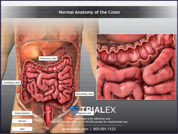
    Colon cancer causes risk factors

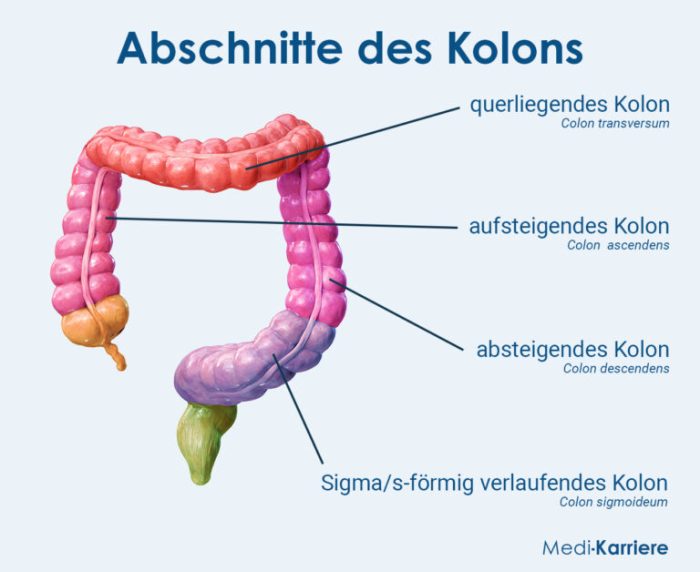
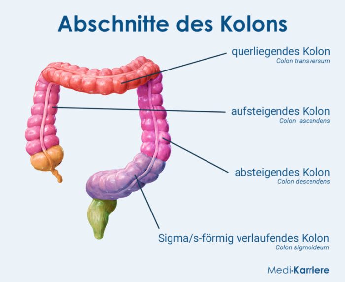
    Colon cancer, a prevalent type of cancer, originates in the large intestine, also known as the colon. This vital part of the digestive system absorbs water and nutrients from food. The disease’s development can significantly impact the body’s ability to function normally, affecting digestion, nutrient absorption, and overall well-being.Colon cancer typically progresses through several stages, each with varying symptoms and levels of severity.

    Early detection is crucial for successful treatment and improved outcomes.

    Stages of Colon Cancer Development

    Understanding the different stages of colon cancer development helps in recognizing potential symptoms and ensuring timely intervention. Each stage signifies a different extent of the disease’s progression.

    • Early Stage: This initial stage often presents few noticeable symptoms, making early detection challenging. Individuals may experience mild changes in bowel habits, such as occasional constipation or diarrhea, or blood in the stool that might be overlooked. Regular screenings are essential for early detection.
    • Intermediate Stage: As the cancer progresses, symptoms might become more pronounced. Changes in bowel habits, such as persistent diarrhea or constipation, along with blood in the stool, abdominal pain, or a feeling of fullness after eating, may appear. The cancer may also have spread to nearby lymph nodes.
    • Advanced Stage: In this stage, the cancer has advanced significantly and spread to other parts of the body, potentially causing more severe symptoms, including significant weight loss, fatigue, and pain. The cancer may have spread to organs such as the liver or lungs.

    Typical Age Range for Diagnoses

    The typical age range for colon cancer diagnoses often falls within a specific timeframe, with risk increasing as individuals age.

    • Most common in older adults: The majority of colon cancer diagnoses occur in individuals aged 50 and older. This reflects the cumulative effects of factors like diet, lifestyle, and genetics that may contribute to the development of the disease over time. For example, a 65-year-old individual may have a higher risk compared to a 45-year-old due to the longer period of potential exposure to risk factors.

    Importance of Early Detection

    Early detection of colon cancer is crucial for successful treatment and improved outcomes.

    • Improved Treatment Outcomes: Early detection allows for more effective treatment options. Cancer at an early stage is often more treatable with less invasive procedures and a higher chance of complete remission. For instance, early detection through screening may lead to surgical removal of the cancerous growth, potentially avoiding more aggressive treatments like chemotherapy.
    • Enhanced Survival Rates: Prompt diagnosis and treatment significantly increase the chances of long-term survival. Studies show that individuals diagnosed with colon cancer in its early stages have higher survival rates compared to those diagnosed at later stages. This underscores the critical role of early detection in improving patient outcomes.

    Genetic Predisposition

    Unveiling the hidden threads of risk, genetic predisposition plays a significant role in the development of colon cancer. While lifestyle factors are crucial, inherited genes can significantly influence an individual’s susceptibility to this disease. Understanding these genetic influences is key to personalized risk assessment and proactive strategies.Inherited mutations in specific genes can disrupt the normal functioning of cells, increasing the risk of uncontrolled growth and tumor formation in the colon.

    These mutations, often passed down through families, can predispose individuals to develop colon cancer at an earlier age or with a higher frequency than those without such genetic predispositions.

    Role of Inherited Genes in Colon Cancer Risk

    Genetic factors significantly contribute to the development of colon cancer. Inherited mutations in genes involved in DNA repair, cell growth, and apoptosis (programmed cell death) can increase the risk of cancerous changes in the colon. These mutations can lead to the accumulation of DNA damage, potentially resulting in the formation of precancerous polyps and eventually cancerous tumors.

    Specific Gene Mutations Increasing Susceptibility

    Several specific gene mutations are associated with an elevated risk of colon cancer. Mutations in genes like APC, MLH1, MSH2, and others are known to disrupt critical cellular processes. For example, mutations in the APC gene are linked to familial adenomatous polyposis (FAP), a condition characterized by the development of numerous precancerous polyps throughout the colon. This condition, if left untreated, can result in a high risk of colon cancer.

    Comparison of Impact of Different Genetic Mutations

    The impact of different genetic mutations on colon cancer risk varies. Some mutations, like those in the APC gene, are strongly associated with a very high risk of developing colon cancer at a young age. Other mutations, such as those in mismatch repair genes (MLH1, MSH2), might increase the risk, but the specific impact depends on the nature of the mutation and other contributing factors.

    While researching colon cancer causes and risk factors, I stumbled upon some interesting connections. It got me thinking about how lifestyle choices can impact health in various ways, like choosing simple home remedies for stuffy nose. For example, maintaining a healthy diet and exercise routine might be just as important for preventing a stuffy nose as it is for reducing colon cancer risk factors.

    So, if you’re looking for some effective home remedies, check out this resource: home remedies for stuffy nose. Ultimately, understanding these interconnected factors can help us make better decisions about our overall well-being, and that includes paying attention to potential colon cancer risk factors.

    This variability necessitates personalized risk assessment to tailor preventative measures.

    Family History as a Risk Factor

    A strong family history of colon cancer is a significant risk factor. Individuals with a first-degree relative (parent, sibling, or child) diagnosed with colon cancer are at a higher risk of developing the disease themselves. This increased risk stems from the shared genetic predisposition within families. The presence of multiple affected relatives, particularly at a younger age, suggests a higher probability of inherited susceptibility.

    Understanding the family history and consulting with a healthcare professional is crucial for assessing personal risk and determining appropriate screening schedules.

    Common Genetic Predispositions and Associated Risks

    Genetic Predisposition Associated Risks Further Considerations
    Familial Adenomatous Polyposis (FAP) High risk of developing numerous precancerous polyps in the colon, leading to a significantly increased risk of colon cancer. Often onset at a younger age. Requires early and frequent screening, often starting in the teens or twenties.
    Lynch Syndrome Increased risk of colon cancer and other cancers, particularly endometrial, ovarian, and stomach cancers. Linked to mutations in mismatch repair genes. Requires close monitoring and potentially prophylactic surgeries.
    Other Genetic Syndromes Certain genetic syndromes, like Peutz-Jeghers syndrome, are associated with a higher risk of colon cancer and other health issues. Requires specialized genetic counseling and monitoring.

    Lifestyle Factors

    Beyond genetics and family history, lifestyle choices play a significant role in colon cancer risk. Dietary habits, physical activity levels, and habits like smoking and alcohol consumption can either protect or increase an individual’s susceptibility to the disease. Understanding these factors is crucial for proactive health management and potentially reducing the risk.

    Dietary Habits

    Dietary patterns have a profound impact on colon health. A diet rich in processed meats, red meats, and foods high in saturated fat is associated with a higher risk of colon cancer. Conversely, a diet rich in fruits, vegetables, and fiber can potentially lower the risk. Fiber, particularly soluble fiber, aids in the digestive process, potentially reducing the amount of time potentially harmful substances remain in contact with the colon lining.

    Crucially, regular consumption of whole grains, legumes, and fruits and vegetables is recommended for a healthier colon.

    Physical Activity

    Regular physical activity is linked to a lower risk of colon cancer. Physical activity helps maintain a healthy weight, and weight management is an important factor in colon cancer prevention. Studies show that individuals who engage in regular moderate-intensity exercise, such as brisk walking, cycling, or swimming, have a lower risk of developing colon cancer. The recommended amount of physical activity for overall health, which can also contribute to reducing colon cancer risk, is at least 150 minutes of moderate-intensity or 75 minutes of vigorous-intensity aerobic activity per week.

    Smoking and Alcohol Consumption

    Both smoking and excessive alcohol consumption are associated with an increased risk of colon cancer. Smoking damages the cells lining the digestive tract, potentially increasing the likelihood of mutations that can lead to cancer. Similarly, excessive alcohol consumption can lead to inflammation and cellular damage, also increasing the risk. Moderation in alcohol consumption is key to minimizing these potential risks.

    Obesity

    Obesity is a significant risk factor for colon cancer. Excess body fat can lead to chronic inflammation in the body, which can damage the colon lining and increase the risk of cancerous growth. Maintaining a healthy weight through a balanced diet and regular exercise is essential in reducing this risk. The association between obesity and colon cancer is well-documented, with research showing a clear correlation.

    Lifestyle Factors and Colon Cancer Risk

    Lifestyle Factor Impact on Colon Cancer Risk
    High intake of processed and red meats Increased risk
    High intake of fruits, vegetables, and fiber Decreased risk
    Regular physical activity Decreased risk
    Smoking Increased risk
    Excessive alcohol consumption Increased risk
    Obesity Increased risk

    Environmental Factors

    Beyond genetics and lifestyle, environmental exposures play a significant role in colon cancer risk. Understanding how these exposures contribute to the development of the disease is crucial for prevention and early detection. Various environmental toxins and occupational hazards can increase a person’s susceptibility to colon cancer, often interacting with genetic and lifestyle factors. Geographic variations in colon cancer rates also highlight the importance of environmental influences.Environmental factors encompass a wide range of exposures, from industrial chemicals to dietary components and even geographical locations.

    These exposures can damage DNA, trigger inflammation, or disrupt cellular processes, ultimately increasing the risk of cancerous transformations in the colon.

    Environmental Toxins and Colon Cancer

    Environmental toxins are substances found in the environment that can harm human health. Some toxins have been linked to an increased risk of colon cancer. These toxins can disrupt cellular processes and increase the likelihood of genetic mutations leading to cancer.

    • Industrial chemicals: Certain industrial chemicals, such as pesticides, solvents, and heavy metals, have been linked to an increased risk of colon cancer. Studies have shown a correlation between exposure to these chemicals and an elevated risk of the disease, particularly in individuals with prolonged or high-level exposure. This suggests a possible causal relationship, although further research is needed to fully understand the mechanisms involved.

    • Air pollution: Exposure to air pollution, particularly particulate matter and certain air pollutants, has been associated with an increased risk of colon cancer. The exact mechanisms are not fully understood, but air pollution may trigger inflammation and oxidative stress, factors implicated in cancer development. This is a significant concern given the widespread nature of air pollution in many urban areas.

      So, colon cancer risk factors – things like family history and a poor diet definitely play a role. But have you ever wondered about the fascinating mechanics behind something seemingly unrelated, like hiccups? Turns out, there are some surprising similarities when exploring the science behind why we get hiccups. Learning more about the complex interplay of nerve signals and muscle contractions, as detailed in this great article about why do we get hiccups , could actually help us better understand the triggers that can increase our chances of developing colon cancer.

      Ultimately, though, understanding risk factors and taking preventative steps are crucial to maintaining a healthy colon.

    • Contaminated water: Water contaminated with certain pollutants, such as industrial waste or agricultural runoff, can increase the risk of colon cancer. The specific compounds in contaminated water and their impact on colon health require further research. This is a critical concern in areas with inadequate water treatment or pollution issues.

    Chemicals and Colon Cancer Risk

    Exposure to certain chemicals can increase the risk of colon cancer. The specific mechanisms through which these chemicals contribute to colon cancer development vary but generally involve DNA damage or disruption of cellular processes.

    • Processed meats: The consumption of processed meats, high in nitrates and nitrites, has been linked to an increased risk of colon cancer. These chemicals can form carcinogenic compounds in the digestive tract. The exact mechanisms and extent of the risk require further research and ongoing studies. This is a critical area of research given the widespread consumption of processed meats in many cultures.

    • Dietary components: Certain dietary components, while not necessarily toxins, can contribute to the risk of colon cancer if consumed in excess. For example, a diet high in red meat may increase the risk of colon cancer, possibly due to the formation of certain compounds during cooking or digestion. However, the exact mechanisms and the level of risk associated with specific dietary components need further investigation.

      This is crucial for developing appropriate dietary guidelines.

    Occupational Hazards and Colon Cancer

    Certain occupational exposures can increase the risk of colon cancer. These exposures may involve prolonged contact with harmful substances or working in environments with high levels of specific chemicals.

    • Exposure to asbestos: Exposure to asbestos, a known carcinogen, is linked to an increased risk of colon cancer. The mechanisms by which asbestos increases colon cancer risk are not fully understood but likely involve DNA damage and inflammation. This is a concern for individuals working in industries where asbestos is present.
    • Exposure to certain chemicals: Workers exposed to certain chemicals in their workplaces, such as benzene or certain pesticides, may have an increased risk of colon cancer. The specific mechanisms and the extent of the increased risk vary based on the type of chemical and the level of exposure. This requires careful monitoring and control measures in workplaces.

    Geographical Variations in Colon Cancer Rates

    Colon cancer rates differ significantly across various geographical regions. Several factors, including environmental exposures and lifestyle choices, may contribute to these differences. These differences underscore the importance of considering environmental factors when evaluating colon cancer risk.

    • Dietary factors: Variations in dietary habits, such as the consumption of processed meats, red meat, and fiber-rich foods, may contribute to these differences. For example, regions with higher rates of red meat consumption may exhibit higher colon cancer rates.
    • Environmental exposures: Variations in environmental exposures, including industrial pollutants and water contamination, may also play a role in the observed differences in colon cancer rates across regions. For instance, regions with higher levels of industrial pollution may experience higher colon cancer rates.

    Medical Conditions

    Beyond lifestyle choices and environmental factors, certain medical conditions significantly increase the risk of developing colon cancer. Understanding these connections is crucial for early detection and preventative measures. These conditions often disrupt the normal cellular processes within the colon, creating an environment conducive to cancerous growth.

    Inflammatory Bowel Disease (IBD)

    Inflammatory bowel disease (IBD) encompasses conditions like Crohn’s disease and ulcerative colitis, characterized by chronic inflammation of the digestive tract. The persistent inflammation associated with IBD can lead to cellular damage and mutations, increasing the risk of colon cancer. This increased risk is directly correlated with the duration and severity of the disease.

    Ulcerative Colitis and Colon Cancer Risk

    Ulcerative colitis, a type of IBD, is particularly linked to an elevated risk of colon cancer. The inflammation in ulcerative colitis primarily affects the colon and rectum, making this region more susceptible to cancerous changes. The longer a person has ulcerative colitis, the higher their risk becomes. Long-term inflammation damages the lining of the colon, potentially leading to dysplasia, a precancerous condition.

    Other Medical Conditions Increasing Colon Cancer Risk

    Several other medical conditions are associated with a heightened risk of colon cancer. These include familial adenomatous polyposis (FAP), a genetic disorder characterized by the development of numerous polyps in the colon, and Peutz-Jeghers syndrome, a genetic condition linked to polyps and other gastrointestinal issues. Certain types of radiation therapy, particularly to the abdomen, can also increase the risk of colon cancer.

    Polyps and Colon Cancer Development

    Colon polyps are abnormal growths in the lining of the colon. While many polyps are benign, some can develop into colon cancer over time. The presence of polyps, especially adenomatous polyps, is a significant risk factor. Regular colonoscopies are vital for identifying and removing these polyps, preventing potential malignant transformations. A high number of polyps or polyps with specific characteristics, like dysplasia, indicate a heightened risk.

    Table of Medical Conditions and Colon Cancer Risk

    Medical Condition Relationship to Colon Cancer Risk
    Inflammatory Bowel Disease (IBD) Chronic inflammation increases risk of cellular damage and mutations.
    Ulcerative Colitis Inflammation primarily in the colon and rectum, increases risk with duration.
    Familial Adenomatous Polyposis (FAP) Genetic predisposition leading to numerous polyps, significantly increasing risk.
    Peutz-Jeghers Syndrome Genetic condition associated with polyps and other GI issues, raising risk.
    Certain Radiation Therapies Specific radiation exposure to the abdomen, may increase risk of colon cancer.
    Colon Polyps (adenomatous) Many polyps are benign, but some can develop into colon cancer.

    Preventive Measures

    Taking proactive steps to reduce your risk of colon cancer is crucial. By understanding the factors that contribute to the disease, you can implement changes that significantly lower your chances of developing it. Early detection is paramount, and preventive measures are vital in achieving this.Adopting a healthy lifestyle, including a balanced diet and regular exercise, along with routine screenings, are essential components of a comprehensive preventive strategy.

    Regular check-ups and screenings play a critical role in early detection, which significantly improves the chances of successful treatment.

    Regular Screening and Diagnostic Tests

    Regular screening is a cornerstone of colon cancer prevention. Early detection through screenings allows for intervention at a stage where treatment is most effective. Different screening methods exist, and the best approach depends on individual factors and medical history. These tests help identify precancerous polyps, which can be removed before they develop into cancer.

    Specific Preventive Actions

    Several actions can be taken to reduce the risk of colon cancer. A balanced diet rich in fruits, vegetables, and whole grains, along with regular physical activity, are fundamental lifestyle choices. Maintaining a healthy weight and avoiding excessive alcohol consumption are also important.

    • Dietary Changes: A diet rich in fiber from fruits, vegetables, and whole grains promotes healthy digestion and can help prevent colon cancer. Limiting processed meats and red meats is also a significant preventative measure.
    • Physical Activity: Regular exercise, such as brisk walking, jogging, or swimming, can help maintain a healthy weight and reduce the risk of colon cancer. Aim for at least 30 minutes of moderate-intensity exercise most days of the week.
    • Maintaining a Healthy Weight: Obesity is a significant risk factor for colon cancer. Maintaining a healthy weight through a balanced diet and regular exercise is crucial in reducing the risk.
    • Limiting Alcohol Consumption: Excessive alcohol consumption has been linked to an increased risk of several cancers, including colon cancer. Moderation is key.

    Importance of a Healthy Diet and Lifestyle, Colon cancer causes risk factors

    A healthy diet and lifestyle are crucial for overall health and reduce the risk of many diseases, including colon cancer. A diet rich in fruits, vegetables, and whole grains, along with regular physical activity, are fundamental elements in preventing colon cancer. These choices support a healthy weight, which is a significant factor in minimizing risk.

    Significance of Regular Check-ups and Screenings

    Regular check-ups and screenings are essential for early detection of colon cancer. Screening tests, such as colonoscopies, can detect precancerous polyps and remove them before they develop into cancer. Early detection significantly improves the chances of successful treatment. This preventive approach saves lives by enabling prompt intervention. By proactively seeking medical attention, individuals can take a crucial step towards reducing their risk.

    • Colon Polyps: These are abnormal growths in the colon that can develop into colon cancer if left untreated. Regular screenings can identify and remove these polyps before they become cancerous. This is a crucial preventive measure.
    • Early Detection: Early detection of colon cancer significantly improves the chances of successful treatment. Regular screenings and check-ups allow for prompt intervention, reducing the risk of advanced stages of the disease.
    • Screening Frequency: The frequency of screening depends on individual risk factors. Consulting a doctor is essential to determine the appropriate screening schedule based on age, family history, and other relevant factors. Regular discussions with your doctor are important for preventative care.

    Risk Factors in Different Demographics

    Colon cancer, while affecting individuals across diverse populations, displays variations in risk factors based on age, ethnicity, socioeconomic status, gender, and other demographic characteristics. Understanding these variations is crucial for targeted prevention and early detection strategies. These differences highlight the complex interplay between genetic predisposition, lifestyle choices, and environmental exposures in shaping colon cancer risk.

    Age-Related Risk Factors

    Age is a significant risk factor for colon cancer. The risk increases substantially with advancing age, primarily due to the accumulation of cellular damage and mutations over time. Individuals over 50 are at heightened risk, and the risk continues to escalate with age. This is partly explained by the increased likelihood of precancerous polyps developing and progressing into cancerous tumors as people age.

    Younger individuals, while less likely to develop the disease, are not immune. Certain genetic syndromes and family histories can significantly increase the risk of colon cancer at younger ages. Early screening and awareness are crucial for all age groups.

    Ethnic and Racial Variations

    Studies suggest that colon cancer risk varies across different ethnic and racial groups. For example, African Americans tend to have a higher incidence and mortality rate compared to other racial groups. This disparity might be attributed to a combination of genetic predisposition, lifestyle factors, access to healthcare, and environmental influences. Other racial and ethnic groups also show variations in risk, highlighting the importance of culturally sensitive screening and prevention strategies.

    Genetic factors, dietary habits, and socioeconomic disparities can all play a role in these variations.

    Socioeconomic Factors and Colon Cancer Risk

    Socioeconomic factors, including income levels, access to healthcare, and educational attainment, can influence colon cancer risk. Lower socioeconomic status often correlates with limited access to preventative screenings, healthy food options, and timely medical care. This can lead to delayed diagnosis and poorer treatment outcomes, thus contributing to higher mortality rates in these groups. Addressing socioeconomic disparities is crucial for improving colon cancer prevention and outcomes across all populations.

    Understanding colon cancer risk factors is crucial for prevention. Diet, genetics, and lifestyle all play a role, but sometimes, advanced screenings like a bone scan for cancer, what is a bone scan for cancer , can reveal if the cancer has spread to the bones. Ultimately, knowing the potential causes and risks of colon cancer empowers us to make healthier choices and seek timely medical attention.

    Gender Differences in Colon Cancer Risk

    While colon cancer can affect both men and women, some studies indicate slight differences in risk factors and outcomes. Men may have a slightly higher incidence of colon cancer compared to women, though this is not a definitive finding across all studies. The reasons for this difference are not fully understood but may be linked to varying hormone levels, lifestyle choices, and other biological factors.

    However, both men and women should prioritize regular screening to detect the disease early.

    Summary Table of Risk Factors Across Demographics

    Demographic Risk Factors
    Age Increased risk with age; accumulation of cellular damage; precancerous polyps; genetic syndromes.
    Ethnicity/Race Higher incidence and mortality in some groups; genetic predisposition; lifestyle factors; access to healthcare; environmental influences.
    Socioeconomic Status Limited access to screenings, healthy food, and timely medical care; delayed diagnosis; poorer outcomes.
    Gender Slight difference in incidence; varying hormone levels; lifestyle choices; biological factors.

    Illustrative Cases: Colon Cancer Causes Risk Factors
    Colon cancer causes risk factors

    Understanding colon cancer requires examining real-world examples to illustrate how various factors contribute to its development. These case studies, while hypothetical, highlight the complex interplay of genetics, lifestyle, environment, and medical conditions in shaping an individual’s risk. They serve as valuable learning tools, emphasizing the importance of preventive measures and early detection.

    Genetic Predisposition

    A 45-year-old woman, Sarah, has a strong family history of colon cancer. Multiple relatives on both sides of her family were diagnosed at relatively young ages. Genetic testing reveals she carries a mutation in the APC gene, a known risk factor for colon cancer. While she maintains a healthy lifestyle, her increased genetic susceptibility necessitates heightened vigilance.

    Regular colonoscopies and close monitoring are crucial to detect any precancerous polyps early, potentially preventing the development of cancer. This illustrates how inherited genetic factors can significantly increase a person’s risk, even in the absence of other risk factors.

    Lifestyle Factors

    Consider Michael, a 55-year-old man who leads a sedentary lifestyle. His diet is high in processed foods, red meat, and saturated fats, while low in fruits and vegetables. He rarely exercises. Over time, Michael develops insulin resistance and obesity. These factors increase his risk of developing colon cancer.

    While genetics might play a role, his lifestyle choices significantly contribute to his elevated risk profile. Making dietary changes and incorporating regular physical activity into his routine would significantly reduce his risk.

    Environmental Exposures

    A 60-year-old farmer, David, has spent his entire career working in fields exposed to pesticides and herbicides. Long-term exposure to these chemicals is linked to an increased risk of colon cancer. While his family history is not strongly suggestive of colon cancer, his occupational exposure significantly raises his risk. Reducing exposure through protective measures, such as wearing appropriate gear, and adopting a healthier diet could mitigate this risk.

    Medical Conditions

    Jane, a 70-year-old woman, has been diagnosed with ulcerative colitis, a chronic inflammatory bowel disease. This condition significantly increases her risk of developing colon cancer. Regular endoscopic surveillance, including colonoscopies, is essential for early detection of precancerous changes. This illustrates how pre-existing medical conditions can act as significant risk factors, requiring proactive management to prevent or detect cancer early.

    Preventive Measures

    A 30-year-old individual, Emily, recognizes her family history of colon cancer. She makes a conscious effort to maintain a healthy lifestyle, including a balanced diet rich in fruits, vegetables, and whole grains, coupled with regular exercise. She participates in routine health screenings, including colonoscopies, at recommended intervals. These proactive steps significantly reduce her risk of developing colon cancer.

    This highlights the potential for individuals to significantly reduce their risk through lifestyle choices and preventive measures.

    Last Point

    In conclusion, colon cancer risk factors are diverse and multifaceted. Genetic predisposition, lifestyle choices, environmental exposures, and underlying medical conditions all contribute to an individual’s risk. While some factors are beyond our control, many lifestyle choices can significantly reduce risk. Early detection through regular screenings is paramount in improving outcomes. By understanding these risk factors, individuals can take proactive steps to protect their health and potentially prevent the development of colon cancer.
  • Weight Loss and Heart Disease Risk Expert Answers

    Weight Loss and Heart Disease Risk Expert Answers

    Ask an expert will losing weight lower my heart disease risk? This question delves into the complex relationship between shedding pounds and reducing cardiovascular risk. We’ll explore the science behind weight loss’s impact on heart health, examining the physiological mechanisms, diverse weight loss methods, and crucial lifestyle factors. Understanding the nuances of this connection is key to making informed decisions about your well-being.

    The discussion will encompass various aspects, from the fundamental correlation between weight and heart disease to the specific considerations for different demographics. We’ll also cover the importance of consulting healthcare professionals, managing underlying medical conditions, and adopting sustainable weight management strategies for long-term heart health.

    Understanding Weight Loss and Heart Disease Risk

    Ask an expert will losing weight lower my heart disease risk

    Losing weight can significantly reduce your risk of heart disease. This is not just about aesthetics; it’s about improving your overall cardiovascular health and potentially extending your lifespan. A healthy weight contributes to a healthier heart, reducing strain on the entire system. This section delves into the crucial connection between weight loss and heart disease prevention, exploring the mechanisms involved and the various weight loss strategies.Weight loss, when achieved through healthy lifestyle modifications, can positively impact various physiological mechanisms that contribute to a reduced risk of heart disease.

    This includes improving blood pressure, cholesterol levels, and blood sugar control. These improvements create a healthier environment for the heart to function effectively, minimizing the risk of developing cardiovascular issues.

    Correlation Between Weight Loss and Reduced Heart Disease Risk

    Weight loss, particularly when focused on visceral fat (the fat surrounding organs), directly correlates with a reduction in cardiovascular risk factors. Visceral fat is metabolically active and releases substances that can damage blood vessels and promote inflammation, which are key factors in the development of heart disease. Losing weight helps to decrease the production of these harmful substances, resulting in a more favorable cardiovascular environment.

    Physiological Mechanisms of Weight Loss Impacting Cardiovascular Health

    Weight loss improves various physiological parameters vital to cardiovascular health. Reduced weight leads to lower blood pressure, as the heart doesn’t have to work as hard to pump blood throughout the body. Improved insulin sensitivity, often a consequence of weight loss, helps regulate blood sugar levels, further decreasing cardiovascular stress. Simultaneously, weight loss can positively influence cholesterol levels, lowering LDL (“bad”) cholesterol and raising HDL (“good”) cholesterol.

    These combined improvements create a protective effect against the development of atherosclerosis (plaque buildup in arteries).

    Role of Excess Weight in Increasing Heart Disease Risk

    Excess weight, particularly abdominal obesity, increases the risk of heart disease through multiple pathways. The increased strain on the heart to pump blood against higher resistance in the vessels, elevated blood pressure, and the production of inflammatory substances by visceral fat are all contributing factors. Furthermore, excess weight is often associated with other conditions like type 2 diabetes and high cholesterol, all of which are significant risk factors for heart disease.

    Types of Heart Disease and Their Risk Factors

    Heart disease encompasses a range of conditions, including coronary artery disease (CAD), heart failure, and arrhythmias. CAD, characterized by the buildup of plaque in the coronary arteries, is a leading cause of heart attacks. Heart failure occurs when the heart cannot pump enough blood to meet the body’s needs. Arrhythmias involve irregular heartbeats, which can lead to various complications.

    Common risk factors for these conditions include high blood pressure, high cholesterol, smoking, diabetes, and a sedentary lifestyle. Obesity is a significant risk factor for all types of heart disease.

    Weight Loss Methods and Their Potential Impact on Heart Health

    Various weight loss methods exist, each with different potential impacts on heart health. A balanced approach, combining dietary changes and regular exercise, is often the most effective strategy for sustainable weight loss and improved cardiovascular health. Diets emphasizing whole foods, fruits, vegetables, and lean protein, along with regular physical activity, promote gradual and healthy weight loss.

    Comparison of Weight Loss Strategies

    Weight Loss Method Potential Impact on Heart Health Effectiveness Rating
    Balanced Diet and Exercise Significant improvements in blood pressure, cholesterol, and insulin sensitivity. High
    Low-Carb Diets Potential for improved blood sugar and cholesterol levels, but potential for negative long-term effects if not balanced. Moderate
    Very Low-Calorie Diets Rapid weight loss, but may have adverse effects on metabolism and cardiovascular health if not medically supervised. Low
    Surgical Procedures Significant weight loss, potentially leading to substantial improvements in cardiovascular risk factors. High (but with risks)

    Factors Influencing Weight Loss and Heart Health Outcomes

    Losing weight can significantly reduce your risk of heart disease, but the journey is multifaceted. It’s not just about the number on the scale; it’s about adopting a holistic approach that considers various lifestyle factors. This involves understanding the complex interplay between diet, exercise, stress, sleep, and specific weight loss programs. This exploration will delve into these crucial components.The path to a healthier heart often involves a profound shift in lifestyle choices.

    Effective weight management isn’t a quick fix; it’s a continuous process of learning, adapting, and consistently making positive changes. Success hinges on understanding the factors that influence both weight loss and heart health. These factors are interconnected and work together to either support or hinder your progress.

    The Role of Diet in Weight Loss and Cardiovascular Health

    A balanced diet is fundamental to both weight loss and heart health. Focus on whole, unprocessed foods like fruits, vegetables, lean proteins, and whole grains. These foods are rich in essential nutrients, fiber, and antioxidants, which support overall health and aid in weight management. Limit processed foods, sugary drinks, and excessive saturated and trans fats. These dietary choices can negatively impact both weight and heart health.

    A registered dietitian can provide personalized dietary guidance.

    Importance of Regular Physical Activity

    Regular physical activity is crucial for achieving and maintaining a healthy weight. Physical activity boosts metabolism, burns calories, and strengthens the cardiovascular system. Aim for at least 150 minutes of moderate-intensity or 75 minutes of vigorous-intensity aerobic activity per week, along with muscle-strengthening activities on two or more days a week. Find activities you enjoy to sustain motivation.

    Consider incorporating activities like brisk walking, swimming, cycling, or dancing.

    Stress and Weight Management

    Chronic stress can significantly impact weight management and heart health. Stress hormones can lead to increased appetite and cravings for unhealthy foods, potentially hindering weight loss efforts. Moreover, stress can negatively impact sleep quality, further complicating the equation. Stress management techniques, such as mindfulness, meditation, yoga, or deep breathing exercises, can help mitigate these effects. Seeking professional support for stress management can be beneficial.

    Sleep Quality and Duration

    Adequate sleep is essential for overall health, including weight management and cardiovascular health. Lack of sleep can disrupt hormones that regulate appetite, leading to increased hunger and cravings for high-calorie foods. Poor sleep quality can also increase inflammation in the body, which is linked to heart disease. Aim for 7-9 hours of quality sleep per night. Establish a regular sleep schedule and create a relaxing bedtime routine.

    Effectiveness of Different Weight Loss Programs

    Various weight loss programs exist, each with its approach and effectiveness. Some programs emphasize diet, others focus on exercise, and some combine both. The most successful programs often incorporate lifestyle changes and provide ongoing support. Consulting with a healthcare professional or registered dietitian is crucial in selecting a program that aligns with your needs and health status.

    So, you’re wondering if shedding those extra pounds will help your heart health? A great question for an expert! While weight loss definitely plays a crucial role in reducing heart disease risk, it’s also important to consider other factors. For example, muscle and joint pain can be a symptom of underlying issues like thyroid disease, which can significantly impact your overall well-being and potentially complicate weight loss efforts.

    If you’re experiencing this, be sure to check out this helpful resource on muscle and joint pain with thyroid disease. Ultimately, consulting a healthcare professional is key to understanding the best approach for your individual situation, whether it’s weight loss or managing other health concerns.

    Comparison of Weight Loss Programs

    Lifestyle Modification Potential Benefits for Heart Health Implementation Tips
    Balanced Diet Reduced risk of high blood pressure, high cholesterol, and type 2 diabetes. Focus on fruits, vegetables, lean proteins, and whole grains. Limit processed foods, sugary drinks, and unhealthy fats.
    Regular Exercise Improved cardiovascular fitness, reduced blood pressure, and increased HDL (“good”) cholesterol. Aim for at least 150 minutes of moderate-intensity or 75 minutes of vigorous-intensity aerobic activity per week. Include muscle-strengthening activities.
    Stress Management Reduced stress hormones, improved sleep quality, and potential weight management support. Practice mindfulness, meditation, yoga, or deep breathing exercises. Seek professional support if needed.
    Adequate Sleep Improved hormone regulation, reduced inflammation, and better overall health. Aim for 7-9 hours of quality sleep per night. Establish a regular sleep schedule and create a relaxing bedtime routine.
    Support System Increased motivation, accountability, and sustained lifestyle changes. Seek support from family, friends, or support groups. Consider professional guidance from a healthcare provider or registered dietitian.

    Specific Populations and Weight Loss

    Weight loss for heart health isn’t a one-size-fits-all approach. Different demographics face unique challenges and opportunities when it comes to shedding pounds and improving cardiovascular well-being. Understanding these variations is crucial for developing effective and sustainable strategies that cater to specific needs and circumstances. Personalized plans are vital for achieving positive outcomes.Effective weight management programs for various groups require careful consideration of individual needs and potential obstacles.

    Strategies must be tailored to address cultural, socioeconomic, and lifestyle factors. This personalized approach recognizes that genetic predispositions and existing health conditions can influence the effectiveness of certain weight loss methods. Acknowledging these factors ensures programs are supportive and promote long-term success.

    Weight Loss in Men

    Men often face different physiological and lifestyle factors compared to women when it comes to weight management. Metabolic differences can lead to varying responses to diet and exercise. Addressing these disparities in weight loss programs is essential for achieving optimal outcomes. Male-specific programs often emphasize strength training and targeted exercise regimes.

    Weight Loss in Women

    Women experience hormonal fluctuations that can impact metabolism and weight regulation. These hormonal changes can make weight loss more challenging and require adjustments in dietary and exercise approaches. Women’s programs often focus on mindful eating and stress management, along with strategies to address potential hormonal imbalances.

    Weight Loss in Older Adults

    Age-related changes in metabolism and muscle mass can affect weight loss effectiveness. Older adults may also have underlying health conditions that influence dietary choices and exercise capacity. Weight loss programs for this demographic need to prioritize safety, gradual progression, and consideration of any existing health issues. These programs often focus on functional fitness and balanced nutrition.

    Weight Loss in Children

    Childhood obesity is a growing concern, and weight loss strategies must be approached with extreme caution. Programs should focus on lifestyle changes rather than rapid weight loss. Encouraging healthy habits, such as balanced nutrition and regular physical activity, is paramount in supporting a child’s overall health and development. Parental involvement and support are crucial components of successful childhood weight management.

    The Role of Genetics in Weight Loss

    Genetic predispositions can influence an individual’s propensity to gain or lose weight. Some people may have a higher predisposition to certain dietary patterns, making it more challenging to lose weight. While genetics play a role, lifestyle factors and environmental influences significantly impact weight loss outcomes. Recognizing genetic predispositions can inform personalized strategies, optimizing the effectiveness of weight loss programs.

    So, you’re wondering if losing weight will lower your heart disease risk? A great question! While shedding pounds definitely has numerous health benefits, it’s also important to consider other factors like managing stress and getting regular exercise. Learning how to treat an itchy scalp can also be beneficial for overall well-being. Many people find relief with remedies like using a gentle shampoo and avoiding harsh products.

    This article delves into the different approaches to managing an itchy scalp. Ultimately, if you’re concerned about your heart health, consulting a doctor is always the best first step in understanding your individual risk factors and how weight loss can play a role.

    Potential Challenges for Weight Loss in Specific Populations

    Weight loss challenges vary across demographic groups. For example, socioeconomic factors can limit access to healthy foods and safe exercise environments. Cultural norms and dietary traditions may present additional obstacles. Recognizing and addressing these specific challenges is essential for creating inclusive and effective weight loss programs.

    Examples of Successful Weight Loss Programs

    Various successful programs cater to diverse populations. Programs like the Mediterranean Diet have proven effective in weight management, particularly for older adults and individuals with heart conditions. Others may use personalized meal plans, exercise programs, and lifestyle coaching tailored to specific needs. Effective programs emphasize sustainability and long-term adherence.

    Table of Potential Differences in Weight Loss Responses

    Population Group Weight Loss Strategies Potential Challenges
    Men Strength training, targeted exercise regimes Metabolic differences, lifestyle factors
    Women Mindful eating, stress management, addressing hormonal imbalances Hormonal fluctuations, lifestyle factors
    Older Adults Functional fitness, balanced nutrition, gradual progression Age-related changes, underlying health conditions
    Children Lifestyle changes, balanced nutrition, regular physical activity, parental involvement Cultural norms, access to healthy foods, safety concerns

    Medical Considerations and Weight Loss: Ask An Expert Will Losing Weight Lower My Heart Disease Risk

    Ask an expert will losing weight lower my heart disease risk

    Before embarking on any weight loss journey, it’s crucial to understand the potential interplay between your health conditions and your approach. A personalized plan, developed in consultation with a healthcare professional, is essential for a safe and effective weight loss strategy. This is particularly important if you have underlying medical conditions that can affect your metabolism, nutrient absorption, or response to different weight loss methods.

    I’ve been getting a lot of questions lately about whether losing weight will lower your heart disease risk. It’s a complex issue, and while shedding pounds definitely has a positive impact on overall health, the specifics depend on individual circumstances. For example, if you have a tooth cracked in half but aren’t experiencing pain, it’s crucial to see a dentist immediately as a cracked tooth can lead to serious issues.

    This can sometimes be overlooked. Still, asking an expert about the specific impact of weight loss on heart disease risk is always a good idea. The link tooth cracked in half no pain is a good starting point for further research, and remember, a healthy lifestyle is key for a healthy heart!

    Importance of Professional Consultation

    Consulting a healthcare professional, such as a doctor or registered dietitian, is paramount before starting any weight loss program. They can assess your overall health, identify any underlying medical conditions, and tailor a plan that aligns with your individual needs and health goals. This personalized approach minimizes potential risks and maximizes the chances of success.

    Medical Conditions and Weight Management

    Certain medical conditions can significantly impact weight management and heart health. For example, diabetes, hypothyroidism, and other hormonal imbalances can affect metabolism and make weight loss more challenging. A healthcare professional can identify these conditions and develop strategies to manage them alongside your weight loss goals.

    Medications and Weight Loss

    Some medications can influence weight. Certain medications for conditions like depression, anxiety, or high blood pressure might have side effects that include changes in appetite or metabolism. A healthcare provider can evaluate the potential impact of any medications you are taking on your weight loss journey and help you adjust your strategy as needed. They can also recommend alternative medications or adjustments to your dosage if necessary.

    Risks of Rapid Weight Loss

    Rapid weight loss can be detrimental to your health. It can lead to nutrient deficiencies, muscle loss, and even electrolyte imbalances. A gradual and sustainable approach, guided by a healthcare professional, is crucial to prevent these potential complications. It’s important to prioritize a balanced approach over quick fixes. For example, extremely restrictive diets can lead to nutrient deficiencies and can be harmful.

    Tailoring Weight Loss Strategies to Individual Needs

    Weight loss strategies should be tailored to individual medical needs. A person with diabetes, for instance, may need a different approach compared to someone with no underlying health conditions. A healthcare professional can determine the most suitable dietary plan, exercise regimen, and overall lifestyle adjustments to meet specific medical needs.

    Potential Medication-Weight Loss Interactions

    Medication Potential Impact on Weight Loss Considerations
    Antidepressants (some types) May increase appetite or affect metabolism Consult your doctor about potential alternatives or adjustments to your dosage.
    Steroids Often lead to weight gain Discuss with your doctor strategies to mitigate weight gain while managing your condition.
    Diabetes medications May affect appetite and metabolism Your doctor can adjust your medication or recommend additional lifestyle changes.
    Thyroid medications May influence metabolism and appetite Adjusting medication doses or dietary changes may be necessary.
    Blood pressure medications Some may lead to fluid retention or appetite changes Consult your doctor for alternative options if necessary.

    Long-Term Maintenance and Weight Management

    Staying at a healthy weight isn’t a sprint; it’s a marathon. Successfully shedding pounds is a significant achievement, but the real challenge lies in maintaining those results for the long haul. This requires a shift in mindset, from a temporary diet to a lifelong commitment to healthy habits. This section will explore the crucial elements of long-term weight management, empowering you to make sustainable choices for your heart health.

    Sustainable Weight Loss Strategies for Long-Term Heart Health, Ask an expert will losing weight lower my heart disease risk

    Maintaining a healthy weight is not about restricting yourself but about making conscious, sustainable lifestyle changes. These changes are more likely to be successful and lead to long-term heart health when approached as an integrated lifestyle rather than a temporary diet. This encompasses a balanced approach that incorporates all facets of your life, from food choices to physical activity and stress management.

    Developing Healthy Habits for Weight Maintenance

    Developing sustainable habits is crucial for weight maintenance. It’s not about perfection, but about consistency. Focus on these key areas:

    • Balanced Diet: Prioritize whole, unprocessed foods like fruits, vegetables, lean proteins, and whole grains. Limit processed foods, sugary drinks, and excessive saturated and unhealthy fats. Pay attention to portion sizes to avoid overeating, even with healthy foods. A registered dietitian can offer personalized dietary guidance.
    • Regular Physical Activity: Incorporate physical activity into your daily routine. Aim for at least 150 minutes of moderate-intensity or 75 minutes of vigorous-intensity aerobic activity per week, along with muscle-strengthening activities twice a week. Find activities you enjoy, whether it’s dancing, hiking, swimming, or team sports. Consistency is key, not just intensity.
    • Stress Management Techniques: Chronic stress can lead to unhealthy coping mechanisms, such as emotional eating. Incorporate stress-reducing activities such as meditation, yoga, deep breathing exercises, or spending time in nature. Stress management is crucial for maintaining a healthy weight and overall well-being.

    The Role of Support Systems in Weight Management

    Weight management is often more successful when you have a supportive network. This could include family members, friends, support groups, or healthcare professionals.

    • Accountability Partners: Sharing your goals with a friend or family member can provide accountability and motivation. Regular check-ins and shared celebrations of progress can significantly boost your commitment.
    • Support Groups: Connecting with others facing similar challenges can offer encouragement, shared experiences, and coping strategies. Online or in-person support groups can provide a valuable network of peers.
    • Healthcare Professionals: Consult your doctor or a registered dietitian for personalized advice and support. They can assess your individual needs and provide tailored recommendations for sustainable weight management.

    Preventing Weight Regain After Achieving Goals

    Maintaining weight loss requires ongoing effort. Strategies to prevent regain include:

    • Consistency: Don’t fall into the trap of “all or nothing.” Consistency in your healthy habits is more effective than occasional extreme measures. Maintain a consistent approach to eating and exercise, adjusting as needed.
    • Addressing Plateaus: Weight loss plateaus are common. Consult your doctor or a registered dietitian to identify potential underlying causes and adjust your plan. Review your current habits and see where adjustments are needed.
    • Long-Term Mindset Shift: Focus on adopting a healthy lifestyle rather than a temporary diet. Weight management is a journey, not a destination. Focus on adopting long-term, sustainable habits.

    Potential Challenges and Solutions for Long-Term Weight Management

    Maintaining a healthy weight is a lifelong commitment, not a quick fix. Some common challenges include:

    • Social Gatherings and Temptations: Learn strategies for managing social situations and temptation. Prepare healthy options in advance or choose smaller portions of tempting foods. Focus on moderation rather than deprivation.
    • Lifestyle Changes and Time Constraints: Make small, manageable changes to fit your lifestyle. Prioritize your well-being by incorporating healthy choices into your daily routine. Be realistic about your time and resources.
    • Emotional Eating: Develop coping mechanisms for emotional eating. Engage in stress-reducing activities or talk to a therapist to address underlying issues.

    Tools and Resources for Weight Management Support

    Numerous tools and resources are available to support your weight management journey.

    • Apps: Many apps offer personalized meal planning, exercise tracking, and support communities.
    • Online Communities: Online forums and social media groups can provide support and motivation from like-minded individuals.
    • Registered Dietitians: Registered dietitians can provide personalized nutrition plans and support.

    Final Wrap-Up

    Ultimately, the answer to “ask an expert will losing weight lower my heart disease risk” is a resounding yes, but not without careful consideration. Weight loss, when approached responsibly and sustainably, can significantly reduce heart disease risk. However, it’s vital to tailor your approach to individual needs, seek professional guidance, and prioritize long-term lifestyle changes for optimal results.

    By understanding the complexities of weight loss and heart health, you can empower yourself to make informed choices and take proactive steps toward a healthier future.

  • Stroke Level Blood Pressure A Deep Dive

    Stroke Level Blood Pressure A Deep Dive

    Stroke level blood pressure sets the stage for this enthralling narrative, offering readers a glimpse into the intricate relationship between high blood pressure and stroke risk. We’ll explore the different types of strokes, the factors that influence blood pressure elevation, and the crucial steps in diagnosing, monitoring, managing, and preventing stroke through blood pressure control. Understanding stroke-level blood pressure is vital for anyone concerned about their health or that of a loved one.

    This comprehensive guide will cover everything from defining stroke-level blood pressure readings and the mechanisms behind its association with stroke to the various factors that contribute to elevated blood pressure. We’ll also examine diagnosis and monitoring techniques, effective management strategies, and preventive measures to reduce the risk of stroke. This information aims to empower individuals with the knowledge and tools needed to take proactive steps toward maintaining healthy blood pressure and overall well-being.

    Defining Stroke Level Blood Pressure

    Stroke level blood pressure

    High blood pressure, or hypertension, is a significant risk factor for stroke. Understanding the relationship between blood pressure and stroke is crucial for preventative measures and timely intervention. This discussion delves into defining stroke-level blood pressure, the mechanisms behind its contribution to stroke, and the different types of strokes.High blood pressure, when consistently elevated, damages the delicate blood vessels in the brain, making them more susceptible to rupture or blockage.

    This increased risk ultimately contributes to the occurrence of stroke. A clear understanding of the connection between blood pressure and stroke is essential for individuals at risk and healthcare professionals.

    Defining Stroke-Level Blood Pressure Readings

    Stroke-level blood pressure signifies a blood pressure reading that is significantly elevated and carries an immediate and substantial risk of a stroke. While there’s no single definitive threshold, readings consistently exceeding 180/120 mmHg are generally considered elevated and warrant immediate medical attention. It’s important to note that individual responses to blood pressure vary, and a reading within this range might not trigger a stroke in every instance.

    However, it signals a high risk and the need for immediate medical intervention. Factors like pre-existing conditions, lifestyle, and other concurrent health issues also play a role in determining the risk.

    Mechanisms of High Blood Pressure and Stroke

    High blood pressure exerts continuous pressure on the blood vessel walls. Over time, this sustained pressure can weaken the vessel walls, making them prone to rupture or damage. Furthermore, the increased pressure can cause the formation of fatty deposits, or plaques, within the arteries, eventually leading to blockages. These blockages can restrict blood flow to the brain, leading to an ischemic stroke.

    Alternatively, the weakened vessel walls can rupture, causing a hemorrhagic stroke. In both cases, the outcome is a disruption in the brain’s blood supply, leading to cell death and neurological impairment.

    High blood pressure, even reaching stroke level, can be a serious concern. Making smart food choices, like swapping out full-fat dairy products for healthier alternatives, can really help. For example, exploring healthy alternatives to full fat dairy products can significantly lower your risk factors, potentially preventing dangerous blood pressure spikes. Ultimately, managing stroke level blood pressure involves a holistic approach, and dietary changes are a crucial part of that strategy.

    Types of Stroke and Blood Pressure Relation

    Strokes are broadly categorized into ischemic and hemorrhagic types. The relationship between blood pressure and these types varies.

    Comparison of Blood Pressure Levels Associated with Different Stroke Types

    Stroke Type Typical Blood Pressure Risk Factors
    Ischemic Stroke Often elevated, but can occur at lower levels in individuals with pre-existing vascular conditions. Readings above 140/90 mmHg are a significant risk factor. High cholesterol, smoking, diabetes, lack of physical activity, obesity, family history of stroke, and previous heart attacks are common risk factors.
    Hemorrhagic Stroke Frequently, but not always, associated with very high blood pressure readings (often exceeding 200/120 mmHg). However, even with elevated readings, some individuals experience hemorrhagic strokes with less extreme blood pressure. High blood pressure is a major risk factor. Other risk factors include aneurysm, arteriovenous malformations (AVMs), blood clotting disorders, and use of blood-thinning medications.

    Factors Influencing Stroke Level Blood Pressure

    High blood pressure, or hypertension, is a significant risk factor for stroke. Understanding the factors that contribute to elevated blood pressure is crucial for prevention and management. This knowledge empowers individuals to make informed lifestyle choices and take proactive steps towards a healthier cardiovascular system.Elevated blood pressure often develops gradually, making it a silent threat. Identifying the various contributing factors can help individuals recognize potential issues and take necessary steps to mitigate their risk.

    Many factors are interconnected and can influence each other, making a holistic approach to blood pressure management essential.

    Lifestyle Choices and Blood Pressure, Stroke level blood pressure

    Lifestyle choices play a pivotal role in maintaining healthy blood pressure levels. A balanced diet, regular physical activity, and stress management techniques are crucial components of a comprehensive approach to cardiovascular health. Adopting these practices can significantly reduce the risk of developing high blood pressure and its associated complications, including stroke.

    • Diet: A diet rich in processed foods, saturated fats, and excessive sodium can contribute to high blood pressure. Conversely, a diet rich in fruits, vegetables, and whole grains can help maintain healthy blood pressure levels. Dietary patterns influence blood pressure by impacting sodium intake, blood vessel health, and overall body weight.
    • Physical Activity: Regular physical activity helps to lower blood pressure and improve cardiovascular health. Consistent exercise strengthens the heart, improves blood flow, and helps to maintain a healthy weight, all of which contribute to healthy blood pressure.
    • Stress Management: Chronic stress can elevate blood pressure. Stress management techniques, such as meditation, yoga, and deep breathing exercises, can help to lower blood pressure and improve overall well-being. Prolonged stress can increase adrenaline and cortisol levels, which can constrict blood vessels and raise blood pressure.

    Risk Factors and Their Impact

    Various factors, both controllable and uncontrollable, can influence blood pressure. Understanding the impact of each factor allows individuals to prioritize preventative measures and adapt their lifestyle choices accordingly.

    • Age: The risk of high blood pressure increases with age. This is often due to age-related changes in blood vessel elasticity and the body’s ability to regulate blood pressure. Older adults are more likely to have high blood pressure, which can affect blood vessels, leading to increased risk of stroke.
    • Genetics: A family history of high blood pressure significantly increases an individual’s risk. Genetic predisposition can influence blood pressure regulation, making some individuals more susceptible to developing high blood pressure than others. Understanding family history is essential for identifying potential risk and taking preventative measures early.
    • Obesity: Excess weight puts extra strain on the cardiovascular system, leading to elevated blood pressure. Maintaining a healthy weight is crucial for preventing high blood pressure and its associated risks. Carrying excess weight puts extra strain on the heart and blood vessels, leading to higher blood pressure.

    Comparison of Risk Factors

    Comparing the effects of different risk factors reveals varying levels of influence on blood pressure. For example, while obesity and smoking directly affect blood vessel health and heart function, age-related changes are less directly controllable. This underscores the importance of adopting a holistic approach to blood pressure management, addressing both modifiable and non-modifiable risk factors. Addressing multiple risk factors simultaneously often yields the most substantial benefits.

    Common Risk Factors and Their Impact

    Risk Factor Impact on Blood Pressure Preventive Measures
    Smoking Smoking damages blood vessels, constricts them, and increases blood clotting, leading to elevated blood pressure. Nicotine in cigarettes also increases heart rate, further contributing to higher blood pressure. Quitting smoking is crucial for reducing blood pressure and improving cardiovascular health. Nicotine replacement therapy, counseling, and support groups can assist in quitting.
    Obesity Excess weight puts strain on the cardiovascular system, increasing blood volume and peripheral resistance, leading to elevated blood pressure. Obesity often leads to other health problems, compounding the risk of high blood pressure. Maintaining a healthy weight through a balanced diet and regular exercise is crucial. Consult a doctor or registered dietitian for personalized guidance.
    Lack of Exercise Lack of regular physical activity can lead to a weakened heart, impaired blood circulation, and a reduced ability to regulate blood pressure. Sedentary lifestyles contribute to unhealthy weight gain and increase the risk of high blood pressure. Regular physical activity, even moderate-intensity exercise, can significantly improve blood pressure and overall cardiovascular health.

    Diagnosing and Monitoring Blood Pressure

    Understanding and managing blood pressure is crucial for preventing stroke and other cardiovascular complications. Accurate diagnosis and consistent monitoring are vital for effective treatment strategies. This section will delve into the methods used to diagnose high blood pressure, detail procedures for long-term monitoring, and explore the tools and equipment employed for precise measurements.Diagnosing high blood pressure often involves a series of steps, starting with a thorough evaluation of your overall health.

    A doctor will consider your medical history, lifestyle factors, and any other conditions that may contribute to elevated blood pressure.

    Methods for Diagnosing High Blood Pressure

    A diagnosis of high blood pressure typically involves repeated measurements over time. This process aims to confirm persistent elevated readings beyond a single, potentially anomalous measurement. Doctors often utilize multiple measurements taken at different times to ensure the accuracy and reliability of the diagnosis. This helps differentiate occasional fluctuations from consistent hypertension.

    Procedures for Monitoring Blood Pressure Over Time

    Regular monitoring of blood pressure is essential for tracking its response to treatment and preventing potential complications. Monitoring methods involve recording blood pressure readings at scheduled intervals. This allows doctors to track trends and adjust treatment plans as needed. This long-term approach is critical for maintaining health and well-being.

    Tools and Equipment for Blood Pressure Measurement

    Accurate blood pressure measurement relies on appropriate tools and equipment. Commonly used tools include manual sphygmomanometers and automatic blood pressure monitors. Each method has unique characteristics that influence the accuracy and efficiency of the measurement process.

    Step-by-Step Procedure for Accurate Blood Pressure Measurement

    This procedure Artikels a standard approach for obtaining accurate blood pressure measurements. Following these steps can significantly improve the reliability of the results:

    1. Preparation: Ensure the patient is relaxed and seated comfortably for at least 5 minutes before the measurement. Avoid caffeine, smoking, and strenuous activity immediately before the measurement.
    2. Equipment Selection: Choose the appropriate device based on the situation. Manual sphygmomanometers are suitable for trained professionals, while automatic monitors are convenient for home use.
    3. Positioning: The patient’s arm should be supported at heart level. Ensure the cuff is positioned correctly, centered over the brachial artery, and snug but not constricting.
    4. Measurement Procedure: Follow the specific instructions for the device used. For manual sphygmomanometers, listen carefully for the sounds of Korotkoff’s sounds. For automatic monitors, allow the device to complete the measurement cycle.
    5. Record Keeping: Document the reading and the date and time of measurement. Record any conditions that might have influenced the measurement, such as stress or recent activity.

    Comparison of Blood Pressure Monitoring Devices

    The following table summarizes the key characteristics of different blood pressure monitoring devices:

    Device Measurement Method Accuracy
    Manual Sphygmomanometer Auscultation (listening to Korotkoff sounds) Highly accurate when used correctly by trained personnel
    Automatic Blood Pressure Monitor Oscillometric (detecting pressure changes) Generally accurate for home use when properly calibrated and used correctly

    Management and Treatment Strategies: Stroke Level Blood Pressure

    Managing stroke-level blood pressure requires a multifaceted approach. Simply put, controlling high blood pressure is crucial for preventing further damage and improving the patient’s overall well-being. This involves a combination of lifestyle adjustments, medication, and potentially more aggressive interventions. The key is to work closely with healthcare professionals to develop a personalized plan that effectively targets the individual’s needs.Effective management of elevated blood pressure involves a coordinated effort.

    It requires a deep understanding of the patient’s specific circumstances and a comprehensive approach encompassing both lifestyle changes and medical interventions. Medication is often necessary, but it should be complemented by lifestyle modifications to achieve optimal results.

    Strategies for Managing High Blood Pressure

    Strategies for managing high blood pressure are multi-pronged. Effective management involves not only medication but also significant lifestyle modifications. These are fundamental components in reducing blood pressure and improving overall health. A proactive approach is key to prevent further complications and promote recovery.

    • Dietary Modifications: A balanced diet rich in fruits, vegetables, and whole grains, along with reduced intake of saturated and trans fats, sodium, and added sugars, is essential. Limiting alcohol consumption is also critical. For example, a diet rich in potassium-rich foods like bananas, avocados, and leafy greens can help regulate blood pressure. Reducing sodium intake is also a key aspect of this strategy.

    • Physical Activity: Regular exercise, such as brisk walking, jogging, swimming, or cycling, can significantly lower blood pressure. The American Heart Association recommends at least 150 minutes of moderate-intensity or 75 minutes of vigorous-intensity aerobic activity per week, along with muscle-strengthening activities at least twice a week. Consistency is key; even short bursts of activity throughout the day can contribute to a healthier blood pressure.

    • Stress Management: Chronic stress can elevate blood pressure. Techniques like yoga, meditation, deep breathing exercises, or spending time in nature can help manage stress levels. Stress management is often overlooked but plays a vital role in overall health and blood pressure regulation.

    Role of Medication in Controlling Blood Pressure

    Medication plays a critical role in managing blood pressure, especially in cases of stroke-level readings. Various classes of antihypertensive medications are available, each with its own mechanism of action and potential side effects. A healthcare professional can determine the most appropriate medication based on individual factors.

    High blood pressure, even reaching stroke levels, can be a serious concern. It’s crucial to understand the long-term effects of such pressure on your overall health, but it’s also important to look at other potential factors that could impact your lifespan. For example, conditions like arthritis can sometimes affect your quality of life and even your life expectancy, as detailed in this informative article on can arthritis shorten your lifespan.

    Ultimately, maintaining healthy blood pressure is key for a longer, healthier life.

    • Different Classes of Medications: Common classes of blood pressure medications include diuretics, beta-blockers, ACE inhibitors, ARBs, and calcium channel blockers. Each class targets different aspects of the body’s blood pressure regulation mechanisms. Understanding the specific mechanism of action of a given medication is crucial for effective treatment and monitoring.
    • Individualized Approach: The selection of medication and dosage is tailored to the individual patient’s specific needs, including age, overall health, other medical conditions, and potential drug interactions. A healthcare professional will monitor the effectiveness and side effects of the medication and adjust the dosage as needed.

    Lifestyle Modifications

    Lifestyle modifications are often the first line of defense in managing high blood pressure. They complement medication and contribute to long-term blood pressure control. These modifications have a lasting positive impact on overall health.

    • Weight Management: Maintaining a healthy weight can significantly lower blood pressure. Even a small amount of weight loss can make a difference. Consult with a registered dietitian or healthcare professional for personalized guidance.
    • Smoking Cessation: Smoking is a major risk factor for high blood pressure and cardiovascular disease. Quitting smoking is crucial for improving overall health and blood pressure control. Consider support groups or counseling programs.
    • Alcohol Limitation: Excessive alcohol consumption can raise blood pressure. Limiting alcohol intake to recommended levels can significantly contribute to better blood pressure management.

    Treatment Options for Stroke-Level Blood Pressure

    Stroke-level blood pressure requires immediate and aggressive intervention. Prompt medical attention is critical to mitigate the risk of further complications and potential stroke. The specific treatment will depend on the underlying cause of the elevated blood pressure.

    • Intensive Blood Pressure Management: In cases of hypertensive emergencies, rapid reduction of blood pressure may be necessary to prevent further damage to vital organs. This often involves intravenous medications administered in a hospital setting.
    • Emergency Medical Care: Prompt consultation with emergency medical services or a healthcare professional is crucial for stroke-level blood pressure. Early diagnosis and treatment are essential to prevent further complications.

    Creating a Personalized Blood Pressure Management Plan

    A personalized blood pressure management plan should be developed in collaboration with a healthcare professional. This plan should consider the patient’s individual circumstances, including medical history, lifestyle, and medication needs.

    • Regular Monitoring: Regular monitoring of blood pressure is essential for tracking progress and adjusting the management plan as needed. This helps to understand how the treatment is affecting blood pressure.
    • Patient Education: Educating the patient about their condition, the importance of adherence to the plan, and the potential risks of uncontrolled blood pressure is crucial for successful management.

    Preventing Stroke through Blood Pressure Control

    Stroke level blood pressure

    Maintaining healthy blood pressure is crucial in preventing strokes. High blood pressure, or hypertension, puts a strain on blood vessels, increasing the risk of damage and the formation of blood clots. Proactive management of blood pressure levels can significantly reduce the likelihood of experiencing a stroke. This proactive approach involves lifestyle modifications and consistent medical monitoring.High blood pressure often has no noticeable symptoms in its early stages.

    Therefore, regular check-ups and monitoring are essential for early detection and timely intervention. By understanding the contributing factors and adopting preventive measures, individuals can significantly lower their risk of stroke. This includes focusing on a healthy lifestyle, which can help maintain a healthy blood pressure and overall well-being.

    Importance of Maintaining Healthy Blood Pressure

    High blood pressure, if left uncontrolled, can lead to serious health complications, including stroke. High blood pressure damages blood vessels, making them more susceptible to blockages or ruptures. This increased risk of clots or aneurysms directly contributes to the development of a stroke. Early detection and management of high blood pressure are critical in preventing long-term health problems.

    Preventive Measures to Reduce Stroke Risk

    Implementing preventive measures can significantly lower the risk of stroke. A proactive approach that includes a combination of lifestyle modifications and regular health checks is crucial.

    • Regular Exercise: Physical activity helps regulate blood pressure by improving cardiovascular health and reducing stress. Aim for at least 150 minutes of moderate-intensity aerobic exercise per week, such as brisk walking, jogging, or cycling. Strength training exercises are also beneficial for overall health and blood pressure management.
    • Healthy Diet: A balanced diet rich in fruits, vegetables, and whole grains, and low in saturated and trans fats, sodium, and added sugars, plays a significant role in maintaining healthy blood pressure. Reducing salt intake is particularly important, as excess sodium can cause the body to retain water, further elevating blood pressure.
    • Stress Management: Chronic stress can elevate blood pressure. Practicing stress-reducing techniques, such as yoga, meditation, or deep breathing exercises, can help regulate blood pressure and improve overall well-being. Stress management is crucial for long-term health and blood pressure control.
    • Maintaining a Healthy Weight: Obesity is a significant risk factor for high blood pressure. Maintaining a healthy weight through a combination of diet and exercise can help lower blood pressure and reduce the risk of stroke.
    • Limiting Alcohol Consumption: Excessive alcohol intake can elevate blood pressure. Moderation in alcohol consumption is crucial for maintaining healthy blood pressure levels.

    Recommendations for Regular Health Check-ups and Blood Pressure Monitoring

    Regular health check-ups and blood pressure monitoring are essential for early detection and management of high blood pressure. Routine check-ups allow healthcare providers to assess overall health, identify potential risks, and recommend appropriate interventions. Monitoring blood pressure at home using a home blood pressure monitor can help track progress and ensure consistency in managing blood pressure.

    • Schedule regular appointments with your doctor: Regular check-ups are essential for preventative care, early detection of potential problems, and timely intervention. This is particularly crucial for individuals with a family history of high blood pressure or other cardiovascular conditions.
    • Utilize home blood pressure monitors: Monitoring blood pressure at home can provide a more comprehensive understanding of blood pressure fluctuations throughout the day. This data can be shared with healthcare providers to help tailor treatment plans.

    Long-Term Benefits of Blood Pressure Control

    Controlling blood pressure offers numerous long-term benefits beyond stroke prevention. It reduces the risk of heart disease, kidney disease, and other serious health conditions. Maintaining healthy blood pressure contributes to a higher quality of life and overall well-being.

    High blood pressure, even reaching stroke level, can be a serious concern. Understanding the various symptoms is crucial, and sometimes those symptoms can be easily confused with other conditions. For example, when dealing with a child, distinguishing between a drool rash and hand-foot-and-mouth disease can be tricky. Luckily, resources like drool rash vs hand foot mouth provide valuable insights.

    Ultimately, if you suspect stroke level blood pressure, seeking immediate medical attention is paramount.

    Controlling blood pressure can significantly reduce the risk of stroke and other cardiovascular diseases.

    Preventive Measures Summary

    Preventive Measure Impact on Blood Pressure Benefits
    Regular Exercise Reduces blood pressure, strengthens heart and blood vessels Improved cardiovascular health, reduced stress
    Healthy Diet Reduces sodium intake, improves overall nutrition Lowered blood pressure, improved overall health
    Stress Management Reduces stress hormones, improves relaxation Lowered blood pressure, reduced risk of cardiovascular issues
    Maintaining a Healthy Weight Reduces strain on blood vessels, improves cardiovascular function Lowered blood pressure, reduced risk of stroke and other diseases
    Limiting Alcohol Consumption Reduces elevated blood pressure Lowered blood pressure, improved overall health

    Blood Pressure and Stroke in Specific Populations

    High blood pressure, or hypertension, significantly increases the risk of stroke, and the impact varies across different demographic groups. Understanding these variations is crucial for developing targeted prevention and management strategies. This section delves into the unique challenges and considerations for managing blood pressure in specific populations, highlighting the importance of culturally sensitive approaches.Stroke-level blood pressure, while a critical risk factor for all, presents distinct challenges for certain groups.

    Factors like socioeconomic status, access to healthcare, and underlying health conditions can significantly influence blood pressure control and outcomes. Moreover, cultural norms and beliefs about health and treatment can impact adherence to prescribed regimens.

    Impact on Different Demographic Groups

    Various demographic factors, such as age, race, ethnicity, socioeconomic status, and geographic location, contribute to variations in blood pressure prevalence and management. For example, certain racial and ethnic groups often experience higher rates of hypertension and associated health complications. Understanding these disparities is critical for targeted interventions.

    Populations at Higher Risk

    Certain populations are inherently more susceptible to stroke due to blood pressure issues. This includes individuals with a family history of stroke or hypertension, those with pre-existing medical conditions like diabetes or kidney disease, and individuals with certain genetic predispositions. Furthermore, those with limited access to quality healthcare or healthy food options are also at greater risk.

    Unique Challenges and Considerations

    Managing blood pressure effectively in diverse populations requires recognizing the unique challenges each group faces. Language barriers, cultural beliefs about medication, and lack of awareness about stroke risk factors can hinder adherence to treatment plans. Moreover, socioeconomic factors, such as limited access to healthy foods and regular exercise, can exacerbate the problem.

    Culturally Sensitive Approaches

    Effective blood pressure management strategies must be culturally sensitive and tailored to the specific needs of each population. This includes employing culturally competent healthcare providers, using community health workers who understand local norms and languages, and educating patients about the importance of regular check-ups and adherence to medication regimens. In addition, promoting health literacy and understanding the significance of early detection and intervention is vital.

    Blood Pressure Trends Across Demographics

    Studies reveal varying blood pressure trends across different demographics. For instance, hypertension prevalence rates often differ significantly between racial and ethnic groups, with some groups experiencing higher rates of high blood pressure than others. These variations underscore the importance of tailored prevention and management strategies.

    Examples of Specific Populations

    Consider the following examples:

    • African Americans: Often exhibit higher rates of hypertension and stroke compared to other racial groups. This disparity may be attributed to a combination of genetic factors, socioeconomic disparities, and environmental influences. Cultural perceptions about medication and healthcare practices also play a role.
    • Individuals with Low Socioeconomic Status: Limited access to healthy food options, regular exercise opportunities, and quality healthcare can contribute to higher blood pressure levels and increase stroke risk. This highlights the importance of addressing socioeconomic factors alongside other risk factors.
    • Elderly Individuals: Age-related physiological changes can affect blood pressure regulation. Chronic diseases and co-morbidities frequently increase blood pressure and stroke risk in older adults.

    Illustrative Case Studies

    Understanding stroke-level blood pressure requires looking at real-world examples. These case studies highlight the impact of uncontrolled hypertension on patients, emphasizing the importance of early diagnosis and effective management strategies. They demonstrate how blood pressure control directly affects recovery and quality of life.This section presents a fictional case study to illustrate the complexities and nuances of stroke-level blood pressure.

    While fictional, the details reflect typical medical scenarios and the challenges faced by patients.

    Patient Profile and Initial Symptoms

    A 65-year-old male patient, Mr. Smith, presented with a history of hypertension, hyperlipidemia, and a family history of stroke. He had been experiencing progressively worsening headaches and dizziness for several weeks. These symptoms were accompanied by numbness and weakness in his left arm and leg, particularly noticeable upon exertion. He reported confusion and difficulty speaking clearly.

    These symptoms, while initially subtle, progressively worsened over a 24-hour period.

    Diagnosis and Treatment

    Mr. Smith was admitted to the hospital. Initial blood pressure readings consistently exceeded 220/120 mmHg. A thorough neurological examination, coupled with diagnostic imaging (CT scan or MRI), confirmed a suspected ischemic stroke. The elevated blood pressure was a critical factor in the diagnostic process, indicating a high risk of further damage.

    Immediate treatment involved aggressive blood pressure reduction, employing a combination of intravenous medications. Close monitoring of vital signs and neurological function was crucial throughout the treatment process. The goal was to lower blood pressure gradually while minimizing the risk of complications.

    Role of Blood Pressure Control in Recovery

    Blood pressure control played a pivotal role in Mr. Smith’s recovery. By rapidly reducing the elevated pressure, the medical team aimed to limit further brain damage. Strict adherence to the prescribed medication regimen, coupled with regular monitoring, ensured consistent blood pressure control. This allowed the brain to receive adequate blood flow, supporting healing and minimizing long-term disability.

    A key aspect was lifestyle modifications to maintain optimal blood pressure long-term, such as diet adjustments and regular exercise.

    Impact on Quality of Life

    The swift and effective management of Mr. Smith’s stroke-level blood pressure significantly impacted his quality of life. While he experienced temporary neurological deficits, ongoing therapy helped him regain lost function and independence. Regular follow-up appointments and lifestyle adjustments contributed to sustained blood pressure control, reducing the risk of future stroke events. He was able to resume many of his daily activities and reintegrate into his social life.

    Mr. Smith’s case highlights the positive impact that proactive blood pressure management can have on the overall well-being of stroke patients.

    Last Recap

    In conclusion, understanding stroke level blood pressure is crucial for preventing and managing this serious health concern. By recognizing the contributing factors, employing effective diagnostic and monitoring techniques, and implementing proactive management and preventative strategies, individuals can significantly reduce their risk of stroke. This discussion has highlighted the importance of a holistic approach to blood pressure management, encompassing lifestyle choices, medical interventions, and ongoing monitoring.

    Maintaining healthy blood pressure is an investment in long-term health and well-being.

  • Can I Stop Wheezing Without an Inhaler?

    Can I Stop Wheezing Without an Inhaler?

    Can I stop wheezing without an inhaler? This question is a common concern for many experiencing this respiratory issue. Understanding the causes, potential home remedies, and when to seek medical attention is key to effectively managing wheezing and improving overall respiratory health. This exploration will delve into the various aspects of wheezing, from its origins to strategies for self-management, offering valuable insights for those seeking relief from this discomfort.

    Wheezing, a whistling sound during breathing, can stem from various factors, including allergies, infections, and asthma. This detailed guide will explore the physiological mechanisms behind wheezing, categorize its common causes, and present a range of potential approaches for managing it without relying solely on inhalers. We’ll also discuss when to seek professional medical advice.

    Understanding Wheezing

    Wheezing, a whistling sound during breathing, is a common symptom experienced by people of all ages. It often indicates a problem with the airways, but it’s not always a serious condition. Understanding the underlying causes and associated symptoms can help individuals and healthcare professionals determine the best course of action.Wheezing occurs when the airways narrow and constrict, making it difficult for air to flow through them smoothly.

    This constriction can be caused by various factors, including inflammation, swelling, or the presence of mucus. The resulting narrowed airways create turbulent airflow, producing the characteristic whistling sound.

    Physiological Mechanisms of Wheezing

    The physiological mechanisms behind wheezing are primarily centered around the narrowing of the airways. This narrowing can stem from several sources, including bronchoconstriction (tightening of the muscles surrounding the airways), edema (swelling), and mucus buildup. These factors lead to a reduction in the cross-sectional area of the airways, creating turbulence and the characteristic wheezing sound.

    Common Causes of Wheezing

    Wheezing can be triggered by a wide array of factors, categorized broadly into allergies, infections, asthma, and other conditions.

    • Allergies: Allergic reactions to substances like pollen, dust mites, or pet dander can trigger inflammation and swelling in the airways, leading to wheezing. This is often accompanied by other allergic symptoms like sneezing, runny nose, and itchy eyes.
    • Infections: Viral or bacterial infections, such as the common cold or bronchitis, can inflame the airways and produce mucus, resulting in wheezing. These infections typically involve other respiratory symptoms like cough and fever.
    • Asthma: Asthma is a chronic respiratory condition characterized by recurring episodes of wheezing, shortness of breath, chest tightness, and coughing. The inflammation and narrowing of the airways are key features of this condition.
    • Other Conditions: Certain medical conditions, such as chronic obstructive pulmonary disease (COPD), cystic fibrosis, and bronchiectasis, can also cause wheezing. These conditions often present with additional symptoms, which can aid in diagnosis.

    Types of Wheezing and Associated Conditions

    Different types of wheezing can provide clues about the underlying cause. A persistent wheeze, for example, may indicate a chronic condition like asthma or COPD, while a sudden onset wheeze might suggest an acute infection.

    • Acute Wheezing: This is often associated with viral infections, allergens, or irritants. The symptoms tend to be temporary and may resolve with treatment.
    • Chronic Wheezing: This type of wheezing suggests an underlying chronic condition, such as asthma, COPD, or cystic fibrosis. Chronic wheezing often requires ongoing medical management.

    Comparison of Wheezing Causes

    Cause Symptoms Treatment
    Allergies (e.g., pollen allergy) Wheezing, sneezing, runny nose, itchy eyes, skin rash Allergen avoidance, antihistamines, decongestants
    Infections (e.g., bronchitis) Wheezing, cough, fever, sore throat, body aches Rest, fluids, over-the-counter pain relievers, antiviral/antibacterial medications (as prescribed)
    Asthma Wheezing, shortness of breath, chest tightness, coughing (especially at night or early morning) Inhalers, medication as prescribed by a doctor, avoiding triggers
    COPD Wheezing, chronic cough, shortness of breath, mucus production, frequent respiratory infections Bronchodilators, inhaled corticosteroids, pulmonary rehabilitation, avoiding irritants

    Wheezing without Inhalers: Possible Approaches

    Managing wheezing without inhalers often involves a multifaceted approach, focusing on identifying and minimizing triggers, exploring lifestyle modifications, and considering natural and over-the-counter remedies. While these methods may provide temporary relief, they should not replace medical advice or treatment from a healthcare professional. Consistent wheezing requires professional evaluation to rule out underlying conditions.Wheezing, a whistling sound during breathing, can stem from various causes, including respiratory infections, allergies, and asthma.

    When inhalers aren’t an immediate option, exploring alternative strategies can be beneficial. These approaches can be helpful in managing symptoms and improving overall respiratory health, but they are not a substitute for appropriate medical care.

    Home Remedies for Wheezing Relief

    Home remedies often offer a first line of defense against wheezing symptoms. These methods can be easily incorporated into daily routines and often involve readily available materials. Many home remedies focus on reducing inflammation and promoting easier breathing.

    • Steam Inhalation: Inhaling steam can help loosen mucus and open up airways, easing breathing. Boiled water, infused with herbs like eucalyptus or peppermint, can be used to create a steamy environment. Ensure the steam is not excessively hot to avoid burns. This technique can provide temporary relief, especially when combined with other methods.
    • Avoiding Triggers: Identifying and avoiding triggers is crucial in managing wheezing. Common triggers include allergens like pollen, dust mites, and pet dander, as well as irritants like smoke and strong odors. Keeping a log of potential triggers can help pinpoint the specific causes and allow for preventative measures.
    • Humidification: Maintaining adequate humidity in the environment can prevent the airways from drying out, which can worsen wheezing. Using a humidifier, especially during dry seasons, can be a beneficial practice to improve breathing comfort.

    Lifestyle Modifications for Wheezing Management

    Certain lifestyle modifications can contribute significantly to managing wheezing symptoms. These changes often require consistency and may not provide immediate results, but their long-term impact can be considerable.

    Trying to stop wheezing without an inhaler can be tricky, but sometimes lifestyle changes and over-the-counter remedies can help. For example, if you’re experiencing diarrhea, addressing that can indirectly impact your breathing. Understanding effective diarrhea relief options, from over-the-counter remedies to prescription medications, can be beneficial for overall health, especially when combined with other treatments. Explore various options for diarrhea relief at diarrhea relief otc and prescription medications.

    Ultimately, if your wheezing persists, consulting a doctor is crucial for a proper diagnosis and personalized treatment plan.

    • Regular Exercise: Engaging in regular physical activity can strengthen lung capacity and improve overall respiratory function. However, consult with a healthcare professional before starting any new exercise regimen, especially if wheezing is persistent or severe.
    • Healthy Diet: A balanced diet rich in fruits, vegetables, and whole grains can support overall health and potentially contribute to improved respiratory function. Reducing processed foods and sugary drinks may also prove beneficial.
    • Stress Management: Stress can exacerbate respiratory symptoms. Implementing stress-reducing techniques, such as meditation or deep breathing exercises, can contribute to managing wheezing and improving overall well-being.

    Natural Remedies for Wheezing

    Several natural remedies, while not scientifically proven for all cases, are often used to alleviate wheezing symptoms. Their effectiveness can vary greatly depending on the individual and the underlying cause.

    • Herbal Supplements: Certain herbal supplements, like licorice root or ginger, are believed to possess anti-inflammatory properties. However, consulting a healthcare professional before using any herbal supplements is crucial, especially if you have pre-existing health conditions or are taking other medications.

    Over-the-Counter Medications for Temporary Relief

    Over-the-counter medications can provide temporary relief from wheezing symptoms. However, they should not be used as a long-term solution and may not be suitable for everyone.

    • Decongestants: Decongestants can help relieve congestion, which may be contributing to wheezing. However, they should be used cautiously, as they can have potential side effects, especially with prolonged use. Consult with a healthcare professional before taking any decongestant.

    Comparison of Home Remedies and Over-the-Counter Medications

    Remedy Mechanism Potential Benefits Potential Drawbacks
    Steam Inhalation Loosens mucus, opens airways Temporary relief, readily available May not be effective for everyone, potential for burns
    Avoiding Triggers Reduces exposure to irritants Long-term symptom management Requires identifying and eliminating specific triggers
    Decongestants Reduces nasal congestion Temporary relief from congestion Potential side effects, not a long-term solution

    When Wheezing is a Concern

    Can i stop wheezing without an inhaler

    Wheezing, while sometimes a mild annoyance, can sometimes signal a more serious underlying condition. Understanding when wheezing warrants immediate medical attention is crucial for ensuring prompt and appropriate treatment. Knowing the difference between a manageable issue and a medical emergency can be lifesaving.Wheezing, a whistling sound during breathing, can stem from various causes, ranging from minor irritants to severe respiratory conditions.

    This section delves into the critical situations where wheezing demands immediate attention, helping you distinguish between manageable symptoms and those requiring urgent medical intervention.

    Identifying Wheezing Emergencies, Can i stop wheezing without an inhaler

    Wheezing that significantly interferes with breathing, accompanied by other concerning symptoms, signifies a potential emergency. Immediate medical intervention is crucial in such cases.

    Trying to stop wheezing without an inhaler can be tricky, and sometimes it’s a symptom of something else, like costochondritis. If you’re experiencing chest pain along with the wheezing, exploring physical therapy exercises, like those outlined in costochondritis physical therapy exercises , could be a beneficial approach. Ultimately, though, if your wheezing persists or worsens, it’s crucial to consult a doctor to determine the underlying cause and get appropriate treatment.

    • Severe difficulty breathing, characterized by rapid or shallow breaths, a bluish tint to the skin (cyanosis), or noticeable exhaustion, necessitates immediate medical attention.
    • Wheezing that suddenly worsens, especially if accompanied by chest pain or tightness, demands immediate evaluation by a medical professional. Sudden onset of severe wheezing often indicates a sudden worsening of a condition requiring immediate treatment.
    • If wheezing is accompanied by a persistent cough producing thick, discolored mucus, along with fever and chills, a medical consultation is essential to rule out potential infections.
    • Wheezing that occurs with other symptoms such as swelling in the face or throat, drooling, or difficulty swallowing, warrants immediate medical attention.

    Wheezing Severity Levels

    Recognizing the severity of wheezing is essential for determining the appropriate course of action. This table categorizes different levels of wheezing severity, providing a framework for understanding the symptoms and actions required in each case.

    Severity Level Symptoms Actions
    Mild Occasional wheezing, noticeable only during exertion or exposure to triggers. Breathing remains relatively easy. Monitor symptoms. If wheezing persists or worsens, consult a doctor. Identify and avoid triggers.
    Moderate Frequent wheezing, noticeably interfering with breathing, but still allowing some conversation. Shortness of breath is noticeable. Seek medical advice promptly. Follow doctor’s instructions regarding medication or treatment.
    Severe Wheezing severely limits breathing, making conversation difficult. Significant shortness of breath, accompanied by rapid, shallow breathing. Bluish discoloration of the skin (cyanosis) may be present. Seek immediate emergency medical care. Call emergency services or rush to the nearest hospital. Administer any prescribed rescue inhaler as directed.

    When to Consult a Doctor

    While mild wheezing might not require immediate medical attention, persistent or worsening symptoms warrant a doctor’s consultation. Regular check-ups are important for managing underlying conditions.

    • If wheezing occurs frequently or consistently, regardless of severity, consulting a doctor is essential for diagnosis and management.
    • If wheezing is triggered by specific allergens or irritants, a doctor can help identify these triggers and recommend appropriate measures for avoidance.
    • If wheezing accompanies other symptoms such as fever, chest pain, or persistent cough, a doctor’s consultation is necessary for proper diagnosis and treatment.

    Factors Influencing Wheezing Management

    Managing wheezing effectively involves understanding and addressing the various factors that contribute to its occurrence. A multifaceted approach, encompassing trigger avoidance, regular medical check-ups, preventative measures, and addressing potential underlying conditions, is crucial for optimal control and well-being.Identifying and eliminating triggers is paramount in managing wheezing. Consistent exposure to these triggers can exacerbate symptoms, making it challenging to maintain a comfortable and healthy lifestyle.

    This requires careful observation and meticulous record-keeping to pinpoint specific environmental factors that might be contributing to wheezing episodes. Regular check-ups with a doctor provide valuable insights into the overall health status, allowing for proactive intervention and the identification of potential underlying conditions. Furthermore, preventative measures can help minimize the likelihood of wheezing episodes, promoting a healthier lifestyle and reducing the need for frequent inhaler use.

    Trying to stop wheezing without an inhaler can be tricky, and sometimes the underlying cause might surprise you. For instance, heartburn can sometimes trigger airway issues. If you’re experiencing frequent heartburn, exploring treatments like gaviscon antacid treatment for heartburn might be a good idea. While this won’t directly stop wheezing, addressing the root cause could potentially lessen the frequency of wheezing episodes.

    Trigger Avoidance

    Recognizing and avoiding triggers is essential for controlling wheezing. Common triggers can range from environmental allergens to specific dietary components. Identifying these triggers allows for proactive steps to minimize exposure, thereby reducing the frequency and severity of wheezing episodes. Consistent vigilance and adaptation of lifestyle choices play a significant role in minimizing wheezing.

    Regular Check-ups

    Regular visits to a healthcare professional are crucial for effective wheezing management. These check-ups provide opportunities to monitor the progression of symptoms, assess the effectiveness of current treatments, and identify any underlying conditions that may be contributing to the wheezing. Furthermore, these check-ups allow for adjustments to treatment plans as needed, optimizing the management of wheezing and ensuring optimal health outcomes.

    Preventative Measures

    Implementing preventative measures can significantly reduce the likelihood of wheezing episodes. These measures can include controlling environmental allergens, maintaining a healthy diet, and practicing stress-reducing techniques. By taking proactive steps, individuals can minimize the risk of wheezing episodes and maintain a healthier lifestyle. These measures are essential for long-term well-being.

    Impact of Allergies on Wheezing

    Allergies play a significant role in triggering wheezing episodes in many individuals. Exposure to allergens, such as pollen, dust mites, or pet dander, can inflame the airways, leading to constriction and wheezing. Understanding and managing allergies is therefore a critical component of wheezing management. Individuals with known allergies should take proactive steps to minimize their exposure to triggers, such as using air purifiers, wearing masks during allergy season, and taking antihistamines as prescribed.

    Effect of Diet on Wheezing

    Diet can influence wheezing in certain individuals. Certain foods, such as processed foods, highly allergenic foods, or foods high in inflammatory compounds, might exacerbate symptoms in susceptible individuals. By incorporating a balanced diet, including plenty of fruits, vegetables, and whole grains, and avoiding known triggers, individuals can potentially improve their overall health and minimize wheezing episodes.

    Common Triggers and Preventive Measures

    Trigger Preventive Measure
    Dust mites Regularly clean bedding and upholstery, use allergen-proof covers, and maintain a clean environment.
    Pet dander Limit pet exposure in the home, wash pets regularly, and consider hypoallergenic breeds.
    Mold Address moisture issues in the home, improve ventilation, and use mold inhibitors.
    Pollen Stay indoors during peak pollen season, use air conditioning, and wear a mask when outdoors.
    Cold air Dress warmly in cold weather, avoid sudden temperature changes, and use a humidifier.
    Certain foods (e.g., dairy, nuts) Identify and avoid known food triggers, and consult with a dietician for guidance on a suitable diet.

    Information for Self-Management

    Managing wheezing without inhalers requires a proactive approach focused on identifying triggers, implementing coping strategies, and monitoring your response. This section details actionable steps for self-management, emphasizing the importance of consistency and careful observation. Understanding your individual triggers and reactions is key to effectively managing your wheezing.

    Do’s and Don’ts for Managing Wheezing

    Effective self-management involves both positive actions and avoidance of certain triggers. This section Artikels crucial do’s and don’ts to help you control your wheezing symptoms.

    • Do: Identify and avoid known triggers. This might include environmental allergens (dust mites, pollen), irritants (smoke, strong odors), or specific foods or activities.
    • Do: Practice relaxation techniques. Deep breathing exercises, mindfulness, or yoga can help manage anxiety and stress, which can sometimes exacerbate wheezing.
    • Do: Maintain a consistent sleep schedule. Adequate rest is vital for overall health and can positively impact respiratory function.
    • Do: Stay hydrated. Drinking plenty of water helps maintain proper mucus consistency, potentially reducing airway irritation.
    • Don’t: Ignore persistent wheezing. If your wheezing worsens or becomes more frequent, seek medical attention immediately.
    • Don’t: Self-treat with medications not prescribed by a healthcare professional. Improper use of over-the-counter medications can lead to complications.
    • Don’t: Neglect your overall health. A healthy diet, regular exercise, and sufficient rest are crucial for a strong immune system and respiratory health.

    Strategies for Managing Wheezing at Home

    Implementing strategies at home can significantly improve comfort and control over wheezing episodes. This section details actionable steps to manage symptoms.

    • Humidifiers: Using a cool-mist humidifier can help increase moisture in the air, reducing irritation to the airways. However, ensure the humidifier is cleaned regularly to prevent the growth of mold and bacteria.
    • Elevated Sleeping Position: Elevating the head of your bed slightly can help improve breathing, especially during sleep. This is particularly helpful for individuals experiencing wheezing during the night.
    • Air Purifiers: Filtering out allergens and irritants from the air can significantly reduce the frequency and severity of wheezing episodes. Choose an air purifier with a HEPA filter for optimal performance.

    Importance of Keeping a Symptom Log

    Regularly recording your wheezing symptoms allows for a comprehensive understanding of patterns, triggers, and the effectiveness of different interventions. This section highlights the value of a symptom log.

    A symptom log provides a detailed record of your wheezing episodes. It includes the date, time, duration, intensity (mild, moderate, severe), any associated symptoms (cough, chest tightness), and any potential triggers you can identify.

    Monitoring Effectiveness of Approaches

    Tracking the effectiveness of different approaches is crucial for tailoring your management plan. This section details how to monitor the effectiveness of your strategies.

    By comparing entries in your symptom log with the implementation of different strategies, you can see which actions are most effective in reducing your wheezing frequency and intensity. For example, if you notice a significant decrease in wheezing episodes after using a humidifier, this indicates that this strategy is effective for you.

    Self-Monitoring Checklist for Wheezing

    This checklist provides a structured approach to self-monitoring. This is vital for managing wheezing without inhalers.

    Item Action
    1 Record the date and time of each wheezing episode.
    2 Note the duration and intensity of the wheezing.
    3 Identify any potential triggers before, during, and after the episode.

    Ending Remarks: Can I Stop Wheezing Without An Inhaler

    Can i stop wheezing without an inhaler

    In conclusion, while some individuals might find temporary relief from wheezing through home remedies and lifestyle modifications, it’s crucial to remember that wheezing can sometimes signal underlying health issues. This guide has provided insights into potential causes, home remedies, and when professional medical attention is necessary. By understanding the nuances of wheezing and implementing appropriate strategies, you can take proactive steps towards managing this condition and improving your respiratory well-being.

    Always consult with a doctor for personalized advice and diagnosis.

  • What is Frontotemporal Dementia? A Deep Dive

    What is Frontotemporal Dementia? A Deep Dive

    What is frontotemporal dementia? It’s a complex and often misunderstood neurological disorder affecting the frontal and temporal lobes of the brain. This article delves into the intricacies of FTD, exploring its various types, symptoms, causes, and the challenges associated with diagnosis and management. We’ll examine the impact on individuals and families, alongside the latest research and potential future directions in treatment.

    Frontotemporal dementia (FTD) isn’t a single disease but rather a spectrum of conditions characterized by progressive changes in personality, behavior, language, and cognitive functions. Understanding its different forms – from the behavioral variant affecting personality to the language variant impacting communication – is crucial for proper diagnosis and support. This article will also discuss the prevalence and typical age of onset, providing a comprehensive overview of this debilitating condition.

    Introduction to Frontotemporal Dementia

    Frontotemporal dementia (FTD) is a group of uncommon, progressive brain disorders that primarily affect the frontal and temporal lobes of the brain. These lobes are crucial for controlling behavior, personality, language, and memory. Unlike Alzheimer’s disease, which typically starts with memory problems, FTD often begins with changes in personality, behavior, or language. This early and varied presentation can make diagnosis challenging.FTD is characterized by a range of symptoms that vary greatly from person to person.

    While memory loss is not the initial presenting feature, it may eventually occur as the disease progresses. The specific symptoms and their severity depend on the affected brain regions and the specific type of FTD. Early diagnosis and intervention are crucial for managing symptoms and supporting those affected.

    Different Types of Frontotemporal Dementia

    FTD encompasses various subtypes, each with its own unique pattern of symptom onset and progression. Understanding these subtypes is vital for accurate diagnosis and personalized care.

    • Behavioral variant FTD (bvFTD): This is the most common type, characterized by significant changes in social behavior, personality, and emotional responses. Individuals may exhibit inappropriate social conduct, apathy, or compulsive behaviors. This often leads to significant interpersonal difficulties and challenges in daily life.
    • Language variant FTD (lvFTD): Also known as primary progressive aphasia, this type is primarily focused on language difficulties. Symptoms can include problems with speaking, understanding, reading, or writing. The progression of language loss can differ considerably between individuals, impacting communication significantly.
    • Semantic variant primary progressive aphasia (svPPA): A subtype of lvFTD, it is specifically characterized by difficulties in understanding the meaning of words and recognizing objects.
    • Logopenic variant primary progressive aphasia (lvPPA): Another subtype of lvFTD, this type focuses on difficulties in word retrieval, fluency, and speech production.

    Prevalence and Demographics

    FTD is less common than Alzheimer’s disease, but it is still a significant neurological concern. The prevalence varies, but it’s estimated to affect around 50,000 to 60,000 individuals in the United States. It’s important to note that FTD affects people of all backgrounds and ethnicities, although specific demographics might show higher or lower rates based on the particular population being studied.

    There are also no notable differences in the incidence of FTD between genders.

    Frontotemporal dementia, a tricky brain disorder, often affects personality and behavior. It’s a tough condition to diagnose, and unfortunately, there’s no known cure. While researching potential treatments, I stumbled across some interesting articles on how vitamin E might help with scar tissue, similar to some of the potential benefits of therapies for frontotemporal dementia. Vitamin E for scars seems to be a popular topic, but more research is needed to see if these findings apply to dementia.

    Understanding the nuances of frontotemporal dementia is crucial, as it can lead to significant challenges for individuals and their families.

    Typical Age of Onset

    FTD typically presents between the ages of 40 and 65, although it can occur earlier or later. This earlier age of onset can make it challenging to differentiate from other neurological conditions. The earlier the onset, the more significant the impact on daily life and relationships. Understanding this age range is essential for timely diagnosis and treatment planning.

    Diagnostic Criteria

    The diagnosis of FTD is complex and often requires a multidisciplinary approach. A combination of clinical evaluations, neurological examinations, and neuropsychological testing is often necessary to differentiate FTD from other conditions. A definitive diagnosis is usually made based on a thorough evaluation of symptoms and their progression.

    Type of FTD Common Symptoms Diagnostic Criteria
    Behavioral Variant FTD (bvFTD) Changes in personality and behavior (e.g., apathy, disinhibition, compulsive behaviors), social inappropriateness, loss of empathy, executive dysfunction. Significant behavioral or personality changes, cognitive deficits in executive functions, and absence of primary language or memory problems.
    Language Variant FTD (lvFTD) Problems with language, such as difficulty speaking, understanding, reading, or writing. This can manifest as word-finding difficulties, anomia (inability to name objects), or comprehension problems. Progressive language impairment as the primary presenting symptom, often with preserved memory and other cognitive functions. Neurological and neuropsychological testing support the diagnosis.

    Symptoms and Diagnosis: What Is Frontotemporal Dementia

    Frontotemporal dementia (FTD) is a complex neurological disorder characterized by progressive deterioration of the frontal and temporal lobes of the brain. Understanding the diverse symptoms and the diagnostic process is crucial for early intervention and improved quality of life for individuals affected by this condition. Early diagnosis allows for access to appropriate support and therapies, which can significantly impact the course of the disease.FTD manifests differently in each individual, and the initial symptoms often mimic other conditions, making diagnosis challenging.

    Neurological assessments, brain imaging, and a thorough evaluation of patient history play vital roles in distinguishing FTD from other possible diagnoses. The following sections detail the common behavioral and cognitive symptoms, the challenges of diagnosis, and the crucial role of various diagnostic tools.

    Frontotemporal dementia, a tricky brain disorder, often affects personality and language skills. While the exact causes are still being unraveled, it’s important to note that, unlike some infections like methicillin resistant staphylococcus aureus infections , it’s not contagious. Understanding this condition’s impact on the brain is crucial for supporting those affected and their families.

    Common Behavioral and Cognitive Symptoms

    FTD presents a range of behavioral and cognitive symptoms, often affecting social conduct, language, and executive functions. These symptoms emerge gradually, initially subtle but progressively worsening.

    • Behavioral changes: Individuals with FTD may experience apathy, loss of empathy, disinhibition, compulsive behaviors, or changes in personality and social conduct. For instance, someone previously reserved might become overly talkative or engage in inappropriate social interactions. Similarly, a person known for their meticulousness might develop a lack of interest in personal hygiene.
    • Cognitive changes: Cognitive decline encompasses difficulties with language, memory, problem-solving, and planning. Aphasia, a language impairment, is a common feature, impacting the ability to understand or use language effectively. Difficulties with word-finding, grammar, or comprehension are common manifestations. Moreover, individuals might struggle with tasks requiring organization, planning, and judgment.

    Challenges in Diagnosing FTD

    Diagnosing FTD can be difficult due to the overlapping symptoms with other neurological disorders and the subtle nature of the initial symptoms. Misdiagnosis is a significant concern, potentially delaying appropriate treatment and support.

    • Symptom overlap: The early symptoms of FTD can closely resemble those of other neurological conditions, such as Alzheimer’s disease, Parkinson’s disease, or other forms of dementia. This overlap makes accurate diagnosis challenging and necessitates careful consideration of the patient’s overall presentation.
    • Subtle onset: The gradual nature of symptom progression can often mask the underlying neurodegenerative process. Initial symptoms might be attributed to stress, depression, or other factors, delaying the correct diagnosis.

    Role of Neuropsychological Assessments

    Neuropsychological assessments are essential in diagnosing FTD. These assessments evaluate cognitive functions, including memory, language, attention, and executive functions. Results from these assessments can help identify specific patterns of cognitive impairment, aiding in the differential diagnosis process.

    • Comprehensive evaluation: Neuropsychological tests provide a detailed profile of the patient’s cognitive abilities. This detailed assessment helps pinpoint the specific cognitive domains affected by the disease.
    • Pattern recognition: By analyzing the pattern of cognitive strengths and weaknesses, clinicians can identify characteristics that are more suggestive of FTD compared to other conditions.

    Use of Brain Imaging Techniques

    Brain imaging techniques, particularly MRI, are valuable in FTD diagnosis. MRI scans can reveal structural changes in the brain that correlate with FTD, such as atrophy (shrinkage) in the frontal and temporal lobes.

    • Structural abnormalities: MRI scans can detect subtle structural changes in the brain, including atrophy of specific brain regions, which are characteristic of FTD. These changes are not always present in the early stages, but they can be helpful for confirming the diagnosis in later stages.
    • Differential diagnosis: The results of MRI scans can help distinguish FTD from other conditions, such as Alzheimer’s disease, which might have different patterns of brain atrophy.

    Comparison of FTD Symptoms with Other Neurological Conditions

    Neurological Condition Key Symptoms (FTD) Key Symptoms (Other Conditions)
    Frontotemporal Dementia Behavioral changes, language impairments, cognitive decline Memory loss, confusion, difficulty with daily tasks
    Alzheimer’s Disease Progressive cognitive decline, memory loss Behavioral changes, personality changes, language problems
    Parkinson’s Disease Movement disorders, cognitive decline Motor impairments, memory issues, executive dysfunction

    Causes and Risk Factors

    Frontotemporal dementia (FTD) is a complex neurodegenerative disorder, and while its precise causes remain largely unknown, research has shed light on the significant role of genetics and potential environmental factors. Understanding these factors is crucial for developing preventative strategies and personalized treatments.The current understanding of FTD’s causes points to a combination of genetic predispositions and environmental influences. Genetic mutations are often a major driver, but environmental triggers may play a supporting role, although their precise impact remains uncertain.

    This interplay between nature and nurture likely contributes to the variability in FTD presentation and progression.

    Genetic Factors Linked to FTD

    Genetic factors are strongly implicated in many cases of FTD. Mutations in specific genes can increase the risk of developing the condition. These mutations disrupt normal cellular processes in the brain, potentially leading to the accumulation of abnormal proteins and neuronal damage. The severity and age of onset of FTD can vary considerably, even within families with similar genetic mutations, suggesting that other factors also contribute to the disease.

    Environmental Risk Factors

    While genetic factors are paramount, the potential influence of environmental factors on FTD remains a subject of ongoing research. Some researchers hypothesize that environmental exposures, such as certain toxins or infections, might contribute to the development of FTD, particularly in individuals with a genetic predisposition. Further research is necessary to determine if specific environmental factors increase the risk of FTD.

    Known Genetic Mutations Associated with FTD

    A variety of genes have been identified as linked to FTD, each associated with different types of the disease. Mutations in these genes disrupt cellular processes, leading to the accumulation of abnormal proteins, ultimately impacting neuronal function. A summary of these mutations is presented in the table below.

    Frontotemporal dementia, a tricky condition, affects the frontal and temporal lobes of the brain, leading to changes in personality, behavior, and language. While it’s a complex issue, understanding how long it takes to pass a kidney stone can be a helpful comparison, especially since both conditions can involve significant physical and emotional challenges. For more information on how long does it take to pass a kidney stone, check out this resource: how long does it take to pass a kidney stone.

    Ultimately, frontotemporal dementia requires careful diagnosis and personalized management strategies.

    Gene Protein FTD Type Description
    MAPT Tau protein FTLD-tau Mutations in the MAPT gene affect tau protein, a crucial component of the neuronal cytoskeleton. Defects in tau protein can cause neurofibrillary tangles, a hallmark of FTD.
    GRN Progranulin FTLD-FUS Mutations in the GRN gene affect progranulin, a protein involved in cell growth and survival. Reduced progranulin levels can disrupt cellular processes and lead to neuronal dysfunction.
    C9orf72 Unknown FTLD-C9orf72 Mutations in the C9orf72 gene are the most frequent genetic cause of FTD. These mutations result in the expansion of a hexanucleotide repeat sequence, leading to the production of abnormal proteins.

    Genetic Pathways in Different FTD Types

    Different types of FTD are linked to specific genetic pathways. For instance, FTLD-tau, often associated with MAPT mutations, involves the abnormal aggregation of tau protein, disrupting neuronal structure and function. In contrast, FTLD-FUS, linked to GRN mutations, affects the production and function of progranulin, impacting cellular processes. The genetic pathways implicated in these different types highlight the intricate interplay between genetic factors and disease manifestation.

    Progression and Impact

    What is frontotemporal dementia

    Frontotemporal dementia (FTD) is a complex and insidious neurological disorder. Understanding its progression is crucial for both individuals living with the disease and their families. The journey is often characterized by a gradual decline in cognitive function, impacting various aspects of daily life and interpersonal relationships. This multifaceted nature necessitates a comprehensive understanding of the challenges involved.The progression of FTD is not uniform; it varies considerably from person to person.

    While there’s no single, predictable path, certain patterns are frequently observed. These patterns can be helpful in anticipating potential challenges and adapting support strategies.

    Typical Progression Timeline

    The initial symptoms of FTD often appear subtly, making early detection difficult. Individuals may experience changes in personality, behavior, or language abilities. These early changes may be attributed to other factors, leading to delays in diagnosis. As the disease progresses, these symptoms become more pronounced and disruptive to daily life. The timeline is not linear and can be influenced by various factors like overall health, treatment, and support systems.

    Impact on Daily Life

    FTD significantly impacts daily activities. Simple tasks that were once routine, like managing finances, shopping, or cooking, can become increasingly challenging. Loss of executive functions, which are essential for planning, organizing, and decision-making, can disrupt daily routines and independence. Furthermore, changes in social behaviour can impact employment and social interactions. For instance, individuals might exhibit inappropriate social conduct, leading to difficulties in maintaining relationships.

    Impact on Interpersonal Relationships

    The progressive nature of FTD can strain interpersonal relationships. Changes in personality and behavior can cause misunderstandings and conflicts with loved ones. For example, individuals with FTD might exhibit apathy, disinhibition, or compulsive behaviours, which can lead to emotional distress for both the person with FTD and their family members. Maintaining close connections becomes a significant challenge as communication difficulties and emotional shifts arise.

    Emotional and Psychological Challenges, What is frontotemporal dementia

    The emotional toll of FTD is substantial for both individuals with the disease and their families. The gradual loss of cognitive abilities, the changes in personality, and the eventual decline in independence can evoke profound sadness, frustration, and anxiety. For caregivers, the emotional burden can be immense, as they witness the gradual deterioration of their loved ones and struggle to adapt to the evolving needs.

    The constant need to adjust to new challenges and the potential for isolation can take a heavy toll on their well-being.

    Stages of FTD Progression

    Stage Symptom Severity Impact on Daily Life
    Early Stage Mild changes in personality, behavior, or language; subtle cognitive decline. Potential difficulties with planning, organization, and decision-making. May still maintain independence in daily tasks.
    Middle Stage More pronounced changes in personality and behavior; increased cognitive impairment. Communication becomes challenging. Significant difficulties with daily tasks; may require assistance with personal care and activities of daily living.
    Late Stage Severe cognitive impairment; significant communication difficulties; loss of independence. Requires full-time care; dependence on others for all aspects of daily life.

    Management and Support

    Navigating frontotemporal dementia (FTD) requires a multifaceted approach that extends beyond medical treatment. Effective management hinges on understanding the specific symptoms and challenges faced by individuals with FTD, and providing comprehensive support for both the person diagnosed and their caregivers. This involves utilizing a variety of therapeutic interventions and fostering a supportive environment.Managing FTD is a continuous process that adapts to the evolving needs of the individual.

    A crucial aspect of this management involves recognizing the progressive nature of the disease and adjusting strategies accordingly. This adaptation is vital for maintaining a positive quality of life for those affected.

    Treatment Options

    Current treatments for FTD primarily focus on managing symptoms rather than curing the disease. Medications may help address certain behavioral or mood changes, although their effectiveness varies greatly from person to person. These medications are typically prescribed to target specific symptoms, such as depression, anxiety, or agitation. A doctor will carefully assess the individual’s needs and consider potential side effects before prescribing any medication.

    Close monitoring and adjustments to the treatment plan are essential.

    Supportive Therapies

    Supportive therapies play a vital role in enhancing the quality of life for individuals with FTD. These therapies provide targeted interventions that can help manage specific symptoms and maintain independence. For example, occupational therapy can help individuals with FTD adapt to daily tasks, while speech therapy can assist with communication challenges.

    Occupational Therapy

    Occupational therapy focuses on adapting daily routines and environments to better accommodate the individual’s changing abilities. This may include modifications to the home environment, assistive devices, or training in new strategies for performing daily activities like eating, dressing, or using the bathroom.

    Speech Therapy

    Speech therapy is crucial for maintaining communication skills. This therapy can help individuals with FTD improve their ability to express themselves, understand others, and participate in conversations. Strategies to maintain communication skills, such as utilizing visual aids or alternative communication methods, may also be implemented.

    Caregiver Support

    Caregivers of individuals with FTD face immense challenges. Providing constant care and support requires significant emotional and physical stamina. Recognizing the burden on caregivers is crucial for effective management. Support groups and resources are essential for caregivers to share experiences, receive emotional support, and gain practical advice.

    Resources and Support Groups

    Numerous resources are available to support individuals with FTD and their families. These include support groups, educational materials, and organizations dedicated to providing information and assistance. Connecting with these resources can provide a vital network of understanding and practical support.

    Table of Therapies and Potential Benefits

    Therapy Type Potential Benefits
    Occupational Therapy Adapting daily routines, maintaining independence, modifying environments, assistive devices
    Speech Therapy Improving communication skills, enhancing expression, understanding, participation in conversations, using visual aids and alternative communication methods
    Medication Management Addressing specific behavioral or mood changes, careful monitoring for potential side effects

    Research and Future Directions

    Unraveling the complexities of frontotemporal dementia (FTD) requires a multifaceted approach, encompassing a deeper understanding of its causes, mechanisms, and potential treatments. Ongoing research efforts are crucial for improving diagnostic accuracy, developing effective therapies, and ultimately enhancing the lives of those affected by this devastating condition.Researchers are actively pursuing various avenues to improve our understanding of FTD, ranging from genetic studies to clinical trials exploring novel therapeutic interventions.

    This commitment to investigation promises to yield significant advancements in the near future.

    Current Research Efforts

    Extensive research is underway to identify the genetic and environmental factors contributing to FTD. Scientists are examining the role of specific genes and mutations, exploring how these factors interact with lifestyle and environmental exposures to increase susceptibility. This intricate interplay is being investigated to pinpoint precise mechanisms behind the disease’s development and progression.

    Areas of Focus for Future Research

    Future research efforts should concentrate on improving diagnostic tools. Early and accurate diagnosis is essential for timely intervention and support. Developing more sensitive and specific biomarkers, including those detectable through blood tests or imaging techniques, will be crucial. Further investigation into the neuropathological changes associated with FTD will refine our understanding of the disease’s progression and the impact on specific brain regions.

    Furthermore, studies focused on the neurochemical imbalances and synaptic dysfunction will illuminate the underlying causes of cognitive and behavioral changes.

    Emerging Therapies and Treatments

    Several emerging therapies hold promise for managing the symptoms of FTD. These include targeted therapies aiming to address specific neurochemical imbalances or synaptic dysfunction. Trials are evaluating drugs that may help slow the progression of the disease or alleviate symptoms like behavioral changes or language difficulties. These experimental treatments are designed to target specific pathways implicated in FTD’s pathogenesis, potentially offering new hope for affected individuals.

    Table of Current Clinical Trials

    Trial Name Intervention Status
    Trial A Novel drug targeting tau protein Recruiting
    Trial B Cognitive rehabilitation program Completed
    Trial C Gene therapy targeting mutated gene Phase 1
    Trial D Experimental drug modulating neurotransmitter levels Recruiting

    Note

    This table is for illustrative purposes only and does not represent an exhaustive list of all current FTD clinical trials. Information on specific trials should be verified through reliable sources.

    Potential Future Impact

    Advancements in FTD research are expected to significantly impact the lives of individuals and families affected by this disease. Improved diagnostic tools will lead to earlier intervention and more personalized care plans. The development of effective therapies could potentially slow disease progression and mitigate the severity of symptoms, enabling individuals with FTD to maintain their independence and quality of life for longer periods.

    For example, a significant improvement in early diagnosis could lead to an earlier introduction of supportive care, allowing individuals to better maintain their daily living skills. These advancements are expected to provide hope and support to those living with FTD, offering a more positive outlook for the future.

    Last Word

    What is frontotemporal dementia

    In conclusion, frontotemporal dementia presents a multifaceted challenge demanding a comprehensive understanding of its various facets. From its varied manifestations and diagnostic complexities to the significant impact on individuals and families, FTD necessitates a holistic approach to care and support. We’ve explored the key aspects of this disorder, highlighting the ongoing research efforts and the importance of early intervention.

    Ultimately, this journey into the world of FTD aims to empower readers with knowledge and compassion.

  • Cancer in Removed Polyp What Happens Next?

    Cancer in Removed Polyp What Happens Next?

    What happens if a polyp that is removed contains cancer? This is a critical question for anyone facing this diagnosis. The process involves careful examination of the removed tissue, determining the extent of the cancer, and then developing a personalized treatment plan. Understanding the steps involved can help patients and their families feel more informed and prepared.

    This comprehensive guide will explore the significance of polyp removal, the analysis of pathology reports, the impact of cancer detection, risk factors, and preventative measures. We’ll also present hypothetical case studies to illustrate real-world scenarios and highlight the importance of early detection.

    Understanding the Significance of Polyp Removal

    What happens if a polyp that is removed contains cancer

    Polyps, small growths that can develop in various parts of the body, including the colon and rectum, often require removal for a variety of reasons. While many polyps are benign, some have the potential to develop into cancer. Understanding the procedure, types, and reasons behind polyp removal is crucial for patients and healthcare providers alike.The removal of polyps is a critical step in preventative healthcare, particularly for individuals at risk of developing colorectal cancer.

    Early detection and removal of precancerous polyps significantly reduce the risk of colorectal cancer progression.

    Polyp Removal Procedure

    Polyp removal procedures are generally performed under endoscopic guidance. A thin, flexible tube with a camera and light source (endoscope) is inserted into the colon or rectum. The endoscope allows the physician to visualize the inside of the digestive tract and identify polyps. Once located, the polyp is typically removed using specialized tools that are advanced through the endoscope.

    The removed polyp is then sent to a pathologist for examination.

    Types of Polyps

    Several types of polyps can develop in the colon and rectum. Some common types include adenomatous polyps, hyperplastic polyps, and inflammatory polyps. Adenomatous polyps are of particular concern as they have the potential to transform into cancerous lesions. Hyperplastic polyps are typically benign, while inflammatory polyps are often associated with inflammatory bowel disease.

    Methods of Polyp Removal

    Endoscopic polypectomy is the most common method of polyp removal. During this procedure, the polyp is carefully removed using specialized instruments through the endoscope. Surgical resection may be necessary in cases where the polyp is large or difficult to remove endoscopically, or if there are concerns about extensive disease.

    Reasons for Polyp Removal

    Polyps are removed for several reasons. The primary reason is to prevent the potential development of cancer. Many polyps, particularly adenomatous polyps, can progress to colorectal cancer over time. Removing them proactively minimizes this risk. Furthermore, polyp removal allows for pathological evaluation, confirming the benign or malignant nature of the growth.

    This helps in determining the best course of action for the patient’s overall health.

    Importance of Early Detection

    Early detection of polyps is crucial for successful treatment and prevention of colorectal cancer. Regular colonoscopies, particularly for individuals at increased risk, allow for the identification of polyps before they have a chance to grow larger or develop cancerous characteristics. Early intervention significantly improves the chances of successful treatment and minimizes the impact on the patient’s health.

    Comparison of Polyp Removal Techniques

    Technique Procedure Advantages Disadvantages
    Endoscopic Polypectomy Utilizes an endoscope to visualize and remove the polyp. Minimally invasive, typically performed under sedation. Less invasive, shorter recovery time, lower risk of complications, typically outpatient procedure. Not suitable for all polyps (e.g., very large or deeply seated polyps), potential for incomplete removal.
    Surgical Resection Involves a surgical incision to remove the polyp and surrounding tissue. Used for larger or more complex cases. Allows for complete removal of the polyp and surrounding tissue, potentially offering better visualization and control during removal. More invasive, longer recovery time, higher risk of complications (e.g., bleeding, infection), often requiring hospitalization.

    Analyzing the Post-Removal Pathology Report

    Understanding the results of a pathology report is crucial in determining the next steps in your health journey after a polyp removal. This report provides a detailed microscopic examination of the removed tissue, offering insights into the polyp’s nature and potential implications. It’s a vital piece of information for both you and your doctor to make informed decisions about ongoing care and potential preventative measures.The pathology report is a comprehensive document that describes the characteristics of the removed tissue.

    It’s not just a simple yes or no for cancer; it’s a detailed account of the polyp’s structure, cellular makeup, and other crucial aspects. This detailed analysis helps to determine the presence or absence of cancerous cells, the aggressiveness of any detected cancer, and the need for further treatment.

    Components of a Typical Pathology Report

    The report typically includes information on the polyp’s size, shape, location within the colon or rectum, and gross appearance. This is followed by a detailed microscopic description, revealing the cellular structure and organization of the tissue. Crucially, the report will specify the presence or absence of cancerous cells and the degree of cellular abnormality if present. This detailed examination is key to understanding the nature of the polyp.

    Describing Polyp Characteristics

    The report might describe the polyp’s tissue as “well-differentiated,” meaning the cells resemble normal cells, or “poorly-differentiated,” indicating a significant deviation from normal cell structure. It might also mention the presence of inflammation, fibrosis, or other tissue changes. The report often uses specific terminology like “adenomatous,” “hyperplastic,” or “inflammatory” to categorize the polyp type. These descriptions provide valuable context for the pathologist’s overall assessment.

    For example, a report might state, “adenomatous polyp with focal high-grade dysplasia.” This indicates the presence of an adenomatous polyp with some areas showing abnormal cell growth, a sign that needs further investigation.

    Criteria for Determining Cancer Presence

    The presence of cancer is determined by the presence of specific cellular changes. These changes are characterized by abnormal cell growth, organization, and cellular features. The report will often describe the grade of dysplasia (precancerous changes) and the extent of invasion into surrounding tissues. High-grade dysplasia, for example, signifies a higher risk of cancerous transformation. The pathologist assesses the degree of cellular atypia and the presence of specific markers associated with malignancy.

    Benign vs. Malignant Polyp Characteristics

    Benign polyps typically show a well-organized structure with relatively normal cell appearance. In contrast, malignant polyps demonstrate significant cellular abnormalities, disorganized growth, and possible invasion into surrounding tissues. The report will note these key differences. For instance, a benign polyp might be described as “hyperplastic,” while a malignant polyp might be described as “adenocarcinoma.”

    Finding out a removed polyp has cancer can be a scary diagnosis. Thankfully, depending on the specifics of the cancer, treatment options are often available and successful. Learning about the monthly dose for Alzheimer’s drug leqembi can be valuable for patients and their families. monthly dose for alzheimers drug leqembi information can provide a better understanding of potential treatment paths.

    Ultimately, the best course of action will depend on the individual case and the advice of medical professionals, but knowing this information can help manage anxieties and allow for informed decisions about treatment options for the polyp cancer.

    Significance of Polyp Margins, What happens if a polyp that is removed contains cancer

    The margins of the removed polyp, the tissue surrounding the excised polyp, are critically important in assessing cancer. If cancer cells are found at the margins, it suggests that the cancer may extend beyond the area removed, highlighting the need for additional treatment, such as further surgery or radiation therapy. The pathologist will document the distance from the cancer to the resection margin.

    Finding out a removed polyp contains cancer can be a scary diagnosis, but it’s important to remember that treatment options are available. Your doctor will likely recommend further tests and discuss a personalized treatment plan. Sometimes, additional procedures like surgery are necessary to address the situation, and understanding the potential for complications like carpal tunnel syndrome is crucial for a comprehensive approach.

    Fortunately, there are early warning signs like tingling and numbness in the hands and wrists that are key to diagnosing carpal tunnel syndrome, signs of carpal tunnel syndrome , which are separate from the cancer treatment but important to monitor. Ultimately, a positive diagnosis doesn’t have to be a death sentence, but proactive management is essential for a successful outcome.

    Types of Cancer in Polyps

    Cancer Type Description Risk Factors
    Adenocarcinoma A type of cancer that develops from glandular tissue. It’s the most common type of colon cancer and often arises from adenomatous polyps. Family history of colorectal cancer, inflammatory bowel disease, older age, smoking, and a diet low in fiber are common risk factors.
    Carcinoma A general term for cancers arising from epithelial tissue. Multiple risk factors are associated with this, similar to Adenocarcinoma.
    Squamous Cell Carcinoma A cancer that develops from squamous cells. This type is less common in the colon. Risk factors often involve chronic irritation or inflammation in the area, although rarer.

    Impact of Cancer Detection in the Removed Polyp

    Discovering cancer within a removed polyp signifies a significant medical event. It necessitates a careful and thorough assessment of the situation, moving beyond the initial relief of the polyp removal to address the potential cancer. This involves understanding the extent of the disease, planning appropriate treatment, and ensuring ongoing monitoring. The implications extend beyond the immediate treatment, potentially affecting long-term health.The immediate implication of finding cancer in a removed polyp is the need for a comprehensive evaluation to determine the stage and extent of the cancer.

    This involves more testing to understand the characteristics of the cancer, such as its grade, size, and location within the polyp. This information is crucial for determining the appropriate course of action.

    Finding out a removed polyp contains cancer can be a scary thing, but it’s important to remember that treatment options are available. While dealing with a medical diagnosis like this is tough, it’s also crucial to maintain good hygiene. For example, if you’re dealing with pesky body lice, checking out resources on how to get rid of body lice can help keep other areas of your health in check.

    Ultimately, the next steps depend on the specifics of the cancer, but it’s vital to follow your doctor’s advice and keep a positive outlook during this process.

    Next Steps in the Diagnostic Process

    The diagnostic process following cancer detection in a removed polyp often includes additional tests to precisely define the cancer. These tests may include imaging studies, such as CT scans or MRIs, to evaluate the surrounding tissues and lymph nodes for any signs of spread. Biopsies of suspicious areas may be taken to confirm the presence and characteristics of the cancer.

    The results from these tests are used to determine the stage of the cancer. This information will inform the treatment plan and the expected outcomes.

    Importance of Follow-up Care and Monitoring

    Ongoing monitoring is critical after cancer detection. Regular check-ups, including physical exams and imaging tests, are necessary to track the progression of the disease and ensure that treatment is effective. Early detection of any recurrence or spread is essential for timely intervention. This proactive approach is essential to managing the potential long-term effects of the cancer and its treatment.

    Treatment Options for Cancer in the Polyp

    Treatment options for cancer detected in a polyp depend on several factors, including the stage of the cancer, the patient’s overall health, and personal preferences. Surgical options might include further resection or removal of affected tissues. Radiation therapy, chemotherapy, or targeted therapy may be employed to reduce or eliminate the cancer cells. A multidisciplinary team, including surgeons, oncologists, and pathologists, will collaboratively develop the most suitable treatment plan.

    Possible Need for Further Testing and Procedures

    Further testing may be required to assess the potential spread of the cancer beyond the polyp. This might involve biopsies of lymph nodes or other tissues. The specific tests will be tailored to the individual case and the results of the initial assessment. The goal is to gather comprehensive information to accurately stage the cancer and guide the treatment strategy.

    Potential Long-Term Effects of Cancer and its Treatment

    The long-term effects of cancer and its treatment can vary widely. Possible side effects of surgery may include pain, discomfort, or scarring. Radiation therapy may lead to fatigue, skin irritation, or other localized side effects. Chemotherapy can have a broader range of side effects, including hair loss, nausea, and fatigue. The specific long-term effects depend on the type and extent of the cancer, the chosen treatment, and the individual’s response.

    Support systems and ongoing care are vital in managing these potential effects.

    Typical Course of Action Flow Chart

    +-----------------+        +-----------------+        +-----------------+
    |     Start       |-------->| Polyp Removal    |-------->| Pathology Report|
    +-----------------+        +-----------------+        +-----------------+
                                           |
                                           V
                                     +-----------------+
                                     | Cancer Detected  |
                                     +-----------------+
                                           |
                                           V
                                     +-----------------+
                                     | Further Testing  |
                                     +-----------------+
                                           |
                                           V
                                     +-----------------+
                                     | Treatment Plan   |
                                     +-----------------+
                                           |
                                           V
                                     +-----------------+
                                     | Monitoring      |
                                     +-----------------+
     

    Risk Factors and Prevention

    What happens if a polyp that is removed contains cancer

    Navigating the world of colon polyps, especially those that might harbor cancer, often feels like a maze.

    Understanding the factors that increase your risk and the preventative steps you can take is crucial. This knowledge empowers you to make informed decisions about your health and take proactive steps towards a healthier future.

    Factors contributing to an increased risk of cancerous polyps are multifaceted, encompassing lifestyle choices, genetics, and certain medical conditions. By identifying these predisposing elements, we can better tailor strategies for risk reduction. A proactive approach to preventative measures, coupled with regular screening, significantly reduces the likelihood of developing cancerous polyps.

    Identifying Risk Factors

    Various factors increase the likelihood of developing polyps containing cancerous cells. Age, a common risk factor, correlates with an elevated chance of developing polyps. Individuals over 50 are more susceptible. A family history of colon polyps or colorectal cancer significantly increases the risk. A sedentary lifestyle, characterized by limited physical activity, can also contribute.

    A diet high in processed foods and low in fiber is another risk factor. Certain medical conditions, such as inflammatory bowel disease, are linked to a heightened risk of developing polyps. Smoking and alcohol consumption can also play a role.

    Lifestyle Modifications for Risk Reduction

    Maintaining a healthy lifestyle is crucial in reducing the risk of developing cancerous polyps. Adopting a balanced diet rich in fruits, vegetables, and whole grains is essential. A diet low in red and processed meats can significantly decrease the risk. Regular physical activity, aiming for at least 30 minutes most days of the week, can greatly impact overall health and reduce the risk of various diseases, including colon cancer.
    Maintaining a healthy weight is also crucial. Quitting smoking and limiting alcohol consumption are vital steps in reducing the risk of numerous health problems.

    Importance of Regular Screening

    Regular screening for colon polyps is paramount. Colonoscopies, for instance, are essential tools for early detection. Screening allows for the identification of polyps, enabling their removal before they potentially develop into cancerous lesions. Regular screening can save lives by detecting and addressing issues early on.

    Recommendations for Reducing Cancerous Polyp Development

    Recommendations for reducing the likelihood of developing cancerous polyps encompass a multi-faceted approach. Prioritize a balanced diet rich in fruits, vegetables, and whole grains. Maintain a healthy weight and engage in regular physical activity. Avoid excessive consumption of red and processed meats. Quit smoking and limit alcohol intake.

    Adhere to a regular screening schedule recommended by your physician.

    Role of Genetics in Polyp Development and Cancer Risk

    Genetics plays a significant role in the development of colon polyps and the risk of colorectal cancer. Individuals with a family history of colon polyps or colorectal cancer are at a higher risk. Certain genetic mutations, such as those in the APC gene, are associated with a predisposition to developing polyps. Understanding one’s family history and genetic predispositions is crucial in proactively managing the risk of developing cancerous polyps.

    Genetic counseling can provide valuable information and guidance in assessing individual risk.

    Illustrative Case Studies (Hypothetical): What Happens If A Polyp That Is Removed Contains Cancer

    Navigating the complexities of polyp removal can be daunting, especially when cancer is involved. These hypothetical case studies aim to illustrate the diverse scenarios encountered in clinical practice, highlighting the importance of prompt diagnosis and tailored treatment plans. Understanding these examples can empower patients and healthcare providers to approach polyp removal with a clearer understanding of potential outcomes.

    Case Study 1: Patient X – Cancerous Polyp

    Patient X, a 55-year-old male with a family history of colorectal cancer, experienced intermittent abdominal discomfort and blood in his stool. These symptoms, though initially mild, persisted for several months. A colonoscopy revealed a polyp in the sigmoid colon. Biopsy results confirmed the presence of adenocarcinoma, a type of colorectal cancer.

    • Symptoms: Patient X presented with intermittent abdominal discomfort, blood in his stool, and a change in bowel habits. These symptoms, while initially subtle, gradually worsened over several months.
    • Diagnosis: A colonoscopy with biopsy was performed. Histopathological analysis of the removed polyp revealed the presence of adenocarcinoma. Staging tests, such as CT scans and blood tests, were conducted to assess the extent of the cancer.
    • Treatment: The treatment plan for Patient X involved a combination of approaches. Surgery was recommended to remove the affected portion of the colon, along with surrounding tissue, to eliminate the cancerous cells. Following surgery, chemotherapy and/or radiation therapy might be considered based on the tumor’s stage and characteristics, potentially improving long-term outcomes.
    • Outcome: Patient X’s outcome depends significantly on the stage of the cancer. Early detection and prompt treatment can significantly improve the chances of successful treatment and a positive prognosis. Long-term follow-up appointments and surveillance are crucial for monitoring for recurrence. Lifestyle changes and adherence to treatment plans are vital for the long-term well-being of the patient.

    Case Study 2: Patient Y – Benign Polyp

    Patient Y, a 42-year-old female, underwent a routine colonoscopy as part of a preventative health screening. The procedure revealed a small, sessile polyp in the ascending colon. Histological examination confirmed its benign nature.

    • Symptoms: Patient Y did not experience any significant symptoms prior to the colonoscopy. The polyp was detected incidentally during the routine screening.
    • Diagnosis: A colonoscopy was performed, and a biopsy of the polyp was taken. The results of the histological examination confirmed the polyp’s benign nature. Further diagnostic tests were not required.
    • Treatment: Surgical removal of the polyp was performed during the same colonoscopy procedure. No further treatment was necessary.
    • Outcome: Patient Y’s outcome was excellent. The removal of the benign polyp eliminated the potential for future complications. Routine colonoscopies are recommended to monitor for any future polyp development.

    Infographic: Importance of Early Detection and Prevention

    (Please note: I cannot create an infographic. However, I can describe the key elements it would include.)

    The infographic would visually represent the importance of early detection and prevention of polyp-related cancers. It would feature a timeline highlighting the progression from healthy tissue to polyp formation, dysplasia, and finally, cancerous polyp. Key elements would include:

    • Visual Representation: A graphic progression from a healthy colon to the development of a polyp, highlighting the stages of dysplasia and cancer.
    • Risk Factors: Icons or text boxes representing modifiable risk factors like diet, exercise, and smoking, as well as non-modifiable factors like age and family history.
    • Early Detection: Emphasis on the role of regular colonoscopies as a crucial preventive measure.
    • Prevention Strategies: Highlighting lifestyle choices like a healthy diet, regular exercise, and avoiding smoking to reduce the risk of polyp formation and cancer.

    Final Summary

    In conclusion, the discovery of cancer within a removed polyp signifies a significant health concern requiring careful attention and proactive management. The journey involves a series of steps, from initial diagnosis to treatment and ongoing monitoring. By understanding the process and potential outcomes, patients and their families can navigate this challenging period with greater confidence and make informed decisions about their health.

    Early detection remains crucial in improving outcomes, and this guide provides essential information for navigating this complex situation.

  • Esophagus Function and Conditions A Deep Dive

    Esophagus Function and Conditions A Deep Dive

    Esophagus function and conditions are a fascinating aspect of human physiology. This exploration delves into the intricate workings of the esophagus, from its anatomy and structure to the various disorders that can affect it. We’ll examine the normal processes of swallowing, motility, and secretions, and then dive into specific conditions like GERD and esophageal cancer. We’ll also cover the diagnostic tools and treatment options available.

    Understanding the esophagus is crucial for comprehending the overall digestive process. Its role in transporting food from the mouth to the stomach is fundamental, and any disruption in this pathway can have significant consequences. This article aims to provide a comprehensive overview, covering everything from the basic anatomy to complex medical conditions, so readers can gain a deeper understanding of the esophagus and its importance.

    Esophageal Anatomy and Structure

    Esophagus function and conditions

    The esophagus, a muscular tube connecting the pharynx to the stomach, plays a crucial role in the digestive process. Understanding its structure and function is vital for comprehending various esophageal conditions. This section delves into the esophagus’s anatomy, from its location and length to the intricate layers of its wall and the critical role of sphincters in controlling its activity.The esophagus, approximately 10 inches (25 cm) long in adults, descends through the thorax, posterior to the trachea, and pierces the diaphragm before reaching the stomach.

    Its precise location and the surrounding structures are important for understanding the potential sites of compression or injury.

    Esophageal Location and Dimensions

    The esophagus’s course through the thorax is crucial for its function. Its location, posterior to the trachea, allows for safe passage of food while avoiding interference with the airway. The length of the esophagus varies slightly among individuals, but the average adult esophagus is approximately 10 inches (25 cm) in length.

    Layers of the Esophageal Wall

    The esophageal wall, like that of other tubular organs in the digestive tract, is composed of four distinct layers. These layers work in concert to facilitate the transport of food and protect against mechanical and chemical stresses.

    • The mucosa, the innermost layer, is a thin layer of epithelium that lines the lumen. It is responsible for secreting mucus, which lubricates the passage of food. It also plays a role in absorbing nutrients and protecting against pathogens.
    • The submucosa, lying beneath the mucosa, contains connective tissue, blood vessels, and nerves. This layer supports the mucosa and provides nourishment to the surrounding tissues. Its elastic nature contributes to the esophagus’s ability to expand and contract.
    • The muscularis, the middle layer, is composed of smooth muscle fibers arranged in circular and longitudinal layers. These layers work together to propel food downwards through peristalsis. This process is critical for efficient swallowing and digestion.
    • The adventitia or serosa, the outermost layer, is a thin layer of connective tissue. In the thoracic portion, the adventitia is present. In the abdominal portion, the serosa (a specialized form of adventitia) lines the esophagus, which is covered by a mesothelial layer, providing a smooth surface that reduces friction during movement.

    Comparison to Other Digestive Tract Organs

    The following table provides a concise comparison of the esophageal structure to other parts of the digestive tract:

    Feature Esophagus Stomach Small Intestine Large Intestine
    Location Thorax, posterior to trachea Upper abdomen Upper abdomen Lower abdomen
    Muscle Layer Two layers of smooth muscle Three layers of smooth muscle Three layers of smooth muscle Two layers of smooth muscle
    Epithelium Stratified squamous non-keratinized Simple columnar epithelium Simple columnar epithelium Simple columnar epithelium
    Function Transport of food bolus Food storage, mixing, and initial digestion Nutrient absorption Water absorption, waste compaction

    Role of Esophageal Sphincters

    Sphincters are crucial for regulating the flow of food and preventing reflux. The presence of these specialized muscle structures maintains the integrity of the digestive system.

    • The upper esophageal sphincter (UES) is located at the junction of the pharynx and esophagus. It prevents air from entering the esophagus during breathing and facilitates the passage of food into the esophagus.
    • The lower esophageal sphincter (LES), also known as the gastroesophageal sphincter, is situated at the junction of the esophagus and stomach. Its primary function is to prevent stomach contents from refluxing back into the esophagus.

    Normal Esophageal Function

    The esophagus, a muscular tube connecting the pharynx to the stomach, plays a crucial role in the digestive process. Its primary function is to transport food and liquids from the mouth to the stomach efficiently and safely. This intricate process involves coordinated muscle contractions, secretions, and nervous system regulation. Understanding these mechanisms is vital for comprehending the various esophageal disorders.The act of swallowing, or deglutition, is a complex, multi-phased process that ensures the smooth transit of bolus (food or liquid) from the mouth to the stomach.

    Swallowing Phases

    The swallowing process is divided into three distinct phases: the oral, pharyngeal, and esophageal phases. Each phase involves specific muscle contractions and neural control to facilitate the movement of the bolus. The oral phase involves voluntary actions, whereas the pharyngeal and esophageal phases are involuntary, ensuring the food reaches the stomach without interruption.

    • Oral Phase: This voluntary phase begins when the tongue pushes the bolus backward toward the oropharynx. The tongue’s movement and the soft palate’s elevation prevent food from entering the nasal cavity.
    • Pharyngeal Phase: This involuntary phase starts when the bolus enters the oropharynx. The larynx elevates, and the epiglottis closes over the trachea to prevent aspiration. Sequential contractions of pharyngeal muscles propel the bolus into the esophagus.
    • Esophageal Phase: This involuntary phase involves the coordinated peristaltic waves in the esophageal wall. Peristalsis propels the bolus through the esophagus and into the stomach. The lower esophageal sphincter (LES) relaxes to allow the bolus to pass into the stomach.

    Muscles Involved in Swallowing

    The coordinated action of various muscles ensures efficient swallowing. The muscles of the mouth, pharynx, and esophagus work together in a precise sequence to propel the bolus.

    • Oral Muscles: These muscles are involved in manipulating the bolus for swallowing.
    • Pharyngeal Muscles: These muscles facilitate the movement of the bolus through the pharynx.
    • Esophageal Muscles: The esophagus contains two types of muscle layers: an inner circular layer and an outer longitudinal layer. Their coordinated contractions, known as peristalsis, propel the bolus down the esophagus.

    Esophageal Motility and Peristalsis

    Peristalsis, a wave-like contraction of the esophageal muscles, is crucial for propelling food through the esophagus. This coordinated muscular activity ensures that the bolus moves in a unidirectional manner, preventing reflux or backflow. This action is controlled by the autonomic nervous system.

    The esophagus, that crucial tube connecting our mouths to our stomachs, plays a vital role in swallowing and getting food to where it needs to go. Various conditions can affect its function, like esophageal spasms or even more serious issues. Interestingly, some research suggests that certain factors linked to stage 4 prostate cancer might also have a subtle impact on esophageal health, though this area requires further investigation.

    Ultimately, understanding esophageal function and conditions is key to maintaining overall digestive health.

    Peristaltic waves are initiated in the upper esophagus and propagate towards the stomach, ensuring efficient transit of the bolus.

    Esophageal Secretions

    The esophagus produces a small amount of mucus to lubricate the passage of the bolus. This mucus plays a crucial role in preventing the bolus from adhering to the esophageal walls. The mucus also aids in the smooth transit of the food material.

    Esophageal Function Across Age Groups

    Esophageal function varies across different age groups. Infants and children have a shorter and wider esophagus compared to adults, impacting the swallowing mechanism. Furthermore, the development of the esophageal sphincters may not be fully matured in infants and young children, which can increase the risk of gastroesophageal reflux. In the elderly, esophageal motility can slow down, potentially leading to dysphagia (difficulty swallowing).

    Autonomic Nervous System Regulation

    The autonomic nervous system, specifically the vagus nerve, plays a vital role in regulating esophageal function. The vagus nerve transmits signals to the esophageal muscles, controlling peristaltic movements and the relaxation of the lower esophageal sphincter.

    Conditions Affecting Esophageal Function

    The esophagus, a vital conduit for food and liquids, can be susceptible to various disorders that disrupt its normal function. These conditions can range from relatively minor issues to life-threatening complications, impacting the quality of life for those affected. Understanding these conditions is crucial for early diagnosis and effective treatment.

    Esophageal Motility Disorders

    Esophageal motility disorders affect the coordinated muscular contractions that propel food through the esophagus. These disorders disrupt the normal peristaltic wave, leading to difficulties with swallowing. Common examples include achalasia, esophageal spasm, and diffuse esophageal spasm.

    • Achalasia: Characterized by impaired relaxation of the lower esophageal sphincter (LES), resulting in difficulty swallowing both solids and liquids. Symptoms often include dysphagia (difficulty swallowing), regurgitation, and chest pain.
    • Esophageal Spasm: This condition involves intermittent, uncoordinated contractions of the esophageal muscles, leading to chest pain that mimics heart attack symptoms. Symptoms can range from mild discomfort to severe pain.
    • Diffuse Esophageal Spasm: A more severe form of esophageal spasm, characterized by non-peristaltic contractions throughout the esophagus. This often causes episodic pain and dysphagia.

    Infections of the Esophagus

    Infections can also compromise esophageal function. Infectious esophagitis, often caused by fungal, viral, or bacterial agents, can inflame the esophageal lining, leading to discomfort and swallowing problems.

    • Candidiasis (Fungal Infection): A common cause of esophageal infection, particularly in immunocompromised individuals. Symptoms include dysphagia, odynophagia (painful swallowing), and retrosternal pain.
    • Viral Esophagitis: While less common than fungal infections, viral infections can also cause inflammation of the esophagus. Symptoms may include fever, sore throat, and dysphagia.
    • Bacterial Esophagitis: Infections caused by bacteria can occur, although less frequently than fungal or viral infections. Symptoms may include inflammation, pain, and difficulty swallowing.

    Structural Abnormalities

    Structural abnormalities in the esophagus can impede the normal passage of food. These abnormalities can result from a variety of causes, including tumors, strictures, or foreign bodies.

    • Esophageal Strictures: Narrowing of the esophageal lumen, often caused by inflammation, scarring, or tumors. Symptoms include dysphagia, initially mild but progressively worsening as the stricture becomes more severe.
    • Esophageal Tumors: Cancerous or non-cancerous growths within the esophagus can obstruct the lumen. Symptoms can include dysphagia, weight loss, and pain.
    • Foreign Body Impaction: A foreign object lodged in the esophagus can cause significant obstruction. Symptoms include sudden and severe dysphagia, and sometimes pain.

    Diagnostic Procedures

    Diagnostic procedures for esophageal disorders vary depending on the suspected cause. Common methods include endoscopy, esophageal manometry, and esophageal pH monitoring.

    • Endoscopy: A flexible tube with a camera is inserted into the esophagus to visualize the lining and identify abnormalities.
    • Esophageal Manometry: Measures the pressure and motility patterns within the esophagus to assess the function of the esophageal muscles.
    • Esophageal pH Monitoring: Tracks the acidity levels in the esophagus to identify reflux or other acid-related problems.

    Treatment Options

    Treatment options for esophageal conditions vary widely, depending on the specific disorder and its severity. Pharmacological interventions, endoscopic procedures, and surgical interventions are common approaches.

    • Pharmacological Treatment: Medications like proton pump inhibitors (PPIs) or H2 blockers can treat acid reflux, while specific medications may be used to address motility disorders.
    • Endoscopic Procedures: Techniques like dilation or endoscopic mucosal resection (EMR) can address strictures, tumors, or other structural abnormalities.
    • Surgical Interventions: Surgical procedures are sometimes necessary to correct severe structural abnormalities or motility disorders.

    Categorization of Esophageal Conditions

    Category Conditions
    Motility Disorders Achalasia, esophageal spasm, diffuse esophageal spasm
    Infections Candidiasis, viral esophagitis, bacterial esophagitis
    Structural Abnormalities Esophageal strictures, esophageal tumors, foreign body impaction

    Motility Disorders

    Esophageal motility disorders are conditions that affect the ability of the esophagus to move food and liquids from the mouth to the stomach. These disorders can cause a range of symptoms, from mild discomfort to severe complications, significantly impacting a person’s quality of life. Understanding the different types, causes, and treatment options is crucial for proper diagnosis and management.Esophageal motility disorders stem from problems with the coordinated contractions and relaxations of the esophageal muscles.

    These rhythmic waves, known as peristalsis, propel food down the esophagus. Disruptions in this process can lead to a variety of symptoms, and recognizing the specific pattern of dysfunction is key to diagnosis and treatment.

    Types of Esophageal Motility Disorders

    Esophageal motility disorders manifest in diverse ways. Different patterns of abnormal esophageal contractions give rise to various types of motility disorders. These can be broadly categorized as follows:

    • Achalasia: Characterized by the failure of the lower esophageal sphincter (LES) to relax properly, achalasia prevents the food from entering the stomach. This results in a buildup of food in the esophagus and difficulties in swallowing.
    • Diffuse esophageal spasm (DES): In DES, the esophageal muscles contract in a disorganized and non-peristaltic manner. This can cause chest pain, often described as squeezing or burning, along with difficulty swallowing.
    • Nutcracker esophagus: In this condition, the esophageal muscles contract with excessive force, causing increased pressure within the esophagus. The symptoms are similar to DES, including chest pain and dysphagia (difficulty swallowing).
    • Sandifer syndrome: Often seen in infants, Sandifer syndrome presents with esophageal motility issues, which are coupled with torticollis (a twisting of the neck). The spasms in the esophagus cause the neck to twist in an effort to relieve the pain.
    • Esophageal spasm: This broad term encompasses various patterns of esophageal muscle contractions. They can manifest in different ways, and the exact cause isn’t always clear. It can lead to episodic chest pain.

    Causes and Risk Factors

    The exact causes of esophageal motility disorders are often unknown, but various factors are thought to play a role.

    • Achalasia: A common theory suggests a problem with the nerves that control the lower esophageal sphincter. Autoimmune responses or infections may also be implicated.
    • Diffuse esophageal spasm (DES): The exact cause is unknown but potential contributors include esophageal inflammation, gastroesophageal reflux disease (GERD), and neurological conditions.
    • Nutcracker esophagus: While the exact mechanism is unknown, potential factors may include esophageal inflammation, stress, and certain medications.
    • Sandifer syndrome: The cause is linked to the interaction between neurological conditions and esophageal motility problems, often stemming from underlying neurological issues. Genetic predispositions may also play a role.
    • Esophageal spasm: In some cases, certain medications, stress, or underlying neurological conditions can trigger or worsen esophageal spasm. The exact mechanism is often unclear.

    Symptoms

    Different esophageal motility disorders can share similar symptoms, yet each type often presents a unique symptom profile.

    • Achalasia: Symptoms frequently include dysphagia (difficulty swallowing), chest pain, regurgitation of food, and a feeling of fullness in the chest after eating.
    • Diffuse esophageal spasm (DES): Chest pain, often described as squeezing or burning, is a hallmark symptom, along with dysphagia, and sometimes heartburn.
    • Nutcracker esophagus: Chest pain, often similar to DES, is common, along with dysphagia and regurgitation.
    • Sandifer syndrome: In addition to esophageal issues, the twisting of the neck is a significant symptom. Vomiting and difficulty feeding are also frequently reported.
    • Esophageal spasm: Symptoms vary but typically include episodic chest pain and difficulty swallowing, sometimes accompanied by a feeling of pressure in the chest.

    Diagnostic Procedures

    Diagnosing esophageal motility disorders requires specialized tests.

    • Upper endoscopy: A flexible tube with a camera is inserted through the mouth to visualize the esophagus, stomach, and duodenum. This can identify structural abnormalities but isn’t specific to motility issues.
    • Esophageal manometry: A thin tube is inserted through the nose or mouth into the esophagus. This measures the pressure and contractions of the esophageal muscles during swallowing. This is a crucial diagnostic tool.
    • Barium swallow: A contrast material is swallowed, and X-rays are taken to visualize the esophagus and identify any structural abnormalities or motility issues. This can help evaluate swallowing function.

    Treatment Options

    Treatment for esophageal motility disorders depends on the specific type and severity of the condition.

    • Achalasia: Treatment options include pneumatic dilation (inflating a balloon to stretch the lower esophageal sphincter) or surgery to create an opening in the sphincter.
    • Diffuse esophageal spasm (DES): Treatments often involve medications to relax the esophageal muscles, such as calcium channel blockers. In severe cases, surgical options may be considered.
    • Nutcracker esophagus: Similar to DES, medications to relax the esophageal muscles are a common treatment approach. Other treatments include lifestyle modifications and, in severe cases, surgery.
    • Sandifer syndrome: Treatment is primarily focused on managing the underlying neurological condition. Esophageal issues are addressed through symptom relief and medications.
    • Esophageal spasm: Treatment approaches are varied and depend on the underlying cause. Medications, lifestyle changes, or, in severe cases, surgical intervention may be necessary.

    Structural Abnormalities

    Esophagus function and conditions

    The esophagus, a crucial part of our digestive system, is susceptible to various structural abnormalities. These abnormalities can arise from congenital defects, acquired diseases, or trauma. Understanding these conditions is vital for accurate diagnosis and effective treatment, as they can significantly impact esophageal function and overall health. Structural issues can lead to swallowing difficulties, pain, and even life-threatening complications.Structural abnormalities of the esophagus can manifest in diverse forms, ranging from subtle narrowing to significant dilatations.

    These variations can be congenital, meaning present at birth, or acquired later in life. Recognizing the specific characteristics of each abnormality, along with their underlying causes and associated symptoms, is paramount in the diagnostic and therapeutic approach. Accurate identification of the precise nature of the structural defect allows for appropriate treatment strategies and improved patient outcomes.

    Congenital Esophageal Atresia and Tracheoesophageal Fistula

    Congenital esophageal atresia (EA) and tracheoesophageal fistula (TEF) are conditions that involve abnormal connections between the esophagus and trachea. These birth defects disrupt the normal development of the esophagus, often resulting in a blockage or a connection between the esophagus and trachea. The severity of these defects varies, and symptoms can range from mild to severe. Babies with these defects frequently exhibit difficulty swallowing and may aspirate, leading to breathing problems and respiratory infections.

    • Causes: The exact causes of EA and TEF are often unknown, but genetic factors and environmental influences during pregnancy are suspected. The precise etiology remains largely elusive. Some cases may be associated with specific genetic syndromes, highlighting the complexity of these conditions.
    • Risk Factors: While precise risk factors are elusive, some research suggests a potential link to maternal factors during pregnancy. For instance, exposure to certain medications or infections during gestation might be implicated. However, definitive risk factors remain unclear.
    • Symptoms: Symptoms in infants include difficulty swallowing (dysphagia), excessive drooling, and coughing or choking during feeding. These symptoms typically appear soon after birth.
    • Diagnostic Procedures: Diagnostic procedures often include X-rays to visualize the esophagus and trachea. Further investigations like endoscopy might be necessary to confirm the diagnosis and assess the extent of the abnormalities. Endoscopy allows for direct visualization of the esophageal and tracheal structures.
    • Treatment Options: Treatment for EA and TEF typically involves surgical intervention to correct the structural abnormalities and restore the normal pathway for swallowing. The surgical approach varies depending on the specific defect.

    Esophageal Strictures

    Esophageal strictures are narrowing of the esophageal lumen, potentially impacting the passage of food. These strictures can be caused by a variety of factors, including chronic acid reflux, radiation therapy, or even certain infections. They may also be a complication of prior esophageal surgery. Esophageal strictures are a frequent cause of dysphagia.

    • Causes: Chronic gastroesophageal reflux disease (GERD) is a significant cause of esophageal strictures, as repeated exposure to stomach acid damages the esophageal lining. Other potential causes include prolonged use of certain medications, radiation therapy to the chest, or infections.
    • Risk Factors: Individuals with a history of GERD, those who have undergone radiation therapy to the chest, and those with a history of esophageal infections are at an elevated risk of developing esophageal strictures. Certain genetic predispositions may also play a role.
    • Symptoms: Symptoms of esophageal strictures typically include progressive dysphagia (difficulty swallowing). Initially, patients may experience difficulty swallowing solid foods, progressing to difficulties with liquids. Pain may also accompany swallowing.
    • Diagnostic Procedures: Diagnostic procedures commonly include endoscopy, a technique that allows visualization of the esophageal lining. Further tests, such as esophageal manometry, might be necessary to assess esophageal motility and function.
    • Treatment Options: Treatment options for esophageal strictures may range from dilation procedures to surgical interventions. Dilation involves widening the narrowed area using specialized instruments. Surgery might be necessary in severe or persistent cases.

    Esophageal Diverticula

    Esophageal diverticula are pouches or sacs that protrude from the esophageal wall. These outpouchings can be caused by a variety of factors and are classified based on their location and structure. Symptoms associated with esophageal diverticula can vary, often manifesting as dysphagia and regurgitation.

    • Causes: The precise causes of esophageal diverticula are not always clear, but factors like chronic esophageal irritation, muscle weakness, and increased pressure in the esophagus are believed to contribute to their development.
    • Risk Factors: Individuals with a history of esophageal motility disorders, those with a history of chronic cough, and those with certain systemic diseases are potentially at increased risk. However, a definitive risk factor profile remains elusive.
    • Symptoms: Symptoms associated with esophageal diverticula can vary depending on the size and location of the diverticulum. These may include dysphagia, regurgitation of undigested food, and pain.
    • Diagnostic Procedures: Diagnostic procedures include endoscopy, barium swallow studies, and esophageal manometry to visualize the esophagus and identify the diverticulum. Barium swallow studies are crucial for visualizing the structure and function of the esophagus.
    • Treatment Options: Treatment for esophageal diverticula is often determined by the severity and location of the diverticulum. Minimally invasive endoscopic procedures are commonly employed to remove or treat the diverticulum. Surgical interventions may be necessary in severe cases.

    Gastroesophageal Reflux Disease (GERD)

    Heartburn, a burning sensation in the chest, is a common symptom, often associated with a sour taste in the mouth. This discomfort is frequently caused by gastroesophageal reflux disease (GERD), a chronic condition that arises when stomach acid frequently flows back into the esophagus. Understanding the pathophysiology, symptoms, diagnosis, and treatment options for GERD is crucial for effective management and prevention of potential complications.

    Pathophysiology of GERD

    GERD occurs when the lower esophageal sphincter (LES), a ring of muscle at the junction between the esophagus and stomach, doesn’t function properly. Normally, the LES prevents stomach acid from refluxing into the esophagus. In GERD, this barrier is weakened or relaxes inappropriately, allowing stomach acid to back up into the esophagus. The prolonged exposure of the esophageal lining to stomach acid is the key driver of GERD symptoms and complications.

    Esophageal acid exposure is a key factor in the development of esophageal damage, ranging from mild inflammation to severe erosion and ulcers.

    Symptoms of GERD

    Common symptoms of GERD include heartburn, a burning sensation in the chest, often radiating to the neck and throat. Other frequent symptoms encompass regurgitation of stomach contents into the mouth, a sour or bitter taste in the mouth, difficulty swallowing (dysphagia), and chest pain, which can mimic angina (heart pain). The severity of symptoms varies among individuals and can fluctuate over time.

    Complications of GERD

    Prolonged and severe GERD can lead to several complications. Esophagitis, an inflammation of the esophagus, is a frequent consequence. Chronic esophagitis can progress to more severe conditions like esophageal strictures (narrowing of the esophagus), Barrett’s esophagus (precancerous changes in the esophageal lining), and esophageal cancer. Careful monitoring and management of GERD are essential to mitigate the risk of these serious complications.

    Understanding the esophagus’s role in swallowing and digestion is crucial, but sometimes things go wrong. For example, acid reflux or esophageal spasms can cause discomfort. While exploring home remedies for spider bites can be helpful, home remedies for spider bites aren’t going to help with these digestive issues. Ultimately, proper medical care is essential for diagnosing and treating esophageal problems.

    Diagnostic Methods for GERD

    Diagnosing GERD involves a combination of patient history, physical examination, and diagnostic tests. A detailed history of symptoms, including frequency, duration, and triggers, is crucial. An upper endoscopy, a procedure that uses a flexible tube with a camera to visualize the esophagus, stomach, and duodenum, is often employed to directly assess the esophageal lining for damage and identify the presence of Barrett’s esophagus.

    In some cases, a 24-hour pH monitoring test may be necessary to measure the amount of acid reflux over a period of time. This test provides a more quantitative assessment of reflux events and the duration of acid exposure.

    Treatment Options for GERD

    Treatment strategies for GERD focus on reducing acid reflux and alleviating symptoms. Lifestyle modifications play a significant role. These include weight loss (if overweight or obese), avoiding trigger foods (e.g., fatty foods, caffeine, chocolate), and elevating the head of the bed to improve nighttime reflux. Medications, such as proton pump inhibitors (PPIs) and H2 blockers, can effectively reduce stomach acid production, providing relief from symptoms.

    In some cases, surgery, such as fundoplication, may be considered for more severe or refractory GERD. Surgery involves wrapping the upper portion of the stomach around the lower esophagus to strengthen the LES and prevent reflux.

    Esophageal Cancer

    Esophageal cancer is a serious disease that develops in the lining of the esophagus, the tube that carries food from the mouth to the stomach. It’s a significant health concern, often requiring aggressive treatment. Early detection and appropriate intervention are crucial for improving outcomes.

    Types of Esophageal Cancers

    Esophageal cancers are broadly categorized based on their cellular structure. Understanding the different types helps in tailoring treatment strategies and predicting prognosis.

    • Adenocarcinoma: This type originates from the glandular cells lining the esophagus. It’s the most common type of esophageal cancer in developed countries, often linked to chronic gastroesophageal reflux disease (GERD). The increased acidity in the esophagus can damage the cells over time, potentially leading to the development of this type of cancer.
    • Squamous Cell Carcinoma: This type originates from the squamous cells that form the outer lining of the esophagus. It’s more prevalent in areas with high rates of tobacco use and alcohol consumption, particularly in parts of Asia and Africa. Exposure to these carcinogens is a key risk factor for the development of this type of cancer.

    Risk Factors for Esophageal Cancer

    Several factors increase the likelihood of developing esophageal cancer. Identifying and managing these risks is crucial for preventative measures.

    • Gastroesophageal Reflux Disease (GERD): Chronic acid reflux can damage the esophageal lining, increasing the risk of adenocarcinoma. Patients with long-standing GERD are at higher risk, particularly if left untreated.
    • Tobacco Use: Smoking is a significant risk factor for both adenocarcinoma and squamous cell carcinoma. The carcinogens in tobacco smoke damage the esophageal cells, leading to uncontrolled cell growth.
    • Alcohol Consumption: Heavy alcohol use is a contributing factor to squamous cell carcinoma. The combination of alcohol and tobacco significantly increases the risk.
    • Obesity: Obesity is associated with a higher risk of esophageal adenocarcinoma. Excess weight can increase pressure on the esophagus and potentially contribute to acid reflux.
    • Diet Deficiencies: A diet lacking in fruits and vegetables and high in processed foods may contribute to the development of esophageal cancer. A balanced diet rich in antioxidants and micronutrients can support overall health.
    • Previous Esophageal Conditions: Individuals with a history of esophageal conditions like Barrett’s esophagus or Plummer-Vinson syndrome are at increased risk. Barrett’s esophagus, in particular, is a precancerous condition that significantly raises the risk of esophageal adenocarcinoma.

    Symptoms of Esophageal Cancer

    Recognizing the symptoms of esophageal cancer early is crucial for timely diagnosis and treatment.

    • Difficulty Swallowing (Dysphagia): This is a common symptom, initially affecting solids and progressing to liquids. As the tumor grows, the difficulty swallowing becomes more pronounced. This is a key indicator for seeking medical attention.
    • Painful Swallowing (Odynophagia): This symptom involves pain or discomfort during the act of swallowing. It is often a later manifestation of the disease as the tumor grows and impinges on surrounding structures.
    • Weight Loss: Unexplained weight loss is a frequent symptom, often due to the difficulty in eating and absorbing nutrients. This can significantly impact the patient’s overall health.
    • Heartburn and Acid Reflux: While common in GERD, persistent and worsening heartburn or acid reflux can be associated with esophageal cancer, particularly adenocarcinoma.
    • Chest Pain: Some individuals may experience chest pain, which can be confused with other cardiac conditions. It’s important to seek medical evaluation for any persistent chest pain.

    Diagnostic Methods for Esophageal Cancer

    Accurate diagnosis is essential for appropriate treatment planning.

    • Upper Endoscopy: A flexible tube with a camera is used to visualize the esophagus. This procedure allows for direct observation of the esophageal lining and potential abnormalities.
    • Biopsy: A small tissue sample is collected from suspicious areas during endoscopy for microscopic examination to determine the presence and type of cancer cells.
    • Imaging Studies (CT, MRI): These imaging techniques provide detailed images of the esophagus and surrounding structures, helping assess the extent of the cancer and potential spread.
    • Esophageal Manometry: This procedure measures the pressure and movement of the esophagus during swallowing to evaluate motility disorders. It is not the primary diagnostic tool for esophageal cancer but can provide important contextual information in conjunction with other tests.

    Treatment Options for Esophageal Cancer

    Treatment strategies vary depending on the stage and type of cancer.

    • Surgery: Surgical removal of the cancerous portion of the esophagus is often a primary treatment option, especially in early-stage cancers. Surgical procedures can involve a portion or complete removal of the esophagus. In advanced cases, a combination of procedures might be needed.
    • Radiation Therapy: Radiation therapy uses high-energy beams to target and destroy cancer cells. It can be used as a standalone treatment or in conjunction with other treatments, like surgery or chemotherapy.
    • Chemotherapy: Chemotherapy uses drugs to kill cancer cells. It can be administered before, during, or after surgery or radiation therapy. It is often combined with other treatments to enhance effectiveness.
    • Targeted Therapy: This type of therapy targets specific molecules or pathways involved in cancer growth and spread. It’s becoming increasingly important in the treatment of esophageal cancer, offering more specific and potentially less damaging treatments.

    Imaging Techniques

    Unveiling the esophageal landscape requires sophisticated imaging techniques. These methods provide detailed views of the esophagus’s structure and function, enabling clinicians to diagnose and monitor various conditions. From visualizing the esophagus’s anatomy to assessing its motility, these techniques are essential tools in modern esophageal medicine.

    Esophageal Imaging Techniques

    Various imaging techniques are employed to evaluate the esophagus, each with unique capabilities. These techniques provide different levels of detail, allowing for a comprehensive assessment of the esophagus’s structure and function.

    The esophagus, that crucial tube connecting your mouth to your stomach, has a pretty important job. It’s responsible for moving food down smoothly. However, various conditions can disrupt this vital process. Considering the complexities of different treatments, like hip labrum surgery is it the right treatment , it’s essential to understand the specific issues and potential solutions.

    Ultimately, understanding the proper function of the esophagus is key to overall health.

    Radiographic Studies

    Radiographic studies, including standard X-rays and fluoroscopy, offer a valuable initial assessment of the esophagus’s structural integrity. These techniques are relatively inexpensive and readily available.

    • Plain X-rays: A plain X-ray uses ionizing radiation to create an image of the esophagus. It primarily assesses the overall shape, size, and position of the esophagus, as well as detecting any significant abnormalities like narrowing or foreign bodies. Contrast agents may be used to enhance visualization.
    • Barium Swallow (Esophagram): This technique involves ingesting a barium suspension, which coats the esophagus. Subsequent X-rays reveal the esophagus’s anatomy and passage of the contrast material. This method allows for detailed visualization of the esophageal lumen, helping to identify strictures, diverticula, or other structural abnormalities. It is often performed in conjunction with fluoroscopy.
    • Fluoroscopy: Fluoroscopy uses continuous X-ray imaging to visualize the esophagus’s function during swallowing. It provides dynamic information about esophageal motility, identifying problems like dysphagia (difficulty swallowing) or esophageal spasms. The real-time nature of fluoroscopy is crucial for assessing the coordination of esophageal contractions.

    Endoscopic Procedures

    Endoscopy provides direct visualization of the esophageal lining and surrounding structures. It allows for the collection of tissue samples (biopsies) for pathological analysis.

    • Esophagogastroduodenoscopy (EGD): EGD involves inserting a flexible endoscope through the mouth and into the esophagus, stomach, and duodenum. This procedure allows for direct visualization of the esophageal mucosa, identifying inflammation, ulcers, or tumors. Biopsies can be taken for diagnostic purposes, and certain interventions, such as dilation of strictures, can be performed simultaneously.

    Manometry

    Manometry measures the pressure changes in the esophagus during swallowing. This technique assesses the esophageal motility patterns, revealing abnormalities in the coordination of contractions.

    • Esophageal Manometry: A thin, flexible catheter with pressure sensors is inserted into the esophagus. The catheter measures the pressure exerted by the esophageal muscles during swallowing, providing data on the timing, strength, and coordination of esophageal contractions. This technique is invaluable for diagnosing motility disorders, such as achalasia or esophageal spasm.

    Other Imaging Techniques

    Other advanced imaging techniques provide more detailed structural information.

    • Computed Tomography (CT) Scan: A CT scan uses X-rays and computer processing to create cross-sectional images of the esophagus. It provides detailed anatomical information, aiding in the detection of tumors, masses, or other structural abnormalities. It can also assess the relationship of the esophagus to surrounding structures.
    • Magnetic Resonance Imaging (MRI): MRI uses magnetic fields and radio waves to create detailed images of the esophagus. It provides superior soft tissue contrast compared to CT scans, allowing for better visualization of subtle structural changes. MRI can be helpful in assessing the extent of esophageal lesions and their relationship to surrounding tissues.

    Comparison of Imaging Techniques

    Technique Advantages Disadvantages
    Plain X-ray Inexpensive, readily available Limited visualization, may require contrast
    Barium Swallow Useful for structural assessment, identifies abnormalities Requires patient cooperation, potential for barium residue
    Fluoroscopy Dynamic visualization of motility Exposure to radiation, limited visualization of mucosal detail
    EGD Direct visualization of mucosa, biopsy capability Invasive procedure, potential for complications
    Esophageal Manometry Assesses motility, diagnoses motility disorders Invasive procedure, limited visualization
    CT Scan Detailed structural information, detects masses Exposure to radiation, less detailed soft tissue visualization
    MRI Superior soft tissue contrast, assesses extent of lesions Longer examination time, potentially less readily available

    Procedures and Risks

    The specific procedures for each technique vary, but generally involve preparing the patient, positioning them appropriately, and using specialized equipment. Potential risks vary depending on the technique. Radiation exposure is a concern with radiographic studies. Complications from endoscopic procedures include bleeding, perforation, and infection. Patient preparation and careful technique minimize these risks.

    Illustrative Examples (Esophageal Conditions): Esophagus Function And Conditions

    Understanding esophageal conditions requires delving into real-world cases to grasp their complexity and impact on patients’ lives. This section presents a detailed case study of a specific esophageal condition, highlighting the symptoms, diagnosis, treatment, prognosis, and long-term implications.A crucial aspect of comprehending esophageal disorders is to consider individual patient experiences, and this case study is designed to provide a concrete example.

    The specific condition detailed below, while not a comprehensive overview of all esophageal pathologies, offers a useful illustration of the challenges and considerations involved in patient care.

    Case Study: Achalasia, Esophagus function and conditions

    Achalasia is a motility disorder of the esophagus, characterized by impaired relaxation of the lower esophageal sphincter (LES) and peristalsis. This leads to difficulty swallowing (dysphagia) and, in severe cases, esophageal dilation and potential complications.

    Patient Medical History

    A 45-year-old female presented with progressive dysphagia over the past 18 months. She initially experienced difficulty swallowing solids, which later progressed to include liquids. She reported occasional chest pain, particularly after meals, and a sensation of food getting stuck in her chest. There was no significant past medical history except for occasional heartburn. She is a non-smoker and has no known allergies.

    Symptoms

    The patient’s primary symptom was progressive dysphagia, initially affecting solids and eventually liquids. She also experienced chest pain, particularly after meals, and a sensation of food becoming lodged in her chest. These symptoms were gradually worsening, impacting her quality of life.

    Diagnosis

    The diagnosis of achalasia was confirmed through a combination of diagnostic tests. Endoscopy revealed an absence of esophageal strictures or other structural abnormalities, while esophageal manometry demonstrated impaired relaxation of the LES. Barium swallow radiography showed a dilated esophagus, a classic feature of achalasia, and a “bird’s beak” appearance at the LES, indicating a narrowing at the lower esophageal sphincter.

    Treatment Plan

    Given the progressive nature of the patient’s condition, a combination of treatment options was implemented. Initially, pharmacologic interventions were attempted to improve LES relaxation, but these proved insufficient. The patient was then advised on dietary modifications, including a softer diet to aid in swallowing. Ultimately, an endoscopic pneumatic dilation procedure was recommended to improve LES function and allow for improved swallowing.

    This procedure involves using a balloon to gently dilate the narrowed LES.

    Prognosis and Long-Term Implications

    The prognosis for achalasia varies depending on the severity of the condition and the effectiveness of treatment. With successful endoscopic dilation, the patient experienced significant improvement in dysphagia. However, the risk of recurrence exists, and the need for periodic follow-up evaluations is essential. Long-term implications include potential esophageal damage if the condition is not managed appropriately. Regular medical check-ups and potential future interventions are necessary to maintain good health.

    Visual Representation

    A visual representation of the condition, achalasia, would demonstrate the following structural changes:

    A radiograph (Barium Swallow) of the esophagus would show a dilated esophageal segment proximal to the LES, and a characteristic “bird’s beak” narrowing at the lower esophageal sphincter. Endoscopic images might also reveal the smooth, muscular esophageal wall without any significant inflammation.

    A diagram comparing a healthy esophagus to one with achalasia could visually depict the differences in esophageal peristalsis, and LES function. A healthy esophagus exhibits rhythmic contractions for effective food transport, while in achalasia, this process is significantly impaired.

    Final Review

    In conclusion, the esophagus, though seemingly a simple conduit, plays a vital role in our digestive health. Its intricate structure and function are crucial for proper nutrition. From the mechanics of swallowing to the various conditions that can impact its performance, this exploration has illuminated the complexities of the esophagus. Hopefully, this overview provides a solid foundation for understanding this critical component of the human body.

  • Does Insulin Cause Weight Gain? A Deep Dive

    Does Insulin Cause Weight Gain? A Deep Dive

    Does insulin cause weight gain? This question sparks intense debate, and this deep dive explores the complex relationship between insulin and weight management. We’ll unravel the intricate mechanisms behind insulin’s actions, examining its effects on different bodily systems and how it impacts weight gain or loss. From the role of diet and exercise to potential clinical considerations, we’ll provide a comprehensive overview of this multifaceted topic.

    Understanding insulin’s role in regulating blood sugar is crucial. High insulin levels can lead to increased fat storage, but the story is far more nuanced than a simple cause-and-effect relationship. Other factors like genetics, lifestyle, and overall health play a vital part. This exploration delves into the factors that influence insulin sensitivity, the impact of chronic high insulin levels, and the ways different treatments and interventions can address the issue.

    Mechanism of Insulin Action

    Insulin, a vital hormone produced by the pancreas, plays a crucial role in maintaining blood glucose homeostasis. It orchestrates a complex series of events to regulate glucose levels, ensuring that cells receive the energy they need while preventing harmful spikes or drops. Understanding this intricate process is essential for comprehending how insulin affects weight management and overall health.Insulin’s primary function is to facilitate the uptake of glucose from the bloodstream into cells.

    This process, triggered by insulin binding to specific receptors on cell surfaces, is crucial for providing cells with the fuel they require for various metabolic activities. The efficiency of this process significantly impacts energy utilization and storage.

    Insulin and Glucose Uptake

    Insulin promotes glucose uptake by stimulating the translocation of glucose transporter proteins (GLUTs) to the cell membrane. These proteins act as channels, enabling glucose to move from the blood into the cells. The most important of these transporters, GLUT4, is particularly sensitive to insulin. In the absence of insulin, GLUT4 primarily resides within intracellular vesicles, limiting glucose entry.

    Upon insulin binding, the vesicles fuse with the cell membrane, bringing GLUT4 to the surface and enabling glucose uptake. This process is crucial for maintaining normal blood glucose levels.

    Effects of Insulin on Different Tissues

    Insulin’s effects differ depending on the tissue type, reflecting the specific metabolic needs and functions of each.

    • Muscle Tissue: Insulin stimulates glucose uptake and utilization in muscle cells for energy production and glycogen synthesis. It also promotes protein synthesis, crucial for muscle growth and repair. This effect is particularly important for individuals engaging in regular physical activity, as it facilitates the delivery of glucose for energy and aids in post-exercise recovery.
    • Liver Tissue: In the liver, insulin inhibits gluconeogenesis (the production of glucose from non-carbohydrate sources) and promotes glycogen synthesis, the process of converting glucose into glycogen for storage. This helps to lower blood glucose levels and maintain a healthy glucose balance.
    • Adipose Tissue (Fat Tissue): Insulin promotes glucose uptake in fat cells and stimulates the synthesis of fatty acids and triglycerides. This process stores excess glucose as fat, contributing to energy reserves. This action is essential for energy storage and utilization, especially during periods of fasting or low-calorie intake.

    Role of Insulin in Glycogen Synthesis and Storage

    Glycogen synthesis is a critical process facilitated by insulin. It involves the conversion of glucose into glycogen, a storage form of glucose, primarily in the liver and muscle. This process is crucial for maintaining blood glucose levels between meals and during periods of fasting. Insulin directly promotes the activation of enzymes involved in glycogen synthesis, ensuring the efficient conversion and storage of glucose.

    Adequate glycogen storage is vital for maintaining energy availability throughout the day.

    Comparison of Insulin Effects on Different Cell Types

    Cell Type Glucose Uptake Glycogen Synthesis Fatty Acid Synthesis Protein Synthesis
    Muscle High High Low High
    Liver Moderate High Moderate Low
    Adipose High Low High Low

    Insulin’s actions on various tissues, as demonstrated in the table, are highly regulated and coordinated to maintain glucose homeostasis. These actions are crucial for overall metabolic health.

    Potential for Weight Gain

    Insulin, a crucial hormone for regulating blood sugar, plays a complex role in weight management. While insulin’s primary function is to facilitate glucose uptake into cells, its actions extend beyond this, influencing fat storage and utilization. Understanding this intricate relationship is essential for managing weight effectively.Insulin’s Influence on Fat StorageInsulin is a key player in the body’s fat storage mechanisms.

    When blood sugar levels rise, insulin signals the body to store excess glucose as glycogen in the liver and muscles, and as fat in adipose tissue. This process is vital for maintaining energy balance. However, chronically elevated insulin levels can lead to a significant increase in fat storage, potentially contributing to weight gain. The body becomes increasingly efficient at storing fat, making it challenging to lose weight.Factors Influencing Insulin Sensitivity and ResistanceInsulin sensitivity refers to the body’s ability to respond effectively to insulin.

    Conversely, insulin resistance is a condition where cells don’t respond adequately to insulin, leading to elevated insulin levels in the bloodstream. Several factors influence insulin sensitivity and resistance, including:

    • Diet: A diet high in processed foods, refined carbohydrates, and saturated fats can contribute to insulin resistance. Conversely, a balanced diet rich in whole foods, fruits, vegetables, and lean proteins can improve insulin sensitivity.
    • Physical Activity: Regular exercise improves insulin sensitivity and helps regulate blood sugar levels. Inactivity often leads to insulin resistance.
    • Genetics: Genetic predisposition can influence insulin sensitivity. Individuals with a family history of diabetes or obesity are more prone to developing insulin resistance.
    • Stress: Chronic stress can disrupt hormonal balance, including insulin regulation, potentially contributing to insulin resistance.
    • Sleep Deprivation: Adequate sleep is crucial for insulin regulation. Sleep deprivation can impair insulin sensitivity and increase the risk of insulin resistance.

    Impact of Chronic High Insulin Levels on Body WeightChronic high insulin levels significantly impact body weight. Elevated insulin promotes fat storage, inhibits fat breakdown, and often results in an overall increase in body fat. This can lead to weight gain, particularly in the abdominal area.Different Ways Insulin Affects Body CompositionInsulin’s influence on body composition extends beyond simply promoting fat storage.

    It also affects protein synthesis and utilization, potentially impacting muscle mass. Additionally, insulin can influence appetite and food cravings, which further contribute to weight gain.Potential Consequences of Insulin Resistance on Weight ManagementThe table below Artikels potential consequences of insulin resistance on weight management.

    Aspect Potential Consequences
    Weight Gain Increased fat storage, particularly in the abdominal region. Difficulty losing weight despite dietary restrictions and exercise.
    Metabolic Health Increased risk of type 2 diabetes, heart disease, and other chronic conditions.
    Hormonal Imbalance Disruption of hormonal balance, leading to further complications in weight management and overall health.
    Food Cravings and Appetite Increased cravings for sugary and high-carbohydrate foods, potentially contributing to overeating and weight gain.
    Physical Activity Reduced physical activity levels due to fatigue and other related symptoms.

    Factors Influencing Weight Changes

    Does insulin cause weight gain

    Understanding weight changes is a complex interplay of various factors. While insulin’s role in metabolism is undeniable, other aspects of lifestyle significantly impact how our bodies store and utilize energy. This includes everything from the foods we consume to the amount of sleep we get, and even the medications we take. This section will explore these influencing factors in detail.Insulin sensitivity isn’t a static condition; it’s dynamic and responsive to numerous external and internal factors.

    The way we eat, exercise, manage stress, and even the medications we take all play crucial roles in modulating insulin’s effect on our bodies. These factors are not independent entities but interconnected pieces of a larger puzzle, and understanding their influence is vital for effective weight management.

    Diet and Exercise in Managing Blood Sugar and Weight

    Diet and exercise are cornerstones of healthy blood sugar management and weight control. A balanced diet rich in fruits, vegetables, whole grains, and lean proteins helps regulate blood sugar levels, preventing significant spikes and promoting steady energy release. Regular physical activity enhances insulin sensitivity, allowing the body to utilize glucose more effectively. Exercise also contributes to calorie expenditure, promoting weight loss and maintenance.

    Individuals with prediabetes or type 2 diabetes can often improve their condition through dietary adjustments and regular exercise, potentially delaying or even preventing the onset of complications.

    Stress, Sleep, and Insulin Sensitivity

    Chronic stress can negatively impact insulin sensitivity. Stress hormones like cortisol can interfere with the body’s ability to utilize insulin effectively, leading to elevated blood sugar levels and potentially hindering weight loss efforts. Adequate sleep is equally important for maintaining healthy insulin sensitivity. Sleep deprivation can disrupt metabolic processes, impacting glucose metabolism and increasing the risk of insulin resistance.

    While the connection between insulin and weight gain is often debated, it’s important to remember that other factors play a crucial role. Certain medications, like some used to treat high blood pressure or other conditions, can sometimes lead to unexpected side effects like tinnitus. For a comprehensive list of medications linked to this auditory issue, check out this resource on medications that can cause tinnitus.

    Ultimately, understanding the complex interplay of factors affecting weight gain, and the potential side effects of various treatments, is key to a healthier lifestyle.

    Prioritizing stress reduction techniques like mindfulness and ensuring sufficient sleep are crucial for optimizing insulin function and weight management.

    Different Types of Carbohydrates and Insulin Release

    Different types of carbohydrates have varying effects on insulin release. Simple carbohydrates, like white bread and sugary drinks, are rapidly digested, causing a rapid surge in blood sugar and insulin. Complex carbohydrates, found in whole grains, fruits, and vegetables, are digested more slowly, leading to a more gradual increase in blood sugar and a more sustained release of insulin.

    Choosing complex carbohydrates over simple ones can be beneficial for maintaining stable blood sugar levels and promoting weight management.

    Medications and Insulin Sensitivity

    Certain medications can impact insulin sensitivity. Some medications, such as corticosteroids, can increase blood sugar levels and reduce insulin effectiveness. Others, such as certain antipsychotics and some blood pressure medications, can also affect insulin sensitivity. Individuals taking medications should discuss their potential impact on blood sugar and insulin sensitivity with their healthcare provider to ensure appropriate monitoring and management.

    It is important to note that this is not an exhaustive list, and specific medications may have unique effects.

    Lifestyle Factors Impacting Insulin Function

    Numerous lifestyle factors can influence insulin function. These include:

    • Dietary Habits: A diet high in processed foods, refined sugars, and saturated fats can contribute to insulin resistance. Conversely, a diet rich in whole foods, fiber, and lean proteins can promote healthy insulin function.
    • Physical Activity: Regular exercise, including both aerobic and strength training, can enhance insulin sensitivity and promote weight management.
    • Stress Management: Chronic stress can impair insulin function. Implementing stress-reduction techniques, such as yoga, meditation, or spending time in nature, can be beneficial.
    • Sleep Quality: Adequate sleep is essential for regulating metabolic processes, including insulin function. Prioritizing sufficient sleep can contribute to improved insulin sensitivity.
    • Alcohol Consumption: Excessive alcohol consumption can negatively affect insulin sensitivity and potentially contribute to weight gain.
    • Smoking: Smoking is associated with insulin resistance and may hinder weight management efforts.
    • Medications: Certain medications can impact insulin sensitivity. Consulting a healthcare provider is essential to understand any potential effects.

    By understanding and addressing these factors, individuals can take proactive steps to optimize their insulin function and achieve better weight management outcomes.

    Clinical Considerations: Does Insulin Cause Weight Gain

    Understanding insulin resistance and its link to weight gain is crucial for effective management. Doctors use a multi-faceted approach, combining assessment, diagnostics, lifestyle modifications, and potentially medications to help patients navigate this complex issue. This section delves into the practical aspects of clinical evaluation and treatment.

    Evaluating Insulin Resistance and Weight Impact

    Doctors assess insulin resistance by considering a patient’s medical history, physical examination findings, and laboratory results. A key aspect is identifying patterns of weight gain, particularly central obesity (fat accumulation around the abdomen), a common characteristic associated with insulin resistance. This assessment often includes detailed discussions about dietary habits, physical activity levels, and any pre-existing conditions that might contribute to the issue.

    Blood tests are essential for measuring various parameters.

    So, does insulin cause weight gain? It’s a complex question, and the answer isn’t a simple yes or no. While insulin’s role in storing glucose is crucial, understanding how it interacts with the body’s systems is key. For instance, the patellofemoral joint, the connection between your kneecap and thigh bone, what is the patellofemoral joint , plays a vital part in movement and overall health, but its function isn’t directly related to insulin’s effect on weight.

    Ultimately, a balanced diet and regular exercise are far more important factors in weight management than focusing solely on insulin.

    Diagnostic Methods for Insulin Resistance

    Several diagnostic methods help identify insulin resistance. Fasting insulin levels and glucose tolerance tests are frequently employed. The oral glucose tolerance test (OGTT) measures how the body responds to a glucose load. An elevated fasting insulin level or impaired glucose tolerance often suggests insulin resistance. Further evaluation might include assessing for specific genetic predispositions or other hormonal imbalances.

    Lifestyle Interventions for Weight Management

    Lifestyle interventions are fundamental in managing weight gain related to insulin resistance. Dietary modifications play a significant role. These include adopting a balanced diet rich in whole grains, fruits, vegetables, and lean proteins, while limiting processed foods, sugary drinks, and excessive saturated fats. Regular physical activity, such as brisk walking, jogging, or cycling, is crucial for improving insulin sensitivity and promoting weight loss.

    Adequate sleep is also essential as sleep deprivation can disrupt metabolic processes and contribute to weight gain.

    Medications to Address Insulin Resistance and Weight

    In some cases, medications can assist in managing insulin resistance and its associated weight gain. Metformin, a commonly prescribed medication for type 2 diabetes, is often used to improve insulin sensitivity and may contribute to weight loss in certain individuals. Other medications might be considered in specific cases, but it’s crucial to consult a healthcare professional to determine the most appropriate course of action.

    These decisions are personalized based on individual patient needs and risk factors.

    Lifestyle vs. Medication-Based Treatments

    Feature Lifestyle-Based Treatment Medication-Based Treatment
    Mechanism Improving insulin sensitivity and promoting healthy metabolic function through diet, exercise, and lifestyle changes. Directly affecting insulin production, action, or sensitivity through pharmaceutical intervention.
    Effectiveness Generally effective when consistently adhered to, demonstrating long-term positive impact on overall health and reducing risk factors. Can be effective but may not address the underlying causes of insulin resistance, potentially leading to dependence or adverse side effects.
    Side Effects Minimal, primarily related to patient compliance with dietary and exercise regimens. Potentially various, ranging from mild gastrointestinal issues to more serious health concerns; carefully monitored by medical professionals.
    Cost Generally lower, primarily requiring investment in healthy foods and gym memberships. Higher, as medications can be expensive and may need long-term commitment.
    Long-term impact Promotes sustained health improvements and long-term weight management. May be effective in the short-term but long-term efficacy can vary depending on the individual and medication.

    Illustrative Examples

    Understanding the complex interplay between insulin, weight, and overall health requires looking at real-world scenarios. These examples illustrate how insulin resistance and weight gain can manifest, and how lifestyle interventions can help manage and reverse these effects. They also highlight the importance of personalized approaches to weight management.Insulin resistance, a condition where the body doesn’t respond effectively to insulin, often leads to weight gain.

    This is not always straightforward; genetics, lifestyle choices, and other health factors influence the relationship. The following examples demonstrate this dynamic.

    While some folks worry about insulin causing weight gain, it’s a bit more complex than that. Focusing on a balanced diet and regular exercise can significantly impact weight management, regardless of insulin levels. This is particularly important during the winter months when seasonal affective disorder (SAD) can impact our mood and motivation. Fortunately, there are some great strategies for coping with SAD, such as light therapy and spending time outdoors.

    Check out these 4 ways to tackle seasonal affective disorder for some helpful tips. Ultimately, a healthy lifestyle encompassing both mental and physical well-being is key to managing weight and overall health.

    Hypothetical Case Study: Insulin Resistance and Weight Gain

    A 45-year-old woman, Sarah, presents with a history of gestational diabetes and a family history of type 2 diabetes. She has gradually gained weight over the past five years, despite maintaining a relatively consistent diet and activity level. Blood tests reveal elevated fasting blood glucose and insulin levels, indicative of insulin resistance. This resistance causes the body to produce more insulin to compensate, leading to a cycle of weight gain and further insulin resistance.

    Sarah’s lifestyle, including her diet and exercise routine, needs to be tailored to address her specific condition.

    Healthy Diet Plan for Optimal Insulin Function

    A healthy diet for optimal insulin function prioritizes whole, unprocessed foods. It emphasizes complex carbohydrates, lean proteins, and healthy fats. This diet plan incorporates the following principles:

    • Prioritize whole grains: Opt for brown rice, quinoa, oats, and whole-wheat bread over refined grains. These complex carbohydrates are digested more slowly, preventing blood sugar spikes and promoting steady insulin release.
    • Include lean protein sources: Incorporate lean meats, poultry, fish, beans, lentils, and tofu. Protein helps stabilize blood sugar levels and promotes satiety.
    • Focus on healthy fats: Include sources like avocados, nuts, seeds, and olive oil. Healthy fats support hormone regulation and satiety.
    • Control portion sizes: Portion control is crucial for maintaining a healthy weight and managing insulin levels. Consider using smaller plates and bowls.
    • Limit processed foods, sugary drinks, and excessive saturated and trans fats: These foods contribute to blood sugar fluctuations and hinder insulin function.

    Exercise Regimen for Improved Insulin Sensitivity

    A regular exercise routine plays a vital role in improving insulin sensitivity and weight management. The following regimen incorporates both aerobic and resistance training:

    • Aerobic exercise: Aim for at least 150 minutes of moderate-intensity aerobic exercise per week, such as brisk walking, jogging, cycling, or swimming.
    • Resistance training: Incorporate strength training exercises two to three times a week. Focus on compound exercises like squats, lunges, push-ups, and rows. This helps build muscle mass, which improves insulin sensitivity.
    • Consistency is key: Regularity in exercise is more effective than sporadic intense workouts. Start slowly and gradually increase the intensity and duration as your fitness level improves.

    Medication-Based Treatments and Weight Management

    Certain medications, such as metformin, can improve insulin sensitivity and help with weight management in individuals with type 2 diabetes or prediabetes. This can improve overall health and well-being, and also positively impact weight loss efforts.

    Lifestyle Interventions for Positive Impact

    Numerous lifestyle interventions can positively impact insulin function and weight loss. These include:

    • Stress management techniques: Chronic stress can negatively impact insulin sensitivity. Practices like meditation, yoga, and deep breathing can help manage stress levels.
    • Adequate sleep: Sufficient sleep is essential for hormone regulation, including insulin function. Aim for 7-9 hours of quality sleep per night.
    • Hydration: Drinking plenty of water is crucial for overall health and can contribute to satiety, aiding in weight management.
    • Support network: Seeking support from friends, family, or a healthcare professional can help individuals stay motivated and accountable on their weight loss journey.

    Misconceptions and Controversies

    The relationship between insulin and weight gain is often shrouded in misconceptions and fueled by conflicting narratives. While insulin’s role in regulating blood sugar is crucial, the idea that it directly and invariably leads to weight gain is a simplification. Understanding the nuances of this relationship requires delving into the common arguments, examining the scientific evidence, and identifying misleading information.Misconceptions surrounding insulin and weight gain often stem from a misunderstanding of its complex interplay with various physiological factors.

    These misunderstandings can lead to anxieties and misinformed dietary choices, potentially hindering effective weight management strategies.

    Common Misconceptions

    Misconceptions about insulin’s role in weight gain are prevalent. A common one is the belief that simply consuming carbohydrates triggers an immediate and excessive insulin release, leading to rapid weight gain. This oversimplification ignores the intricate processes that govern carbohydrate metabolism and the body’s capacity to regulate blood sugar.

    Prevalent Arguments

    One prevalent argument is that high-carbohydrate diets inevitably lead to elevated insulin levels, promoting fat storage. Another is that insulin’s primary function is to facilitate fat storage, regardless of other factors. These arguments, while seemingly logical, often lack the context of individual metabolic responses, the role of other hormones, and the significance of lifestyle factors.

    Scientific Evidence

    The scientific evidence suggests a more nuanced picture. While insulin does play a role in glucose metabolism and fat storage, it’s not the sole determinant of weight gain. Other factors, such as genetics, physical activity, and overall caloric intake, significantly influence body weight. The impact of insulin on weight gain is highly dependent on individual metabolic profiles, which vary considerably.

    Misleading Information, Does insulin cause weight gain

    Misleading information about insulin and weight gain is widespread. Some sources imply a direct correlation between insulin and weight gain, neglecting the complex interplay of factors. Others may focus solely on the hormonal aspect while ignoring the importance of diet and exercise. These simplifications often result in an inaccurate understanding of the mechanisms involved.

    Accurate vs. Inaccurate Information

    Aspect Accurate Information Inaccurate Information
    Insulin’s Role Insulin is essential for glucose uptake and utilization, and plays a role in fat storage, but it’s not the sole determinant of weight gain. Insulin is the primary driver of fat storage, regardless of other factors.
    Carbohydrate Consumption High carbohydrate intake, if combined with excess calories and insufficient physical activity, can contribute to weight gain, but it doesn’t automatically lead to excessive insulin secretion or weight gain in everyone. High carbohydrate intake invariably leads to excessive insulin release and weight gain.
    Individual Variation Metabolic responses to carbohydrates and insulin vary significantly between individuals, influenced by genetics, lifestyle, and health conditions. Metabolic responses are uniform, and everyone reacts similarly to insulin and carbohydrate intake.

    Future Research Directions

    Unraveling the intricate relationship between insulin, weight, and related factors demands further exploration. Current research provides a solid foundation, but significant knowledge gaps remain, particularly regarding the complex interplay of genetics, lifestyle, and environmental influences. Future studies can refine our understanding and ultimately lead to more effective strategies for weight management and prevention of insulin resistance.

    Investigating the Role of Gut Microbiota

    The human gut microbiome plays a crucial role in metabolism and energy regulation. Emerging evidence suggests a strong correlation between gut microbiota composition and insulin sensitivity. Future research should focus on characterizing the specific microbial communities associated with insulin resistance and weight gain. Studies employing metagenomic sequencing and functional analyses of gut microbiota can shed light on the mechanisms by which these communities influence insulin action.

    For instance, research could investigate the impact of prebiotics and probiotics on insulin sensitivity in individuals with varying degrees of weight and insulin resistance.

    Exploring the Epigenetic Mechanisms

    Epigenetic modifications, such as DNA methylation and histone modifications, can influence gene expression without altering the underlying DNA sequence. These modifications can be influenced by environmental factors, lifestyle choices, and even stress. Future research should investigate the role of epigenetic factors in mediating the link between insulin, diet, and weight. Studies examining the epigenetic profiles of individuals with different weight statuses and insulin sensitivities could reveal potential biomarkers for early detection and targeted interventions.

    Developing Personalized Interventions

    Recognizing the diversity in individual responses to interventions is crucial. Future research should move towards personalized approaches to weight management and insulin resistance prevention. This includes developing predictive models that identify individuals at high risk for developing insulin resistance based on their genetic predispositions, lifestyle factors, and gut microbiome profiles. These models could then be used to tailor interventions, such as dietary recommendations, exercise regimens, and pharmacological strategies, to maximize effectiveness.

    This approach is already evident in cancer treatment and could be equally beneficial in managing metabolic disorders.

    Addressing the Challenges in Research

    Conducting research on insulin, weight, and related factors presents several challenges. These include the complexity of the human metabolic system, the difficulty in isolating specific factors, and the ethical considerations associated with long-term studies. Furthermore, the cost of advanced technologies like metagenomic sequencing and sophisticated modeling can pose a barrier to progress. Robust and longitudinal studies are needed to adequately address these complexities.

    Potential Implications for Insulin Resistance and Weight Management

    Advancements in understanding the mechanisms linking insulin, weight, and related factors will have significant implications for insulin resistance and weight management. This knowledge can lead to the development of more targeted and effective interventions, reducing the risk of developing chronic diseases associated with these conditions. Improved understanding of the intricate interactions between diet, lifestyle, and the human metabolic system may lead to more personalized and sustainable weight management strategies.

    For example, individuals at risk of developing insulin resistance could receive personalized dietary recommendations and lifestyle modifications to prevent disease progression.

    Last Word

    Does insulin cause weight gain

    In conclusion, the relationship between insulin and weight gain is a multifaceted one, far beyond a simple yes or no answer. Insulin’s role in regulating blood sugar and influencing fat storage is undeniable. However, lifestyle factors, genetics, and overall health significantly impact how insulin functions in the body. The key takeaway is that a balanced approach involving diet, exercise, and potentially medical interventions is crucial for managing weight and maintaining overall health.

    Understanding the nuances of this relationship is essential for individuals seeking to manage their weight effectively.

  • PFAS Exposure Fatty Liver Disease Risk

    PFAS Exposure Fatty Liver Disease Risk

    PFAS exposure fatty liver disease is an emerging concern, linking the pervasive presence of these chemicals to the development of this potentially serious condition. PFAS, or per- and polyfluoroalkyl substances, are a group of man-made chemicals found in various products, from non-stick cookware to firefighting foam. Understanding how these substances might contribute to fatty liver disease is crucial for both public health and individual well-being.

    This article delves into the complex relationship between PFAS exposure and the development of fatty liver, examining potential mechanisms, epidemiological evidence, clinical manifestations, and future research directions.

    A comprehensive overview will provide insights into the possible pathways through which PFAS might disrupt liver function, leading to the accumulation of fat within the organ. Different routes of exposure, such as contaminated water or food, will be explored. The discussion will also cover the limitations of existing studies and the challenges in establishing a direct cause-and-effect relationship.

    We’ll also examine potential treatment and prevention strategies, along with future research needs.

    Introduction to PFAS Exposure and Fatty Liver Disease

    Per- and polyfluoroalkyl substances (PFAS) are a group of man-made chemicals used in various products, including non-stick cookware, firefighting foam, and food packaging. Their widespread use has led to widespread environmental contamination and human exposure. These persistent chemicals don’t readily break down in the environment, accumulating in water sources, soil, and eventually, our bodies.PFAS exposure is increasingly recognized as a potential contributor to various health issues, and fatty liver disease is one area of concern.

    The unique properties of PFAS, such as their ability to interfere with metabolic processes and disrupt hormone function, may play a role in the development of this condition. Understanding the relationship between PFAS and fatty liver disease is crucial for developing effective prevention and treatment strategies.

    PFAS and Their Sources

    PFAS are a large family of chemicals with varying structures and properties. Their widespread use in industrial applications and consumer products has led to their presence in numerous environmental matrices, including water, soil, and air. Sources include industrial discharges, firefighting foam applications, and the manufacturing of various consumer products. The persistence of PFAS in the environment and their ability to bioaccumulate in living organisms poses a significant health risk.

    Mechanisms of PFAS-Induced Fatty Liver Disease

    PFAS exposure can potentially disrupt normal metabolic pathways, potentially contributing to the development of fatty liver disease. Studies suggest that PFAS can affect the liver’s ability to process and store fats, leading to an accumulation of lipids within the liver cells. This accumulation, known as hepatic steatosis, is a key feature of fatty liver disease. Furthermore, PFAS may interfere with the body’s natural hormone regulation, impacting insulin sensitivity and potentially contributing to metabolic dysregulation.

    PFAS exposure is increasingly linked to fatty liver disease, a serious health concern. One potential avenue for support in managing this condition might involve exploring different magnesium forms, like magnesium glycinate vs citrate. Understanding the nuances of these forms, as detailed in this resource on magnesium glycinate vs citrate , could potentially offer valuable insights into supporting liver health and overall well-being, especially in the context of PFAS exposure.

    Further research is needed to definitively connect these supplement choices to PFAS-related fatty liver disease outcomes.

    Current Understanding of PFAS Exposure and Fatty Liver

    Current research suggests a potential correlation between PFAS exposure and the development of fatty liver disease. While definitive causal links are still being established, epidemiological studies have shown an association between PFAS levels in blood and liver fat content in some populations. The complex interplay of genetic predisposition, environmental factors, and lifestyle choices complicates the identification of specific mechanisms and the extent of this correlation.

    It is important to note that more research is needed to fully understand the extent and nature of the association.

    Different Types of Fatty Liver Disease and Their Relation to PFAS

    Fatty liver disease encompasses a spectrum of conditions, ranging from simple hepatic steatosis (fatty liver) to non-alcoholic steatohepatitis (NASH). NASH is a more severe form of fatty liver disease characterized by inflammation and liver damage. While the exact role of PFAS in the development of NASH is still unclear, the potential disruption of metabolic pathways and immune responses suggests a possible link.

    More research is required to determine the extent to which PFAS exposure contributes to the progression of NASH.

    Routes of PFAS Exposure

    Understanding the various routes of PFAS exposure is crucial for implementing effective preventive measures. Exposure can occur through various pathways.

    Route of Exposure Description
    Drinking Water PFAS contamination in drinking water sources is a significant concern, particularly in areas with industrial or firefighting activities.
    Food Consumption PFAS can be found in various food sources, including meat, poultry, fish, and dairy products.
    Air Inhalation Inhalation of PFAS-contaminated air, particularly in areas with industrial discharges or firefighting activities, is a potential route of exposure.
    Skin Contact Skin contact with PFAS-containing products or contaminated surfaces is also a possible route of exposure.
    Occupational Exposure Workers in industries handling PFAS-containing products are at higher risk of occupational exposure.

    Epidemiological Studies and Evidence

    Unraveling the link between per- and polyfluoroalkyl substance (PFAS) exposure and fatty liver disease requires a deep dive into epidemiological studies. These studies, examining populations potentially exposed to PFAS, provide crucial insights into potential correlations, but also highlight the challenges in establishing a definitive causal relationship. Understanding the methodologies, limitations, and existing data is essential for interpreting the available evidence and guiding future research.

    PFAS exposure and fatty liver disease are definitely a concern, and understanding the underlying mechanisms is crucial. While the connection between these isn’t directly about eye health, it’s interesting to note that some similar diagnostic methods for conditions like macular degeneration, which you can learn more about in this article on how macular degeneration is diagnosed , could potentially be adapted to help researchers pinpoint the specific effects of PFAS exposure on the liver.

    Further research is needed to establish these links, but understanding how similar conditions are diagnosed could be key to unraveling the impacts of PFAS exposure on fatty liver disease.

    Existing Epidemiological Studies

    Numerous studies have explored the potential association between PFAS exposure and non-alcoholic fatty liver disease (NAFLD). These studies employ diverse methodologies, including analyzing blood serum PFAS concentrations in participants alongside liver function tests, imaging techniques like ultrasound to assess liver fat content, and detailed lifestyle questionnaires. These varied approaches contribute to a complex picture, with some studies showing a potential correlation between PFAS levels and NAFLD prevalence, while others report no significant association.

    This variability highlights the need for more robust and consistent research designs.

    Data on PFAS Levels and Fatty Liver Prevalence

    Some epidemiological studies have reported an association between higher PFAS levels in blood and a higher prevalence of fatty liver. For instance, one study might find a statistically significant correlation between serum PFAS concentrations and liver fat content measured via ultrasound in a cohort of individuals living near a PFAS-contaminated water source. However, it’s crucial to acknowledge that these findings are not always consistent across all studies, potentially due to differences in study populations, exposure levels, and measurement methodologies.

    PFAS exposure and its link to fatty liver disease is a serious concern, impacting many communities. Navigating the complexities of healthcare, especially when considering the unique needs of diverse populations, is crucial. This includes understanding the specific challenges faced by those seeking transgender healthcare and health insurance coverage, which can often be a significant hurdle. Transgender healthcare and health insurance often requires navigating complex systems and advocating for equitable access.

    Ultimately, addressing PFAS exposure and its impact on fatty liver disease requires comprehensive and accessible healthcare solutions for everyone.

    Limitations of Epidemiological Studies, Pfas exposure fatty liver disease

    Several limitations hamper the conclusive interpretation of epidemiological studies on PFAS exposure and fatty liver disease. Confounding factors, such as diet, physical activity, and overall lifestyle, can influence both PFAS exposure and liver health. Furthermore, the varying exposure levels and routes of exposure among study participants make it challenging to isolate the specific impact of PFAS. The relatively short duration of many studies may not fully capture the long-term effects of PFAS exposure on liver health.

    Moreover, the inherent difficulty in accurately assessing PFAS exposure over time and the potential for measurement errors further complicate the analysis.

    Comparison of Study Methodologies

    Study Exposure Assessment Liver Disease Assessment Confounding Factors Limitations
    Study A Blood serum PFAS levels, questionnaire on water source Liver ultrasound, liver function tests Dietary habits, alcohol consumption Limited sample size, short follow-up period
    Study B Biomonitoring of PFAS in blood, hair Liver biopsy, MRI Genetic predisposition, medication use High cost of liver biopsy
    Study C Environmental PFAS levels in drinking water, food Liver enzyme tests, CT scans Socioeconomic status, smoking Potential for recall bias in questionnaires

    The table above illustrates the diversity of approaches in assessing PFAS exposure and fatty liver disease. The different methods used to evaluate exposure, assess liver health, and account for confounding factors contribute to the varying results observed across studies.

    Challenges in Establishing a Causal Link

    Establishing a definitive causal link between PFAS exposure and fatty liver disease presents significant challenges. The complex interplay of factors, including the aforementioned confounding variables, necessitates sophisticated statistical modeling to isolate the effect of PFAS exposure. Longitudinal studies, tracking individuals over extended periods, are needed to fully understand the long-term impact of PFAS exposure on liver health. Furthermore, the variability in PFAS exposure levels, the diverse mechanisms by which PFAS might affect the liver, and the limited understanding of the underlying biological pathways all contribute to the complexity of this research area.

    Biological Mechanisms

    PFAS exposure’s impact on the liver, leading to fatty liver disease, involves intricate biological pathways. Understanding these mechanisms is crucial for developing effective prevention and treatment strategies. The interference of PFAS with normal metabolic processes in the liver plays a significant role in the development of this condition. This section delves into the potential biological pathways, focusing on the role of inflammation, oxidative stress, and disruptions in lipid metabolism.

    Potential Pathways of PFAS-Induced Fatty Liver Disease

    PFAS compounds, due to their persistent nature, can accumulate in various tissues, including the liver. This accumulation can trigger a cascade of events, ultimately contributing to the development of fatty liver disease. The precise mechanisms are still under investigation, but emerging research points to several key pathways.

    Role of Inflammation

    Chronic inflammation is a hallmark of many diseases, including fatty liver disease. PFAS exposure has been linked to inflammatory responses within the liver. This inflammatory response likely involves the activation of immune cells and the release of pro-inflammatory cytokines. These inflammatory mediators can disrupt the normal functioning of hepatocytes (liver cells), leading to impaired lipid metabolism and increased fat accumulation.

    Role of Oxidative Stress

    Oxidative stress, characterized by an imbalance between reactive oxygen species (ROS) production and antioxidant defenses, is another key factor in liver injury. PFAS exposure may increase ROS production, potentially by impairing antioxidant systems or by directly generating ROS. This increased oxidative stress can damage cellular components, including lipids, proteins, and DNA, contributing to liver damage and fat accumulation.

    Interference with Lipid Metabolism

    PFAS compounds may directly interfere with various steps in lipid metabolism. This disruption can manifest in several ways, including impaired lipid uptake, transport, and storage within the liver. For instance, PFAS might alter the activity of enzymes involved in lipid synthesis or oxidation, leading to an accumulation of triglycerides (fat) in hepatocytes.

    Effects on Liver Cell Function and Structure

    PFAS exposure can affect the structure and function of liver cells (hepatocytes). Possible effects include alterations in mitochondrial function, endoplasmic reticulum stress, and impaired detoxification pathways. These changes in liver cell function can ultimately lead to increased fat accumulation and damage to liver tissue.

    Summary of Biological Mechanisms

    Mechanism Description
    Inflammation PFAS exposure triggers inflammatory responses in the liver, disrupting normal hepatocyte function and contributing to fat accumulation.
    Oxidative Stress PFAS may increase reactive oxygen species (ROS) production, damaging cellular components and further exacerbating liver damage.
    Lipid Metabolism Disruption PFAS can interfere with enzymes involved in lipid synthesis, transport, and storage, leading to triglyceride accumulation in hepatocytes.
    Liver Cell Dysfunction PFAS exposure can alter mitochondrial function, endoplasmic reticulum stress, and detoxification pathways, impacting liver cell health and potentially contributing to fat accumulation.

    Clinical Manifestations and Diagnosis

    PFAS exposure, a growing concern, has been linked to various health issues, including fatty liver disease. Understanding the clinical manifestations and diagnostic methods is crucial for early detection and appropriate management. Identifying the specific effects of PFAS on the liver, and differentiating them from other causes of fatty liver, remains a challenge. This section delves into the potential signs, symptoms, diagnostic approaches, and the intricacies of distinguishing PFAS-related liver damage from other etiologies.

    Clinical Signs and Symptoms

    The clinical presentation of fatty liver disease, whether related to PFAS exposure or other factors, can be subtle. Early stages often lack specific symptoms. As the disease progresses, individuals might experience fatigue, abdominal discomfort, and in severe cases, jaundice or swelling in the abdomen. The presence or absence of these symptoms, and their severity, vary significantly depending on the degree of liver damage and individual factors.

    Importantly, these symptoms are not unique to PFAS-related fatty liver disease and can be seen in other liver conditions.

    Diagnostic Methods for Fatty Liver

    Several methods are employed to diagnose fatty liver, regardless of the underlying cause. These include blood tests to assess liver function and identify potential markers of liver damage. Specific markers related to PFAS exposure might be emerging but are not yet standardized for routine clinical use. Non-invasive imaging techniques, such as ultrasound, are commonly used to visualize the liver and detect fat accumulation.

    More advanced techniques, such as magnetic resonance imaging (MRI) or computed tomography (CT) scans, can provide more detailed images, offering greater accuracy in assessing the extent of liver damage.

    Challenges in Distinguishing Causes of Fatty Liver

    Differentiating fatty liver caused by PFAS exposure from other causes, such as obesity, alcohol consumption, or metabolic disorders, presents a significant challenge. This is because many risk factors overlap. A comprehensive medical history, including lifestyle factors and potential exposure to PFAS, is essential. Further investigation into environmental exposure, including water sources and product use, may be necessary.

    In cases where PFAS exposure is suspected, advanced analytical methods for PFAS detection in biological samples, like blood or urine, may be required to confirm exposure levels. This is often necessary to establish a causal link between PFAS exposure and fatty liver.

    Comparison of Diagnostic Imaging Techniques

    Different imaging techniques offer varying levels of detail and accuracy. Ultrasound is a readily available, relatively inexpensive, and non-invasive technique. It provides a good initial assessment of liver morphology and fat accumulation. MRI and CT scans offer higher resolution images, allowing for more precise quantification of fat infiltration. MRI may also provide insights into liver tissue composition, which can be helpful in characterizing the disease.

    However, MRI may be more costly and less readily available than ultrasound.

    Summary Table of Clinical Features and Diagnostic Criteria for Fatty Liver Disease

    Clinical Feature Description Diagnostic Criterion
    Fatigue Persistent tiredness Common, but not specific
    Abdominal Discomfort Pain or uneasiness in the abdomen Variable severity
    Jaundice Yellowing of the skin and eyes Indicates advanced liver damage
    Swelling (Ascites) Fluid buildup in the abdomen Severe liver impairment
    Blood Tests Liver function tests (LFTs) Elevated enzymes suggest liver damage
    Imaging Ultrasound, MRI, CT scan Visually assess liver fat and structure

    Treatment and Management

    Pfas exposure fatty liver disease

    Managing fatty liver disease, particularly when linked to PFAS exposure, is a multifaceted challenge requiring a holistic approach. Current strategies focus on mitigating the liver damage, addressing the underlying PFAS burden, and improving overall health. A combination of lifestyle modifications, potentially novel detoxification methods, and supportive medical interventions is crucial for optimal outcomes.Addressing PFAS-related fatty liver requires a personalized approach.

    While no single cure exists, early intervention and consistent lifestyle changes significantly enhance the prospects for liver health improvement and long-term well-being. The key lies in understanding the specific impact of PFAS exposure on individual patients and tailoring a treatment plan accordingly.

    Lifestyle Modifications

    Lifestyle changes play a vital role in managing fatty liver disease, regardless of its cause. A healthy diet, regular exercise, and stress management techniques contribute significantly to liver health and overall well-being. These modifications, when integrated into daily routines, can positively impact liver function and reduce the severity of the disease.

    • Dietary Changes: A balanced diet low in saturated and processed fats, with an emphasis on fruits, vegetables, and lean protein, is essential. Reducing sugar intake is also crucial, as high sugar consumption can exacerbate fatty liver conditions. Specific dietary guidelines should be developed in consultation with a healthcare professional or registered dietitian to address individual needs and PFAS exposure levels.

    • Physical Activity: Regular exercise, such as brisk walking, jogging, swimming, or cycling, can significantly improve liver health by enhancing insulin sensitivity and promoting healthy weight management. Exercise routines should be tailored to individual fitness levels and health conditions.
    • Stress Management: Chronic stress can negatively impact liver function. Implementing stress-reducing techniques, such as meditation, yoga, or spending time in nature, can be beneficial in mitigating the negative effects on liver health. Stress management strategies should be tailored to individual preferences and needs.

    PFAS Detoxification Strategies

    Research into PFAS detoxification methods is ongoing. Several promising avenues are being explored, including the use of specific dietary supplements, chelating agents, and targeted medications. The efficacy and safety of these approaches are still under investigation, and further research is needed to determine their long-term effects.

    • Dietary Supplements: Some studies suggest that specific dietary supplements, such as certain antioxidants or specific types of plant extracts, may aid in PFAS elimination from the body. However, the evidence supporting these claims remains preliminary, and further investigation is required.
    • Chelating Agents: Chelating agents are compounds that can bind to PFAS molecules, facilitating their removal from the body. Clinical trials are evaluating the safety and effectiveness of specific chelating agents for PFAS detoxification. However, their long-term effects and potential side effects require careful monitoring.
    • Pharmacological Interventions: Researchers are exploring the potential of specific medications to enhance PFAS metabolism and excretion. While promising results have emerged from laboratory studies, human trials are necessary to validate their effectiveness and safety profile.

    Early Intervention

    Early intervention is crucial in managing PFAS-related fatty liver disease. Early diagnosis and treatment can prevent the progression of liver damage and improve patient outcomes. Prompt action can minimize the long-term consequences of PFAS exposure and significantly improve the chances of a positive health trajectory.

    Treatment Approaches Summary

    Treatment Approach Description Considerations
    Lifestyle Modifications Dietary changes, exercise, stress management Tailored to individual needs, crucial for long-term management
    PFAS Detoxification Strategies Dietary supplements, chelating agents, pharmacological interventions Preliminary research, further studies required
    Medical Interventions Treatment of complications, monitoring liver function Individualized treatment plans based on disease severity

    Prevention and Mitigation Strategies

    PFAS exposure poses a significant threat to public health, particularly concerning the development of fatty liver disease. Proactive measures are crucial to minimize exposure and mitigate the long-term health risks associated with these persistent chemicals. Effective prevention strategies involve a multifaceted approach, targeting both environmental contamination and individual exposure.Implementing comprehensive prevention and mitigation strategies is vital for safeguarding public health and reducing the burden of PFAS-related diseases.

    This involves a coordinated effort between government agencies, industry, and individuals to identify and address contamination sources, implement robust treatment and monitoring programs, and promote awareness and responsible practices.

    Preventive Measures to Minimize PFAS Exposure

    Strategies to minimize PFAS exposure focus on reducing contact with contaminated sources. This includes limiting exposure to PFAS-contaminated water, food, and soil. Individuals can take proactive steps to protect themselves, and public health initiatives play a critical role in informing and educating the public.

    • Reducing Exposure Through Water Consumption: The use of alternative water sources, such as bottled water or water filters, can help mitigate PFAS exposure. Installing water filters that specifically target PFAS contaminants is an important step. Furthermore, promoting awareness of potential contamination in local water supplies and encouraging responsible water management practices are critical for public health.
    • Minimizing Exposure Through Food Consumption: Consuming a diet rich in fruits, vegetables, and whole grains can potentially lower PFAS exposure. Reducing consumption of potentially contaminated foods like fish and certain meats is also important. The development of clear guidelines on food safety and PFAS testing in agricultural products is critical for ensuring public health.

    Strategies to Reduce PFAS Contamination in Water and Food Sources

    Addressing PFAS contamination in water and food requires a multifaceted approach. This includes improving water treatment processes, regulating PFAS use in industrial and agricultural settings, and developing effective remediation techniques.

    • Improving Water Treatment Processes: Implementing advanced water treatment technologies, such as membrane filtration and activated carbon adsorption, can effectively remove PFAS from drinking water sources. Regular monitoring of PFAS levels in water supplies and implementing early intervention strategies are vital.
    • Regulating PFAS Use in Industries and Agriculture: Restricting the use of PFAS in various industries and agricultural practices is crucial. This includes promoting the development and use of PFAS-free alternatives and enforcing strict regulations on PFAS emissions and disposal. The development of legislation to ban PFAS use in certain products and applications is essential for environmental protection.
    • Developing Effective Remediation Techniques: Developing and implementing effective remediation techniques for PFAS-contaminated water sources and soil is critical. This includes using advanced technologies to remove or break down PFAS compounds. The development of cost-effective and efficient remediation strategies is important for addressing existing contamination.

    Personal Protective Measures to Minimize PFAS Exposure

    Personal protective measures are essential to minimize individual exposure to PFAS. This includes awareness, avoidance, and appropriate safety precautions.

    • Awareness and Avoidance: Educating the public about PFAS sources and potential exposure routes is critical for preventive measures. Individuals should be informed about PFAS contamination in their surroundings and take necessary steps to avoid exposure. Avoiding contact with potentially contaminated soil, water, or products is an important step in minimizing personal exposure.
    • Appropriate Safety Precautions: Using protective equipment, such as gloves and clothing, when handling PFAS-containing materials is crucial. This includes proper disposal of contaminated materials and adhering to safety guidelines when working in environments with potential PFAS exposure. Regular monitoring of PFAS levels in one’s personal environment is also important.

    Role of Public Health Initiatives in Preventing PFAS-Related Fatty Liver Disease

    Public health initiatives play a critical role in preventing PFAS-related fatty liver disease. These initiatives should focus on raising awareness, providing education, and implementing preventative measures.

    • Raising Awareness: Public health campaigns should focus on raising awareness about PFAS and their potential health effects. This includes educating the public about PFAS exposure routes, potential health risks, and preventive measures. Promoting public awareness campaigns is essential to educating individuals about the risks associated with PFAS exposure.
    • Providing Education: Public health initiatives should provide education on PFAS exposure and its potential health effects, especially concerning fatty liver disease. This includes information on recognizing symptoms, seeking medical attention, and adopting preventive strategies. Promoting accessible educational resources on PFAS and related health concerns is critical.
    • Implementing Preventative Measures: Public health agencies should collaborate with policymakers and industry to implement preventative measures to reduce PFAS exposure. This includes promoting responsible industrial practices, supporting water treatment improvements, and promoting awareness of the risks. Implementing policies and regulations to minimize PFAS exposure is a crucial part of preventative measures.

    Preventative Measures and Strategies for PFAS Exposure Reduction

    Preventive Measure Strategy
    Reduce Water Exposure Use filtered water, bottled water, or alternative water sources.
    Minimize Food Exposure Consume a diet rich in fruits, vegetables, and whole grains. Limit intake of potentially contaminated foods.
    Improve Water Treatment Implement advanced water treatment technologies (e.g., membrane filtration, activated carbon adsorption).
    Regulate PFAS Use Restrict PFAS use in various industries and agricultural practices.
    Develop Remediation Techniques Develop and implement effective remediation techniques for contaminated water sources and soil.
    Personal Protective Measures Use protective equipment (gloves, clothing), proper disposal of contaminated materials.
    Public Health Initiatives Raise awareness, provide education, implement preventative measures.

    Future Research Directions: Pfas Exposure Fatty Liver Disease

    Pfas exposure fatty liver disease

    Unraveling the complex relationship between PFAS exposure and fatty liver disease requires a multi-faceted approach. Current research highlights a strong correlation, but the underlying mechanisms and the extent of the problem remain largely unknown. Further investigation is crucial to develop effective prevention and treatment strategies, and ultimately protect public health.Future research efforts must focus on addressing critical knowledge gaps to improve our understanding of this emerging health concern.

    This includes meticulously designed epidemiological studies, in-depth mechanistic investigations, and the development of novel diagnostic tools. Furthermore, the development of preventative and therapeutic interventions will be paramount to mitigating the impact of PFAS exposure on liver health.

    Critical Areas for Epidemiological Studies

    Robust epidemiological studies are essential to confirm the association between PFAS exposure and fatty liver disease and to quantify the risk. These studies must go beyond simply correlating exposure levels with disease prevalence. They must incorporate detailed information on PFAS exposure pathways, including sources of exposure (e.g., drinking water, food, consumer products), and exposure duration and intensity. Such studies should also investigate potential confounding factors, such as diet, lifestyle, and pre-existing medical conditions, to accurately assess the independent effect of PFAS exposure.

    Mechanistic Investigations of PFAS Effects

    Further research is necessary to elucidate the specific molecular mechanisms by which PFAS exposure leads to fatty liver disease. This includes investigating how PFAS disrupt metabolic pathways, alter lipid homeostasis, and induce oxidative stress. Studies should focus on identifying specific PFAS compounds that have the strongest association with hepatic steatosis. In addition, investigations should examine the impact of PFAS exposure on the gut microbiome, given the crucial role of the gut-liver axis in metabolic regulation.

    Development of Novel Diagnostic Tools

    Early detection of fatty liver disease associated with PFAS exposure is vital for timely intervention and prevention of potential complications. Research should focus on developing sensitive and specific diagnostic tools, potentially utilizing advanced imaging techniques or biomarkers. These tools should be cost-effective and readily accessible for widespread application, especially in vulnerable populations. The development of a rapid, non-invasive diagnostic test would be highly beneficial.

    Potential Avenues for Preventative and Therapeutic Interventions

    Identifying effective strategies to prevent and treat PFAS-induced fatty liver disease is crucial. This includes exploring potential avenues for reducing PFAS exposure, such as implementing stricter regulations on PFAS use and promoting water purification technologies. Research should also investigate the effectiveness of existing therapies for non-alcoholic fatty liver disease (NAFLD), and evaluate whether these therapies are effective in mitigating PFAS-induced liver damage.

    Table of Key Areas of Future Research

    Research Area Specific Focus
    Epidemiological Studies Detailed PFAS exposure assessment (sources, duration, intensity), Confounding factor analysis (diet, lifestyle, pre-existing conditions), Population-based studies in high-exposure areas
    Mechanistic Investigations Molecular mechanisms of PFAS-induced metabolic disruption, Impact of PFAS on lipid metabolism, Effect of PFAS on oxidative stress, Role of gut microbiome in PFAS-induced liver damage
    Diagnostic Tools Development of sensitive and specific biomarkers, Non-invasive imaging techniques for early detection, Cost-effective and readily accessible diagnostic tests
    Preventative Interventions Reducing PFAS exposure through stricter regulations, Promoting water purification technologies, Identifying dietary and lifestyle modifications
    Therapeutic Interventions Evaluating existing NAFLD therapies for PFAS-induced liver damage, Developing targeted therapies to address PFAS-induced metabolic pathways, Exploring the use of antioxidants and other protective agents

    Final Review

    In conclusion, the association between PFAS exposure and fatty liver disease is a complex issue demanding further investigation. While epidemiological studies show potential correlations, more research is needed to establish definitive causal links and identify the precise mechanisms involved. Understanding the various pathways of exposure, the biological effects of PFAS on the liver, and the clinical manifestations of this condition are crucial for developing effective prevention strategies and treatment approaches.

    The future of research should focus on robust epidemiological studies, mechanistic investigations, and the development of novel diagnostic tools and therapeutic interventions. Ultimately, this knowledge will help protect public health and provide targeted interventions for those potentially affected by PFAS exposure.

  • How to Prevent and Manage Osteopenia A Comprehensive Guide

    How to Prevent and Manage Osteopenia A Comprehensive Guide

    How to prevent and manage osteopenia is a crucial topic for maintaining bone health. Osteopenia, a condition characterized by lower bone density than normal, often precedes osteoporosis, making early detection and management vital. This guide delves into the various facets of preventing and managing osteopenia, exploring dietary strategies, lifestyle modifications, medical interventions, and monitoring techniques. We’ll examine the factors contributing to its development, from genetics to lifestyle choices, to provide a complete understanding of this often overlooked health concern.

    Understanding the prevalence and impact of osteopenia on public health is essential. This comprehensive guide covers dietary strategies, lifestyle modifications, medical interventions, and monitoring techniques, allowing readers to take proactive steps towards bone health. It will empower individuals to take charge of their bone health, making informed decisions about their diet, exercise, and medical care.

    Introduction to Osteopenia

    Osteopenia is a condition characterized by lower bone density than normal, but not low enough to be classified as osteoporosis. Essentially, it represents a transitional stage between healthy bone mass and the more serious bone loss associated with osteoporosis. While osteopenia itself might not cause immediate symptoms, it significantly increases the risk of fractures later in life. Understanding the factors that contribute to its development and recognizing the importance of early intervention are crucial for maintaining skeletal health.

    Defining Osteopenia and its Distinction from Osteoporosis

    Osteopenia is diagnosed when bone mineral density (BMD) falls below the average peak bone density, but not to the extent of osteoporosis. The World Health Organization (WHO) uses bone density measurements to categorize these conditions. A T-score between -1 and -2.5 indicates osteopenia, while a T-score below -2.5 indicates osteoporosis. This distinction is important because while osteopenia increases fracture risk, osteoporosis represents a more advanced stage with a higher fracture risk.

    Factors Contributing to Osteopenia Development

    Several factors can contribute to the development of osteopenia. Genetics plays a significant role, as individuals with a family history of osteoporosis are more susceptible. Dietary deficiencies in calcium and vitamin D are crucial contributors, as these nutrients are essential for bone health. Lifestyle factors like inadequate physical activity, smoking, excessive alcohol consumption, and certain medications (e.g., corticosteroids) can also negatively impact bone density, potentially leading to osteopenia.

    Importance of Early Detection and Management

    Early detection of osteopenia is vital for preventive measures and management. Regular bone density screenings, particularly for individuals at higher risk, can help identify the condition early. Early management strategies can help slow or halt the progression of bone loss, thereby reducing the risk of fractures. Dietary adjustments, lifestyle modifications, and potential medication (if prescribed by a physician) can all play a role in managing osteopenia effectively.

    Prevalence and Public Health Impact

    Osteopenia affects a substantial portion of the population, particularly as individuals age. Studies suggest a high prevalence of osteopenia in postmenopausal women and older adults, highlighting the significant public health impact. This condition can lead to increased healthcare costs associated with fractures and related complications. The impact extends beyond individual well-being, affecting the overall healthcare system’s capacity and resources.

    Preventing and managing osteopenia involves a multifaceted approach, including a healthy diet rich in calcium and vitamin D. Supplementing with nutrients like those found in the benefits of royal jelly might also play a role, as some studies suggest it can support bone health. Ultimately, a balanced lifestyle incorporating regular exercise and appropriate medical guidance is crucial for maintaining strong bones and preventing further bone loss.

    Understanding Osteopenia Stages

    Stage Symptoms Risk Factors Prevention Strategies
    Early Osteopenia Typically asymptomatic. May include mild back pain or discomfort. Family history of osteoporosis, low calcium intake, lack of weight-bearing exercise, smoking, excessive alcohol consumption. Increase calcium and vitamin D intake, engage in regular weight-bearing exercises (e.g., walking, jogging, dancing), quit smoking, limit alcohol consumption.
    Moderate Osteopenia May experience more pronounced back pain, reduced height, and increased susceptibility to falls. Prolonged use of corticosteroids, hormonal imbalances, inadequate sun exposure, low body weight, sedentary lifestyle. Seek professional medical advice, focus on a balanced diet rich in calcium and vitamin D, engage in regular weight-bearing exercises and strength training, consider medication if prescribed.
    Severe Osteopenia Increased risk of fractures with minimal trauma. Significant bone pain. Advanced age, certain medical conditions (e.g., celiac disease, hyperthyroidism), lack of physical activity, malnutrition. Close monitoring by healthcare professionals, a highly tailored nutrition plan, targeted physical therapy and rehabilitation, adherence to medication as prescribed.

    Dietary Strategies for Prevention and Management

    Nourishing your body with the right foods is crucial in preventing and managing osteopenia. A balanced diet rich in bone-building nutrients can significantly impact bone density and reduce the risk of fractures. Understanding the role of specific nutrients and incorporating them into your daily meals is key to optimal bone health.

    Importance of Calcium and Vitamin D

    Calcium and vitamin D work synergistically to support bone health. Calcium is the primary building block of bones and teeth, while vitamin D aids in calcium absorption. Adequate intake of both nutrients is essential for maintaining bone density throughout life. Insufficient calcium and vitamin D intake can lead to weaker bones, increasing the risk of fractures, particularly as we age.

    High-Calcium Foods and Dietary Recommendations

    A varied diet rich in calcium-rich foods is a cornerstone of maintaining strong bones. Milk and dairy products are excellent sources, but many other foods contain calcium.

    Taking steps to prevent and manage osteopenia is crucial for long-term bone health. A balanced diet rich in calcium and vitamin D, coupled with regular weight-bearing exercise, is key. Interestingly, while researching the connection between lifestyle choices and bone density, I stumbled upon an intriguing question: is there a link between sneezing and migraines? This article delves into the potential connection, which, while fascinating, doesn’t directly relate to preventing osteopenia.

    Ultimately, focusing on those crucial lifestyle factors remains the best approach to building strong bones and managing osteopenia.

    • Dairy products (milk, yogurt, cheese): These are excellent sources of calcium and are often fortified with vitamin D. Choose low-fat or non-fat options to minimize saturated fat intake.
    • Leafy green vegetables (kale, collard greens, bok choy): These provide calcium and other essential nutrients. Cooking methods can affect calcium absorption, so consider steaming or lightly sautéing these greens.
    • Fortified foods (orange juice, cereals, plant-based milks): Many foods are fortified with calcium and vitamin D, providing a convenient way to increase intake.
    • Calcium-set tofu: A good source of calcium for those following a plant-based diet. The calcium content varies depending on the specific preparation.
    • Sardines and canned salmon with bones: These provide both calcium and vitamin D, as well as other essential nutrients.

    Vitamin D-Rich Foods and Supplements

    Vitamin D is crucial for calcium absorption. Sunlight exposure is a natural source, but dietary intake is also important.

    • Fatty fish (salmon, tuna, mackerel): These are excellent sources of vitamin D and omega-3 fatty acids.
    • Egg yolks: A moderate source of vitamin D.
    • Fortified foods (some cereals, plant-based milks): Many foods are fortified with vitamin D, offering a convenient way to increase intake.
    • Vitamin D supplements: If dietary intake is insufficient, supplements can be beneficial. Consult with a healthcare professional before starting any new supplement regimen.

    Comparison of Calcium Supplements, How to prevent and manage osteopenia

    Various calcium supplements are available. Choosing the right one depends on individual needs and preferences.

    • Calcium carbonate: Often less expensive but requires stomach acid for absorption, making it best taken with meals.
    • Calcium citrate: Better absorbed even without stomach acid, making it suitable for those with low stomach acid or who take medications that reduce stomach acid.
    • Calcium gluconate: Generally well-absorbed but may be less potent than other forms.

    Role of Protein, Magnesium, and Other Micronutrients

    Protein, magnesium, and other micronutrients play a vital role in bone health, supporting bone structure and strength. A balanced diet rich in various nutrients is key.

    Nutrient Table

    Nutrient Function Food Sources Recommended Intake
    Calcium Essential component of bones and teeth Dairy products (milk, yogurt, cheese), leafy green vegetables, fortified foods 1000-1200 mg/day (adults)
    Vitamin D Aids in calcium absorption Fatty fish, egg yolks, fortified foods, sunlight exposure 600 IU/day (adults)
    Protein Supports bone structure and strength Meat, poultry, fish, beans, lentils, eggs 0.8 g/kg of body weight/day (adults)
    Magnesium Supports bone metabolism and calcium absorption Leafy green vegetables, nuts, seeds, whole grains 310-400 mg/day (women), 400-420 mg/day (men)

    Lifestyle Modifications for Prevention and Management

    Osteopenia, while not a disease, is a significant risk factor for osteoporosis, a condition characterized by weakened bones. Lifestyle modifications play a crucial role in preventing bone loss and managing osteopenia. These changes, often simple and sustainable, can have a profound impact on bone health and overall well-being.

    Impact of Exercise on Bone Density

    Regular exercise is essential for maintaining bone density and preventing bone loss. Weight-bearing exercises, in particular, stimulate bone growth and remodeling, making bones stronger and denser. Resistance training also plays a significant role in increasing bone mineral density. The body responds to stress by building stronger bones, similar to how muscles become stronger with resistance training.

    Taking steps to prevent and manage osteopenia involves a healthy diet rich in calcium and vitamin D, regular weight-bearing exercise, and avoiding smoking. While dealing with a rash can be frustrating, sometimes a simple topical treatment like calamine lotion for rash calamine lotion for rash can offer relief. Ultimately, focusing on these lifestyle choices is key to maintaining bone health and preventing further complications.

    Weight-Bearing Exercises for Bone Health

    Weight-bearing exercises involve activities that force the bones to work against gravity. These activities stimulate bone formation and help maintain bone density.

    • Walking: A simple and accessible exercise, brisk walking is an excellent way to strengthen bones and improve cardiovascular health.
    • Running: A more intense form of weight-bearing exercise, running can significantly impact bone density, but proper form and pacing are essential to prevent injuries.
    • Hiking: Hiking on varied terrain provides a good weight-bearing workout, promoting bone health and offering opportunities for fresh air and scenery.
    • Dancing: Many dance forms involve weight-bearing movements, making them effective for bone strengthening. Consider ballroom dancing or other rhythmic dance styles.
    • Stair Climbing: Climbing stairs is a convenient and effective way to incorporate weight-bearing exercise into daily routines.

    Resistance Exercises for Bone Health

    Resistance exercises, often performed with weights or resistance bands, build muscle strength and indirectly contribute to bone density.

    • Lifting Weights: Using weights of varying resistance challenges the muscles and bones, promoting bone growth and preventing bone loss.
    • Bodyweight Exercises: Push-ups, squats, lunges, and planks are examples of bodyweight exercises that provide resistance and stimulate bone density.
    • Resistance Band Exercises: Resistance bands provide a convenient and versatile way to perform a variety of resistance exercises, especially suitable for those with limited access to weights or for home workouts.

    Maintaining a Healthy Weight

    Maintaining a healthy weight is crucial for bone health. Being underweight can increase the risk of bone loss, while being overweight or obese can also put stress on bones and increase the risk of fractures.

    A balanced diet and regular exercise are key components of a healthy weight management strategy. Consult with a healthcare professional or registered dietitian for personalized recommendations.

    Smoking and Alcohol Consumption

    Smoking and excessive alcohol consumption significantly contribute to bone loss. Smoking reduces bone density, while excessive alcohol intake interferes with calcium absorption and bone formation.

    “Smoking and heavy alcohol use are major contributors to bone loss and an increased risk of fractures.”

    Stress Management Techniques

    Chronic stress can negatively impact bone health by increasing the release of hormones that contribute to bone loss. Stress management techniques can help mitigate these effects.

    • Mindfulness Meditation: Practicing mindfulness meditation can help reduce stress levels and promote relaxation, positively influencing bone health.
    • Yoga: Yoga combines physical postures, breathing techniques, and meditation, promoting relaxation and stress reduction.
    • Deep Breathing Exercises: Deep breathing exercises can calm the nervous system and reduce stress responses, potentially benefiting bone health.
    • Spending Time in Nature: Spending time outdoors in nature can promote relaxation and reduce stress, potentially contributing to improved bone health.
    • Social Connection: Maintaining strong social connections and supportive relationships can act as a buffer against stress and promote overall well-being, including bone health.

    Medical Interventions for Osteopenia

    How to prevent and manage osteopenia

    Osteopenia, while often manageable through lifestyle and dietary changes, may require medical intervention in some cases. This section explores the medications and other medical approaches used to treat osteopenia, emphasizing the benefits, potential risks, and factors influencing treatment decisions. Understanding these aspects is crucial for making informed choices about your health.

    Medications for Osteopenia

    Various medications can help manage and potentially reverse osteopenia. Their effectiveness and appropriateness depend on individual circumstances and the severity of the condition. A healthcare professional will assess your specific needs before recommending any medication.

    Bisphosphonates

    Bisphosphonates are a common class of medication used to treat osteopenia and osteoporosis. They work by inhibiting the breakdown of bone tissue, thereby encouraging bone growth and density.

    • Mechanism of Action: Bisphosphonates bind to bone mineral and inhibit the activity of osteoclasts, the cells responsible for bone resorption. This reduces bone turnover and allows for increased bone density.
    • Benefits: Bisphosphonates can effectively increase bone mineral density, reducing the risk of fractures. They are generally well-tolerated and available in various forms.
    • Potential Side Effects: Common side effects include gastrointestinal issues such as heartburn, nausea, and abdominal pain. More serious, though less common, side effects can include osteonecrosis of the jaw (ONJ) and atypical femoral fractures. The risk of these side effects is often influenced by factors such as dosage, duration of use, and overall health.

    Hormone Therapy

    Hormone therapy, often estrogen replacement therapy, plays a role in bone health, particularly for women after menopause. Estrogen is crucial for maintaining bone density, and its decline after menopause can contribute to bone loss.

    • Mechanism of Action: Estrogen helps maintain bone density by inhibiting bone resorption and stimulating bone formation. It also helps regulate calcium absorption.
    • Benefits: Estrogen therapy can effectively slow or reverse bone loss, improving bone density and reducing the risk of fractures. It can also help alleviate menopausal symptoms.
    • Potential Side Effects: Potential side effects include an increased risk of blood clots, stroke, and breast cancer. The risks and benefits of hormone therapy must be carefully considered on a case-by-case basis.

    Other Medical Interventions

    Beyond medications, other medical interventions may be considered, depending on the individual’s needs and the underlying causes of osteopenia.

    • Calcium and Vitamin D Supplements: These are often recommended in conjunction with other treatments to ensure adequate calcium and vitamin D intake. Calcium is crucial for bone health, and vitamin D helps the body absorb calcium.
    • Parathyroid Hormone (PTH) Analogs: These medications stimulate bone formation. They are typically used for more severe cases of osteopenia or osteoporosis.
    • Calcitonin: This hormone can slow bone loss, but it’s less commonly used than other treatments due to potential side effects.

    Medication Comparison Table

    Medication Mechanism of Action Benefits Potential Side Effects
    Bisphosphonates (e.g., Alendronate) Inhibit osteoclast activity, reducing bone resorption. Increased bone density, reduced fracture risk. Gastrointestinal issues (heartburn, nausea), osteonecrosis of the jaw (ONJ), atypical femoral fractures.
    Estrogen Replacement Therapy Maintains bone density by inhibiting bone resorption. Improved bone density, reduced fracture risk, alleviation of menopausal symptoms. Increased risk of blood clots, stroke, breast cancer.
    Parathyroid Hormone (PTH) Analogs (e.g., Teriparatide) Stimulates bone formation. Increased bone formation, potentially more rapid bone density improvement. Potential for hypercalcemia, nausea, leg cramps.

    Monitoring and Evaluation

    How to prevent and manage osteopenia

    Staying on top of osteopenia requires proactive monitoring and evaluation. Regular assessments help track progress and adjust strategies as needed. This proactive approach allows for timely intervention and helps maintain bone health, potentially preventing the progression to osteoporosis.

    Importance of Regular Bone Density Scans

    Bone density scans, or DEXA scans, are crucial tools for monitoring osteopenia. They provide a quantitative measure of bone mineral density (BMD), allowing for the detection of changes over time. These scans are particularly vital in identifying whether interventions are effective in improving bone density. Regular scans enable clinicians to adjust treatment plans based on individual responses.

    Frequency and Interpretation of Bone Density Test Results

    The frequency of bone density scans is determined by individual risk factors and the severity of osteopenia. For individuals with moderate to severe osteopenia, scans may be performed annually or every two years. Those with milder cases might have scans every two to three years. Interpreting the results involves examining the T-score and Z-score.

    Interpretation of T-score and Z-score

    The T-score compares an individual’s bone density to the average bone density of a healthy young adult of the same sex. A T-score between -1.0 and -2.5 indicates osteopenia. The Z-score, on the other hand, compares bone density to the average bone density of people in the same age group, sex, and ethnicity. A low Z-score might suggest an underlying condition or inadequate calcium intake.

    Role of Other Diagnostic Tests

    While DEXA scans are the primary diagnostic tool for bone density, other tests can provide additional insights. These include blood tests to assess vitamin D levels, calcium, and parathyroid hormone (PTH). These tests help determine if other factors are contributing to bone loss. For instance, if vitamin D levels are low, supplementation can be recommended.

    Monitoring the Effectiveness of Interventions

    Monitoring the effectiveness of interventions involves regularly reviewing bone density scan results. Improvements in T-scores, along with improvements in lifestyle factors, such as diet and exercise, indicate that the interventions are working. If no improvement is seen, the healthcare provider might adjust the treatment plan or explore other underlying causes.

    Diagnostic Methods for Osteopenia Monitoring

    • DEXA Scan (Dual-energy X-ray absorptiometry): This is the gold standard for assessing bone mineral density. The procedure involves lying on a table while an X-ray machine passes over the body. Interpretation involves comparing the patient’s BMD to the average BMD of a healthy young adult of the same sex, producing a T-score. A T-score between -1.0 and -2.5 indicates osteopenia.

      Expected outcome: Detection of bone density changes over time, guiding treatment adjustments.

    • Blood Tests: Blood tests can assess vitamin D, calcium, and parathyroid hormone (PTH) levels. This procedure involves drawing a blood sample. Interpretation involves evaluating the levels of these markers. Low vitamin D levels might indicate a need for supplementation. Expected outcome: Identification of potential underlying factors contributing to bone loss.

    • Urine Tests: Urine tests can measure markers of bone turnover, helping assess the rate of bone resorption and formation. This procedure involves collecting a urine sample. Interpretation involves assessing the levels of bone resorption markers. High markers might suggest increased bone loss. Expected outcome: Understanding the dynamics of bone turnover to fine-tune treatment.

    Summary Table of Diagnostic Methods

    Diagnostic Method Procedure Interpretation Expected Outcome
    DEXA Scan Lying on a table while an X-ray machine scans the body Comparing patient’s BMD to a healthy young adult’s BMD; resulting in a T-score Identifying bone density changes and adjusting treatment plans
    Blood Tests Drawing a blood sample Evaluating vitamin D, calcium, and PTH levels Identifying potential underlying factors like vitamin D deficiency
    Urine Tests Collecting a urine sample Assessing bone resorption and formation markers Understanding bone turnover dynamics to refine treatment strategies

    Prevention Strategies for Specific Populations: How To Prevent And Manage Osteopenia

    Osteopenia prevention isn’t a one-size-fits-all approach. Different populations face varying degrees of risk, influenced by factors like age, genetics, and underlying medical conditions. Tailoring strategies to these specific needs is crucial for maximizing effectiveness. This section will delve into targeted prevention methods for high-risk groups.

    Postmenopausal Women

    Postmenopausal women are at increased risk of osteopenia due to hormonal changes. Estrogen deficiency plays a significant role in bone loss. Strategies to mitigate this risk encompass lifestyle modifications and potential medical interventions. A balanced diet rich in calcium and vitamin D, coupled with regular weight-bearing exercise, is fundamental. Hormone replacement therapy (HRT) might be considered in some cases, but it’s crucial to discuss the potential benefits and risks with a healthcare provider.

    Medication options like bisphosphonates, depending on individual factors, could be part of a comprehensive strategy.

    Individuals with Certain Medical Conditions

    Several medical conditions can negatively impact bone health, increasing the risk of osteopenia. Conditions like celiac disease, inflammatory bowel disease, and hyperthyroidism can interfere with nutrient absorption and bone metabolism. A proactive approach is vital in these situations. Working closely with healthcare professionals is essential to identify and address the underlying medical condition and to create a tailored prevention plan that includes dietary adjustments, potential medications, and regular monitoring.

    For example, individuals with inflammatory bowel disease might require specific dietary supplements and medications to manage nutrient deficiencies and maintain bone health.

    Role of Genetics in Susceptibility

    Genetic predisposition significantly influences an individual’s susceptibility to osteopenia. Family history of osteoporosis is a strong indicator of potential risk. While genetics can’t be altered, understanding this factor empowers individuals to proactively implement preventive measures, particularly those with a family history of the condition.

    Prevention Strategies for Individuals with a Family History of Osteoporosis

    Individuals with a family history of osteoporosis benefit from early and aggressive preventive measures. This involves a holistic approach that encompasses dietary strategies, lifestyle modifications, and, in some cases, medical interventions. A diet rich in calcium and vitamin D, regular weight-bearing exercise, and smoking cessation are key elements. Early screening and monitoring can help detect any signs of bone loss and allow for timely interventions.

    Genetic counseling can also play a role in understanding individual risk and developing tailored prevention plans.

    Prevention Strategies for Individuals with Certain Medical Conditions

    Prevention strategies for individuals with specific medical conditions require careful consideration of the interplay between the underlying condition and bone health. For example, individuals with celiac disease might need to strictly follow a gluten-free diet to prevent nutrient malabsorption, which can impact bone density. Individuals with hyperthyroidism need to manage their thyroid hormone levels effectively to maintain healthy bone metabolism.

    Consultations with endocrinologists and registered dietitians are crucial in developing individualized strategies.

    Table: Prevention Strategies for Specific Populations

    Population Group Risk Factors Prevention Strategies Key Considerations
    Postmenopausal Women Estrogen deficiency, decreased bone density, age Increased calcium and vitamin D intake, weight-bearing exercises, potential hormone replacement therapy (HRT), medication (bisphosphonates) Individualized assessment of HRT risks, regular bone density screenings
    Individuals with Celiac Disease Nutrient malabsorption, decreased calcium and vitamin D levels Gluten-free diet, calcium and vitamin D supplements, regular bone density screenings Close monitoring with gastroenterologist and dietitian
    Individuals with Hyperthyroidism Increased bone turnover, potential calcium loss Effective thyroid hormone management, calcium and vitamin D supplementation, regular bone density screenings Close monitoring with endocrinologist
    Individuals with Family History of Osteoporosis Genetic predisposition to low bone density High calcium and vitamin D intake, weight-bearing exercises, smoking cessation, regular bone density screenings, early intervention Genetic counseling may be beneficial to understand risk

    Illustrative Examples of Osteopenia

    Osteopenia, a condition characterized by low bone mineral density, often progresses silently until it leads to more serious issues like osteoporosis. Understanding the practical aspects of prevention and management is crucial. This section provides real-world examples to illustrate the importance of early detection and proactive measures.The following examples showcase the diverse strategies that can be employed to effectively manage and prevent osteopenia.

    These approaches highlight the significance of a holistic approach, integrating diet, exercise, stress reduction, and early intervention.

    Healthy Diet Plan for Preventing and Managing Osteopenia

    A healthy diet rich in calcium, vitamin D, and other bone-supporting nutrients is fundamental to maintaining optimal bone health. This dietary plan emphasizes nutrient-dense foods and portion control.

    • Breakfast (Example): Oatmeal with berries, a sprinkle of chia seeds, and a glass of milk. This provides complex carbohydrates for sustained energy, antioxidants, and calcium from the milk.
    • Lunch (Example): A salad with grilled chicken or fish, leafy greens, and a light vinaigrette dressing. Leafy greens are excellent sources of vitamins and minerals, and protein from chicken or fish supports overall health and bone density.
    • Dinner (Example): Baked salmon with roasted vegetables like broccoli and carrots. Salmon is a great source of vitamin D and omega-3 fatty acids, while vegetables provide essential vitamins and minerals.
    • Snacks (Examples): Hard-boiled eggs, yogurt with fruit, or a handful of almonds. These provide protein, calcium, and healthy fats.

    Sample Weekly Exercise Program for Building Bone Density

    Regular weight-bearing and resistance exercises are vital for stimulating bone growth and preventing bone loss. This sample program focuses on exercises that specifically target bone density.

    • Monday: Walking (30-45 minutes) and light weight training (using dumbbells or resistance bands). Weight-bearing activities stimulate bone formation.
    • Tuesday: Yoga (focus on poses that strengthen core muscles and improve balance). Balance exercises are important to reduce the risk of falls.
    • Wednesday: Swimming (30-45 minutes). Swimming is a low-impact exercise that is gentle on the joints while providing cardiovascular benefits.
    • Thursday: Hiking (1-2 hours) or brisk walking. Hiking provides both weight-bearing exercise and a change of scenery.
    • Friday: Strength training (using bodyweight exercises like squats, lunges, push-ups). Strength training is essential for building muscle mass, which helps support bones.
    • Saturday & Sunday: Rest or low-impact activities like cycling or gentle stretching. Rest days are crucial for allowing muscles and bones to recover.

    Stress-Reduction Plan for Promoting Bone Health

    Chronic stress can negatively impact bone health. This plan emphasizes strategies for managing stress and promoting relaxation.

    • Mindfulness and Meditation: Practicing mindfulness and meditation techniques can help reduce stress hormones and promote a sense of calm. Even a few minutes of daily meditation can significantly impact stress levels.
    • Yoga and Tai Chi: These practices combine physical postures with breathing techniques, promoting relaxation and reducing stress.
    • Sufficient Sleep: Adequate sleep is essential for overall health and bone repair. Aim for 7-9 hours of quality sleep each night.
    • Spending Time in Nature: Exposure to nature can have a calming effect, reducing stress levels and promoting overall well-being.

    Illustrative Scenarios Showcasing Early Detection and Management

    Early detection and management of osteopenia are crucial for preventing progression to osteoporosis. A patient with a family history of osteoporosis, experiencing frequent back pain, and having low bone density on a DEXA scan should consider early intervention.

    • Scenario 1: A 45-year-old woman experiencing back pain and noticing a decrease in height. A DEXA scan reveals osteopenia. She can start a tailored exercise plan, improve her diet, and reduce stress. This early intervention can prevent further bone loss.
    • Scenario 2: A 65-year-old man with a history of low calcium intake. A DEXA scan reveals osteopenia. He can be advised to increase his calcium intake, take vitamin D supplements, and engage in weight-bearing exercises.

    Real-World Case Studies Highlighting Positive Outcomes

    Numerous case studies demonstrate the effectiveness of preventive measures.

    • Case Study 1: A patient diagnosed with osteopenia, following a personalized exercise and dietary plan, experienced a significant increase in bone density within 6 months. This highlights the effectiveness of early intervention.
    • Case Study 2: A patient diagnosed with osteopenia implemented a stress reduction plan involving mindfulness meditation. This led to a decrease in stress hormones and improvements in bone density, showcasing the positive impact of holistic strategies.

    Importance of a Holistic Approach

    Managing osteopenia effectively requires a comprehensive approach that addresses multiple factors. A holistic approach combines diet, exercise, stress management, and medical interventions, to optimize bone health and prevent further bone loss.

    Closing Notes

    In conclusion, managing and preventing osteopenia requires a multifaceted approach. A balanced diet rich in calcium and vitamin D, regular weight-bearing exercise, and a healthy lifestyle are fundamental to building and maintaining strong bones. While medical interventions may be necessary in certain cases, adopting preventative measures early on can significantly reduce the risk of developing more severe conditions like osteoporosis.

    This guide has provided a roadmap for understanding and addressing osteopenia, empowering individuals to make informed decisions about their bone health.