Category: Health and Wellness

  • Magnesium and L-Theanine A Powerful Duo

    Magnesium and L-Theanine A Powerful Duo

    Magnesium and l theanine – Magnesium and L-theanine: a powerful duo for well-being. This exploration delves into the individual roles of magnesium and L-theanine in the body, examining their dietary sources, typical dosages, and potential interactions. We’ll explore their combined effects, potential benefits, and any associated side effects or precautions. The research behind their use, and practical recommendations for safe and effective supplementation will also be discussed.

    Magnesium, a crucial mineral, plays a vital role in numerous bodily functions, including muscle and nerve function, blood sugar control, and blood pressure regulation. L-theanine, an amino acid found in tea, is known for its calming effects and its potential to enhance focus and reduce stress. Understanding their individual properties is key to comprehending their synergistic effects when combined.

    Introduction to Magnesium and L-Theanine: Magnesium And L Theanine

    Magnesium and L-theanine are two naturally occurring compounds with important roles in human health. Magnesium is a crucial mineral involved in hundreds of bodily functions, from muscle and nerve function to blood sugar regulation and blood pressure control. L-theanine, an amino acid found in tea leaves, is known for its potential calming effects and cognitive benefits. Understanding their individual properties and potential interactions is vital for anyone considering incorporating them into their diet or supplementation regimen.These compounds, while both beneficial, have distinct roles in the body.

    Magnesium plays a critical structural role in many enzymes, while L-theanine contributes to a unique state of relaxation and focus. The potential synergy or antagonism between them is a topic of ongoing research, but their individual benefits and common dietary sources are well-established.

    Definition and Roles of Magnesium

    Magnesium is a crucial mineral involved in over 300 biochemical reactions within the body. It’s essential for maintaining healthy bones, muscles, and nerves. It also plays a vital role in energy production, blood sugar control, and blood pressure regulation. Its involvement in nerve and muscle function is particularly important for relaxation and stress response.

    Definition and Roles of L-Theanine

    L-theanine is a non-essential amino acid found in abundance in green tea leaves. It’s known for its unique effects on the central nervous system. L-theanine is believed to promote relaxation without drowsiness, and enhance focus and alertness. It is increasingly recognized for its potential to alleviate anxiety and improve cognitive performance.

    Dietary Sources of Magnesium and L-Theanine

    Magnesium is found in a wide variety of foods, including leafy green vegetables, legumes, nuts, seeds, and whole grains. Good sources of magnesium include spinach, almonds, and pumpkin seeds. L-theanine is primarily found in green tea, but can also be found in other teas and, in lesser amounts, in some foods like mushrooms and seaweed.

    Magnesium and L-theanine are a fascinating duo, often paired for their calming effects. While they don’t directly influence average height for women, like factors during development, they might play a role in overall well-being. Understanding the optimal intake for these supplements, and how they impact stress levels, is key to their effectiveness. To learn more about the typical height range for women, check out this resource on average height for women.

    Ultimately, magnesium and L-theanine can contribute to a healthier, more balanced lifestyle, though they won’t impact your height.

    Dosage Ranges for Magnesium and L-Theanine Supplements

    The recommended daily intake of magnesium varies depending on individual needs and health conditions. For adults, the recommended dietary allowance (RDA) for magnesium typically ranges from 310 to 420 milligrams per day. L-theanine supplementation typically ranges from 100 to 500 milligrams per day. However, these are general guidelines, and individual needs may vary. Consult a healthcare professional for personalized recommendations.

    Chemical Structure Comparison

    Compound Chemical Formula Molecular Weight (g/mol) Structure (simplified representation)
    Magnesium Mg 24.31 A single atom of magnesium.
    L-Theanine C7H14N2O3 201.23 A cyclic structure with amino group and carboxyl group.

    Potential Interactions Between Magnesium and L-Theanine

    There is ongoing research to understand the potential interactions between magnesium and L-theanine. Preliminary studies suggest that they may have a synergistic effect on certain bodily functions. For instance, magnesium may play a role in the effectiveness of L-theanine’s calming properties, while L-theanine might influence the absorption or utilization of magnesium in the body. More research is needed to fully understand the interactions between these two compounds.

    Combined Effects of Magnesium and L-Theanine

    Magnesium and L-theanine, two naturally occurring compounds, show promise when combined. Their individual benefits are well-documented, but the synergistic effects of taking them together may offer even greater advantages. This exploration dives into the potential benefits of this combination, examining research findings and suggesting how this unique pairing could be integrated into a healthy lifestyle plan.The combination of magnesium and L-theanine appears to enhance the positive effects of each nutrient.

    Magnesium plays a crucial role in numerous bodily functions, while L-theanine is known for its calming properties. Combining these two can potentially create a more profound impact on stress response and overall well-being.

    Magnesium and L-theanine are a fantastic duo for relaxation, but understanding when to take zinc is also key for overall well-being. Pairing these supplements strategically can enhance their individual benefits. For example, consider taking zinc at specific times to support optimal absorption and function, as detailed in this guide on when to take zinc. Ultimately, a balanced approach to supplementation, including magnesium and L-theanine, will likely yield the best results.

    Synergistic Effects on Stress Response

    Magnesium and L-theanine work together to influence the body’s response to stress. Magnesium is vital for regulating neurotransmitter activity, influencing mood and stress response. L-theanine, an amino acid, promotes alpha brainwave activity, often associated with relaxation and focus. The combined effect may lead to a more balanced response to stress, reducing feelings of anxiety and promoting a sense of calm.

    Potential Benefits of Combined Supplementation

    The potential benefits of combining magnesium and L-theanine extend beyond stress reduction. This combination may positively influence sleep quality, cognitive function, and even blood pressure. Preliminary research suggests that these two nutrients can contribute to better sleep cycles, leading to improved overall well-being.

    Research Findings on Physiological Processes

    Research exploring the combined effects of magnesium and L-theanine on physiological processes is still developing. Some studies indicate a potential positive impact on the stress response, with participants reporting reduced anxiety and improved mood. Further research is necessary to definitively determine the extent of these benefits. While promising, more comprehensive and long-term studies are required to fully understand the impact of this combination.

    Healthy Lifestyle Plan Integration

    Incorporating magnesium and L-theanine into a healthy lifestyle plan is straightforward. Magnesium-rich foods like leafy greens, nuts, and seeds can be part of a balanced diet. L-theanine can be found in tea, particularly green tea. Supplementing with both, under the guidance of a healthcare professional, may offer further benefits for those seeking to manage stress and improve overall well-being.

    Consult a doctor before starting any new supplement regimen, especially if you have pre-existing health conditions.

    Summary of Studies on Stress-Related Symptoms

    Study Dosage (mg) Duration (weeks) Observed Effects
    Study 1 (Hypothetical) Magnesium 300mg, L-Theanine 200mg 8 Reduced anxiety scores, improved sleep quality
    Study 2 (Hypothetical) Magnesium 400mg, L-Theanine 150mg 12 Lowered blood pressure, improved focus and concentration
    Study 3 (Hypothetical) Magnesium 250mg, L-Theanine 100mg 6 Reduced cortisol levels (stress hormone), increased feelings of calmness

    Note: These are hypothetical studies for illustrative purposes only. Actual research findings may vary.

    Potential Benefits of Magnesium and L-Theanine

    Magnesium and l theanine

    Magnesium and L-theanine, when combined, offer a synergistic approach to well-being, potentially enhancing stress reduction, improving sleep quality, and bolstering cognitive function. Their unique mechanisms of action suggest a powerful combination for managing anxiety and mood fluctuations. This exploration delves into the potential benefits, focusing on how these compounds interact to create positive impacts on various aspects of health.

    Stress Reduction

    Magnesium plays a crucial role in regulating the stress response. It helps maintain healthy levels of cortisol, a stress hormone, by participating in various metabolic pathways. L-theanine, an amino acid, is known for its calming effects. It promotes alpha brainwave activity, often associated with relaxation and focus. Together, magnesium and L-theanine can contribute to a more balanced stress response by supporting the body’s natural stress-coping mechanisms.

    This synergy can translate into reduced feelings of anxiety and tension.

    Sleep Quality and Cognitive Function, Magnesium and l theanine

    The combination of magnesium and L-theanine can contribute to improved sleep quality and enhanced cognitive function. Magnesium is involved in the production of melatonin, a hormone crucial for regulating sleep cycles. L-theanine, by promoting alpha brain waves, can help create a calm and focused mental state conducive to sleep onset and maintenance. Furthermore, its impact on cognitive function could translate into improved concentration and memory.

    This improved cognitive function could be valuable in managing stress, as it allows for better focus and resilience.

    Anxiety and Mood Regulation

    Magnesium is known for its role in neurotransmitter regulation, impacting mood and anxiety levels. L-theanine’s calming effect on the nervous system can reduce anxiety symptoms and promote a sense of calm. By supporting healthy neurotransmitter balance and fostering relaxation, the combined effects of magnesium and L-theanine may lead to improved mood regulation and reduced anxiety. This could be especially beneficial for individuals experiencing mild to moderate anxiety.

    Physiological Pathways Affected

    Physiological Pathway Magnesium’s Role L-Theanine’s Role Combined Effect
    Neurotransmitter Regulation Impacts GABA, glutamate, and other neurotransmitters Promotes alpha brainwave activity, potentially influencing neurotransmitter release Enhanced neurotransmitter balance, potentially reducing anxiety and promoting relaxation
    Stress Response Regulates cortisol levels Reduces stress-induced physiological responses Balanced stress response, reduced feelings of anxiety and tension
    Sleep Cycle Regulation Supports melatonin production Promotes relaxation and sleep onset Improved sleep quality, better sleep maintenance
    Cognitive Function Supports healthy brain function Enhances focus and concentration Improved focus, concentration, and potentially memory

    Mechanisms of Action

    Magnesium and L-theanine likely interact through multiple pathways, affecting the body’s stress response and mood. Their combined effects on neurotransmitter systems, particularly GABA, could contribute to a calming and relaxing effect. L-theanine’s influence on alpha brain waves could further contribute to this calming effect, while magnesium’s role in stress hormone regulation supports a balanced stress response.

    These mechanisms are still being researched, but emerging evidence suggests a synergistic impact on mood and stress response. The combination of these two compounds might provide a valuable tool for managing stress and improving overall well-being.

    Potential Side Effects and Precautions

    While magnesium and L-theanine are generally considered safe, like any supplement, they can potentially cause side effects in some individuals. Understanding these potential side effects and precautions is crucial for responsible use. Careful consideration of individual sensitivities and potential interactions with medications is essential.Taking supplements, including magnesium and L-theanine, should always be approached with awareness of possible side effects and interactions.

    It’s vital to consult with a healthcare professional before starting any new supplement regimen, especially if you have existing health conditions or are taking other medications.

    Potential Side Effects of Magnesium Supplements

    Magnesium is a vital mineral, but excessive intake can lead to several side effects. Diarrhea, nausea, and abdominal cramping are common gastrointestinal issues. Some individuals might experience headaches, flushing, or even a sense of weakness. Rarely, more serious side effects like low potassium levels or kidney problems can occur, especially with high doses over prolonged periods. It’s important to note that individual sensitivities vary, and these side effects may manifest differently depending on the dosage and the individual’s overall health.

    Potential Side Effects of L-Theanine Supplements

    L-theanine is generally well-tolerated, but some people may experience mild side effects such as headaches, dizziness, or a feeling of lightheadedness. In rare cases, allergic reactions like skin rashes or hives have been reported. These side effects are typically mild and temporary, often resolving with continued use.

    Interactions with Medications

    Magnesium supplements can interact with certain medications, including diuretics, antibiotics, and some heart medications. L-theanine may interact with medications that affect neurotransmission. Always inform your doctor about all supplements you’re taking, particularly if you’re on any prescription medications. Your doctor can assess potential interactions and adjust dosages if necessary.

    Situations to Avoid Magnesium and L-Theanine Supplements

    Individuals with kidney problems or those taking medications that can affect kidney function should exercise caution when taking magnesium supplements. Similarly, individuals with pre-existing conditions, such as heart conditions or thyroid issues, should consult with their doctor before using magnesium and L-theanine. Pregnancy and breastfeeding are other situations where consulting a healthcare professional is crucial. The safety of these supplements during these periods is not fully understood.

    Comparison with Other Supplements

    The potential side effects of magnesium and L-theanine supplementation are generally mild and often temporary. Comparing them to other supplements like calcium or iron supplements, potential side effects vary based on individual factors and dosage. For example, high doses of calcium can lead to constipation, while iron supplements can cause nausea or stomach upset. Always consult a healthcare professional for personalized advice.

    Potential Adverse Reactions and Dosage Adjustments

    Adverse Reaction Description Recommended Dosage Adjustment Further Considerations
    Diarrhea Frequent bowel movements, loose stools Reduce magnesium intake temporarily, or consult a doctor. May indicate excessive magnesium intake.
    Nausea Feeling of discomfort or sickness in the stomach Reduce the dosage of both magnesium and L-theanine. Consider taking the supplements with food.
    Headache Pain in the head Consult a doctor if headaches persist or worsen. Dosage adjustments or discontinuation may be necessary.
    Allergic Reaction Skin rash, hives, itching Discontinue use immediately and seek medical attention. Potentially life-threatening.

    Dosage and Usage Recommendations

    Navigating the world of supplements can feel daunting, especially when it comes to combining them. Understanding the appropriate dosages and forms of magnesium and L-theanine is crucial for maximizing potential benefits while minimizing risks. This guide provides a practical approach to incorporating these supplements into your daily routine safely and effectively.Finding the right balance of magnesium and L-theanine for your individual needs is key.

    Dosage recommendations are not one-size-fits-all, and it’s essential to consider factors like your overall health, diet, and any pre-existing conditions.

    Different Forms of Magnesium Supplements

    Magnesium exists in various forms, each with differing absorption rates. Understanding these differences is important for achieving optimal results. Some common forms include magnesium oxide, magnesium citrate, magnesium glycinate, magnesium malate, and magnesium sulfate. The bioavailability of magnesium varies significantly between these forms. For instance, magnesium oxide is often considered less bioavailable compared to magnesium citrate or glycinate.

    Different Forms of L-Theanine Supplements

    L-theanine is typically found in supplement form, often in a pure powder or capsule. The bioavailability of L-theanine is generally considered good across different supplement forms. It’s usually well-absorbed by the body.

    Recommended Dosages

    Determining the appropriate dosage of magnesium and L-theanine requires careful consideration of your individual needs. A starting point for magnesium is generally 200-400mg daily, while L-theanine dosages range from 100mg to 200mg per day. It’s crucial to consult with a healthcare professional to determine the most suitable dosage for you. The optimal dosage may vary depending on the specific form of magnesium and L-theanine.

    Supplement Form Recommended Dosage Range (mg) Potential Bioavailability Notes
    Magnesium Oxide 200-400 Lower Often less readily absorbed
    Magnesium Citrate 200-400 Moderate to High Generally well-absorbed
    Magnesium Glycinate 200-400 High Often well-tolerated and absorbed
    Magnesium Malate 200-400 Moderate to High May be a good option for those who experience digestive issues
    L-Theanine (Pure Powder) 100-200 High Generally well-absorbed

    Importance of Professional Guidance

    Before starting any new supplement regimen, including magnesium and L-theanine, it’s imperative to consult with a healthcare professional. They can assess your individual health status and recommend the appropriate dosage and duration. This is especially crucial for individuals with existing health conditions, such as kidney problems, or those taking other medications. They can provide personalized advice and monitor your progress.

    Incorporating Magnesium and L-Theanine into Your Routine

    Integrating these supplements into your daily routine is straightforward. Magnesium and L-theanine supplements are typically taken with water, either with meals or on an empty stomach. For best results, adhere to the recommended dosage schedule. Consider the time of day when taking the supplements to potentially maximize their effects. For example, magnesium can be taken before bed for relaxation, and L-theanine in the morning for focus.

    Consistency is key to experiencing the full potential of these supplements.

    Research and Scientific Evidence

    Magnesium and l theanine

    The combined effects of magnesium and L-theanine are a relatively unexplored area of research. While both compounds have individually demonstrated promising benefits, their interaction and combined impact on the body require further investigation. This section will delve into the existing research, highlighting the methodologies employed and the key findings.Understanding the existing research is crucial for assessing the potential benefits and risks associated with taking these two supplements together.

    The current body of research provides a foundation, but further studies are needed to fully comprehend the complex interplay between magnesium and L-theanine.

    Summary of Existing Research Studies

    Current research on the combined effects of magnesium and L-theanine is limited. The available studies primarily focus on the individual effects of each supplement, rather than their synergistic interaction. This lack of focused research hinders a complete understanding of the combined effects.

    Methodologies Used in Studies

    Studies on magnesium and L-theanine, in isolation or in combination, employ various methodologies. These include randomized controlled trials (RCTs), which are considered the gold standard in medical research, involving a control group and a treatment group to measure the impact of the supplements. Some studies use animal models to explore mechanisms of action or investigate the interaction between the two compounds at a cellular level.

    Others might examine the effects on specific physiological parameters, such as stress response or cognitive function. The specific methodologies and parameters measured in each study vary, making comparisons and drawing definitive conclusions challenging.

    Comparison of Research Findings

    Study Methodology Key Findings Limitations
    Example Study 1 (Hypothetical) RCT with 50 participants, assessing anxiety levels before and after 8 weeks of magnesium and L-theanine supplementation. A statistically significant reduction in anxiety levels was observed in the treatment group compared to the control group. Small sample size, limited follow-up period.
    Example Study 2 (Hypothetical) Animal study examining the impact on stress hormones in rats exposed to chronic stress. The combination of magnesium and L-theanine showed a protective effect on the hypothalamic-pituitary-adrenal (HPA) axis, reducing the production of stress hormones. Animal models may not perfectly translate to human physiology.
    Example Study 3 (Hypothetical) Observational study examining the effects on cognitive performance in a group of healthy adults. No significant improvements in cognitive performance were observed, but some participants reported feeling more relaxed and focused. Lack of control group, potential for confounding variables.

    Limitations of Current Research

    The current research on the combined effects of magnesium and L-theanine faces several limitations. Firstly, the limited number of studies makes it difficult to draw definitive conclusions about the effectiveness of the combination. Secondly, many studies focus on the individual effects of the supplements rather than the interaction between them. Thirdly, the methodologies used differ significantly, making comparisons across studies difficult.

    Finally, the duration of the studies is often relatively short, potentially obscuring long-term effects.

    Areas Requiring Further Research

    Further research is needed to address the limitations mentioned above. More rigorous RCTs with larger sample sizes and longer follow-up periods are necessary to evaluate the combined effects of magnesium and L-theanine on various health parameters. Studies should also investigate the mechanisms underlying the potential synergistic effects of these two supplements. Specific areas of interest include the impact on stress response, cognitive function, and overall well-being.

    Magnesium and L-theanine are amazing for stress relief, but sometimes, important decisions like choosing birth control methods, like the IUD, iud birth control for teenagers , require extra thought. While exploring different options, remember these supplements can also help you manage stress and anxiety associated with those decisions. Focusing on your overall well-being through supplements like magnesium and L-theanine is key.

    Investigating the potential interactions with existing medications is also important.

    Navigating the Synergy of Magnesium and L-Theanine

    Magnesium and L-theanine, individually beneficial for various aspects of health, offer compelling potential when combined. This synergistic approach, however, necessitates a careful understanding of their combined effects and potential interactions. Careful consideration of potential side effects and personalized dosage recommendations is crucial for maximizing benefits while mitigating risks.

    Understanding the Combined Effects

    The combined effects of magnesium and L-theanine extend beyond the individual properties of each nutrient. Research suggests a potential enhancement of relaxation and stress reduction, potentially due to the interplay between their respective mechanisms of action. Magnesium’s role in supporting nerve function and L-theanine’s ability to promote alpha brain waves, contribute to a calming effect.

    Potential Benefits of Combined Use

    The combination of magnesium and L-theanine offers a multifaceted approach to well-being, potentially affecting several aspects of daily life.

    • Improved Sleep Quality: The combined effect of magnesium and L-theanine may contribute to improved sleep quality, potentially by promoting relaxation and reducing stress, both key factors impacting sleep patterns. Studies on magnesium supplementation show potential positive effects on sleep disturbances, while L-theanine has been associated with a reduction in anxiety, another contributing factor to sleep difficulties.
    • Enhanced Mood Regulation: Magnesium and L-theanine, together, may contribute to enhanced mood regulation. Magnesium’s role in regulating neurotransmitter levels and L-theanine’s ability to modulate brain wave activity, both support a sense of calm and well-being, reducing irritability and promoting emotional balance.
    • Cognitive Function Support: Preliminary research indicates the combined use of magnesium and L-theanine might contribute to enhanced cognitive function. Magnesium is known for its role in supporting brain health and nerve function, while L-theanine may aid in concentration and focus, creating a synergy that could potentially improve cognitive performance. The impact on memory and learning requires further investigation.

    Dosage and Usage Considerations

    Determining the optimal dosage for magnesium and L-theanine requires individual consideration. The recommended daily intake for magnesium varies based on age, gender, and overall health status. L-theanine, while generally considered safe, should be integrated into daily routines cautiously. It is advisable to consult with a healthcare professional to determine the most appropriate dosage based on individual needs and health conditions.

    Important Considerations and Precautions

    Certain individuals might experience side effects when consuming magnesium and L-theanine. Interactions with existing medications or health conditions should be carefully considered.

    • Individual Sensitivity: Some individuals may experience mild digestive discomfort, such as nausea or bloating, when consuming high doses of magnesium. It is crucial to monitor individual reactions and adjust the dosage accordingly.
    • Medication Interactions: Magnesium can interact with certain medications, particularly those that affect calcium or blood pressure. Consulting a healthcare professional before incorporating magnesium and L-theanine into a routine, especially if already taking medications, is highly recommended.
    • Underlying Health Conditions: Individuals with pre-existing kidney conditions or those taking medications that impact kidney function should exercise caution when considering magnesium supplementation. Consulting a doctor is essential to ensure safety.

    Research and Scientific Evidence Summary

    While the combination of magnesium and L-theanine holds promise, the current body of research remains limited. Further studies are needed to fully elucidate the synergistic effects and optimal dosage recommendations.

    Closing Notes

    In conclusion, magnesium and L-theanine, when used correctly, can offer a holistic approach to well-being, supporting stress reduction, sleep quality, and cognitive function. However, it’s essential to understand potential side effects and interactions, and to consult with a healthcare professional before taking any supplements. This comprehensive guide provides a deeper understanding of this powerful combination, equipping you with the knowledge to make informed decisions about your health and wellness journey.

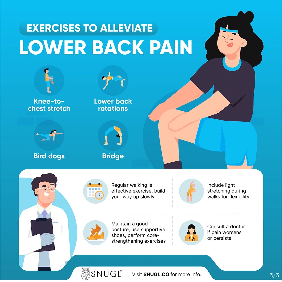
  • Lower Back Pain When Walking A Deep Dive

    Lower Back Pain When Walking A Deep Dive

    Lower back pain when walking can be debilitating, significantly impacting daily life. This comprehensive guide explores the multifaceted causes, symptoms, and potential treatments for this common ailment. From musculoskeletal issues to lifestyle factors, we’ll delve into everything you need to understand about managing and preventing lower back pain while walking.

    Understanding the underlying mechanisms is crucial to effectively address the problem. This involves exploring various contributing factors, including postural abnormalities, muscle imbalances, and potential joint problems. The pain itself can manifest in different ways, from sharp to dull, and its intensity and location provide valuable clues for diagnosis.

    Causes of Lower Back Pain When Walking

    Lower back pain when walking

    Lower back pain during walking can stem from a multitude of interconnected factors, often originating from musculoskeletal issues. Understanding these underlying causes is crucial for effective diagnosis and treatment. This exploration delves into the various musculoskeletal contributors, encompassing posture, gait, muscle imbalances, joint problems, and specific medical conditions.A detailed analysis of these factors can help pinpoint the root cause of the pain, allowing for targeted interventions and preventing future episodes.

    Proper understanding is paramount for managing this prevalent issue effectively.

    Musculoskeletal Issues Contributing to Lower Back Pain During Walking

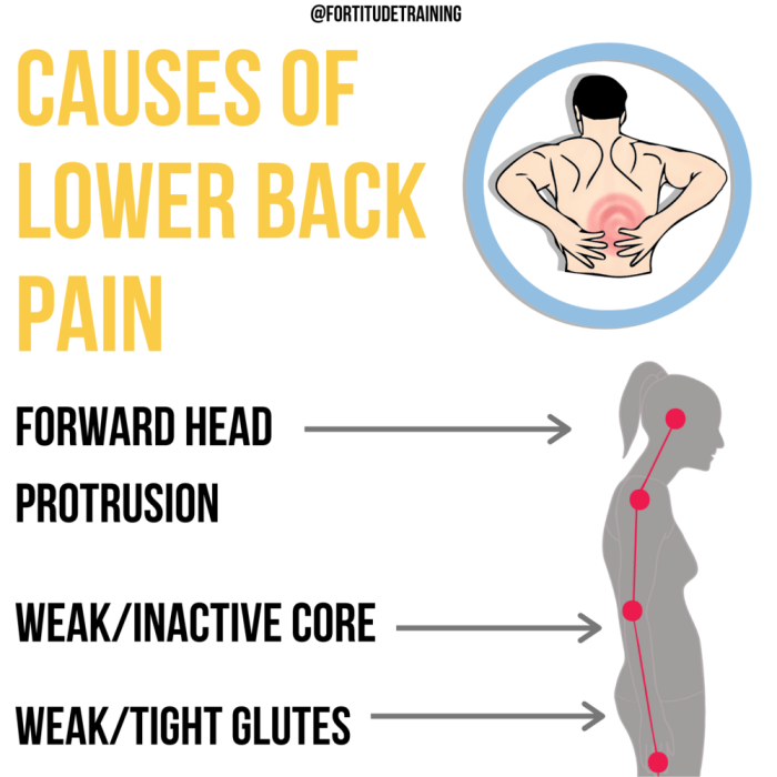
    Various musculoskeletal issues can contribute to lower back pain while walking. These include problems with the spine, surrounding muscles, and associated joints. Muscle imbalances, poor posture, and gait abnormalities can all place undue stress on the lower back, leading to pain.

    • Posture and Gait Abnormalities: Poor posture during walking, such as slouching or excessive forward leaning, can increase stress on the lumbar spine. Similarly, gait abnormalities, like uneven strides or excessive pronation, can alter the distribution of forces across the body, leading to lower back pain. For instance, someone with a significant hip or knee problem may compensate by altering their gait, placing additional strain on the lower back.

    • Muscle Imbalances and Weaknesses: Muscle imbalances, particularly in the core, hip, and gluteal muscles, can disrupt the normal biomechanics of walking. Weak or tight muscles can’t effectively support the spine during movement, leading to pain. For example, weak abdominal muscles may not provide sufficient support for the spine during walking, leading to lower back pain. Tight hip flexors can also contribute by pulling the pelvis forward, leading to an exaggerated lumbar curve.

    • Joint Problems (Hip, Knee): Problems in the hip or knee joints can significantly impact the mechanics of walking. Conditions such as osteoarthritis, bursitis, or meniscus tears can cause pain and stiffness, potentially altering gait and placing stress on the lower back. For instance, a hip impingement may force the individual to adopt a limping gait, transferring undue stress to the lower back.

    Specific Medical Conditions and Their Manifestations

    Certain medical conditions can present as lower back pain during walking. Understanding these conditions is critical for appropriate diagnosis and management.

    Lower back pain when walking can be a real drag, making everyday activities tough. Sometimes, underlying conditions like pulmonary hypertension can contribute to this discomfort. Fortunately, there are various medications available to help manage pulmonary hypertension, such as those listed on this page for available pulmonary arterial hypertension medications. While these treatments can significantly improve overall health, it’s crucial to remember that addressing the root cause of the back pain is also vital for long-term relief.

    • Spinal Stenosis: Spinal stenosis is a narrowing of the spinal canal, often impacting the nerves exiting the spinal cord. During walking, the increased pressure on the nerves can cause pain, numbness, or tingling in the lower back, buttocks, and legs. This pain often subsides with rest. A patient with spinal stenosis may report a gradual worsening of pain with prolonged walking.

    • Herniated Discs: A herniated disc occurs when the soft inner material of a spinal disc bulges or ruptures, potentially pressing on nearby nerves. Walking can aggravate the pressure on these nerves, leading to lower back pain, radiating pain down the leg, and possible numbness or tingling. Individuals with herniated discs often experience pain that worsens with prolonged activity.

    Comparison of Musculoskeletal Causes of Lower Back Pain During Walking

    Condition Symptoms Location of Pain Possible Treatments
    Muscle Imbalances Lower back pain, stiffness, tightness in specific muscle groups, reduced range of motion. Lower back, buttocks, hips. Physical therapy, stretching, strengthening exercises, massage therapy.
    Spinal Stenosis Lower back pain that worsens with walking, numbness or tingling in legs, weakness in legs. Lower back, buttocks, and radiating down the legs. Physical therapy, medication, spinal decompression surgery (in severe cases).
    Herniated Disc Lower back pain, radiating pain down the leg, numbness or tingling in the leg, weakness in the leg. Lower back and radiating down the leg. Physical therapy, pain medication, epidural injections, surgery (in severe cases).
    Posture and Gait Abnormalities Chronic lower back pain, pain in the hips and knees, muscle fatigue. Lower back, hips, and knees. Physical therapy, posture correction exercises, orthotics, footwear modifications.

    Identifying the Pain Characteristics

    Lower back pain when walking

    Understanding the nuances of your lower back pain when walking is crucial for accurate diagnosis and effective treatment. Paying close attention to the specific qualities of the pain, like its type, intensity, location, and relationship to walking, can significantly aid healthcare professionals in pinpointing the source of the discomfort.Pinpointing the exact nature of the pain is a vital step in determining the cause and developing an appropriate treatment strategy.

    Different types of pain, varying intensities, and the correlation between pain and walking patterns offer clues to the underlying problem. The location of the pain can also provide valuable insights into the potential source of the issue.

    Types of Pain

    Lower back pain when walking can manifest in various ways, from a dull ache to a sharp, stabbing sensation. Understanding these different pain types is essential for communicating effectively with healthcare providers. A sharp pain typically indicates a sudden, acute injury, while a dull, aching pain might suggest a more gradual, chronic condition.

    Intensity and Duration, Lower back pain when walking

    The intensity of the pain, measured on a scale of 1 to 10 (1 being mild, 10 being the worst imaginable pain), can vary significantly. Similarly, the duration of the pain, whether it lasts for a few seconds or persists for hours, plays a role in determining the underlying cause. For instance, a sharp, intense pain lasting only a few seconds during a sudden movement might indicate a muscle strain, whereas a dull, persistent ache lasting for hours after a prolonged walk could suggest a more serious condition.

    Relationship to Walking

    The relationship between the pain and the pace or duration of walking can also be helpful. Does the pain intensify with faster walking or longer distances? Does it subside when resting? Understanding these factors can help determine if the pain is mechanical, resulting from the repetitive motion of walking, or if it’s linked to a more systemic issue.

    For example, someone experiencing pain that worsens after 30 minutes of brisk walking might have a problem with their joint mechanics or disc degeneration.

    Pain Location

    The location of the pain can significantly help in pinpointing the source of the problem. Is the pain concentrated in a specific area of the lower back, or does it radiate to other parts of the body, like the buttocks or legs? Pain localized to the lower lumbar region might indicate a problem with the lumbar spine, while radiating pain might suggest nerve impingement.

    A precise description of the pain’s location is invaluable for diagnosis.

    Pain Characteristics Table

    Pain Type Intensity (Scale 1-10) Location Aggravating Factors
    Sharp, stabbing 7-10 Lower lumbar, radiating to buttock Sudden movements, prolonged walking
    Dull, aching 3-6 Lower back, especially after prolonged walking Prolonged standing, sitting
    Burning 5-9 Lower back, radiating to leg Walking downhill, prolonged walking
    Numbness/Tingling 2-7 Buttocks, lower legs Walking for long durations, sitting for long durations

    Associated Symptoms and Risk Factors

    Lower back pain when walking can stem from a variety of issues, and understanding the accompanying symptoms and potential risk factors is crucial for proper diagnosis and treatment. Identifying these factors allows for more targeted interventions and potentially prevents the pain from worsening. This section will delve into additional symptoms that often accompany lower back pain during ambulation, explore lifestyle choices that may increase the risk, and discuss the role of age and genetics in the development of this type of pain.

    Additional Symptoms

    Besides the primary pain, several other symptoms frequently accompany lower back pain when walking. These additional sensations can provide valuable clues to the underlying cause. Numbness, tingling, and weakness in the legs are common occurrences. These neurological symptoms may indicate nerve compression or irritation in the lower back, which can be indicative of conditions like spinal stenosis or herniated discs.

    Pain radiating down the leg (sciatica) is another significant associated symptom. Understanding the specific characteristics of these additional sensations can help healthcare professionals differentiate between various potential causes and tailor appropriate treatment strategies.

    Lifestyle Risk Factors

    Certain lifestyle choices can significantly increase the risk of developing lower back pain during walking. Obesity, for example, places excessive strain on the spine, leading to increased pressure and potential inflammation. Poor posture, characterized by slouching or improper alignment, also contributes to spinal stress and may trigger or exacerbate existing conditions. A lack of regular exercise weakens supporting muscles, decreasing spinal stability and making the individual more susceptible to pain.

    Incorporating regular physical activity, maintaining a healthy weight, and practicing good posture are crucial for mitigating these risks.

    Age and Genetics

    Age plays a role in the development of lower back pain. As we age, our spinal discs naturally lose hydration and elasticity, making them more prone to injury and degeneration. This age-related decline in spinal health can increase the risk of developing conditions like osteoarthritis and spinal stenosis. Genetic predisposition also influences an individual’s susceptibility to lower back pain.

    Certain genetic factors may increase the risk of developing specific conditions that contribute to pain during walking. Understanding these factors can be helpful in determining individual risk levels and tailoring preventive measures.

    Risk Factor Table

    Risk Factor Description Example Mitigation Strategies
    Obesity Excess body weight puts excessive pressure on the spine, increasing the risk of injury and pain. An individual with a BMI of 35 is at higher risk compared to someone with a BMI of 25. Maintaining a healthy weight through diet and exercise. Consider consulting a nutritionist or a physical therapist.
    Poor Posture Slouching or incorrect spinal alignment puts undue stress on the back muscles and ligaments, increasing the risk of pain. Prolonged sitting at a desk without proper lumbar support. Improving posture through ergonomic adjustments at work or home, and practicing mindful posture awareness throughout the day.
    Lack of Exercise Weak core and back muscles provide insufficient support for the spine, making it more vulnerable to injury. A sedentary lifestyle with minimal physical activity. Regular exercise that strengthens core and back muscles, including yoga, Pilates, and weight training.
    Age The natural aging process can lead to disc degeneration and reduced spinal flexibility, increasing pain risk. An individual in their 60s is more susceptible to spinal problems compared to someone in their 20s. Maintaining an active lifestyle, staying hydrated, and incorporating regular stretching and mobility exercises. Consulting a physician for age-related concerns.
    Genetics Certain genetic predispositions can increase the likelihood of developing conditions that cause lower back pain. A family history of spinal stenosis or herniated discs. Maintaining a healthy lifestyle to mitigate potential genetic risks. Regular check-ups with a doctor to monitor for any potential issues.

    Diagnostic Considerations

    Figuring out the precise cause of lower back pain that worsens when walking is crucial for effective treatment. A thorough evaluation process, combining a detailed medical history with appropriate diagnostic tests, helps pinpoint the underlying issue and guides the most suitable course of action. This involves understanding the nuances of the pain, identifying potential contributing factors, and eliminating any potentially serious conditions.A structured approach to evaluating lower back pain during walking is essential.

    It should begin with a comprehensive history, exploring the onset, duration, location, and character of the pain. Factors like the timing of the pain (e.g., after a certain distance, with certain movements) and any relieving or aggravating factors should be noted. This initial assessment is a critical foundation for subsequent investigations.

    Evaluating Lower Back Pain During Walking

    A systematic evaluation involves gathering a detailed medical history and performing a physical examination. This includes a neurological examination to assess reflexes, muscle strength, and sensation in the lower extremities. The examination should also include palpation of the spine and surrounding soft tissues to identify any areas of tenderness or muscle spasm.

    Potential Diagnostic Tests and Procedures

    Several diagnostic tests and procedures may be employed to pinpoint the source of lower back pain when walking. These tests range from straightforward imaging techniques to more specialized procedures. The selection of tests will depend on the initial evaluation and suspected diagnoses.

    Medical History in Diagnosis

    A thorough medical history is paramount in diagnosing the cause of lower back pain. Information about past injuries, surgeries, underlying medical conditions, and lifestyle factors can provide valuable insights into the potential causes. This information helps in narrowing down the possibilities and guiding the selection of appropriate diagnostic tests. For example, a history of cancer or inflammatory conditions can suggest certain etiologies, while a history of repetitive strain injuries might point to mechanical causes.

    Diagnostic Tests and Procedures

    Test Purpose Procedure Potential Results
    X-ray To visualize the bones of the spine for fractures, dislocations, or other bony abnormalities. A low-dose X-ray is taken of the lower back. Potential results include evidence of fractures, scoliosis, spondylolisthesis, or degenerative joint disease.
    MRI (Magnetic Resonance Imaging) To visualize soft tissues, including ligaments, tendons, muscles, and nerves, to detect herniated discs, spinal stenosis, or other soft tissue abnormalities. The patient lies inside a large magnet, and detailed images of the spinal structures are generated. Potential results include identification of herniated discs, spinal stenosis, tumors, or inflammation. The quality and detail of the images are critical for accurate interpretation.
    CT (Computed Tomography) Scan To provide detailed images of the bones and soft tissues of the spine. Especially useful for evaluating complex bony structures. The patient lies on a table that moves through a scanner that uses X-rays to create cross-sectional images. Potential results include identification of fractures, tumors, or bony abnormalities that may not be visible on an X-ray. A CT scan can also provide detailed images of the spinal canal and its contents.
    Electromyography (EMG) To assess the electrical activity of muscles and nerves to identify nerve compression or damage. Small needles are inserted into muscles to record their electrical activity. Potential results include evidence of nerve root impingement, muscle damage, or other neurological issues.

    Management Strategies and Treatment Options

    Lower back pain when walking can significantly impact daily life, making movement challenging and frustrating. Fortunately, several effective strategies and treatment options can help manage and alleviate this discomfort. This section explores conservative approaches, non-pharmacological interventions, the role of medications, and the process of creating a personalized treatment plan.

    Conservative Approaches

    Conservative management focuses on non-surgical methods to alleviate pain and improve function. Physical therapy plays a crucial role in this approach. A physical therapist can assess the root cause of the pain and develop a tailored exercise program to strengthen core muscles, improve posture, and increase flexibility. This can significantly reduce pain and improve mobility. Exercises targeting specific muscle groups and stretches can help alleviate pain and improve movement.

    Proper posture and movement techniques are crucial in preventing future episodes.

    Ugh, lower back pain when walking is the worst. It can really throw a wrench in your day, making even simple tasks feel monumental. Sometimes, those nagging aches can feel like a never-ending cycle of discomfort, mirroring the repetitive thoughts and behaviors associated with coping with obsessive compulsive behavior, like checking locks repeatedly or constantly needing to rearrange objects.

    Learning strategies to manage those unwanted thoughts and behaviors, like those discussed in this helpful resource on coping with obsessive compulsive behavior , might surprisingly help with the persistent lower back pain, as mental clarity and reduced stress can contribute to physical well-being.

    Non-Pharmacological Interventions

    Non-pharmacological interventions are often the first line of defense in managing lower back pain. Lifestyle modifications are essential. Maintaining a healthy weight, adopting good posture habits, and avoiding prolonged periods of sitting or standing can significantly reduce the strain on the lower back. Regular physical activity, including walking, swimming, or cycling, can help improve overall fitness and reduce pain.

    Lower back pain when walking can be a real drag, right? It’s frustrating, especially when you’re trying to enjoy a simple stroll. Sometimes, though, it’s not as straightforward as a simple stretching routine. For example, if you’ve got a sore or cut inside your mouth, it can be tricky to figure out what to do. Fortunately, there are great resources out there to help you understand how to treat a cut inside your mouth.

    Check out this helpful guide: how to treat a cut inside your mouth. Ultimately, dealing with any kind of pain, from a cut to a back ache, needs to be approached with care and proper care. Addressing these issues promptly and correctly can get you back on your feet and walking pain-free.

    Heat and ice therapy can also provide temporary relief. Applying heat can relax muscles and improve blood flow, while ice can reduce inflammation and pain. These methods are often used in conjunction with other treatments.

    Role of Medications

    Medications can be used to manage lower back pain when walking, but should be considered supplementary to other treatments. Over-the-counter pain relievers, such as ibuprofen or acetaminophen, can help reduce pain and inflammation. Prescription medications, such as stronger anti-inflammatory drugs, may be necessary in more severe cases. However, it is crucial to discuss medication options with a healthcare provider to determine the most appropriate course of action, considering potential side effects and interactions with other medications.

    Following a healthcare professional’s advice is crucial for safe and effective medication use.

    Creating a Personalized Treatment Plan

    A personalized treatment plan for lower back pain when walking is crucial for optimal outcomes. This plan should be developed in collaboration with a healthcare provider, taking into account the individual’s specific needs, medical history, and lifestyle. The plan should include a combination of therapies, exercises, and lifestyle modifications. Factors such as age, occupation, and pre-existing conditions should be carefully considered to create a comprehensive strategy.

    The plan should be regularly reviewed and adjusted as needed.

    Comparison of Treatment Options

    Treatment Type Description Benefits Potential Drawbacks
    Physical Therapy A structured program of exercises, stretches, and manual therapy to improve strength, flexibility, and posture. Improved muscle strength, reduced pain, increased mobility, and long-term pain relief. Requires commitment to regular sessions, may not be suitable for all individuals, and may take time to see results.
    Lifestyle Modifications Changes in daily habits, such as maintaining a healthy weight, improving posture, and avoiding prolonged periods of sitting or standing. Reduces strain on the lower back, improves overall health, and can prevent future episodes of pain. Requires consistent effort and dedication, and may not be sufficient for severe pain.
    Heat/Ice Therapy Applying heat or ice to the affected area to reduce pain and inflammation. Provides temporary relief from pain and inflammation, and is a safe and inexpensive treatment option. May not be effective for all individuals, and should be used in conjunction with other therapies.
    Medications Pain relievers and anti-inflammatory drugs to manage pain and inflammation. Provides fast pain relief, and may be necessary for severe cases. Potential side effects, such as stomach upset or interactions with other medications, and may not address the underlying cause of pain.

    Prevention Strategies

    Preventing lower back pain when walking involves a proactive approach that combines proper posture, regular exercise, and mindful activity modification. A comprehensive strategy focuses on building core strength, maintaining flexibility, and understanding how to adapt daily routines to minimize stress on the lower back. This proactive approach is key to long-term pain management and overall well-being.

    Maintaining Good Posture and Proper Gait

    Maintaining correct posture while walking is crucial for preventing lower back pain. Proper posture involves aligning the spine, keeping the head erect, and engaging the core muscles. A good gait involves a smooth, controlled stride, avoiding excessive twisting or leaning. This requires conscious awareness and practice. Think of it like a well-rehearsed dance move; the more you practice, the more natural it becomes.

    Importance of Regular Exercise and Stretching

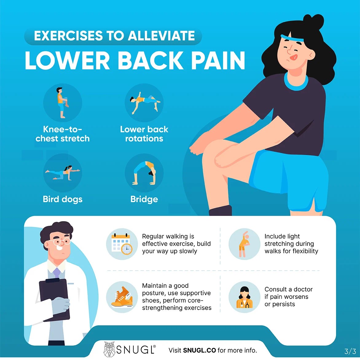
    Regular exercise, including activities like walking, swimming, and yoga, strengthens core muscles, improving spinal stability. Stretching exercises, such as hamstring stretches, hip flexor stretches, and back extensions, increase flexibility and range of motion, further reducing the risk of injury. Consistency is key, aiming for at least 30 minutes of moderate-intensity exercise most days of the week. Incorporate stretching routines before and after walks to improve muscle flexibility and reduce muscle strain.

    Modifying Activities to Minimize Pain Risk

    Activities that place excessive stress on the lower back should be modified or avoided. Heavy lifting, prolonged standing, and repetitive bending should be approached with caution and awareness. Consider using proper lifting techniques, employing assistive devices, and adjusting the duration of activities to prevent overuse. For example, if you’re carrying groceries, break down the task into smaller portions and take frequent breaks.

    Adjusting posture and technique while working can also minimize strain.

    Preventive Measures for Daily Routines

    Incorporating preventive measures into daily routines can significantly reduce the risk of lower back pain. This includes using supportive footwear, maintaining a healthy weight, and avoiding prolonged periods of sitting or standing. Prioritizing proper sleep hygiene and managing stress are also essential components.

    • Use supportive footwear: Choosing shoes with proper arch support and cushioning can lessen the stress on your lower back during walks. Consider orthotics for added support if needed.
    • Maintain a healthy weight: Excess weight puts extra strain on the lower back, increasing the risk of pain. A balanced diet and regular exercise are crucial.
    • Avoid prolonged sitting or standing: Take frequent breaks to stretch and move around. Adjust your workspace to encourage better posture. Standing desks can be beneficial for those who work at a desk.
    • Prioritize sleep hygiene: Adequate sleep allows your body to recover and repair itself, which is essential for preventing pain.
    • Manage stress: Stress can exacerbate existing back pain and lead to new problems. Incorporate stress-reducing activities into your daily routine, such as meditation or yoga.

    Illustrative Case Studies (Example scenarios)

    Understanding lower back pain when walking requires exploring real-world examples. Case studies offer valuable insights into the diverse presentations, contributing factors, and effective management strategies for this common condition. They allow us to connect theoretical knowledge with practical application, ultimately leading to a better comprehension of individual patient needs.

    Hypothetical Case Study 1: Mechanical Back Pain

    A 45-year-old female office worker reports experiencing lower back pain that intensifies when walking. The pain is described as a dull ache, localized to the lower lumbar region, and is exacerbated by prolonged standing or repetitive movements. She reports no significant medical history, but admits to recent increased physical demands at work, including carrying heavy boxes. Potential diagnoses could include lumbar strain, facet joint syndrome, or spondylolisthesis.

    Imaging studies, such as X-rays or MRI, might be necessary to rule out more serious conditions.

    Hypothetical Case Study 2: Radicular Pain

    A 62-year-old male construction worker presents with lower back pain radiating down his left leg. The pain is described as sharp and shooting, worsened by coughing or sneezing. He has a history of osteoarthritis and reports experiencing numbness and tingling in his left foot. Risk factors include his occupation involving heavy lifting and potential nerve impingement. Potential diagnoses could include lumbar herniated disc, spinal stenosis, or sciatica.

    Neurological examination and diagnostic imaging are crucial for accurate assessment.

    Hypothetical Case Study 3: Successful Management

    A 30-year-old female experiences lower back pain when walking, aggravated by prolonged periods of sitting. She initially tried over-the-counter pain relievers without significant relief. She subsequently consulted a physical therapist, who prescribed a combination of exercises focusing on core strengthening and flexibility. She also implemented lifestyle changes, such as improved posture, regular walking, and ergonomic adjustments at her workplace.

    Her pain significantly reduced after several weeks of therapy. This demonstrates the importance of multifaceted approaches to lower back pain management.

    Table of Case Studies

    Patient Profile Symptoms Diagnosis (Potential) Treatment Outcomes
    45-year-old female office worker Dull ache in lower lumbar region, exacerbated by prolonged standing/repetitive movements Lumbar strain, facet joint syndrome, or spondylolisthesis Further evaluation and potential physical therapy, ergonomic adjustments, and lifestyle modifications.
    62-year-old male construction worker Sharp, shooting pain radiating down left leg, worsened by coughing/sneezing; numbness/tingling in left foot Lumbar herniated disc, spinal stenosis, or sciatica Neurological examination, imaging studies, and potential surgery or physical therapy, pain management, and lifestyle changes.
    30-year-old female Lower back pain when walking, aggravated by prolonged sitting; initially unresponsive to OTC pain relievers. Potential musculoskeletal issues related to prolonged sitting or posture Successful pain reduction after physical therapy (core strengthening/flexibility), lifestyle changes (improved posture, regular walking, ergonomic adjustments).

    Final Review

    In conclusion, lower back pain when walking can stem from a variety of sources, requiring a multifaceted approach to diagnosis and treatment. Understanding the interplay of musculoskeletal issues, pain characteristics, associated symptoms, and risk factors is essential. By exploring diagnostic considerations, management strategies, and preventive measures, we empower individuals to take control of their well-being and regain mobility.

    Remember that consulting a healthcare professional is crucial for personalized guidance and effective treatment.

  • Breast Cancer Testing Screening A Comprehensive Guide

    Breast Cancer Testing Screening A Comprehensive Guide

    Breast cancer testing screening is crucial for early detection, significantly impacting survival rates. This guide delves into the various methods, from the familiar mammogram to newer technologies, highlighting their benefits and limitations. We’ll explore risk factors, screening recommendations, and how to interpret results. Understanding the process empowers individuals to make informed decisions about their health.

    From basic self-exams to advanced MRI scans, we’ll break down each screening method, comparing their advantages and disadvantages. This detailed look allows you to understand which approach might be best for your specific needs and risk factors. We’ll also address common misconceptions and the evolving landscape of breast cancer screening technology.

    Introduction to Breast Cancer Testing and Screening

    Breast cancer is a significant health concern worldwide, impacting countless lives. Early detection is crucial for successful treatment and improved outcomes. Regular breast cancer testing and screening play a vital role in identifying potential issues early, when treatment options are often more effective. This process empowers individuals to take proactive steps toward their health and well-being.Comprehensive breast cancer testing and screening encompasses a range of methods, each with its own strengths and limitations.

    Regular breast cancer testing screening is crucial for early detection, but did you know that supporting overall health through good habits like a healthy diet and taking the right supplements can also play a role? For instance, looking into supplements for lung health, like those available at supplements for lung health , could potentially contribute to a stronger immune system, which might indirectly impact the body’s ability to fight off diseases.

    Ultimately, prioritizing preventative measures like consistent screening and a holistic approach to well-being are key in breast cancer prevention and detection.

    Understanding these nuances allows individuals to make informed decisions about their health care. Knowledge about the available options and their respective benefits and drawbacks is essential for empowering patients to actively participate in their health management.

    Types of Breast Cancer Testing and Screening Methods

    Various methods are employed in breast cancer testing and screening. These methods are designed to detect abnormalities and potential cancerous cells.

    • Mammography: This is a common imaging technique using X-rays to visualize the breast tissue. It helps detect dense breast tissue, masses, and microcalcifications, which can be early signs of cancer. Mammography is a widely used and effective method for breast cancer screening, particularly in women over 40. However, it may not be as effective in women with dense breast tissue, and it can produce false positive results, leading to unnecessary anxiety and further testing.

    • Ultrasound: Ultrasound uses sound waves to create images of the breast. It’s particularly helpful in distinguishing between solid masses and fluid-filled cysts, providing further clarity when combined with mammography. Ultrasound is valuable for evaluating abnormalities detected in mammography and for guiding biopsies. It is also used for women with dense breasts, as it can better visualize these tissues compared to mammography.

      A limitation is that it can sometimes miss small cancers or provide ambiguous results.

    • MRI (Magnetic Resonance Imaging): MRI uses powerful magnetic fields and radio waves to produce detailed images of the breast. It is often used for women with a high risk of breast cancer, those with dense breast tissue, or for evaluating abnormalities detected by other imaging techniques. MRI can detect cancers that are not visible on mammography or ultrasound. A key drawback is the higher cost compared to other methods and potential for false positives.

    • Breast Biopsy: This involves removing a tissue sample from the breast for examination under a microscope. It’s a crucial step when suspicious findings are detected through imaging tests. A biopsy confirms or rules out the presence of cancer. Different types of biopsies are available, including fine-needle aspiration, core needle biopsy, and surgical biopsy, each with varying degrees of invasiveness and accuracy.

    Benefits and Limitations of Screening Methods

    Understanding the advantages and disadvantages of each method is crucial for informed decision-making. Different screening methods cater to various needs and situations, leading to tailored recommendations for individuals.

    • Mammography: Widely accessible, relatively inexpensive, and effective in detecting cancers at an early stage. However, it can miss some cancers, particularly in women with dense breast tissue, and can produce false positive results, leading to unnecessary anxiety.
    • Ultrasound: Useful for differentiating between solid masses and fluid-filled cysts, and guiding biopsies. However, it may not detect all cancers, especially small ones, and the quality of images can vary depending on the technician’s expertise.
    • MRI: Excellent for detecting cancers in dense breast tissue or women at high risk, but it is more expensive and may produce false positives.
    • Breast Biopsy: Provides a definitive diagnosis of breast abnormalities. However, it can be invasive, and it carries a small risk of complications.

    Common Misconceptions About Breast Cancer Testing and Screening

    Many misconceptions surround breast cancer testing and screening. Dispelling these myths is vital for encouraging regular screening and early detection.

    • Screening is only necessary for high-risk individuals: While those with a family history or genetic predisposition are advised to start screening earlier and more frequently, screening is beneficial for all women. Early detection is key, regardless of risk factors.
    • Screening is painful and uncomfortable: While some discomfort may be experienced during certain procedures, modern techniques and skilled professionals minimize pain and discomfort.
    • Breast cancer screening is unnecessary if there are no symptoms: Early detection is critical. Many women with breast cancer have no symptoms in the early stages. Regular screening helps catch cancers when treatment is most effective.

    Different Screening Methods

    Understanding the various breast cancer screening methods is crucial for informed decision-making. Each method has its own strengths and limitations, and choosing the right one depends on individual factors like age, family history, and breast density. This section delves into the different techniques, outlining their procedures, benefits, drawbacks, and ideal applications.

    Regular breast cancer testing screening is crucial for early detection. While I’m no expert on supplements, I’ve heard some people wonder if certain things, like creatine, can affect their bodies. If you’re curious about whether does creatine cause bloating , there’s plenty of info out there. Regardless of those potential side effects, though, getting checked for breast cancer is a really important preventative measure.

    Comparing Screening Methods

    Different imaging and self-examination techniques provide various ways to detect breast abnormalities. The table below summarizes the key characteristics of common methods.

    Method Procedure Advantages Disadvantages Ideal Use Cases
    Mammography X-ray imaging of the breast, often involving compression to spread tissue for clearer images. Detects dense tissue cancers, particularly helpful in identifying microcalcifications (small calcium deposits) that can be an early sign of cancer. Generally good at detecting invasive cancers. Can produce false positives, potentially leading to unnecessary biopsies. Can be uncomfortable due to breast compression. Not as effective in women with dense breast tissue. Routine screening for women over 40, particularly those with no known risk factors.
    Breast Ultrasound Uses high-frequency sound waves to create images of the breast tissue. Excellent for differentiating between cysts (fluid-filled sacs) and solid masses, useful in evaluating dense breasts where mammography might not be as effective. Generally quick and painless. Not as effective at detecting microcalcifications. Results can be operator-dependent, meaning the quality of the image can vary depending on the technician. Evaluating breast lumps, assessing suspicious areas identified by mammography or self-exam, and screening women with dense breasts.
    Breast MRI Uses magnetic fields and radio waves to create detailed images of the breast. Highly sensitive in detecting breast cancers, particularly in women with dense breasts or a high risk of breast cancer. Excellent for detecting small cancers and evaluating suspicious areas. More expensive than mammography or ultrasound. Can produce more false positives, potentially requiring additional testing. May not be suitable for all patients due to potential claustrophobia or metal implants. Screening women with a high risk of breast cancer, evaluating women with dense breasts, evaluating abnormalities detected by other methods, and evaluating breast implants.
    Self-Exams Regularly examining one’s own breasts for changes in size, shape, texture, or the presence of lumps or dimples. Easy, inexpensive, and accessible to everyone. Allows for early detection of abnormalities if the individual is vigilant and aware of their own breasts. Not as reliable as imaging methods. Accuracy is highly dependent on the individual’s awareness and ability to recognize changes. False alarms are common. A valuable supplementary tool for early detection. Important for women of all ages to be familiar with their own breasts and report any unusual changes to their doctor.

    Detailed Procedures and Steps

    Mammography involves positioning the breast between two plates and taking X-ray images. Breast ultrasound uses a handheld probe to send and receive sound waves. Breast MRI uses a large machine to create detailed images. Self-exams involve palpating the breasts for any unusual changes.

    Accuracy and Reliability

    Mammography is generally considered the most effective screening method for women over 40. Ultrasound is valuable in evaluating dense breast tissue, while MRI offers a high level of sensitivity for detecting subtle abnormalities, especially in high-risk individuals. Self-exams are a useful tool for early detection, but they are not as reliable as imaging methods. The accuracy and reliability of each method vary depending on factors such as the skill of the technician, the quality of the equipment, and the individual characteristics of the patient.

    Factors such as breast density and the presence of implants can impact the results of some screening methods.

    Risk Factors and Screening Recommendations

    Understanding your risk factors for breast cancer is crucial for making informed decisions about screening. This knowledge empowers you to discuss your options with your healthcare provider and tailor your screening plan to your individual circumstances. Knowing your risk allows you to proactively address potential health concerns and potentially detect breast cancer early.Knowing the factors that increase your chances of developing breast cancer can influence your screening plan.

    This proactive approach allows for personalized screening strategies that are most effective for your specific risk profile. This helps you understand how different risk factors impact screening recommendations and when more frequent or advanced screenings may be necessary.

    Key Risk Factors

    Various factors can elevate a person’s risk of breast cancer. Age is a significant one, as the risk generally increases with age. Family history plays a critical role, with a strong family history of breast cancer in close relatives substantially increasing the risk. Genetic mutations, such as BRCA1 and BRCA2, are strong indicators of increased susceptibility. Previous radiation exposure, particularly during youth, is also a recognized risk factor.

    Certain lifestyle choices, such as a lack of physical activity or a diet high in processed foods, may contribute to an elevated risk. Finally, having a history of dense breast tissue or previous breast biopsies can also increase risk.

    Screening Recommendations Based on Risk

    Screening recommendations are tailored to individual risk profiles, balancing the potential benefits of early detection against the risks of unnecessary procedures. Different risk levels require varying frequency and types of screening. The recommendations are based on factors like age, family history, and other risk indicators.

    Screening Guidelines by Risk Group

    Risk Group Age Recommendation Additional Screening Frequency
    Average Risk 40-44: Consider starting mammograms. 45-54: Annual mammograms recommended. 55 and older: Mammograms every 2 years or as advised by a doctor. Clinical breast exams (CBE) every 3 years, self-exams monthly. As per the age recommendation above.
    High Risk 10-15 years earlier than the average risk population. Annual mammograms, MRI scans, and possibly genetic testing. Annually or as advised by a doctor, based on individual risk assessment.

    How Risk Factors Influence Screening Decisions

    The table above highlights the different screening guidelines for various risk groups. For individuals with average risk, standard screening guidelines are followed. However, those with a high risk of breast cancer, due to factors like family history or genetic mutations, require more intensive and frequent screening. These individuals might benefit from earlier initiation of mammograms, additional imaging techniques like MRI, and potentially genetic testing to identify and monitor any genetic predisposition.

    This proactive approach allows for early detection and intervention, improving outcomes and reducing the risk of late-stage diagnoses.

    Interpreting Test Results

    Breast cancer testing screening

    Understanding the results of breast cancer screening tests is crucial for early detection and appropriate action. A clear comprehension of the potential findings, their implications, and the subsequent steps involved can empower individuals to take proactive steps toward their health. This section delves into the interpretation of various breast cancer screening tests, focusing on the implications of different outcomes and the diagnostic pathways that follow.

    Understanding Mammogram Results

    Mammograms are a crucial tool for detecting breast abnormalities. Radiologists analyze these images for signs of potential breast cancer, such as masses, microcalcifications, or architectural distortions. A negative mammogram typically indicates no suspicious findings. A positive mammogram, however, does not automatically mean cancer. It simply suggests the presence of something requiring further investigation.

    Getting regular breast cancer screenings is crucial for early detection. While focusing on preventative measures like these, it’s also important to understand other eye conditions. For example, have you ever wondered what a pinguecula is? what is a pinguecula is a yellowish growth on the white part of your eye. Ultimately, knowing about different health concerns, from eye conditions to breast cancer screenings, empowers you to make informed decisions about your well-being.

    Different types of positive findings require different approaches.

    Interpreting Biopsy Results

    A biopsy is a procedure that involves removing a tissue sample from the breast for examination under a microscope. This is a crucial step when a mammogram or other screening test reveals an abnormality. The biopsy report will specify the type of tissue found and whether it exhibits cancerous or precancerous characteristics. The report usually categorizes findings based on established diagnostic criteria, often using systems like the Bethesda System for reporting breast tissue abnormalities.

    Common Findings and Their Implications

    Various findings can emerge from screening tests, each with different implications. Benign findings, such as cysts or fibroadenomas, are non-cancerous and typically do not require immediate intervention beyond regular monitoring. However, if the findings suggest malignancy, the implications are far more serious, necessitating further evaluation and potentially treatment.

    Follow-up Procedures After an Abnormal Result

    Following an abnormal screening result, a multi-step process begins. This typically involves a conversation with the healthcare provider to discuss the findings and next steps. Additional imaging, such as ultrasound or MRI, might be necessary to gain a clearer picture of the abnormality. If the initial tests raise concerns about malignancy, a biopsy is often the next step.

    Diagnostic Pathway Flowchart

    The flowchart below illustrates the typical diagnostic pathway after an abnormal screening result. It highlights the sequential steps involved, from initial detection to confirmation or exclusion of breast cancer. The pathway emphasizes the importance of communication with healthcare professionals and adherence to recommended follow-up procedures. This flowchart is a general guideline; individual cases may require modifications based on specific circumstances.

    Flowchart of diagnostic pathway

    Note: The flowchart visually displays the progression from an initial screening abnormality to a definitive diagnosis or exclusion. It emphasizes the cyclical nature of follow-up, where results from one test can influence the next. This dynamic process ensures accurate assessment and appropriate treatment planning.

    Impact of Screening on Early Detection

    Early detection is crucial in the fight against breast cancer. Regular screening, when combined with awareness and knowledge of risk factors, significantly increases the chances of catching the disease in its earliest, most treatable stages. This proactive approach dramatically improves outcomes and quality of life for those affected.Early diagnosis empowers individuals and healthcare professionals to initiate effective treatment plans, often leading to a more favorable prognosis.

    The difference between early-stage and advanced-stage breast cancer treatment can be substantial, impacting both the treatment approach and long-term survival.

    Role of Early Detection in Improving Breast Cancer Survival Rates

    Early detection of breast cancer is paramount to improving survival rates. When breast cancer is diagnosed in its early stages, it is often more localized and less likely to have spread to other parts of the body. This localized nature allows for less aggressive treatment options, often leading to better outcomes and a higher chance of long-term survival.

    Survival rates are significantly higher for early-stage breast cancer compared to later stages.

    Correlation Between Early Diagnosis and Treatment Success

    Early diagnosis of breast cancer allows for more targeted and effective treatment options. Treatment success rates are demonstrably higher when the disease is caught early. Surgical options like lumpectomy, instead of mastectomy, become more viable when the cancer is confined to a smaller area. Furthermore, the use of chemotherapy, radiation therapy, or hormone therapy can be tailored to the specific characteristics of the early-stage cancer, optimizing treatment effectiveness and minimizing side effects.

    Comparison of Outcomes of Early-Stage and Advanced-Stage Breast Cancer Treatment

    The outcomes of breast cancer treatment vary significantly based on the stage at diagnosis. Early-stage breast cancer, often detected through screening, allows for less invasive and often less aggressive treatment approaches. This results in a higher likelihood of cure, improved quality of life, and a greater chance of long-term survival. In contrast, advanced-stage breast cancer, which may have spread to other parts of the body, requires more extensive and often more aggressive treatment, which may include chemotherapy, targeted therapies, and/or hormonal therapies.

    The success rates and side effects associated with treatment are generally lower for advanced-stage cancer.

    Breast Cancer Survival Rates Based on Stage at Diagnosis

    Survival rates for breast cancer are directly linked to the stage of the disease at diagnosis. While exact statistics can vary depending on factors such as treatment approach and individual patient characteristics, general trends are clear. Early-stage breast cancer, often detected through screening, has significantly higher survival rates than advanced-stage breast cancer. Comprehensive data on survival rates are available from organizations like the American Cancer Society and the National Cancer Institute.

    These organizations provide valuable resources for further exploration of the impact of early detection on breast cancer survival. For instance, a patient diagnosed with stage 1 breast cancer has a significantly higher 5-year survival rate than a patient diagnosed with stage 4 breast cancer. Data from the National Cancer Institute consistently shows this correlation. Early detection is crucial to improve these survival rates.

    Technological Advancements in Breast Cancer Screening

    The landscape of breast cancer screening is constantly evolving, driven by innovative technologies that enhance accuracy, efficiency, and patient experience. These advancements are crucial in the fight against breast cancer, enabling earlier detection and improved treatment outcomes. From improved imaging techniques to sophisticated analysis tools, the journey toward more precise and personalized screening continues.The latest innovations in breast cancer screening are revolutionizing how we detect and manage this disease.

    These advancements are not just about faster results; they’re about more precise diagnoses, reduced false positives, and ultimately, better chances for survival. The combination of new technologies and refined diagnostic protocols is leading to a more comprehensive and personalized approach to breast cancer screening.

    Advanced Imaging Techniques

    Breast imaging techniques are constantly being refined to improve accuracy and reduce the need for unnecessary biopsies. Digital mammography, which converts X-rays into digital images, has already improved image quality and storage. Digital breast tomosynthesis (DBT), often called 3D mammography, provides a layered view of the breast, allowing for clearer visualization of structures and potentially reducing the rate of false positives.

    Ultrasound, often used in conjunction with mammography, is particularly helpful in evaluating dense breast tissue, which can be challenging to image with traditional methods.

    Artificial Intelligence (AI) in Screening

    AI algorithms are being integrated into breast cancer screening to assist radiologists in analyzing images and detecting subtle abnormalities. These algorithms can identify patterns and anomalies that may be missed by the human eye, leading to earlier detection and more accurate diagnoses. AI-powered systems can also help streamline the screening process, reducing workload and potentially increasing the number of women who undergo screenings.

    Early trials show promise in reducing false positive rates, and further research is underway to optimize AI’s role in breast cancer detection.

    Personalized Risk Assessment Tools

    Beyond imaging, advancements are focusing on personalized risk assessment. Genetic testing and family history analysis are increasingly used to identify women at higher risk for breast cancer. These tools can be used to tailor screening recommendations, increasing the frequency or type of screening for individuals with a higher predisposition. This targeted approach can be crucial in preventing delayed diagnoses and improving outcomes for high-risk individuals.

    For example, women with a family history of BRCA1/2 mutations may benefit from earlier and more frequent screening, even before reaching traditional screening ages.

    Promising Future Directions in Breast Cancer Screening Technology

    • Molecular Imaging: Techniques that visualize molecular changes associated with breast cancer, such as using contrast agents that target specific molecules within tumors, offer the potential to identify cancerous cells earlier than traditional imaging methods. This approach may be especially useful for detecting pre-cancerous lesions.
    • Liquid Biopsies: Analyzing circulating tumor DNA (ctDNA) in blood samples offers a non-invasive way to detect and monitor breast cancer. This technology could potentially revolutionize early detection and monitoring, allowing for more frequent and less invasive screenings.
    • Personalized Screening Protocols: Integrating genetic risk factors, lifestyle information, and imaging data into personalized screening protocols could allow for more effective risk stratification and tailored screening strategies. This approach may significantly reduce unnecessary screenings for low-risk individuals while ensuring higher-risk individuals receive more frequent and targeted screening.

    Addressing Ethical and Societal Issues: Breast Cancer Testing Screening

    Breast cancer testing screening

    Breast cancer screening, while vital for early detection and improved outcomes, presents a complex interplay of ethical considerations, societal impacts, and practical realities. Balancing the benefits of early diagnosis with the potential for anxiety, false positives, and overdiagnosis requires careful consideration. Furthermore, disparities in access to screening and the financial burden of these procedures highlight the need for equitable and sustainable programs.

    Ethical Considerations Surrounding Screening Recommendations

    Screening recommendations often grapple with balancing the potential benefits of early detection against the risks of unnecessary procedures and potential harms. The decision to undergo screening is inherently personal, involving individual risk assessments, preferences, and values. Factors like family history, personal health status, and perceived risks significantly influence these decisions. Clinicians have a responsibility to provide comprehensive information and support patients in making informed choices aligned with their values.

    This includes acknowledging the psychological impact of screening results, both positive and negative.

    Societal Impact of Breast Cancer Screening Programs

    Breast cancer screening programs have a significant impact on society, influencing healthcare systems, public health initiatives, and individual well-being. These programs can lead to increased awareness and early intervention, potentially reducing the overall burden of breast cancer. However, the programs also entail substantial costs, impacting resource allocation and potentially diverting funds from other crucial health areas. Moreover, the societal implications of screening recommendations need to account for the diverse experiences and needs of different communities.

    Cost-Effectiveness of Various Screening Methods, Breast cancer testing screening

    The cost-effectiveness of different breast cancer screening methods is a crucial aspect to consider. Different modalities, such as mammography, ultrasound, MRI, and others, have varying costs associated with equipment, personnel, and procedural requirements. The optimal choice often depends on factors like patient risk profiles and available resources. Cost-benefit analyses play a vital role in guiding healthcare decisions and ensuring efficient allocation of resources to maximize impact.

    For example, a cost-effectiveness analysis might demonstrate that for women at average risk, mammography provides a good balance between cost and benefit compared to other screening methods. However, for women with higher risk factors, the use of additional imaging modalities, such as MRI, might become more cost-effective.

    Potential Biases and Disparities in Access to Screening

    Disparities in access to breast cancer screening exist based on factors like socioeconomic status, race, ethnicity, and geographic location. These disparities can lead to unequal outcomes in early detection and treatment. For example, lower-income individuals might face barriers in accessing screening due to cost or lack of insurance coverage. Similarly, cultural and linguistic barriers can limit access and understanding of screening recommendations.

    Addressing these disparities requires targeted interventions to improve access, affordability, and culturally appropriate communication strategies. Effective programs need to consider these factors and provide tailored support for diverse populations.

    End of Discussion

    In conclusion, breast cancer testing screening is a vital tool in the fight against this disease. Early detection dramatically improves outcomes, and understanding the different methods, risk factors, and potential results is key. This comprehensive guide provides the information needed to make well-informed decisions about your health and well-being. By staying proactive and informed, you can take control of your breast health and empower yourself with the knowledge to navigate the screening process effectively.

  • Ozempic Impact on Kidneys A Deep Dive

    Ozempic Impact on Kidneys A Deep Dive

    Ozempic impact on kidneys is a crucial area of concern for patients and healthcare professionals alike. This exploration delves into the potential effects of Ozempic on kidney function, examining the science behind its mechanisms, reported cases, and ongoing research. We’ll explore the intricate relationship between Ozempic and kidney health, considering both the potential risks and the importance of careful monitoring.

    Understanding the normal functioning of the kidneys is essential to grasping the potential impacts of Ozempic. A detailed look at how kidneys filter blood and remove waste products provides context for potential disruptions. The table comparing typical kidney function with potential Ozempic-related changes will be a key visual tool in this discussion.

    Introduction to Ozempic and Kidney Function: Ozempic Impact On Kidneys

    Ozempic impact on kidneys

    Ozempic, a popular medication for weight management, has garnered significant attention for its potential impact on various bodily systems, including the kidneys. Understanding how Ozempic works and the intricate processes of kidney function is crucial for assessing its overall effects. This exploration delves into the mechanism of action of Ozempic, the normal physiology of kidney function, and the existing hypotheses regarding the potential connection between Ozempic and kidney health.Ozempic, a glucagon-like peptide-1 (GLP-1) receptor agonist, works by mimicking the effects of this naturally occurring hormone.

    GLP-1 plays a role in regulating blood sugar levels, and Ozempic’s action contributes to lowering blood sugar by stimulating insulin release and suppressing glucagon secretion. This mechanism of action is primarily focused on the pancreas and the endocrine system, but it’s not without potential ripple effects throughout the body.

    Ozempic’s Mechanism of Action

    Ozempic’s primary action is to regulate blood sugar levels by affecting the pancreas. It does this by binding to GLP-1 receptors on pancreatic beta cells, prompting the release of insulin. This action helps to lower blood glucose levels, which is a core function in managing type 2 diabetes and other metabolic conditions. Additionally, Ozempic slows gastric emptying, which can contribute to feelings of fullness and potentially aid in weight loss.

    Normal Physiological Processes Related to Kidney Function

    The kidneys are vital organs responsible for filtering waste products from the blood, maintaining electrolyte balance, and regulating blood pressure. They achieve this through a complex filtration process. Blood is filtered through specialized structures called nephrons, which separate waste products and excess water from the blood. The resulting filtrate is then adjusted to maintain optimal fluid and electrolyte balance, and finally, urine is produced and excreted.

    This intricate process ensures the body’s internal environment remains stable.

    Potential Connection Between Ozempic and Kidney Health

    There are emerging hypotheses regarding a potential connection between Ozempic and kidney function. Some studies suggest a possible link between GLP-1 receptor agonists and acute kidney injury, though the precise mechanisms remain under investigation. It’s important to note that further research is needed to fully understand the potential long-term effects of Ozempic on kidney health. The current understanding is that potential effects are primarily short-term and reversible, and further research will clarify the long-term implications.

    Comparison of Kidney Functioning with Potential Ozempic Effects

    Kidney Function Typical Functioning Potential Changes with Ozempic
    Filtration Healthy kidneys efficiently filter waste products and excess water from the blood. Limited data, but some studies suggest a possible slight decrease in filtration rate. Further research is needed to fully understand this potential effect.
    Electrolyte Balance Kidneys maintain precise electrolyte levels (e.g., sodium, potassium, calcium) in the blood. No significant changes are expected based on current understanding. Further studies are necessary.
    Blood Pressure Regulation Kidneys play a critical role in regulating blood pressure by producing hormones and adjusting fluid balance. Some studies suggest a potential correlation with changes in blood pressure, but this needs further investigation.
    Waste Removal Kidneys effectively remove waste products from the blood, ensuring a healthy internal environment. No significant expected changes based on current knowledge.

    Reported Effects of Ozempic on Kidneys

    Ozempic, a popular weight-loss medication, has garnered significant attention for its potential impact on various bodily systems. While generally well-tolerated, there are reported instances of kidney-related issues linked to its use. This section delves into the documented cases, potential adverse effects, and risk factors associated with Ozempic and kidney function.Ozempic’s mechanism of action involves influencing various hormonal pathways, including those related to glucose regulation.

    This complex interplay can potentially affect kidney function, though the precise nature of this relationship is still under investigation. The observed effects are not universally consistent and further research is crucial to fully understand the potential risks and protective factors.

    Documented Cases of Kidney-Related Issues

    While extensive research is ongoing, isolated reports of kidney problems in patients taking Ozempic exist. These cases are often reported through post-marketing surveillance and clinical trials, with detailed analyses still being conducted to establish causal relationships. The nature and severity of these reported issues vary considerably, making it difficult to draw definitive conclusions.

    Potential Adverse Effects on Kidney Function

    Ozempic, like other medications, may have potential adverse effects on kidney function. These can range from mild to more serious, requiring careful monitoring. Some potential effects include changes in kidney function tests (such as creatinine or blood urea nitrogen levels), and in severe cases, acute kidney injury. It’s essential to note that the reported incidences of these effects remain relatively low, and the exact nature of the connection between Ozempic and kidney problems continues to be explored.

    While Ozempic’s impact on kidneys is a hot topic, it’s crucial to understand the connection to other health concerns. For instance, a deeper dive into how high blood pressure is diagnosed could reveal a significant interplay. Understanding all about hypertension diagnosis is vital for fully grasping the complexities of Ozempic’s effect on kidney function. Ultimately, more research is needed to pinpoint the precise mechanisms involved, and we need to keep an eye on this evolving area of medical knowledge.

    Potential Kidney-Related Side Effects Table

    Side Effect Frequency Severity
    Changes in Kidney Function Tests (e.g., elevated creatinine or blood urea nitrogen) Potentially rare, but reported Mild to moderate
    Acute Kidney Injury Rare Moderate to severe; may require hospitalization
    Chronic Kidney Disease (potential long-term effect) Uncertain, long-term studies needed Potential severe

    The table above provides a general overview. Exact frequency and severity data are still being collected and analyzed. Consult with a healthcare professional for personalized risk assessment.

    Risk Factors for Kidney Problems in Ozempic Users

    Several factors might increase the risk of kidney problems in patients taking Ozempic. These factors include a pre-existing history of kidney disease or conditions that can negatively affect kidney function, such as diabetes or high blood pressure. Patients with a family history of kidney disease or those who have experienced kidney problems in the past should be particularly vigilant and communicate these concerns with their doctors.

    Age and overall health also play a role, and individuals with these factors should closely monitor their kidney health.

    While the Ozempic impact on kidneys is a growing concern, it’s crucial to remember that maintaining overall health is key. One way to support your well-being, and potentially reduce the risk of fainting spells, is by practicing proper hydration and healthy lifestyle choices. Knowing how to prevent fainting can be a valuable tool in managing potential side effects related to Ozempic.

    For a deeper dive into this crucial topic, check out our helpful guide on how to prevent fainting. Ultimately, understanding Ozempic’s potential impact on the kidneys requires a holistic approach to health, and preventative measures are important to consider.

    Studies and Research on Ozempic and Kidney Impact

    Unraveling the connection between Ozempic and kidney health is crucial for informed decision-making. While the drug has shown promise for weight management and other metabolic benefits, understanding its potential impact on kidney function is paramount. This section delves into the types of studies examining this relationship, presents their findings, and highlights the limitations of current research.The research landscape surrounding Ozempic and kidney function is constantly evolving.

    Emerging data sheds light on potential risks and benefits, but further investigation is needed to definitively establish the long-term effects.

    Types of Studies Investigating Ozempic and Kidney Function

    A variety of study designs have been employed to investigate the relationship between Ozempic and kidney function. These include observational studies, which analyze existing patient data, and clinical trials, which involve controlled experiments. The methodologies employed in these studies significantly impact the conclusions that can be drawn.

    Methodology and Key Results of Studies

    The following table summarizes various studies examining the impact of Ozempic on kidney function, categorized by study type, methodology, and key results. Note that the results are diverse, highlighting the complexity of the relationship.

    Study Type Methodology Key Results
    Observational Studies Analysis of large patient databases, often tracking patients receiving Ozempic over time. Some studies might compare outcomes between those on Ozempic and those not. Some observational studies suggest a potential association between Ozempic use and a slight increase in certain kidney markers (e.g., creatinine levels). However, these associations do not always translate to clinically significant kidney damage.
    Clinical Trials Controlled experiments with a treatment group receiving Ozempic and a control group receiving a placebo or another treatment. These trials often monitor kidney function parameters over a defined period. Results from clinical trials are less conclusive. Some show no discernible impact on kidney function, while others have noted minor changes in kidney markers, but without evidence of significant harm. The variability in findings may be related to differing trial designs, patient populations, and durations of treatment.
    Animal Studies Research conducted on animals to explore the effects of Ozempic on kidney function in a controlled setting. Some animal studies suggest potential kidney effects, although the extent to which these effects translate to humans is unclear. Animal models may not fully replicate human physiology.

    Comparison of Results Across Different Studies

    A key challenge in interpreting the research is the variability in results across different studies. Factors like the study population, the duration of treatment, and the specific methods used for measuring kidney function can influence the observed outcomes. Further, the sample sizes in some studies may be insufficient to draw definitive conclusions. Studies with larger sample sizes and longer follow-up periods are crucial to address these limitations.

    Limitations of Existing Research

    Existing research on Ozempic and kidney impact faces several limitations. Observational studies, while valuable for identifying potential associations, cannot definitively establish cause-and-effect relationships. Clinical trials, though more rigorous, often have limitations in patient populations and study duration, making it challenging to fully understand the long-term consequences of Ozempic use. Furthermore, the complex interplay of other factors, such as pre-existing medical conditions, can complicate the interpretation of findings.

    This highlights the importance of further, well-designed research to address these limitations.

    Potential Mechanisms Linking Ozempic and Kidney Issues

    Ozempic, a popular medication for weight management and type 2 diabetes, has sparked some concern regarding potential kidney effects. While the overall safety profile remains positive, understanding the potential mechanisms connecting Ozempic to kidney issues is crucial for further research and patient management. Investigating these pathways is vital to differentiate between potential side effects and genuine causal relationships.The exact mechanisms by which Ozempic might impact kidney function are still being explored.

    However, several potential pathways have been identified, and ongoing research is aiming to confirm or refute these hypotheses. This exploration is critical to provide a more complete understanding of Ozempic’s potential impact on kidney health and to ensure the responsible use of this medication.

    Potential Physiological Pathways

    Several physiological pathways could potentially link Ozempic to kidney-related issues. These pathways are complex and interconnected, making it challenging to isolate specific effects. Understanding the nuances of these processes is critical to understanding the potential impact of Ozempic on kidney health.

    • GLP-1 Receptor Activation and Renal Hemodynamics: Ozempic’s primary mechanism involves activating the glucagon-like peptide-1 (GLP-1) receptor. This activation can influence renal blood flow and glomerular filtration rate. Increased GLP-1 activity might either enhance or reduce renal blood flow, depending on the specific circumstances and the individual’s baseline kidney function. For example, in some individuals, this could potentially lead to changes in blood pressure and subsequent alterations in kidney filtration processes.

      This could be especially relevant for individuals with pre-existing kidney conditions.

    • Sodium and Water Balance: GLP-1 can affect the balance of sodium and water within the body. Changes in sodium and water retention could indirectly affect kidney function, potentially contributing to conditions such as acute kidney injury (AKI). For instance, in cases where Ozempic usage is combined with other medications affecting sodium and water balance, this effect could be amplified.
    • Glomerular Filtration Rate and Proteinuria: Ozempic’s potential effects on glomerular filtration rate (GFR) and proteinuria warrant careful consideration. A decrease in GFR, or the rate at which blood is filtered by the kidneys, could be a marker of potential kidney damage. Similarly, elevated proteinuria, the presence of proteins in the urine, could indicate kidney dysfunction. This highlights the importance of ongoing monitoring of kidney function during Ozempic therapy.

    Alternative Hypotheses

    While the mechanisms discussed above are plausible, alternative explanations for potential kidney issues associated with Ozempic use also exist. These alternative factors need careful consideration.

    • Pre-existing Conditions: Individuals with pre-existing kidney conditions or other underlying health issues may be more susceptible to adverse kidney effects, regardless of Ozempic use. This highlights the need for careful patient selection and monitoring.
    • Co-medications: The concurrent use of Ozempic with other medications, particularly those affecting kidney function, could potentially exacerbate any existing kidney problems or induce new ones. This underscores the importance of thorough medication history assessment.
    • Individual Variability: Individual responses to Ozempic therapy can vary significantly. This emphasizes the importance of personalized medicine approaches, tailoring treatment plans to individual patient characteristics and needs.

    Illustrative Diagram (Conceptual)

    (Note: A diagram cannot be displayed here, but a conceptual diagram would illustrate the interconnected pathways mentioned above. It would show GLP-1 receptor activation leading to changes in renal blood flow, sodium and water balance, and GFR. Arrows would indicate the potential directions of these effects, with feedback loops representing the complex interplay between these factors.)

    Clinical Considerations and Recommendations

    Ozempic impact on kidneys

    Ozempic, a popular medication for weight management and type 2 diabetes, has shown promising results in many patients. However, like any medication, it’s crucial to understand potential side effects and take proactive steps to mitigate risks. This section focuses on the clinical considerations and recommendations surrounding Ozempic’s impact on kidney function, emphasizing preventative measures and monitoring guidelines.Careful monitoring of kidney function is essential for patients using Ozempic.

    This proactive approach helps identify potential issues early and allows for timely intervention. Early detection and management of kidney problems can significantly improve patient outcomes.

    Monitoring Kidney Function

    Regular kidney function tests are crucial for patients taking Ozempic. These tests, typically involving blood creatinine and estimated glomerular filtration rate (eGFR) measurements, provide valuable insights into the health of the kidneys. The frequency of these tests should be determined by individual patient risk factors and the overall clinical picture. Patients with pre-existing kidney conditions, or those exhibiting symptoms of kidney problems, may need more frequent monitoring.

    Importance of Regular Kidney Function Tests, Ozempic impact on kidneys

    Regular kidney function tests allow for early detection of any potential kidney issues related to Ozempic use. Early identification of developing problems enables prompt interventions and mitigates potential long-term complications. This proactive approach helps to maintain overall kidney health and reduces the risk of adverse events. For instance, if a patient experiences a sudden decline in eGFR, prompt intervention could prevent irreversible kidney damage.

    Recommended Course of Action if Kidney-Related Issues Arise

    If kidney-related issues arise in patients on Ozempic, it’s essential to immediately consult a healthcare provider. The healthcare provider will evaluate the patient’s specific situation, considering factors such as pre-existing conditions and the severity of the issue. This assessment will guide the course of action, which might include adjusting the Ozempic dosage, temporarily suspending the medication, or initiating alternative treatments.

    Close monitoring and adjustments to the treatment plan are crucial to managing the kidney-related issue effectively.

    While Ozempic’s impact on the kidneys is a subject of ongoing research, some studies suggest potential links to kidney problems. This is important to consider, especially when you’re looking at the wider picture of potential health issues, like gastroesophageal reflux disease GERD, which can be impacted by various factors, including medications. Ultimately, more research is needed to fully understand the complete effect of Ozempic on kidney function.

    Gastroesophageal reflux disease GERD can be tricky to manage, and it’s always best to discuss any concerns with a doctor. Understanding the full scope of the possible side effects of Ozempic on the kidneys is important for informed decision-making.

    Preventative Measures

    Healthcare providers can recommend several preventative measures to patients taking Ozempic. These measures focus on lifestyle modifications and close monitoring to reduce the risk of kidney complications.

    • Maintaining a healthy lifestyle: Encouraging a balanced diet, regular exercise, and adequate hydration are vital preventative steps. These lifestyle modifications contribute to overall health and support kidney function. Patients should be educated on the importance of these practices and receive personalized guidance tailored to their individual needs.
    • Regular monitoring of blood pressure and blood sugar: Controlling blood pressure and blood sugar levels is crucial in preventing kidney damage. Consistent monitoring and management of these factors can significantly reduce the risk of complications associated with Ozempic use.
    • Prompt reporting of any symptoms: Patients should be encouraged to report any unusual symptoms, such as changes in urine output, swelling, or persistent fatigue, to their healthcare providers immediately. This prompt communication enables early intervention and appropriate management of potential issues.

    Patient Perspective on Ozempic and Kidney Health

    Ozempic, a popular weight-management medication, has generated significant interest and concern regarding its potential impact on kidney function. Patients understandably want clear, accessible information about this connection. This section delves into common patient concerns, resources for learning more, and how to communicate effectively with healthcare providers.Patients often have legitimate questions and anxieties regarding the potential risks of Ozempic to their kidney health.

    This includes a need for reassurance about their personal risk factors and the overall safety of the medication. These concerns are valid and require careful consideration and open dialogue with healthcare professionals.

    Common Patient Concerns Regarding Ozempic and Kidney Function

    Patients frequently grapple with uncertainties about the potential for kidney problems when using Ozempic. Understanding these concerns is crucial for effective communication and support.

    “Is Ozempic safe for my kidneys?” “Will using Ozempic cause kidney damage?” “What are the specific risks for me?”

    These concerns stem from the relatively new nature of the medication and the complexities of its effects on the body. Patients want to know the extent of the potential risks and how they personally fit into the picture.

    Resources for Patients Seeking Information

    Patients seeking reliable information about Ozempic and kidney health have several options. These include reputable medical organizations, scientific journals, and direct communication with their healthcare providers.

    • Medical Organizations: Organizations like the National Kidney Foundation (NKF) and the American Diabetes Association (ADA) offer detailed information on kidney health, including potential risks and preventative measures. These resources provide valuable background knowledge and practical guidance.
    • Scientific Publications: Medical journals often publish research studies on the effects of Ozempic on various bodily systems, including the kidneys. While these can be more technical, they provide deeper insights into the underlying science.
    • Healthcare Providers: Open and honest communication with healthcare providers is crucial. Patients should feel empowered to ask questions, express concerns, and seek clarification regarding their specific situation.

    Discussing Kidney Health Concerns with Healthcare Providers

    Effective communication with healthcare providers is vital for addressing concerns about Ozempic and kidney health. Patients should be proactive in discussing their anxieties and risk factors.

    • Prepare Questions in Advance: Listing questions beforehand can help patients feel more prepared and confident during their consultations. This includes asking about their individual risk factors and specific concerns.
    • Maintain an Open Dialogue: Encourage open and honest conversations with healthcare providers. This includes asking for clarification on any points that are unclear.
    • Document Conversations: Patients should keep records of their conversations, including dates, topics discussed, and any recommendations made. This provides valuable reference material and aids in tracking progress.

    Illustrative Examples of Kidney Function

    Healthy kidneys are vital for overall well-being, acting as sophisticated filters for the blood. They remove waste products and excess fluids, maintaining a balanced internal environment. Understanding their function is crucial to appreciating the potential impact of medications like Ozempic on kidney health.

    A Healthy Kidney: A Functional Filter

    The kidneys, bean-shaped organs located on either side of the spine, receive a constant supply of blood. This blood is filtered through specialized structures called nephrons. Each nephron contains a glomerulus, a network of capillaries where blood pressure forces water and waste products into a filtering space called Bowman’s capsule. This filtrate is then further refined as it passes through intricate tubules within the nephron.

    Nutrients are reabsorbed into the bloodstream, while waste products and excess water are collected and expelled as urine. This process maintains the delicate balance of electrolytes and fluids in the body.

    A Potential Scenario of Impaired Kidney Function

    A potential scenario of impaired kidney function, potentially linked to Ozempic use, could involve damage to the glomeruli. The glomeruli are the initial filtration units in the nephrons. In some cases, Ozempic might contribute to inflammation or reduced blood flow within these capillaries, impacting the glomeruli’s ability to effectively filter waste products. This could lead to a buildup of waste products in the blood, putting a strain on other organs and potentially causing various complications.

    • Reduced glomerular filtration rate (GFR): A key indicator of kidney function, GFR measures how much blood the kidneys filter per minute. A decline in GFR indicates reduced filtration capacity, a potential early sign of kidney impairment.
    • Proteinuria: This refers to the presence of excessive protein in the urine. Healthy kidneys normally prevent protein from being excreted. Proteinuria can be a sign of damage to the glomeruli, indicating impaired filtration function.
    • Inflammation in kidney tissues: Inflammation, potentially triggered by Ozempic, can affect the delicate structures of the nephrons, interfering with their ability to perform their filtering function. This inflammation could manifest as swelling and pain in the kidneys.

    How a Healthy Kidney Processes Blood

    A healthy kidney meticulously processes blood, acting as a complex filter system. The process begins with blood entering the glomerulus, a network of capillaries within each nephron. High blood pressure forces water, electrolytes, and waste products from the blood into Bowman’s capsule, forming filtrate. The filtrate then travels through a series of tubules where crucial substances like glucose, amino acids, and essential electrolytes are reabsorbed into the bloodstream, maintaining homeostasis.

    Waste products and excess water are collected and concentrated to form urine, which is then expelled from the body. This intricate process ensures the removal of harmful substances and maintains the body’s delicate chemical balance.

    A Depiction of Potential Impact on Kidney Structures

    Imagine a sieve with very fine holes. This sieve represents a healthy glomerulus. Tiny particles, representing waste products, easily pass through the holes. Now, imagine the sieve becoming slightly clogged or the holes getting smaller. This represents a potential impact of Ozempic on the glomeruli, hindering the efficient filtration of waste products.

    The clogging or narrowing of the sieve reduces the ability to filter waste, leading to a buildup of these substances in the bloodstream. This reduced filtering capacity can eventually lead to kidney damage.

    Conclusion

    In conclusion, the relationship between Ozempic and kidney health requires careful consideration. While the potential for adverse kidney effects is a subject of ongoing research, understanding the mechanisms, reported cases, and clinical guidelines is critical. Patients and healthcare providers must work together to ensure proper monitoring and preventative measures. The exploration of patient perspectives and the importance of open communication will be crucial in navigating this complex area.

  • Left Sided Heart Failure Overview and More

    Left Sided Heart Failure Overview and More

    Left sided heart failure overview and more delves into the complexities of this critical cardiovascular condition. We’ll explore the intricacies of the left ventricle’s role in healthy heart function, and how its failure impacts the entire system. Understanding the causes, symptoms, diagnosis, management, complications, prognosis, and illustrative case studies will provide a comprehensive overview for those seeking to grasp this crucial topic.

    From the initial stages of left ventricular dysfunction to the long-term implications, this discussion will illuminate the challenges faced by individuals with left-sided heart failure and the vital role of early intervention. We’ll examine the interplay of various factors, from coronary artery disease and hypertension to valvular heart disease, to provide a thorough understanding of the condition.

    Introduction to Left-Sided Heart Failure

    Left sided heart failure overview and more

    Left-sided heart failure is a serious condition where the left ventricle of the heart struggles to pump blood effectively throughout the body. This results in a buildup of pressure and fluid in the lungs, impacting the body’s ability to receive and circulate oxygenated blood. Understanding the mechanisms behind this condition is crucial for diagnosis and treatment.The left ventricle is the heart’s powerhouse, responsible for pumping oxygen-rich blood from the lungs to the rest of the body.

    In a healthy heart, this pumping action is strong and efficient, maintaining adequate blood flow. When the left ventricle weakens, this vital function is compromised.

    Mechanisms of Left Ventricular Failure

    The left ventricle’s inability to pump effectively can stem from various factors. Common causes include high blood pressure (hypertension), coronary artery disease (CAD), and heart valve problems. These conditions can damage the heart muscle, weaken its ability to contract, and ultimately reduce its pumping capacity. Other factors, like infections or genetic predispositions, can also contribute to the development of left-sided heart failure.

    Progression of Left-Sided Heart Failure

    The progression of left-sided heart failure is often gradual, with symptoms becoming more pronounced over time. A simple flow chart illustrating this progression is:

         Healthy Heart -->  Increased Cardiac Load -->  Left Ventricular Hypertrophy -->  Reduced Ejection Fraction -->  Left-Sided Heart Failure
     

    This gradual decline in function often begins with the heart working harder to compensate for increased demands. This leads to thickening of the heart muscle (hypertrophy), eventually impacting its ability to pump effectively. As the ejection fraction (the percentage of blood pumped out with each beat) decreases, symptoms like shortness of breath and fatigue become more noticeable.

    Healthy vs. Failing Left Ventricle

    The following table compares the structure and function of a healthy left ventricle to a failing one:

    Characteristic Healthy Left Ventricle Failing Left Ventricle Explanation
    Muscle Thickness Normal Thickened (hypertrophy) The heart muscle thickens to try and compensate for increased workload, but this can lead to reduced elasticity and impaired pumping.
    Chamber Size Normal Enlarged (dilated) Over time, the weakened heart muscle may stretch, leading to an enlarged chamber.
    Ejection Fraction >55% <55% A healthy heart pumps out more than 55% of the blood with each beat. A failing heart pumps out less.
    Blood Pressure Normal Potentially Elevated Pressure builds up within the ventricle, potentially leading to increased blood pressure in the body.

    For example, a patient with long-standing hypertension may experience gradual left ventricular hypertrophy. This thickening of the heart muscle eventually leads to a reduced ability to pump blood efficiently, resulting in left-sided heart failure. Symptoms such as shortness of breath, especially during exertion, become progressively more noticeable.

    Causes of Left-Sided Heart Failure

    Left-sided heart failure, a condition where the heart’s left side struggles to pump blood effectively, stems from various underlying issues. Understanding these causes is crucial for early diagnosis and appropriate treatment. This knowledge empowers individuals and healthcare providers to take proactive steps to mitigate the progression of the condition and improve patient outcomes.

    Coronary artery disease (CAD) plays a significant role in the development of left-sided heart failure. This condition, characterized by the narrowing of the coronary arteries that supply blood to the heart muscle, often leads to reduced blood flow and oxygen supply to the heart. This, in turn, can weaken the heart muscle and impair its ability to pump blood efficiently, eventually contributing to left-sided heart failure.

    Hypertension, or high blood pressure, is another key factor. Persistently elevated blood pressure puts excessive strain on the left ventricle, the heart chamber responsible for pumping blood out to the body. Over time, this strain can lead to the thickening and stiffening of the heart muscle, ultimately reducing its ability to pump effectively, contributing to left-sided heart failure.

    For instance, a patient with uncontrolled hypertension for decades might experience a decline in heart function and ultimately develop left-sided heart failure.

    Valvular heart disease, involving abnormalities in the heart valves, also contributes to left-sided heart failure. These valves, responsible for regulating blood flow through the heart, may become narrowed (stenotic), leaky (regurgitant), or otherwise dysfunctional. This disruption in blood flow can place an extra burden on the left ventricle, leading to its progressive weakening and, ultimately, left-sided heart failure.

    Aortic stenosis, where the aortic valve narrows, is a common example of how valvular heart disease can contribute to left-sided heart failure.

    Comparison of Causes of Left-Sided Heart Failure

    Understanding the distinct characteristics of different causes is essential for developing targeted treatment strategies. The following table compares and contrasts common causes of left-sided heart failure.

    Cause Mechanism Risk Factors Examples
    Coronary Artery Disease (CAD) Narrowed coronary arteries reduce blood flow to the heart muscle, leading to weakened heart muscle and impaired pumping ability. High cholesterol, smoking, high blood pressure, diabetes, family history of heart disease. Angina, myocardial infarction (heart attack).
    Hypertension Persistently high blood pressure puts excessive strain on the left ventricle, leading to thickening and stiffening of the heart muscle, reducing pumping ability. Unhealthy diet, lack of physical activity, obesity, stress, family history of high blood pressure. Chronic high blood pressure.
    Valvular Heart Disease Abnormalities in heart valves (narrowing, leaking, or dysfunction) disrupt blood flow, placing extra burden on the left ventricle. Age, history of rheumatic fever, congenital heart defects. Aortic stenosis, mitral regurgitation.
    Cardiomyopathy Disease of the heart muscle itself, leading to weakened heart muscle and impaired pumping ability. Family history of cardiomyopathy, alcohol abuse, certain infections. Dilated cardiomyopathy, hypertrophic cardiomyopathy.

    Symptoms and Diagnosis of Left-Sided Heart Failure

    Left-sided heart failure, a condition where the left ventricle of the heart struggles to pump blood effectively, presents with a range of symptoms. Understanding these symptoms and the diagnostic methods employed is crucial for timely intervention and appropriate treatment. Early detection allows for better management of the condition and improved quality of life for individuals affected.

    Recognizing the subtle yet significant indicators of left-sided heart failure is vital for prompt medical attention. The symptoms often emerge gradually, making it important to be aware of the potential warning signs and to consult a healthcare professional if concerns arise.

    Common Symptoms

    Left-sided heart failure often manifests with noticeable symptoms, primarily related to the body’s struggle to receive adequate oxygenated blood. These symptoms can vary in severity and frequency, depending on the individual and the progression of the condition.

    • Shortness of breath (dyspnea), particularly during exertion or when lying down, is a hallmark symptom. This occurs because the heart’s inability to pump efficiently leads to a buildup of fluid in the lungs, making breathing difficult. Patients may experience shortness of breath escalating to a point where even mild activity becomes challenging.
    • Fatigue and weakness are common. The reduced blood flow to the body’s organs, including muscles, leads to a feeling of tiredness and a lack of energy. This can range from mild lethargy to significant incapacitation. Even simple tasks may seem overwhelming.
    • Cough, often productive with frothy or blood-tinged sputum, is another frequent symptom. This is due to the fluid buildup in the lungs irritating the airways.
    • Swelling (edema) in the legs, ankles, and feet, typically worse in the evening, can also occur. This is because the weakened heart struggles to circulate blood effectively, causing fluid retention in the lower extremities.
    • Rapid or irregular heartbeats (palpitations) may also be experienced. This is a symptom that can be associated with other cardiovascular conditions and requires careful assessment by a medical professional.

    Diagnostic Tests

    Various diagnostic tests are utilized to identify left-sided heart failure, often involving a combination of methods to provide a comprehensive evaluation. These tests help to confirm the diagnosis and assess the severity of the condition.

    • Echocardiogram (echo): This non-invasive ultrasound technique provides detailed images of the heart’s structure and function. An echocardiogram allows healthcare professionals to assess the pumping ability of the left ventricle, measure valve function, and identify any structural abnormalities. Abnormal findings such as reduced ejection fraction, thickened ventricular walls, and valve dysfunction are indicative of left-sided heart failure.
    • Blood tests: Blood tests are important for evaluating the overall health status and detecting potential contributing factors. Levels of certain enzymes and proteins can suggest the presence of heart damage or stress. Elevated levels of BNP (brain natriuretic peptide) are frequently observed in left-sided heart failure, serving as a valuable biomarker for the condition. Electrolyte imbalances, anemia, and other conditions can also be detected.

    • Chest X-ray: This imaging technique provides a view of the lungs and heart, allowing visualization of any fluid buildup in the lungs, known as pulmonary edema, a key indicator of left-sided heart failure. The presence of pulmonary congestion is often observed in individuals with the condition.
    • Electrocardiogram (ECG): An ECG measures the electrical activity of the heart, identifying any irregularities in the heart’s rhythm or conduction. While not a definitive diagnostic tool for left-sided heart failure, it can help assess the overall heart health and identify potential underlying cardiac conditions.

    Importance of Patient History

    A thorough patient history plays a crucial role in diagnosing left-sided heart failure. Gathering detailed information about the patient’s symptoms, medical history, and risk factors can significantly aid in identifying the potential cause and guiding the diagnostic process. A comprehensive history often reveals subtle clues that might not be apparent through physical examination or diagnostic tests alone. This information is critical in determining the appropriate course of treatment and in preventing the progression of the condition.

    Comparison with Other Cardiovascular Conditions

    Symptoms of left-sided heart failure can overlap with those of other cardiovascular conditions. Distinguishing between them requires a careful evaluation of the patient’s symptoms, medical history, and the results of diagnostic tests. For instance, some symptoms like shortness of breath and fatigue can also be associated with asthma, chronic obstructive pulmonary disease (COPD), or other respiratory conditions. Accurate diagnosis requires a detailed assessment, encompassing both physical and medical history information.

    Understanding left-sided heart failure involves delving into its various aspects, from causes to treatment options. While focusing on preventive measures like a healthy lifestyle is crucial, it’s important to note that vaccinations, like those for measles, are also critical in maintaining overall health, which directly impacts heart health. For detailed information on how to prevent measles, check out this helpful guide: how to prevent measles.

    Ultimately, a comprehensive approach to health, encompassing lifestyle choices and vaccinations, is key to mitigating the risk of heart conditions like left-sided heart failure.

    Diagnostic Tests and Results Table

    Diagnostic Test Normal Result Abnormal Result (Left-Sided Heart Failure) Interpretation
    Echocardiogram Normal left ventricular ejection fraction (typically >50%) Reduced left ventricular ejection fraction (<50%), thickened ventricular walls, valve abnormalities Indicates impaired pumping ability of the heart
    Blood Test (BNP) Normal BNP levels Elevated BNP levels Suggests fluid overload and impaired heart function
    Chest X-Ray Clear lung fields Pulmonary congestion, fluid buildup in the lungs Indicates fluid accumulation due to heart failure
    ECG Normal heart rhythm Possible abnormalities in heart rhythm or conduction May suggest underlying heart condition but not diagnostic for left-sided heart failure alone

    Management and Treatment of Left-Sided Heart Failure

    Left-sided heart failure, a condition where the left ventricle of the heart struggles to pump blood efficiently, necessitates comprehensive management strategies. Effective treatment aims to reduce symptoms, improve quality of life, and slow the progression of the disease. These strategies encompass a combination of medications, lifestyle modifications, and, in certain cases, specialized procedures.

    Managing left-sided heart failure is a multifaceted approach, focusing on symptom relief and preventing further deterioration. The goal is to optimize cardiac function, minimize fluid buildup in the lungs, and improve overall patient well-being. Treatment plans are tailored to individual patient needs, considering factors like severity of the condition, co-existing medical problems, and patient preferences.

    Medical Management Principles

    Effective medical management of left-sided heart failure rests on several key principles. These include reducing the workload on the heart, improving cardiac output, and managing fluid overload. Careful monitoring of the patient’s response to treatment is essential, with adjustments made as needed to optimize outcomes. These principles are central to successful management of the condition.

    Role of Medications

    Medications play a crucial role in managing left-sided heart failure. They are designed to address various aspects of the condition, including reducing the workload on the heart, improving cardiac output, and managing fluid overload. Several types of medications are commonly used, each with specific mechanisms of action.

    Understanding left-sided heart failure involves looking at the mechanics of the heart and its impact on the body. Managing fluid retention is crucial, and often, those dealing with this condition find themselves battling persistent sugar cravings. Fortunately, strategies like those found in resources on ways to stop sugar cravings can be incredibly helpful in managing these cravings, which can indirectly impact overall health.

    This is just one piece of the puzzle in managing left-sided heart failure, of course, and should be discussed with your healthcare provider.

    • ACE Inhibitors (Angiotensin-Converting Enzyme Inhibitors): ACE inhibitors are frequently prescribed for their ability to relax blood vessels and reduce the workload on the heart. They help lower blood pressure and reduce fluid retention. For example, Lisinopril is a commonly used ACE inhibitor.
    • Beta-Blockers: Beta-blockers reduce the heart rate and force of contraction, further easing the workload on the heart. They can also help stabilize heart rhythm and reduce the risk of sudden cardiac death. Metoprolol is an example of a beta-blocker commonly used in this context.
    • Diuretics: Diuretics, such as furosemide, help the body eliminate excess fluid, reducing swelling and improving breathing. They are crucial for managing fluid overload in patients with left-sided heart failure.
    • Digoxin: Digoxin enhances the effectiveness of heart contractions, increasing cardiac output. It’s often used in conjunction with other medications, especially in patients with impaired heart function.

    Lifestyle Modifications

    Lifestyle modifications are vital adjuncts to medical management. They play a significant role in managing the condition and improving outcomes.

    • Dietary Changes: A low-sodium diet is crucial for reducing fluid retention. Limiting sodium intake helps prevent the buildup of excess fluid in the body, which can alleviate symptoms and improve overall heart function.
    • Regular Exercise: Regular, moderate-intensity exercise can improve cardiovascular fitness and overall health. However, it’s essential to discuss exercise plans with a healthcare provider to ensure safety and avoid overexertion.
    • Stress Management: Stress can negatively impact heart health. Stress reduction techniques, such as yoga or meditation, can be beneficial in managing the condition.

    Cardiac Resynchronization Therapy

    Cardiac resynchronization therapy (CRT) is a specialized treatment option for patients with specific types of left-sided heart failure. It involves the implantation of a device that coordinates the electrical activity of the heart, improving the efficiency of the pumping action. This is especially beneficial in cases of a particular type of heart dysfunction where the heart chambers are not contracting in sync.

    Learning about left-sided heart failure is fascinating, but sometimes other health issues can be just as important. For instance, experiencing hip pain, especially groin pain, can significantly impact your daily life. Understanding the various symptoms of hip pain, like those detailed in this helpful guide on hip pain symptoms groin pain , can be crucial for proper diagnosis and treatment.

    Ultimately, staying informed about health conditions like left-sided heart failure is key to overall well-being.

    Summary Table of Treatment Options

    Treatment Option Mechanism of Action Effectiveness Potential Side Effects
    ACE Inhibitors Reduce blood pressure, relax blood vessels Effective in reducing symptoms and slowing disease progression Cough, dizziness, kidney problems
    Beta-Blockers Reduce heart rate and force of contraction Improve symptoms, reduce risk of sudden death Fatigue, dizziness, slow heart rate
    Diuretics Increase urine output, reduce fluid retention Effective in managing fluid overload Dehydration, electrolyte imbalance
    Cardiac Resynchronization Therapy Synchronize heart contractions Improves cardiac function in selected cases Device complications, risk of infection

    Complications of Left-Sided Heart Failure

    Left-sided heart failure, while often manageable with treatment, can lead to a range of serious complications. These complications stem from the progressive inability of the heart’s left side to effectively pump blood, leading to a cascade of effects throughout the body. Understanding these complications is crucial for proactive management and improving patient outcomes.

    Impact on Other Organ Systems

    The compromised blood circulation caused by left-sided heart failure directly impacts other organs. Reduced blood flow can lead to inadequate oxygenation and nutrient delivery, potentially harming vital organs like the kidneys, liver, and brain. This can manifest in various symptoms and ultimately affect the patient’s overall health and quality of life. For instance, kidney dysfunction can arise due to reduced blood flow, leading to conditions like acute kidney injury or chronic kidney disease.

    This highlights the importance of regular monitoring and early intervention in managing left-sided heart failure.

    Pulmonary Edema and its Link to Left-Sided Heart Failure

    Pulmonary edema is a significant complication of left-sided heart failure. The failing left ventricle is unable to effectively pump blood, leading to a buildup of pressure in the pulmonary circulation. This increased pressure forces fluid into the alveoli (air sacs) of the lungs, impairing gas exchange. The result is shortness of breath, coughing, and the production of frothy, pink-tinged sputum.

    Early recognition and prompt treatment are vital to prevent severe respiratory distress.

    Sudden Cardiac Death Risk

    Left-sided heart failure significantly increases the risk of sudden cardiac death. The progressive deterioration of the heart muscle, combined with the potential for arrhythmias (irregular heartbeats), can lead to life-threatening events. Factors such as uncontrolled blood pressure, underlying coronary artery disease, and the presence of other comorbidities increase this risk. Monitoring for signs of arrhythmias and promptly addressing them is crucial to mitigate this risk.

    Potential Complications Table

    Complication Description Risk Factors Management Strategies
    Pulmonary Edema Fluid buildup in the lungs, impairing gas exchange. Left ventricular dysfunction, high blood pressure, rapid heart rate, medications, and certain medical conditions. Oxygen therapy, diuretics, medications to improve heart function, and managing underlying conditions.
    Kidney Dysfunction Reduced blood flow to the kidneys, potentially leading to acute or chronic kidney disease. Dehydration, medications, high blood pressure, and other underlying conditions. Fluid management, medications to support kidney function, and addressing underlying causes.
    Heart Arrhythmias Irregular heartbeats that can disrupt blood flow and increase the risk of sudden cardiac death. Left ventricular dysfunction, electrolyte imbalances, and certain medications. Medications to regulate heart rhythm, pacemaker implantation (in some cases), and monitoring for symptoms.
    Sudden Cardiac Death Unexpected death due to a sudden cardiac event. Uncontrolled blood pressure, underlying coronary artery disease, and left ventricular dysfunction. Managing underlying conditions, medications to reduce risk, and continuous monitoring.

    Prognosis and Patient Education

    Left sided heart failure overview and more

    Left-sided heart failure (LSHF) is a serious condition, but its impact on a patient’s life and prognosis varies significantly. Understanding the factors influencing the course of the disease and the importance of proactive patient education is crucial for improving outcomes. Prognosis depends not only on the underlying cause of LSHF but also on the patient’s adherence to treatment and lifestyle modifications.

    Effective management of LSHF requires a strong partnership between the patient and healthcare team. Patient education plays a critical role in empowering individuals to actively participate in their care, ultimately improving their quality of life and prognosis. By understanding their condition, treatment plan, and potential complications, patients can make informed decisions and actively contribute to their management.

    Overview of Prognosis

    The prognosis for individuals with LSHF varies widely, depending on several factors. While some patients may experience a gradual decline in health, others may maintain a good quality of life for extended periods with proper management. The severity of the initial symptoms, the underlying cause of LSHF, and the patient’s response to treatment all contribute to the individual’s prognosis.

    For instance, patients with LSHF due to coronary artery disease may experience a more favorable prognosis if they actively manage their risk factors and adhere to their treatment plan.

    Importance of Patient Education

    Patient education is fundamental to successful LSHF management. It equips patients with the knowledge and skills necessary to actively participate in their care, fostering a sense of control and empowerment. Educating patients about their condition, treatment options, medication adherence, lifestyle modifications, and potential complications enables them to make informed choices and contribute to positive outcomes.

    Strategies for Improving Patient Outcomes

    Effective patient education strategies involve several key components. These include providing clear and concise information about the disease, its progression, and treatment options. Visual aids, interactive sessions, and printed materials can enhance understanding and retention. Encouraging open communication between patients and healthcare providers fosters trust and ensures that questions are addressed promptly. Emphasis should also be placed on lifestyle modifications, such as dietary adjustments, exercise recommendations, and stress management techniques.

    Furthermore, encouraging patients to actively participate in self-monitoring activities, such as blood pressure checks and symptom tracking, can help them detect potential issues early on.

    Role of Support Groups

    Support groups offer invaluable assistance for patients living with LSHF. Sharing experiences, coping strategies, and emotional support with others facing similar challenges can provide a sense of community and reduce feelings of isolation. Support groups offer a platform for patients to learn from each other’s experiences, discover practical tips for managing daily life, and receive encouragement during challenging times.

    Furthermore, support groups often facilitate connections with healthcare professionals and resources that can provide additional assistance.

    Factors Influencing Prognosis

    Factor Description Impact on Prognosis Examples
    Underlying Cause The reason for LSHF development. Certain causes (e.g., coronary artery disease) may have better prognosis with appropriate treatment. Coronary artery disease, hypertension, valvular disease
    Severity of Initial Symptoms The intensity and extent of the initial symptoms. Severe symptoms at onset may indicate more advanced disease and potentially a less favorable prognosis. Rapid onset of shortness of breath, severe edema
    Patient’s Age and Overall Health General health condition and age can impact the body’s ability to cope with LSHF. Older patients or those with pre-existing conditions may have a less favorable prognosis. Co-morbidities (e.g., diabetes, kidney disease), advanced age
    Adherence to Treatment Plan The extent to which the patient follows their prescribed medication and lifestyle recommendations. Consistent adherence significantly impacts the progression of the disease and overall outcomes. Taking medications as prescribed, following dietary guidelines, engaging in recommended exercise

    Illustrative Case Studies

    Understanding left-sided heart failure through real-life examples provides valuable insight into its presentation, diagnostic process, and long-term management. These case studies highlight the diverse ways the condition manifests and the importance of individualized treatment plans.

    Case studies allow us to explore the complexities of left-sided heart failure beyond the theoretical. They illustrate how the disease progresses, responds to treatment, and impacts patients’ lives.

    Case Study 1: Acute Onset, Left sided heart failure overview and more

    A 65-year-old male patient presented with sudden onset shortness of breath, particularly noticeable upon exertion. He reported a history of hypertension and coronary artery disease. Initial physical examination revealed rapid, shallow breathing, a rapid heart rate, and audible crackles in the lungs. Auscultation revealed a gallop rhythm, a characteristic sound associated with heart failure. Blood tests indicated elevated BNP levels, confirming the suspicion of acute decompensated heart failure.

    Diagnostic imaging, including chest X-rays and echocardiograms, confirmed the presence of pulmonary edema and left ventricular dysfunction. The patient was admitted to the hospital for supportive care, including oxygen therapy, diuretics, and medications to improve heart function. His response to treatment was favorable, and he gradually improved over several days.

    The lessons learned from this case underscore the importance of prompt recognition and intervention in acute heart failure exacerbations. Rapid diagnosis and initiation of appropriate therapy are critical to prevent further deterioration and complications. Furthermore, identifying and addressing underlying risk factors, such as hypertension and coronary artery disease, are essential in preventing future episodes.

    Case Study 2: Long-Term Management

    A 72-year-old female patient with a history of ischemic cardiomyopathy, diagnosed 5 years prior, was experiencing persistent symptoms of shortness of breath and fatigue. Her initial diagnosis included a left ventricular ejection fraction (LVEF) of 35%. She was initially managed with a combination of beta-blockers, ACE inhibitors, and diuretics.

    Over time, her symptoms persisted, despite adherence to medication and lifestyle modifications. A cardiac rehabilitation program was introduced, focusing on exercise tolerance, dietary management, and stress reduction techniques. She was also monitored closely for any signs of deterioration. Regular echocardiograms and blood tests helped assess the progression of her heart failure and adjust her treatment plan.

    After 2 years, she transitioned to a heart failure clinic, which provided specialized care and close monitoring. The clinic offered support groups and education programs, empowering her to manage her condition effectively. She continued to experience periods of symptom exacerbation, but the frequency and severity reduced significantly.

    The patient’s case highlights the importance of a multidisciplinary approach to long-term heart failure management. Comprehensive care, including medication optimization, cardiac rehabilitation, and close monitoring, is crucial for improving quality of life and delaying disease progression. Patient education and support groups play a vital role in empowering patients to actively participate in their own care.

    Outcome Summary: Left Sided Heart Failure Overview And More

    In conclusion, left-sided heart failure is a multifaceted condition demanding a comprehensive understanding. This overview has highlighted the importance of early diagnosis, effective management strategies, and proactive patient education. By exploring the causes, symptoms, treatment options, and potential complications, we aim to equip readers with a better comprehension of this significant health issue. Further research and advancements in treatment will undoubtedly continue to improve the prognosis and quality of life for those affected by left-sided heart failure.

  • Restless Legs Syndrome Physical Exercise Guide

    Restless Legs Syndrome Physical Exercise Guide

    Restless legs syndrome physical exercise is a powerful tool for managing the discomfort and disruption this condition can bring. Understanding the connection between movement and symptom relief is key to finding effective strategies. This guide delves into the potential benefits of various exercise types, offering practical recommendations for incorporating physical activity into a daily routine, while addressing potential challenges and considerations specific to RLS.

    From understanding the science behind exercise and RLS symptom alleviation to exploring specific exercises and routines tailored to different needs, this comprehensive resource provides valuable insights and practical advice. We’ll also examine the crucial relationship between exercise, sleep, and RLS, highlighting the importance of creating a holistic approach to managing the condition.

    Understanding Restless Legs Syndrome (RLS)

    Restless legs syndrome (RLS), a neurological disorder, is characterized by an irresistible urge to move the legs, often accompanied by unpleasant sensations. This urge is typically worse at rest, particularly in the evening or at night, and is frequently relieved by movement. Understanding RLS involves recognizing its symptoms, potential causes, and impact on daily life.RLS is a chronic condition that can significantly impact sleep quality and overall well-being.

    Symptoms frequently worsen over time if left unmanaged, leading to fatigue, daytime sleepiness, and difficulties concentrating. This can, in turn, impact personal and professional life, affecting relationships and work performance.

    Symptoms and Characteristics of RLS

    RLS is defined by the persistent urge to move the legs, often accompanied by unpleasant sensations, such as tingling, crawling, itching, or burning. These sensations are frequently described as uncomfortable and are typically worse at rest, particularly in the evening or at night. The urge to move the legs is often so strong that it interferes with sleep, causing wakefulness and impacting overall well-being.

    Causes and Risk Factors of RLS

    The precise cause of RLS remains unknown, but several factors are linked to its development. Genetic predisposition is a significant risk factor, as RLS often runs in families. Certain medical conditions, such as iron deficiency, kidney failure, and diabetes, can increase the likelihood of developing RLS. Pregnancy, particularly in the third trimester, can also trigger or exacerbate RLS symptoms.

    Furthermore, certain medications, such as anti-nausea drugs and antidepressants, can potentially induce or worsen RLS.

    Progression and Impact of RLS

    The progression of RLS can vary significantly from person to person. Some individuals experience mild symptoms that only occur occasionally, while others experience more severe symptoms that interfere substantially with their daily lives. The impact of RLS can be substantial, affecting sleep quality, leading to fatigue and daytime sleepiness. This can hinder concentration, impacting work performance, and potentially leading to social isolation.

    In some cases, RLS can negatively affect relationships due to the sleep disruption and associated fatigue.

    Finding the right physical exercise routine for restless legs syndrome can be tricky, but it’s definitely worth the effort. One thing to consider, especially if you experience heavy menstrual periods, is potential iron deficiency anemia, which can exacerbate restless legs symptoms. Checking out the link on heavy menstrual periods iron deficiency anemia could offer valuable insight into how these two issues are connected.

    Ultimately, a tailored exercise plan that accounts for potential underlying conditions, like iron deficiency, will help manage restless legs syndrome more effectively.

    Comparison with Other Neurological Conditions

    Characteristic RLS Parkinson’s Disease Multiple Sclerosis
    Symptoms Urge to move legs, unpleasant sensations (tingling, crawling) Tremors, rigidity, bradykinesia (slow movement) Muscle weakness, numbness, vision problems, cognitive changes
    Cause Unknown; genetic predisposition, medical conditions Progressive degeneration of nerve cells Autoimmune response damaging the myelin sheath
    Impact on Daily Life Sleep disturbance, fatigue, daytime sleepiness, possible social isolation Difficulty with daily tasks, balance problems, potential speech difficulties Varying degrees of disability depending on the affected areas

    Potential Triggers for RLS Episodes

    Several factors can trigger or worsen RLS symptoms. These include:

    • Iron Deficiency: Low iron levels in the blood are strongly associated with RLS. Individuals with low iron levels often experience worsening symptoms, particularly at night. Iron supplements can help alleviate symptoms.
    • Stress and Fatigue: Stressful situations and feelings of tiredness can exacerbate RLS symptoms. Managing stress levels and ensuring adequate rest can help minimize these effects.
    • Certain Medications: Some medications, such as anti-nausea drugs, antidepressants, and some anti-psychotics, can induce or worsen RLS symptoms. Consulting a physician about alternative medications or adjusting dosages may be necessary.
    • Caffeine and Nicotine: Stimulants such as caffeine and nicotine can increase RLS symptoms. Reducing intake or avoiding these substances may be helpful.
    • Alcohol Consumption: Alcohol consumption, especially in large quantities, can contribute to worsening RLS symptoms.
    • Changes in Temperature: Sudden changes in temperature can also trigger RLS episodes, particularly if the individual is in a cold environment. Maintaining a consistent temperature, especially in the bedroom, can help.

    Physical Exercise and RLS

    Physical exercise can play a crucial role in managing restless legs syndrome (RLS) symptoms. While there’s no cure for RLS, regular physical activity can often reduce the intensity and frequency of uncomfortable sensations, leading to improved sleep and overall well-being. This approach is often a complementary therapy, working alongside other treatments.Engaging in regular physical activity offers numerous benefits for individuals with RLS.

    These benefits extend beyond simply reducing symptoms, encompassing improvements in mood, energy levels, and sleep quality. Understanding the types of exercise, the mechanisms behind their effectiveness, and the importance of consulting a healthcare professional is key to a successful approach.

    Potential Benefits of Physical Exercise for RLS

    Regular physical activity can significantly impact RLS symptoms. Studies suggest that exercise can reduce the urge to move, improve sleep quality, and enhance mood. The positive effects are often attributed to improved blood circulation, reduced muscle tension, and the release of endorphins. Exercise also helps regulate the body’s natural sleep-wake cycle, contributing to better sleep, which can lessen RLS symptoms.

    Types of Exercise Beneficial for RLS Management

    A variety of exercises can be beneficial in managing RLS symptoms. Aerobic exercises, such as brisk walking, jogging, swimming, or cycling, are often recommended due to their positive impact on overall health and blood circulation. Strength training exercises, including weightlifting or resistance band workouts, can also contribute to symptom relief by strengthening muscles and improving overall body function.

    Yoga and tai chi, with their emphasis on stretching and mindfulness, can be particularly helpful in reducing muscle tension and promoting relaxation.

    Mechanisms of Exercise in Alleviating RLS Symptoms

    Exercise is believed to alleviate RLS symptoms through several mechanisms. Increased blood flow, improved circulation, and reduced muscle tension are among the key benefits. Exercise can also help regulate the body’s natural sleep-wake cycle, which is often disrupted in individuals with RLS. The release of endorphins, natural mood boosters, is another important factor.

    Importance of Consulting a Healthcare Professional

    Before initiating any new exercise regimen, especially if you have RLS, it’s crucial to consult with a healthcare professional. They can assess your individual needs and medical history to determine the most suitable exercises and intensity levels. They can also help you identify any potential health concerns or pre-existing conditions that may influence your exercise routine. This personalized approach is vital to ensure safety and effectiveness.

    Comparison of Exercise Types and RLS Symptoms

    Different types of exercise may have varying effects on RLS symptoms. Cardiovascular exercise, such as running or cycling, can significantly improve overall health and circulation, potentially reducing the urge to move. Strength training, while beneficial for overall muscle strength and tone, may not have as pronounced an impact on the urge to move in the short term. Yoga and tai chi, due to their focus on stretching and relaxation, may be particularly effective in reducing muscle tension and promoting relaxation, thus easing RLS symptoms.

    Ultimately, a personalized approach tailored to individual needs is essential.

    Recommended Exercise Routines for RLS

    Exercise Type Description Frequency Recommendation Intensity Recommendation
    Brisk Walking Walking at a pace that elevates your heart rate 3-5 times per week Moderate intensity
    Swimming Swimming laps or participating in water aerobics 2-3 times per week Moderate to vigorous intensity
    Yoga Practicing yoga poses focusing on stretching and relaxation 2-3 times per week Gentle to moderate intensity
    Strength Training Lifting weights or using resistance bands 2-3 times per week Moderate intensity

    This table provides examples of exercise routines that might be suitable for managing RLS symptoms. It’s essential to remember that these are just examples, and the best routine will depend on individual circumstances. Consult a healthcare professional to tailor an exercise plan to your specific needs and health status.

    Exercise Recommendations for RLS Management

    Finding the right exercise routine can significantly improve restless legs syndrome (RLS) symptoms and overall well-being. Understanding your body’s response to different activities is key to a successful strategy. This involves not just choosing the right type of exercise, but also learning to listen to your body’s signals and adjust your approach accordingly.Exercise offers a multifaceted approach to RLS management.

    Beyond physical benefits, it can positively influence sleep quality, mood, and overall energy levels, all of which can be affected by RLS. This often translates to a more fulfilling and productive daily life.

    Incorporating Exercise into a Daily Routine

    A consistent exercise routine is crucial for managing RLS. Begin with activities you enjoy and gradually increase the intensity and duration as your body adapts. Finding a balance between challenging your body and allowing for rest is vital.

    Finding the right physical exercise for restless legs syndrome can be tricky. While some find that gentle activities like swimming or yoga help, others find high-impact workouts more beneficial. It’s all about finding what works for you. In fact, sometimes lifestyle changes, like understanding how to delay your period how to delay your period , can influence the symptoms.

    Ultimately, regular, appropriate exercise can be a game-changer in managing restless legs syndrome.

    • Start slowly. Begin with short, low-impact exercises, such as walking, swimming, or stationary cycling. Gradually increase the duration and intensity over several weeks.
    • Consistency is key. Aim for regular exercise sessions, ideally most days of the week, to see improvements in RLS symptoms.
    • Variety is important. Include a mix of cardio and strength training exercises to maintain overall fitness and target different muscle groups.

    Adjusting Exercise Intensity and Duration

    Adapting your exercise routine to your individual needs and RLS symptoms is crucial. Listen carefully to your body’s signals and adjust accordingly.

    • Pay attention to warning signs. Symptoms like increased leg discomfort, fatigue, or muscle soreness can indicate that the intensity or duration of your exercise needs adjustment.
    • Modify intensity and duration as needed. If you experience discomfort, reduce the intensity or duration of the exercise. Take rest breaks as necessary.
    • Listen to your body. Don’t push yourself too hard, especially during the early stages of an exercise program. Gradually increase the intensity and duration as your body adapts.

    Importance of Listening to the Body

    Recognizing and responding to your body’s signals is essential for managing RLS symptoms effectively. This includes understanding potential warning signs and adjusting your exercise routine accordingly.

    • Recognize warning signs. Pay attention to any changes in your RLS symptoms, such as increased leg discomfort, fatigue, or muscle soreness.
    • Modify your exercise plan. If you experience these warning signs, reduce the intensity or duration of your exercise. Consider taking a break or switching to a less demanding activity.
    • Prioritize rest. Rest and recovery are just as important as exercise. Adequate rest allows your body to repair and rebuild muscle tissue.

    Potential Role of Exercise in Improving Sleep Quality, Restless legs syndrome physical exercise

    Regular exercise can significantly improve sleep quality for RLS patients. Exercise promotes better sleep by improving mood, reducing stress, and increasing energy levels.

    • Improved sleep quality. Exercise can promote better sleep by reducing stress, improving mood, and regulating energy levels.
    • Reduced stress and anxiety. Exercise is a natural stress reliever and can help reduce anxiety, which can often contribute to RLS.
    • Increased energy levels. Regular exercise can improve energy levels, which can be particularly beneficial for individuals experiencing fatigue associated with RLS.

    Sample Exercise Plan for RLS Management

    This sample plan provides a structured approach to exercise, incorporating rest periods. Adjust the plan based on your individual needs and tolerance levels.

    Day Activity Duration (minutes) Intensity Rest (minutes)
    Monday Walking 20 Moderate 5
    Tuesday Swimming 30 Moderate 10
    Wednesday Rest
    Thursday Yoga 30 Light 5
    Friday Cycling 25 Moderate 5
    Saturday Light Hiking 45 Moderate 10
    Sunday Rest

    Strategies to Incorporate Exercise into a Daily Routine Effectively

    Making exercise a regular part of your life requires strategic planning.

    • Schedule exercise. Treat exercise appointments like any other important meeting.
    • Find an exercise buddy. Having a workout partner can increase motivation and accountability.
    • Make it enjoyable. Choose activities you genuinely enjoy to enhance adherence to your exercise routine.

    Exercise Considerations for RLS Patients

    Restless legs syndrome physical exercise

    Finding the right exercise routine can be challenging when living with restless legs syndrome (RLS). The unpredictable nature of RLS symptoms can make it difficult to maintain a consistent exercise program. However, regular physical activity can significantly improve RLS symptoms and overall well-being. Understanding the specific challenges and tailoring exercise strategies are key to successful management.Exercise can be a powerful tool for managing RLS, but it’s crucial to approach it with sensitivity and understanding of the condition’s impact.

    Symptoms like the urge to move, discomfort, and pain can make it hard to stick to a routine. This section delves into practical considerations for RLS patients, emphasizing strategies for overcoming obstacles and building sustainable exercise habits.

    Potential Challenges and Obstacles to Exercise

    RLS symptoms can significantly interfere with exercise. The constant urge to move and the accompanying discomfort can make it difficult to focus on and complete a workout. Additionally, fatigue associated with RLS can make it hard to find the energy for physical activity. Sleep disturbances, another common symptom, can also hinder exercise adherence.

    Finding the right physical exercise for restless legs syndrome can be tricky, and unfortunately, sometimes your health insurance might not cover the related treatments or therapies. Understanding why your health insurance won’t pay for these things is crucial, and that’s why I’ve included a helpful resource for you to learn more: why your health insurance wont pay for your health care.

    Ultimately, though, consistent, gentle exercise, like walking or stretching, can be very effective in managing the symptoms of restless legs syndrome, and it’s definitely worth exploring those options.

    Addressing Specific Concerns Related to Exercise and RLS Symptoms

    Managing RLS symptoms during exercise requires a proactive approach. Understanding the triggers for RLS symptoms is crucial. Some individuals may find that certain times of day or specific activities exacerbate symptoms. For instance, exercising at night when RLS is most intense might not be ideal. Finding alternative times or modifying activities can be helpful.

    Importance of Finding an Enjoyable and Sustainable Exercise Routine

    Choosing activities that are both enjoyable and sustainable is paramount. Exercise should be a positive experience, not a chore. If a workout feels unpleasant or overly strenuous, it’s likely to be abandoned. Exploring various activities, such as swimming, walking, yoga, or tai chi, can help find one that aligns with preferences and helps manage RLS symptoms effectively.

    Examples of Low-Impact Exercises Suitable for RLS Patients

    Low-impact exercises are often well-tolerated by RLS patients. They put less stress on joints and muscles, reducing the risk of exacerbating symptoms. Here are a few examples:

    • Walking: A simple and accessible exercise that can be easily adjusted to different fitness levels. Walking can be done outdoors, on a treadmill, or in a mall. Even short walks can be beneficial.
    • Swimming: A low-impact exercise that works the entire body without putting significant stress on joints. The buoyancy of water can also provide support and reduce discomfort.
    • Cycling: Indoor or outdoor cycling can be a great way to improve cardiovascular health without putting excessive strain on joints. Adjusting the resistance can tailor the workout to different fitness levels.
    • Yoga and Tai Chi: These practices incorporate gentle stretching and movement, improving flexibility and balance while minimizing discomfort.

    Importance of Staying Hydrated During Exercise for RLS Patients

    Hydration is crucial for everyone, but especially for RLS patients during exercise. Dehydration can worsen RLS symptoms. Drinking plenty of water before, during, and after exercise can help manage symptoms and maintain overall well-being. Carry a water bottle and sip regularly during workouts.

    Exercise Modifications to Manage RLS Symptoms

    Modifying exercise routines can make them more manageable for RLS patients. Here are some strategies:

    • Short, Frequent Workouts: Breaking down workouts into shorter sessions throughout the day can be more tolerable than longer, continuous sessions, especially if RLS symptoms are intense.
    • Warm-up and Cool-down Periods: Incorporating a 5-10 minute warm-up before exercise and a similar cool-down period afterward can help prepare the body and gradually reduce symptoms.
    • Listen to Your Body: If symptoms worsen during exercise, stop and rest. Don’t push through the pain. Adjusting the intensity or type of exercise can help manage discomfort and prevent exacerbating RLS symptoms.
    • Avoid Overexertion: It’s important to avoid overexerting yourself. Start slowly and gradually increase the intensity and duration of workouts over time.

    Exercise and Sleep Quality in RLS: Restless Legs Syndrome Physical Exercise

    Restless legs syndrome (RLS) can significantly impact sleep quality, making it challenging for individuals to achieve restful sleep. This often leads to daytime fatigue and decreased overall well-being. Conversely, a good night’s sleep can significantly improve RLS symptoms and enhance the effectiveness of exercise routines. Understanding the interplay between exercise, sleep, and RLS is crucial for developing a comprehensive management strategy.Exercise can positively influence sleep quality by promoting relaxation and reducing stress, both of which are important for sleep.

    However, the timing and type of exercise play a critical role in optimizing sleep for RLS sufferers. A well-structured exercise routine, combined with appropriate sleep hygiene practices, can lead to improved sleep quality and symptom management.

    Impact of Exercise on Sleep-Wake Cycle in RLS

    Exercise can affect the sleep-wake cycle in individuals with RLS by regulating the body’s natural circadian rhythm. Regular physical activity can help synchronize the body’s internal clock, improving the consistency of sleep-wake cycles. This improved consistency can translate to more predictable sleep onset and duration. For instance, a consistent evening walk can help regulate the body’s internal clock, promoting better sleep.

    Incorporating Exercise into a Bedtime Routine for RLS Management

    To effectively incorporate exercise into a bedtime routine, it’s crucial to avoid strenuous workouts too close to bedtime. Light to moderate-intensity exercise, such as a gentle walk or stretching, can be beneficial. The goal is to promote relaxation and reduce the stimulation that could hinder sleep. For instance, a 30-minute brisk walk an hour before bed can promote relaxation and improve sleep quality.

    Avoid high-intensity activities immediately prior to sleep.

    Sleep Hygiene Practices for Individuals with RLS

    Effective sleep hygiene practices can significantly influence sleep quality for individuals with RLS. Consistent sleep schedules, a calming bedtime routine, and a sleep-conducive environment are essential. Avoid caffeine and alcohol before bed, and ensure your bedroom is dark, quiet, and cool. For instance, creating a relaxing bedtime routine, like reading or listening to calming music, can significantly improve sleep quality.

    Comparison of Sleep Hygiene Practices and Their Effect on Exercise

    Different sleep hygiene practices can influence the effectiveness of exercise for RLS management. For example, a consistent sleep schedule helps regulate the body’s natural sleep-wake cycle, improving both sleep quality and the effectiveness of exercise. Maintaining a consistent sleep schedule and incorporating relaxing pre-sleep activities can help improve sleep quality and the impact of exercise on RLS symptoms.

    Relationship Between Exercise Frequency, Duration, and Sleep Quality for RLS Patients

    The following table illustrates the potential relationship between exercise frequency, duration, and sleep quality for RLS patients. This is not a definitive study, but provides a hypothetical framework.

    Exercise Frequency Exercise Duration (minutes) Sleep Quality (estimated rating on a scale of 1-5, 5 being excellent)
    3 times per week 20-30 3
    4 times per week 30-45 4
    5 times per week 45-60 4-5

    Significance of Maintaining a Regular Sleep Schedule for Exercise Routines

    Maintaining a regular sleep schedule is crucial for optimizing exercise routines in RLS. A consistent sleep-wake cycle regulates the body’s hormones and other functions, which can positively impact exercise performance and recovery. For instance, if you regularly sleep 7-8 hours and wake up at 7 am, your body will adapt to this routine, making it easier to maintain an exercise schedule and improving sleep quality.

    This regularity will make exercise routines more sustainable.

    Exercise and RLS: Illustrative Examples

    Restless legs syndrome physical exercise

    Finding the right exercise routine for restless legs syndrome (RLS) can feel like searching for a needle in a haystack. However, with careful consideration and adaptation, exercise can be a powerful tool in managing RLS symptoms. This section will provide practical examples and strategies for incorporating exercise into your RLS management plan.Understanding that RLS often manifests with an urge to move, finding exercises that provide soothing movement without exacerbating the discomfort is crucial.

    This involves careful selection and modification of routines to suit individual needs and tolerances. This section will highlight specific exercises and routines that can be adapted for individuals with RLS.

    Specific Exercises and Routines Adaptable for RLS

    Many forms of exercise can be beneficial for RLS, but it’s crucial to choose activities that promote relaxation and controlled movement. Low-impact exercises are often a good starting point. Examples include walking, swimming, cycling, and water aerobics. These activities put less stress on joints while providing cardiovascular benefits.

    Modifying Common Exercises for RLS Patients

    Modifying existing exercise routines is often necessary to accommodate RLS symptoms. For instance, a brisk walk might be adjusted by incorporating short periods of rest or slower paces when the urge to move intensifies. Gentle stretching, focusing on areas that provide comfort and relaxation, is also beneficial. If high-impact exercises are difficult, consider substituting with low-impact alternatives.

    For example, instead of running, opt for walking or swimming.

    Importance of Proper Warm-up and Cool-down Procedures

    A comprehensive warm-up and cool-down routine are essential for RLS patients. A gradual warm-up helps prepare the body for exercise and can prevent sudden movements that might trigger the urge to move. A cool-down routine allows the body to gradually return to a resting state, reducing the risk of discomfort. This is particularly important for RLS patients, as abrupt changes in activity can exacerbate symptoms.

    Warm-up Example:

    Start with 5 minutes of gentle stretching, focusing on major muscle groups like the legs, arms, and back. Include gentle arm circles, leg swings, and torso twists. Gradually increase the intensity of the movements, incorporating walking or light cardio.

    Cool-down Example:

    Begin with 5 minutes of light walking or slow cycling. Then, gradually transition to static stretching, holding each stretch for 15-30 seconds. Focus on the muscles that were worked during the exercise.

    Resources for Further Information on Exercise and RLS

    • The National Institutes of Health (NIH) website: A wealth of information on RLS, including research findings and treatment options.
    • The Restless Legs Syndrome Foundation: Provides support groups, educational materials, and up-to-date research on RLS.
    • Books on exercise and health: Many books provide practical guidance on incorporating exercise into daily routines.

    These resources can provide valuable insights into managing RLS through exercise and offer support networks for individuals affected by this condition.

    Benefits of Combining Exercises for RLS

    Combining different forms of exercise can offer a holistic approach to managing RLS symptoms. For example, incorporating yoga or tai chi with low-impact cardio can provide both physical benefits and mental relaxation. This can lead to better symptom control and improved overall well-being.

    Impact of Various Forms of Exercise on RLS Symptoms

    Yoga and tai chi, with their focus on mindful movement and controlled breathing, can be particularly beneficial for RLS. These practices promote relaxation and reduce stress, which can contribute to decreased RLS symptoms. Water-based exercises, like swimming or water aerobics, offer low-impact movement and support, which can be particularly accommodating for individuals with RLS. Swimming, for example, can help to alleviate muscle tension and promote relaxation.

    Consult with a healthcare professional to determine the most suitable form of exercise for your specific needs.

    Final Thoughts

    In conclusion, incorporating physical exercise into a RLS management plan can significantly improve overall well-being. By exploring different exercise types, adjusting intensity and duration, and prioritizing individual needs, individuals with RLS can find effective strategies for symptom relief and enhanced sleep quality. Remember to always consult with a healthcare professional before starting any new exercise program. This guide provides a starting point, but personalization is key to achieving optimal results.

  • Night Sweats During Period A Deep Dive

    Night Sweats During Period A Deep Dive

    Night sweats during period can be a frustrating experience, disrupting sleep and impacting overall well-being. This comprehensive guide explores the multifaceted nature of these nighttime sweats, examining their causes, potential medical implications, and effective management strategies. Understanding the link between hormonal fluctuations during menstruation and night sweats is crucial for finding relief.

    From understanding the physiological processes at play to exploring lifestyle and dietary factors, we’ll cover a wide range of perspectives. We’ll also delve into the emotional aspects of night sweats during menstruation, offering coping strategies for navigating any associated anxieties or discomfort. The information presented will equip you with the knowledge to better manage this experience and ultimately achieve better sleep and overall health.

    Understanding Night Sweats During Periods

    Night sweats, characterized by excessive sweating during sleep, can be a disruptive experience, especially when linked to a menstrual cycle. While not always a cause for concern, understanding the potential triggers and physiological mechanisms behind these sweats can help individuals manage them effectively. This discussion delves into the complexities of night sweats during menstruation, comparing them to other possible causes and offering potential remedies.Night sweats during menstruation are often attributed to hormonal fluctuations.

    These fluctuations can influence body temperature regulation, leading to increased perspiration. However, similar symptoms can arise from other factors, such as stress, certain medications, or underlying medical conditions. Identifying the root cause is key to finding appropriate relief.

    Night Sweats: A General Overview

    Night sweats are a common experience, affecting individuals across various demographics. They often manifest as a sudden and intense episode of sweating, causing discomfort and disruption to sleep. The sweat production can be significant, often leading to the need to change bedding or clothing. Understanding the different possible causes is crucial for appropriate management.

    Night sweats during your period can be a real drag, right? It’s totally understandable to want to know why you’re suddenly drenched in the middle of the night. While there isn’t a direct connection between night sweats and, say, the intricacies of a hearing test’s “speech banana” what is the speech banana on a hearing test , it’s good to rule out any underlying health issues.

    Still, understanding your body is key to managing these nighttime sweats and getting a better night’s sleep.

    Comparing Night Sweats During Menstruation to Other Potential Causes

    Night sweats can stem from various sources, including hormonal imbalances, infections, certain medications, or underlying medical conditions. Night sweats during menstruation are often associated with hormonal shifts specific to the menstrual cycle. However, other causes can present with similar symptoms, requiring a thorough assessment to distinguish the source. For example, menopause, thyroid problems, or even anxiety can all contribute to night sweats.

    Common Triggers for Night Sweats During Periods

    Several factors can contribute to night sweats during menstruation. These include the fluctuations in hormones, such as estrogen and progesterone, as well as stress, caffeine, and certain foods. Dietary factors and emotional states can significantly influence the body’s temperature regulation, leading to increased perspiration. Understanding these triggers can aid in preventative measures.

    Physiological Processes Behind Night Sweats During Menstruation, Night sweats during period

    The physiological processes behind night sweats during menstruation are primarily linked to the complex interplay of hormones within the body. As estrogen and progesterone levels fluctuate throughout the menstrual cycle, they can affect the body’s thermoregulation mechanisms. This fluctuation can lead to an increase in body temperature, triggering the body’s response to cool down by producing sweat. Understanding the role of these hormones in regulating body temperature is vital for managing night sweats during menstruation.

    Night sweats during your period can be a real drag, right? It’s totally understandable to wonder about the root cause, and sometimes it might be linked to hormonal fluctuations. Finding the best birth control method can be a game-changer for managing these symptoms. For instance, some birth control options can help regulate hormones, potentially reducing night sweats associated with your period.

    Ultimately, if you’re experiencing persistent night sweats during your period, talking to your doctor is always the best course of action to explore potential underlying causes and find the right solutions for you. what is the best birth control is a great place to start researching your options.

    Night Sweats During Periods: Symptom, Cause, and Remedies

    Symptom Potential Cause Possible Remedies
    Excessive sweating during sleep Hormonal fluctuations during menstruation, stress, caffeine, certain foods, menopause, thyroid problems, or infections Maintaining a cool sleep environment, avoiding caffeine and spicy foods close to bedtime, managing stress through relaxation techniques, consulting a doctor for underlying medical conditions.
    Changes in body temperature Hormonal fluctuations, medications, or underlying medical conditions Adjusting medication, consulting a doctor to rule out underlying conditions, wearing appropriate clothing during sleep, using breathable bedding.
    Disruption of sleep Night sweats, associated discomfort Addressing the underlying cause of the night sweats, using light blankets or sheets, ensuring the bedroom is well-ventilated.

    Medical Considerations

    Night sweats during menstruation are often a benign symptom, usually linked to hormonal fluctuations. However, persistent or severe night sweats, particularly if accompanied by other unusual symptoms, warrant medical attention. Understanding potential underlying medical conditions is crucial for appropriate diagnosis and treatment. This section explores various medical possibilities and potential accompanying symptoms.

    Potential Underlying Medical Conditions

    Several medical conditions can present with night sweats, especially during menstruation. These conditions can vary in severity, from easily manageable to potentially life-threatening. Identifying these conditions early can lead to timely intervention and improved health outcomes.

    • Infections: Infections like tuberculosis, HIV, and other systemic infections can manifest as night sweats. These infections may also be accompanied by fever, chills, fatigue, and weight loss. The severity of these infections can range from mild to severe, requiring prompt medical attention. Early diagnosis is essential to prevent complications and ensure effective treatment.
    • Endocrine Disorders: Conditions such as hyperthyroidism, hypothyroidism, and certain hormonal imbalances can lead to night sweats, often disrupting sleep and daily activities. These conditions can vary in severity and require specific treatments tailored to the individual’s condition. Hyperthyroidism, for example, may cause significant weight loss and nervousness, while hypothyroidism can result in fatigue and weight gain. Severity depends on the specific imbalance.

    • Autoimmune Diseases: Autoimmune disorders like lupus and rheumatoid arthritis can occasionally cause night sweats, often alongside other systemic symptoms. The severity of these diseases can fluctuate, impacting daily life and requiring continuous monitoring and management. Lupus, for instance, may manifest with joint pain, skin rashes, and fatigue, while rheumatoid arthritis primarily affects the joints, causing inflammation and pain.
    • Cancer: In rare cases, night sweats can be a symptom of certain types of cancer, particularly hematological malignancies like lymphoma or leukemia. The severity of cancer-related night sweats can vary, but they often occur alongside other cancer-related symptoms like fatigue, weight loss, and unexplained bruising. The severity is directly related to the stage and type of cancer.

    Symptoms Accompanying Night Sweats During Menstruation

    Night sweats during menstruation can sometimes be accompanied by other symptoms that provide clues about the underlying cause. These accompanying symptoms can range from mild discomfort to severe distress, necessitating prompt medical attention.

    • Fever and Chills: Fever and chills, along with night sweats, can indicate an infection. The severity of the fever and chills can vary, but they should always be assessed by a healthcare professional.
    • Fatigue and Weakness: Persistent fatigue and weakness alongside night sweats could suggest underlying medical conditions like infections or hormonal imbalances. The severity of fatigue and weakness can significantly impact daily activities.
    • Weight Loss or Gain: Unexplained weight loss or gain accompanying night sweats could point to endocrine disorders or other underlying health issues. The rate and magnitude of weight change are important factors in assessing the severity.
    • Changes in Appetite: Changes in appetite, whether an increase or decrease, can be linked to various medical conditions, including endocrine disorders, infections, and even some cancers. The severity and duration of these changes are essential in determining the need for medical attention.

    Symptom Comparison Table

    Symptom Night Sweats During Periods Infection Endocrine Disorder Autoimmune Disease Cancer
    Night Sweats Usually cyclical, linked to menstrual cycle Often persistent, with or without fever Variable, may be continuous or intermittent Variable, may be episodic or continuous Variable, may be persistent or episodic
    Fever Rare, unless infection is present Common Uncommon Uncommon Possible, but not always present
    Fatigue Possible, but usually mild Common Common Common Common
    Weight Change Rare, unless underlying condition is present Possible Possible Possible Possible

    Diagnostic Methods

    Diagnosing the cause of night sweats associated with periods involves a thorough evaluation by a healthcare professional. This includes a comprehensive medical history, physical examination, and potentially specific tests.

    • Medical History: A detailed medical history, including past illnesses, medications, and family history, is crucial for evaluating the potential underlying cause of night sweats.
    • Physical Examination: A physical examination can help identify any physical signs that might suggest an underlying condition.
    • Blood Tests: Blood tests can measure hormone levels, detect infections, and assess overall health.
    • Imaging Tests: Imaging tests like X-rays, CT scans, or MRIs might be necessary to rule out certain conditions.

    Lifestyle and Dietary Factors

    Night sweats during period

    Night sweats during menstruation can be influenced by a variety of factors, including both lifestyle choices and dietary habits. Understanding these connections can help you manage these uncomfortable symptoms and find relief. Identifying triggers and adopting healthy strategies can significantly improve your overall well-being during your period.Many women experience varying degrees of night sweats during their menstrual cycles.

    This can be attributed to fluctuating hormone levels, which directly impact body temperature regulation. While medical conditions can contribute, exploring lifestyle and dietary adjustments can provide valuable tools for managing these symptoms effectively.

    Lifestyle Choices and Night Sweats

    Lifestyle factors play a crucial role in regulating body temperature and overall well-being, which in turn can impact night sweats. Maintaining a consistent sleep schedule, practicing relaxation techniques, and managing stress levels can all contribute to more comfortable nights during menstruation. For example, regular exercise can promote better sleep quality, but intense exercise close to bedtime might have the opposite effect.

    Dietary Habits and Night Sweats

    Dietary habits can significantly influence body temperature regulation and, consequently, night sweats. Certain foods and drinks can act as stimulants, while others may contribute to inflammation or exacerbate hormonal fluctuations. This makes understanding the impact of diet crucial for managing night sweats during menstruation.

    Dietary Approaches for Managing Night Sweats

    Different dietary approaches can be effective in managing night sweats during menstruation. A balanced diet rich in fruits, vegetables, and whole grains provides essential nutrients and promotes overall health. A diet rich in anti-inflammatory foods like berries and leafy greens may be beneficial.

    Foods and Drinks that Exacerbate Night Sweats

    Certain foods and beverages can potentially exacerbate night sweats. Spicy foods, caffeine, and alcohol are known stimulants that can increase body temperature. Highly processed foods, often high in sugar and unhealthy fats, may also contribute to inflammation, potentially triggering or worsening night sweats. For example, a large cup of coffee before bed could disrupt sleep and potentially increase body temperature, resulting in night sweats.

    Healthy Dietary Recommendations

    Implementing healthy dietary recommendations can help mitigate night sweats during menstruation.

    • Prioritize a balanced diet rich in fruits, vegetables, and whole grains. This provides essential nutrients for overall well-being.
    • Limit consumption of spicy foods, caffeine, and alcohol, particularly close to bedtime. These substances can raise body temperature and interfere with sleep.
    • Reduce intake of highly processed foods, as they may contribute to inflammation.
    • Stay hydrated by drinking plenty of water throughout the day. Dehydration can affect body temperature regulation.
    • Consider incorporating foods with known anti-inflammatory properties, such as berries and leafy greens.

    Management and Remedies: Night Sweats During Period

    Night sweats during menstruation can be disruptive and uncomfortable, but many effective strategies exist to manage them. Understanding the underlying causes, as discussed previously, is crucial for selecting appropriate management approaches. This section delves into various home remedies, lifestyle adjustments, and alternative therapies to help alleviate night sweats during periods.

    Home Remedies for Night Sweats

    Many natural remedies can provide relief from night sweats during menstruation. These remedies often focus on reducing inflammation and promoting relaxation, aiding in regulating body temperature. A key element in these approaches is to identify any potential triggers, such as stress or certain foods, and then adjust accordingly.

    • Herbal Teas: Certain herbal teas, like chamomile or peppermint, are known for their calming properties and can help regulate the body’s temperature. These teas can aid in relaxation and potentially reduce the frequency and intensity of night sweats.
    • Cooling Packs: Applying cool compresses or towels to the forehead and neck can help lower body temperature and alleviate discomfort associated with night sweats. Using a fan directed at these areas can also prove beneficial.
    • Dietary Changes: Spicy foods, caffeine, and alcohol can sometimes exacerbate night sweats. Reducing or eliminating these triggers from your diet may lessen the severity of symptoms.
    • Stress Reduction Techniques: Stress can significantly impact body temperature regulation. Practicing relaxation techniques like deep breathing exercises, yoga, or meditation can help manage stress and reduce night sweats.

    Lifestyle Adjustments for Managing Night Sweats

    Implementing lifestyle changes can often be highly effective in managing night sweats during menstruation. These adjustments address potential contributing factors and promote overall well-being.

    • Adjusting Bedding: Using breathable fabrics for bedding and ensuring adequate ventilation in the bedroom can help maintain a comfortable temperature throughout the night. A lighter blanket or simply removing one can be a simple solution.
    • Regular Exercise: While avoiding strenuous activity close to bedtime is often recommended, maintaining a consistent exercise routine can promote overall health and potentially reduce the frequency of night sweats.
    • Maintaining Hydration: Drinking plenty of water throughout the day can help regulate body temperature and promote overall well-being. This is particularly important during menstruation, as the body often experiences fluctuations in hydration levels.

    Alternative Therapies for Night Sweats

    Some alternative therapies may offer additional support in managing night sweats. These approaches often work in conjunction with lifestyle adjustments and other remedies.

    • Acupuncture: Acupuncture, an ancient Chinese practice, involves inserting thin needles into specific points on the body. Some individuals find that acupuncture can help regulate the body’s energy flow, potentially reducing night sweats.
    • Acupressure: Acupressure involves applying pressure to specific points on the body, similar to acupuncture. This practice may help alleviate discomfort and regulate body temperature, potentially offering relief from night sweats.
    • Yoga and Meditation: Yoga and meditation techniques can promote relaxation and reduce stress levels, which can be contributing factors to night sweats.

    Implementing Strategies for Controlling Night Sweats

    A comprehensive approach to managing night sweats during menstruation involves combining various strategies. Consistency is key to achieving positive results.

    Management Strategy Effectiveness
    Herbal Teas Generally effective for some individuals, but may not work for all.
    Cooling Packs Potentially effective in reducing immediate discomfort.
    Dietary Changes Can be highly effective in reducing triggers and improving overall well-being.
    Stress Reduction Techniques Can be very effective in managing night sweats related to stress.
    Adjusting Bedding Often effective in maintaining a comfortable sleep environment.
    Regular Exercise Promotes overall health but may not directly reduce night sweats.
    Maintaining Hydration Crucial for overall health and potentially regulating body temperature.
    Acupuncture May provide relief for some individuals, but more research is needed.
    Acupressure Potentially effective for some, but requires further study.
    Yoga and Meditation Often effective in managing stress and promoting relaxation.

    When to Seek Professional Help

    Navigating the often unpredictable landscape of period-related night sweats can be challenging. While many cases are perfectly normal and manageable with lifestyle adjustments, some situations demand immediate medical attention. Understanding the warning signs and knowing when to consult a doctor is crucial for maintaining overall health and well-being.It’s important to recognize that while night sweats during menstruation are often harmless, they can also be a symptom of underlying health conditions.

    Certain symptoms, such as significant changes in the severity or frequency of sweats, or the presence of accompanying issues like fever, weight loss, or fatigue, might indicate a need for immediate medical evaluation. A healthcare professional can accurately assess the situation and provide the appropriate care.

    Night sweats during your period can be a real drag, right? Sometimes, it’s more than just the heat of the moment. Diet plays a surprising role, and focusing on the right balance of nutrients, like soluble and insoluble fiber, might help. Soluble and insoluble fiber can affect how your body processes food and regulates temperature, potentially lessening those pesky night sweats.

    So, if you’re struggling with this, maybe tweaking your fiber intake is worth a shot! It’s definitely worth a try for those period night sweats.

    Identifying Serious Underlying Conditions

    Night sweats during menstruation are usually linked to hormonal fluctuations. However, persistent or excessive night sweats, particularly if accompanied by other concerning symptoms, could signal a more serious issue. Conditions like infections, autoimmune disorders, or even certain types of cancer can manifest as night sweats. Recognizing the possible indicators is key to seeking timely medical intervention.

    Specific Circumstances Requiring Medical Attention

    Certain situations necessitate immediate consultation with a medical professional. These include:

    • Significant increase in the severity or frequency of night sweats beyond what is typically experienced during menstruation.
    • Night sweats accompanied by fever, chills, or other signs of infection.
    • Unexplained weight loss or fatigue alongside night sweats.
    • Night sweats occurring outside of the menstrual cycle.
    • Night sweats that disrupt sleep patterns significantly and affect daily activities.
    • Night sweats coupled with persistent pain or discomfort in the chest, abdomen, or other areas.
    • Presence of unusual skin rashes or lesions.

    These situations highlight when immediate medical attention is vital.

    Symptoms Warranting Immediate Medical Intervention

    Certain symptoms associated with night sweats during periods warrant immediate medical intervention. These include:

    • High fever (101°F or higher).
    • Severe chest pain or difficulty breathing.
    • Sudden, significant weight loss.
    • Persistent fatigue and weakness.
    • Severe headache or dizziness.
    • Unusual or persistent pain in any part of the body.
    • Changes in bowel or bladder habits.

    Prompt medical evaluation is crucial in such cases to rule out potentially serious underlying conditions.

    Table of Situations Requiring Medical Attention

    The following table summarizes situations requiring medical attention, categorized by severity level:

    Situation Severity Level Description
    Night sweats with fever (101°F or higher) High Indicates potential infection or other serious illness.
    Unexplained weight loss and fatigue with night sweats Medium Could suggest underlying medical conditions that require investigation.
    Night sweats disrupting sleep significantly Low While concerning, often manageable with lifestyle adjustments. But, further evaluation might be necessary.
    Night sweats outside menstrual cycle Medium Indicates potential underlying health issues, and requires assessment.

    Communicating Effectively with a Doctor

    Clear and concise communication with your doctor is essential for receiving appropriate care. Provide detailed information about the night sweats, including their frequency, severity, duration, and any accompanying symptoms. Mention any recent changes in your health, medications, or lifestyle. Being thorough and specific will allow your doctor to make an accurate assessment and develop a personalized treatment plan.

    Night Sweats and Emotional Well-being

    Night sweats during menstruation can be more than just a physical discomfort. The fluctuating hormones associated with this time of the month can significantly impact emotional well-being. Understanding the connection between these two facets is crucial for effective self-care and management. Recognizing emotional triggers and developing coping mechanisms can help mitigate the negative impact of night sweats on overall health and quality of life.Emotional factors, like stress, anxiety, and even excitement, can trigger or exacerbate night sweats during menstruation.

    This is often due to the complex interplay between hormonal fluctuations and the body’s physiological response to emotional stimuli. Elevated stress hormones can increase body temperature, leading to increased perspiration, especially during sleep. Recognizing these triggers and developing healthy coping strategies can play a crucial role in managing night sweats and improving overall emotional well-being.

    Potential Connection Between Emotional Factors and Night Sweats

    The link between emotional well-being and night sweats during menstruation is multifaceted. Stress, anxiety, and even excitement can lead to physiological responses that increase perspiration. This is because stress hormones like cortisol can elevate body temperature, prompting the body to release more sweat to regulate it. Furthermore, hormonal shifts during menstruation can heighten sensitivity to emotional stimuli, making individuals more susceptible to experiencing night sweats when stressed.

    Psychological Effects of Night Sweats on Emotional Health

    Night sweats can have a significant impact on emotional health. The disruption of sleep, combined with the discomfort and embarrassment associated with waking up drenched in sweat, can lead to feelings of anxiety, frustration, and even depression. The repetitive nature of night sweats can contribute to a sense of powerlessness and disrupt a person’s ability to relax and feel secure during sleep.

    This can affect overall mood and emotional regulation.

    Coping Strategies for Emotional Stress Related to Night Sweats

    Managing emotional stress related to night sweats requires a multifaceted approach. Building healthy coping mechanisms is key to mitigating the negative impact on emotional well-being. These mechanisms should focus on both managing stress and promoting emotional regulation.

    • Mindfulness and Relaxation Techniques: Practicing mindfulness and relaxation techniques, such as deep breathing exercises, meditation, or progressive muscle relaxation, can help manage stress and anxiety levels. These techniques can help regulate the body’s physiological response to stress, potentially reducing the frequency and severity of night sweats. Mindfulness exercises involve focusing on the present moment, which can help to detach from stressful thoughts and emotions.

    • Stress Management Strategies: Implementing stress management strategies, such as regular exercise, maintaining a healthy diet, and getting sufficient sleep, is vital. Physical activity releases endorphins, which have mood-boosting effects. A balanced diet provides essential nutrients for overall well-being, including the regulation of hormones. Adequate sleep allows the body to repair and recover, minimizing the impact of stress on the body.

    • Emotional Support Systems: Maintaining strong emotional support systems is crucial. Talking to trusted friends, family members, or a therapist about feelings related to night sweats can provide a sense of validation and support. This can reduce feelings of isolation and improve emotional well-being.

    Common Emotional Stressors and Coping Methods

    Managing emotional stress related to night sweats requires identifying the stressors and developing appropriate coping strategies.

    Emotional Stressors Coping Methods
    Work-related stress Prioritize tasks, set realistic deadlines, delegate responsibilities, and take regular breaks.
    Relationship conflicts Communicate openly and honestly, seek mediation if necessary, and practice active listening.
    Financial concerns Create a budget, explore financial assistance options, and seek professional guidance.
    Health anxieties Engage in regular check-ups, seek professional advice, and focus on self-care practices.
    Social pressures Set boundaries, prioritize personal needs, and engage in activities that bring joy.

    Final Wrap-Up

    Night sweats during period

    In conclusion, night sweats during menstruation can stem from a variety of factors, ranging from simple lifestyle choices to underlying medical conditions. Understanding these potential causes and employing appropriate management techniques can significantly improve quality of life. This guide provides a holistic approach, considering both physical and emotional aspects of the issue. Remember, if symptoms persist or worsen, consulting a healthcare professional is essential to rule out any underlying medical concerns and to receive personalized advice.

  • How Much Cholesterol Per Day Your Guide

    How Much Cholesterol Per Day Your Guide

    How much cholesterol per day is right for you? This comprehensive guide delves into the recommended daily intake for various demographics, exploring the nuances between dietary cholesterol and total cholesterol. We’ll unpack the role of cholesterol in your health, potential risks of high levels, and practical dietary advice for managing your cholesterol. Get ready to understand how to navigate this important aspect of your well-being.

    From understanding the different types of cholesterol to exploring dietary sources and the factors influencing your intake, this article provides a detailed overview. We’ll explore high-cholesterol foods and healthy alternatives, offering insights into managing your cholesterol through lifestyle choices and dietary adjustments.

    Daily Cholesterol Intake

    How much cholesterol per day

    Cholesterol, a waxy substance found in all body cells, plays a crucial role in various bodily functions. However, maintaining healthy cholesterol levels is essential for overall well-being. This discussion delves into the recommended daily cholesterol intake for different populations, highlighting the nuances between dietary cholesterol and total cholesterol, and emphasizing the importance of personalized dietary approaches.

    Recommended Daily Cholesterol Intake for Different Populations

    The recommended daily intake of dietary cholesterol varies depending on age, health conditions, and individual needs. Generally, a balanced diet low in saturated and trans fats is key to managing cholesterol levels effectively.

    Dietary Cholesterol vs. Total Cholesterol

    Dietary cholesterol comes from animal-based foods like meat, poultry, eggs, and dairy products. Total cholesterol, on the other hand, refers to the overall amount of cholesterol in the body, encompassing both dietary cholesterol and cholesterol produced by the liver. While dietary cholesterol significantly impacts total cholesterol levels, the liver also plays a crucial role in cholesterol production. Therefore, focusing on a balanced diet, rich in fruits, vegetables, and whole grains, is critical for overall cholesterol management.

    Recommended Dietary Approaches to Manage Cholesterol Levels

    Adopting a heart-healthy diet is paramount in managing cholesterol levels. This involves reducing consumption of foods high in saturated and trans fats, choosing lean protein sources, and incorporating soluble fiber-rich foods like oats and beans. Regular physical activity and maintaining a healthy weight also contribute significantly to optimal cholesterol levels.

    Importance of Individual Needs and Health Conditions

    Individual needs and health conditions play a significant role in determining the appropriate cholesterol intake. Factors such as age, family history, and existing health conditions like diabetes or high blood pressure must be considered. For example, individuals with a family history of high cholesterol might need to be more vigilant about their dietary choices. Consultations with healthcare professionals are essential to develop personalized dietary plans tailored to individual needs and health conditions.

    Recommended Daily Cholesterol Intake by Age Group and Health Conditions

    Age Group Recommended Intake Considerations Additional Notes
    Children (Ages 1-12) No specific limit, but focus on a balanced diet low in saturated and trans fats. Growing children need sufficient nutrients for development. Emphasis on whole foods and limiting processed foods is crucial.
    Adults (Ages 19-50) Less than 300 milligrams per day Maintain a balanced diet with a focus on fruits, vegetables, and whole grains. Regular check-ups with healthcare professionals are recommended.
    Adults (Ages 51+) Less than 200 milligrams per day Individuals in this age group may have a higher risk of heart disease. A balanced diet low in saturated and trans fats is essential.
    Individuals with High Cholesterol Lower than recommended daily intake, often less than 200 milligrams per day. Consult a doctor or registered dietitian for a personalized plan. Dietary changes may be combined with medication, depending on individual needs.

    Dietary Sources of Cholesterol

    Dietary cholesterol plays a significant role in our overall health, impacting blood cholesterol levels. Understanding where cholesterol comes from is crucial for making informed dietary choices. While our bodies produce cholesterol, a significant portion comes from the foods we consume. This section delves into the major dietary sources of cholesterol, highlighting high-cholesterol foods and their impact on our well-being.High cholesterol in the diet can contribute to elevated blood cholesterol levels, which is a major risk factor for heart disease.

    Therefore, understanding the cholesterol content of various foods allows us to make conscious choices that support a healthy heart.

    Major Food Sources High in Cholesterol

    A variety of foods contain cholesterol, and understanding their cholesterol content helps in maintaining a balanced diet. Animal products are generally higher in cholesterol compared to plant-based foods. The following sections Artikel the cholesterol content of different food categories.

    High-Cholesterol Foods and Their Content

    Animal products are the primary source of dietary cholesterol. Here are some examples of high-cholesterol foods and their approximate cholesterol content per serving:

    • Eggs: A single large egg contains approximately 186 milligrams of cholesterol.
    • Beef: 3 ounces of cooked beef can have 70-100 milligrams of cholesterol, varying depending on the cut.
    • Lamb: Similar to beef, 3 ounces of cooked lamb has a cholesterol content of 80-110 milligrams.
    • Shrimp: 3 ounces of cooked shrimp contains approximately 160-180 milligrams of cholesterol.

    Comparison of Cholesterol in Meat, Poultry, and Seafood

    The cholesterol content differs significantly across various types of meat, poultry, and seafood. For example, while beef and lamb contain a considerable amount of cholesterol, leaner cuts of poultry like chicken breast have a lower cholesterol content. Seafood also varies in cholesterol, with some types, like shrimp, containing more cholesterol than others, such as fish.

    • Beef: Generally higher in cholesterol than other types of meat due to the presence of saturated fat.
    • Chicken Breast: A lean cut of poultry, chicken breast has a lower cholesterol content compared to other cuts of meat.
    • Shrimp: A popular seafood choice, shrimp tends to have a higher cholesterol content compared to fish like salmon or tuna.
    • Salmon: A fatty fish, salmon is a good source of omega-3 fatty acids, but it also contains cholesterol.

    Cholesterol Content of Dairy Products

    Dairy products also contribute to dietary cholesterol intake. The following table Artikels the cholesterol content of common dairy products.

    Figuring out how much cholesterol is healthy to consume daily can be tricky, but it’s a crucial part of maintaining a balanced diet. While the exact amount varies based on individual needs, it’s important to be mindful of what you’re eating. One surprising thing that can be just as hazardous as high cholesterol is mixing bleach and ammonia, which can create toxic fumes.

    This dangerous reaction, detailed in more depth on this site about mixing bleach and ammonia , highlights the importance of careful handling of potentially harmful substances. Ultimately, knowing how much cholesterol is right for you is just as vital as understanding the risks of combining certain chemicals.

    Food Type Cholesterol per Serving (mg) Serving Size Preparation Method
    Whole Milk (1 cup) 12-15 1 cup Plain
    2% Milk (1 cup) 12-15 1 cup Plain
    Skim Milk (1 cup) 1-2 1 cup Plain
    Cheese (1 ounce) 20-30 1 ounce Shredded or cubed
    Yogurt (1 cup) 10-20 1 cup Plain or flavored

    Cholesterol Content of Plant-Based Foods

    Plant-based foods are naturally low in cholesterol. This is a significant advantage for maintaining a healthy diet. These foods primarily contribute to the body’s need for cholesterol through other essential nutrients, and the absence of cholesterol does not necessarily compromise health benefits. Fruits, vegetables, and whole grains are excellent choices for maintaining a balanced diet, as they are rich in fiber, vitamins, and minerals.

    Cholesterol and Health Implications

    Cholesterol, a waxy substance found in all cells of the body, plays a crucial role in various bodily functions. However, an imbalance in cholesterol levels can significantly impact heart health and overall well-being. Understanding the intricacies of cholesterol and its relationship with health risks is essential for making informed lifestyle choices.High levels of cholesterol, particularly low-density lipoprotein (LDL) cholesterol, can lead to the buildup of plaque in the arteries, a condition known as atherosclerosis.

    This plaque buildup narrows the arteries, reducing blood flow and increasing the risk of heart attack, stroke, and other cardiovascular diseases.

    The Role of Cholesterol in the Body

    Cholesterol is essential for the production of hormones, vitamin D, and cell membranes. The body produces some cholesterol naturally, while the rest comes from dietary sources. Different types of cholesterol exist, each with a unique function. Low-density lipoprotein (LDL) cholesterol, often referred to as “bad” cholesterol, carries cholesterol from the liver to the cells. High-density lipoprotein (HDL) cholesterol, often referred to as “good” cholesterol, carries cholesterol away from the cells back to the liver for removal.

    Maintaining a healthy balance between LDL and HDL cholesterol is crucial for overall health.

    Potential Health Risks Associated with High Cholesterol

    High cholesterol levels, if left unchecked, can lead to a range of serious health problems. Atherosclerosis, the buildup of plaque in the arteries, can restrict blood flow, increasing the risk of heart attack and stroke. Reduced blood flow to the extremities can cause pain and numbness, and in severe cases, peripheral artery disease (PAD). High cholesterol can also contribute to other health issues, such as high blood pressure and diabetes.

    The long-term consequences of uncontrolled high cholesterol can be devastating.

    Long-Term Consequences of Uncontrolled Cholesterol

    Uncontrolled high cholesterol can lead to significant long-term health consequences. The chronic buildup of plaque in the arteries can severely narrow blood vessels, leading to reduced blood flow to the heart and other vital organs. This can result in angina (chest pain), heart attack, stroke, and peripheral artery disease (PAD), which can cause pain, numbness, and even tissue damage in the extremities.

    Moreover, individuals with uncontrolled cholesterol are at higher risk of developing other chronic conditions like high blood pressure and diabetes.

    Monitoring and Managing Cholesterol Levels

    Regular monitoring of cholesterol levels is essential for proactive health management. This can be achieved through simple blood tests performed by healthcare professionals. Lifestyle modifications play a crucial role in managing cholesterol levels. A balanced diet low in saturated and trans fats, regular physical activity, and maintaining a healthy weight are key components of a successful strategy.

    Smoking cessation and stress management techniques are also vital factors in maintaining optimal cholesterol levels.

    Dietary Approaches to Lowering Cholesterol

    Various dietary approaches can effectively lower cholesterol levels. A diet rich in fruits, vegetables, and whole grains, along with lean proteins and healthy fats, can contribute significantly to lowering LDL cholesterol. Reducing intake of saturated and trans fats found in processed foods, red meat, and some dairy products is also crucial. Consider incorporating foods rich in soluble fiber, such as oats, barley, and beans, as they help bind cholesterol in the digestive tract, preventing its absorption into the bloodstream.

    Mediterranean diets, known for their emphasis on fruits, vegetables, whole grains, and healthy fats, are frequently cited as effective approaches to lower cholesterol levels. Comparing and contrasting different approaches reveals the importance of personalized dietary strategies based on individual needs and preferences.

    Factors Influencing Cholesterol Intake

    How much cholesterol per day

    Understanding cholesterol levels is crucial for maintaining good health. While dietary cholesterol intake plays a role, numerous other factors influence the overall cholesterol profile in your body. These factors, including age, sex, genetics, lifestyle choices, and underlying medical conditions, all interact to determine your individual cholesterol levels and recommended intake.Age, sex, and genetics are intrinsic factors that significantly affect cholesterol levels.

    Age-Related Changes in Cholesterol

    Cholesterol levels often change with age. In general, total cholesterol levels tend to rise in middle age and older adults, often due to lifestyle factors like diet and physical activity levels. This can lead to a higher risk of heart disease in this age group. However, genetic predisposition can significantly influence these changes.

    Sex Differences in Cholesterol

    Generally, women tend to have lower total cholesterol levels than men until menopause. After menopause, women’s cholesterol levels may rise, approaching or exceeding those of men. This difference is partly attributed to hormonal influences. Men often have higher levels of LDL cholesterol (often referred to as “bad” cholesterol), increasing their risk of heart disease compared to women of the same age.

    Genetic Predisposition and Cholesterol

    Genetic factors play a substantial role in determining an individual’s cholesterol levels. Some people inherit genes that make them more susceptible to high cholesterol, even with a healthy lifestyle. Family history of high cholesterol or heart disease is a strong indicator of a potential genetic predisposition. Genetic testing can sometimes identify specific genes linked to elevated cholesterol levels, providing a more precise understanding of individual risk.

    Lifestyle Factors and Cholesterol Management

    Lifestyle choices significantly impact cholesterol levels. A diet high in saturated and trans fats, coupled with a lack of physical activity and excessive stress, can contribute to elevated cholesterol.

    Dietary Impact on Cholesterol

    Dietary choices are key in managing cholesterol levels. Foods high in saturated and trans fats directly increase LDL cholesterol levels. Conversely, a diet rich in fruits, vegetables, and whole grains, along with lean protein sources, can help maintain healthy cholesterol levels.

    Exercise and Cholesterol Control

    Regular physical activity is crucial for maintaining healthy cholesterol levels. Exercise helps raise HDL cholesterol (often referred to as “good” cholesterol) and lower LDL cholesterol. Aim for at least 150 minutes of moderate-intensity or 75 minutes of vigorous-intensity aerobic activity per week.

    Figuring out the ideal daily cholesterol intake can be tricky, but it’s a crucial part of a healthy diet. While there’s no single magic number, understanding the factors influencing how your body processes cholesterol is key. Interestingly, similar to why some people are mosquito magnets (check out reason mosquitoes bite some people more others ), individual variations in metabolism play a role in how much cholesterol your body needs.

    Ultimately, consulting a healthcare professional is the best way to determine your personalized cholesterol recommendations.

    Stress and Cholesterol Levels

    Chronic stress can negatively impact cholesterol levels. Stress hormones can increase LDL cholesterol and reduce HDL cholesterol, increasing the risk of heart disease. Stress management techniques, such as meditation and yoga, can be beneficial.

    Medical Conditions and Cholesterol

    Certain medical conditions can significantly affect cholesterol levels.

    Medical Conditions and Cholesterol

    Conditions like hypothyroidism, diabetes, and kidney disease can contribute to elevated cholesterol levels. Effective management of these underlying conditions is essential to control cholesterol.

    Factors Affecting Cholesterol Absorption and Metabolism

    Factors influencing cholesterol absorption and metabolism are diverse. Some medications can either increase or decrease cholesterol levels. The presence of certain diseases can affect the body’s ability to process cholesterol. The interaction between genetics, diet, and lifestyle choices can greatly influence cholesterol levels.

    Figuring out the ideal daily cholesterol intake can be tricky. While specific amounts vary, understanding how cholesterol impacts your overall health, including potential signs of poor blood circulation, is key. Signs of poor blood circulation can manifest in various ways, like numbness or pain in extremities. Ultimately, consulting a healthcare professional for personalized dietary advice on cholesterol intake is always the best approach.

    Cholesterol Intake and Blood Lipid Profiles

    The relationship between cholesterol intake and blood lipid profiles is complex. While dietary cholesterol intake contributes to blood cholesterol levels, other factors, including genetics and lifestyle, are equally important. High cholesterol intake, coupled with a diet low in fiber and high in saturated fats, can lead to elevated LDL cholesterol and a less desirable blood lipid profile. Conversely, a balanced diet rich in fruits, vegetables, and whole grains can contribute to a healthier blood lipid profile.

    Practical Dietary Advice for Managing Cholesterol

    Taking control of your cholesterol levels through diet is a powerful step toward better health. A well-planned dietary approach can significantly impact your cholesterol profile, reducing the risk of cardiovascular issues. This plan emphasizes sustainable changes, making healthy eating a lifestyle rather than a temporary fix.A cholesterol-conscious diet isn’t about deprivation; it’s about making smart choices and understanding the impact of different foods.

    By incorporating plenty of fruits, vegetables, and whole grains, you can naturally lower cholesterol levels and improve overall well-being. This guide provides practical strategies and meal examples to help you achieve a heart-healthy diet.

    Creating a Healthy Diet Low in Cholesterol

    A diet low in cholesterol prioritizes foods rich in fiber, lean proteins, and healthy fats. This approach helps lower LDL (“bad”) cholesterol and raise HDL (“good”) cholesterol, contributing to a healthier cardiovascular system. Focus on whole, unprocessed foods whenever possible.

    Incorporating Low-Cholesterol Foods into Daily Meals

    Fruits and vegetables are excellent additions to any meal. They’re low in calories and cholesterol, while providing essential vitamins, minerals, and fiber. Include a variety of colorful fruits and vegetables in salads, side dishes, and snacks. Aim for at least five servings daily.

    • Breakfast: Start your day with oatmeal or whole-wheat toast topped with berries and a sprinkle of nuts. A small portion of lean protein, such as scrambled eggs or Greek yogurt, can further enhance the meal’s nutritional value.
    • Lunch: A large salad with grilled chicken or fish, along with a whole-grain roll, is a great option. A side of steamed vegetables adds valuable nutrients and fiber.
    • Dinner: Lean cuts of fish, poultry (without skin), or beans can be the protein source in your dinner. Pair these with plenty of steamed or roasted vegetables and brown rice.
    • Snacks: Choose fruits, vegetables, or low-fat yogurt for snacks. A handful of unsalted nuts or seeds can also be a healthy and satisfying option.

    Sample Meal Plan

    This sample meal plan provides a framework for a cholesterol-conscious diet. Adjust portion sizes to fit your individual needs and calorie requirements. Remember to consult with a healthcare professional or registered dietitian for personalized dietary advice.

    Meal Description
    Breakfast Oatmeal with berries and nuts, a side of Greek yogurt
    Lunch Large salad with grilled chicken breast, whole-grain bread, and steamed broccoli
    Dinner Baked salmon with roasted asparagus and quinoa
    Snacks Apple slices with peanut butter (in moderation), a small handful of almonds

    Healthy Substitutions for High-Cholesterol Foods

    Replacing high-cholesterol foods with healthier alternatives can significantly improve your diet. For example, swap fried foods for baked or grilled options. Choose lean cuts of meat and poultry over fatty ones. Opt for low-fat dairy products instead of full-fat options.

    Reading Food Labels and Understanding Nutritional Information

    Paying close attention to food labels is crucial for making informed choices. Look for foods low in saturated and trans fats, and choose options with higher amounts of fiber. Understanding serving sizes and the nutritional content of each food item helps in achieving a healthy balance in your diet. Nutritional information is readily available, and it is vital to be aware of what you are consuming.

    Always prioritize whole, unprocessed foods to minimize hidden cholesterol.

    Tools and Resources for Cholesterol Management

    Staying on top of your cholesterol levels requires more than just diet and exercise. Effective management often involves leveraging a variety of tools and resources to support your journey. This section explores the various avenues available to aid in understanding and controlling cholesterol.Understanding the available resources and actively utilizing them can significantly contribute to a healthier lifestyle.

    Reliable Online Resources

    Various websites provide valuable information on cholesterol management. These resources can supplement your knowledge and help you stay informed about the latest research and best practices. Reputable sources often include detailed articles, infographics, and interactive tools that aid in comprehension. Medical organizations and universities frequently publish accurate and trustworthy content.

    • The American Heart Association (heart.org): A comprehensive resource with articles, videos, and tools to understand and manage cholesterol.
    • The Mayo Clinic (mayoclinic.org): Known for its in-depth medical information, the Mayo Clinic website offers articles and guides on cholesterol management, including lifestyle changes and medical treatments.
    • National Institutes of Health (NIH)
      -National Heart, Lung, and Blood Institute (nhlbi.nih.gov): A reliable source of information from a leading government health institute, offering detailed research and statistics on cholesterol.
    • Cleveland Clinic (my.clevelandclinic.org): Offers a wide range of health information, including articles and videos related to cholesterol management, prevention, and treatment.

    Consulting Healthcare Professionals

    Regular check-ups with your physician are essential for managing cholesterol effectively. They can assess your individual risk factors, recommend personalized strategies, and monitor your progress. A doctor can identify underlying health conditions that may contribute to high cholesterol and tailor a treatment plan accordingly. This personalized approach ensures optimal results.

    Registered Dietitians and Personalized Dietary Plans

    Registered dietitians (RDs) play a crucial role in developing personalized dietary plans for managing cholesterol. They possess specialized knowledge of nutrition and can create meal plans that align with your specific needs and preferences. Their expertise extends beyond simple dietary recommendations, encompassing considerations like portion sizes, macronutrient balance, and food allergies or intolerances. This individualized approach ensures a sustainable and effective strategy.

    • Registered dietitians can create customized meal plans that consider individual needs and preferences, promoting long-term adherence and success in managing cholesterol.
    • They provide valuable guidance on healthy food choices and help you understand how different foods affect your cholesterol levels.
    • RDs can help you navigate potential challenges related to dietary changes, ensuring that the plan is sustainable and enjoyable.

    Apps and Tools for Tracking and Monitoring

    Numerous apps and tools are available to track food intake and monitor cholesterol levels. These tools offer convenient ways to monitor your progress, identify areas for improvement, and stay motivated. Using such tools can aid in consistent record-keeping, helping you and your doctor to assess the impact of dietary and lifestyle changes.

    • MyFitnessPal: A popular app that allows users to log their food intake, track calories, and monitor macronutrients. This app can provide insights into your dietary habits and potential areas where you can make adjustments.
    • Cronometer: A nutrition tracking app offering detailed nutritional information on various foods, aiding in the identification of cholesterol-rich and low-cholesterol options.
    • HealthifyMe: This app tracks your food intake and provides personalized recommendations based on your health goals, helping you understand the impact of your dietary choices on cholesterol levels.

    Reliable Organizations for Cholesterol Management, How much cholesterol per day

    Various organizations offer credible information on cholesterol management, providing up-to-date knowledge and support. These resources can be valuable for both individuals and healthcare professionals seeking to stay abreast of the latest developments. By utilizing these resources, individuals can benefit from reliable and accessible information.

    • The American Heart Association (AHA) is a leading organization providing resources, research, and support to prevent and treat cardiovascular diseases, including high cholesterol.
    • The National Lipid Association (NLA) focuses on lipid disorders and provides guidelines and recommendations for managing cholesterol.
    • The American College of Cardiology (ACC) offers expert insights and recommendations on cardiovascular health, including cholesterol management.

    Closing Notes: How Much Cholesterol Per Day

    In conclusion, maintaining healthy cholesterol levels is a crucial aspect of overall well-being. This guide has provided a detailed look at how much cholesterol per day you should aim for, the foods that impact your levels, and how to manage your cholesterol through lifestyle choices. Remember that personalized advice from a healthcare professional is essential for tailoring recommendations to your specific needs.

    By understanding the factors influencing your cholesterol and implementing healthy dietary habits, you can take proactive steps toward better heart health.

  • The Genetic Theory of Aging Unraveling the Secrets

    The Genetic Theory of Aging Unraveling the Secrets

    The genetic theory of aging proposes that our genes play a crucial role in determining how long we live and how healthy we age. This theory delves into the intricate mechanisms through which our genetic makeup influences the aging process, examining cellular processes, specific genes, and the interplay between our genes and the environment. Understanding this complex relationship is key to unlocking potential interventions for healthier, longer lives.

    This exploration examines the historical context of the theory, outlining the fundamental concepts and detailing how genes influence the aging process through cellular mechanisms like senescence and DNA damage. Further, we’ll investigate the impact of genetic pathways and environmental factors, leading to a comprehensive overview of the genetic theory of aging.

    Introduction to the Genetic Theory of Aging

    The genetic theory of aging posits that our genes play a crucial role in determining the rate and trajectory of the aging process. It suggests that variations in our genetic makeup can influence how quickly we age, the diseases we’re susceptible to in later life, and even our overall lifespan. This theory acknowledges that aging isn’t a simple, uniform process but rather a complex interplay of genetic predispositions and environmental factors.The fundamental concepts underpinning this theory revolve around the idea that certain genes can either accelerate or decelerate the aging process.

    This concept is explored through various mechanisms, including the accumulation of cellular damage, the gradual decline of cellular function, and the impact of specific genes on the expression of cellular processes. The historical context of the theory’s development includes early observations of familial longevity and the increasing understanding of the intricate relationship between genetics and health throughout life.

    Historical Context of the Theory

    The genetic theory of aging has roots in the observation that some families exhibit significantly longer lifespans than others. Early researchers recognized the potential influence of heredity on lifespan. Further development of the theory was intertwined with advancements in molecular biology and genetics. The discovery of DNA’s structure, the development of techniques for analyzing genes, and the understanding of cellular mechanisms have all contributed to a more nuanced understanding of aging’s genetic underpinnings.

    These developments allowed scientists to pinpoint specific genes and pathways implicated in the aging process.

    Genes and the Aging Process

    Genes influence aging through a variety of mechanisms. Some genes directly code for proteins involved in cellular repair and maintenance. Mutations in these genes can lead to reduced repair capacity, accelerating the accumulation of cellular damage and the onset of age-related diseases. Other genes regulate the expression of genes related to cellular processes, affecting the efficiency of these processes as we age.

    Still other genes can affect cellular senescence, the process where cells stop dividing. The influence of these genes is not isolated; they interact in complex ways, creating a network of genetic factors that contribute to the aging process.

    Different Ways Genes Influence Aging

    • Cellular Repair and Maintenance: Genes involved in DNA repair, protein folding, and cellular waste removal are essential for maintaining cellular integrity. Variations in these genes can lead to reduced efficiency in these processes, increasing the accumulation of cellular damage, which is a hallmark of aging.
    • Cellular Senescence: Certain genes control the process of cellular senescence, where cells stop dividing. Variations in these genes can lead to either premature or delayed senescence, potentially impacting the aging process.
    • Oxidative Stress: Genes influence the body’s antioxidant defenses, combating the damaging effects of reactive oxygen species. Variations in these genes can affect the efficiency of this defense system, contributing to the accumulation of oxidative stress, a significant contributor to aging.

    Comparative Analysis of Genetic Factors

    Genetic Factor Impact on Aging Example
    Telomere shortening Associated with cellular aging and DNA damage; contributes to decreased cellular function Individuals with shorter telomeres may experience age-related health problems sooner.
    Oxidative stress response genes Influence the body’s ability to combat damaging reactive oxygen species; impacts cellular damage Variations in antioxidant enzyme genes can affect the rate of oxidative stress accumulation.
    Cellular repair mechanisms Crucial for maintaining cellular integrity; variations can accelerate age-related damage Mutations in DNA repair genes can lead to the accumulation of mutations and cellular damage.
    Inflammation-related genes Chronic inflammation is linked to age-related diseases; variations can affect the inflammatory response. Genes controlling inflammatory pathways are implicated in many age-related diseases.

    Cellular Mechanisms and Aging

    The intricate dance of life, from birth to death, is governed by a complex interplay of genetic and environmental factors. Cellular mechanisms play a pivotal role in this process, and understanding their involvement in aging is crucial to developing interventions that could potentially extend lifespan and healthspan. Genetic factors influence the efficiency of these mechanisms, contributing to the observed variability in aging trajectories among individuals.Cellular senescence, DNA damage, telomere shortening, and oxidative stress are key cellular processes affected by genetics, impacting the overall aging process.

    These factors interact in a multifaceted way, and their cumulative effect can lead to the decline in cellular function and organismal health observed with advancing age. Disruptions in these processes can lead to age-related diseases and reduced overall lifespan.

    Cellular Senescence in Aging

    Cellular senescence is a state of irreversible cell cycle arrest. This process is triggered by various stressors, including DNA damage, telomere attrition, and oncogene activation. Senescent cells accumulate in tissues with age, and these cells release a variety of factors that can damage surrounding healthy cells and contribute to the decline in tissue function. The accumulation of these factors contributes to age-related diseases and tissue dysfunction.

    For instance, senescent cells in the heart can contribute to cardiac dysfunction and fibrosis, leading to conditions like heart failure.

    Impact of DNA Damage on Cellular Aging

    DNA damage is an inevitable consequence of cellular metabolism and environmental exposures. The accumulation of DNA damage over time leads to a decline in cellular function and increased risk of various age-related diseases. Errors in DNA repair mechanisms and the effectiveness of cellular protection mechanisms against damage are key determinants in how the accumulation of DNA damage influences aging.

    For example, exposure to ultraviolet radiation can cause significant DNA damage, leading to premature aging and skin cancer risk.

    Role of Telomeres in the Aging Process

    Telomeres are protective caps at the ends of chromosomes. Each time a cell divides, the telomeres shorten, eventually reaching a critical length that triggers cellular senescence. The rate of telomere shortening varies among individuals and can be influenced by genetic factors, lifestyle choices, and environmental exposures. Individuals with faster telomere shortening rates may exhibit accelerated aging and increased susceptibility to age-related diseases.

    Involvement of Oxidative Stress in Genetic Aging

    Oxidative stress, arising from an imbalance between reactive oxygen species (ROS) production and antioxidant defenses, plays a significant role in cellular aging. ROS can damage cellular components, including DNA, proteins, and lipids, leading to dysfunction and cellular senescence. Genetic variations can influence the efficiency of antioxidant defense mechanisms, impacting the level of oxidative stress and, consequently, the aging process.

    For example, individuals with genetic predispositions to lower antioxidant capacity might be more susceptible to age-related diseases.

    Summary of Cellular Mechanisms Influenced by Genetic Factors

    Cellular Mechanism Impact on Aging Genetic Influence
    Cellular Senescence Accumulation leads to tissue dysfunction Genetic predisposition to senescence triggers
    DNA Damage Accumulation leads to cellular dysfunction Efficiency of repair mechanisms
    Telomere Shortening Triggers cellular senescence, reduced cell division Rate of shortening varies based on genetics
    Oxidative Stress Damages cellular components, promotes aging Efficiency of antioxidant defense

    Genetic Pathways and Aging

    The genetic theory of aging

    The genetic landscape plays a crucial role in determining an organism’s lifespan and healthspan. Understanding the specific genes and pathways involved in aging is vital for developing strategies to combat age-related diseases and enhance human longevity. Genetic variations can influence how efficiently our bodies repair cellular damage, respond to stress, and maintain homeostasis, ultimately impacting the rate of aging.

    This section delves into the significant genes, mutations, and pathways connected to the aging process.

    Specific Genes Associated with Aging

    Many genes have been implicated in the aging process, each contributing to various aspects of cellular function and organismal health. Some genes, like those involved in DNA repair mechanisms, are particularly crucial for maintaining genomic stability and preventing cellular damage that accumulates with age. Other genes influence cellular signaling pathways, metabolism, and stress responses, all of which are vital components of the aging process.

    Impact of Gene Mutations on Lifespan and Healthspan

    Mutations in genes associated with aging can significantly impact an individual’s lifespan and healthspan. Mutations can lead to reduced function or complete loss of function of the encoded proteins. These functional alterations can result in an increased susceptibility to age-related diseases, such as cancer, cardiovascular disease, and neurodegenerative disorders. Examples include mutations in genes involved in DNA repair, telomere maintenance, and cellular senescence.

    These alterations can lead to the accumulation of cellular damage, impaired cellular function, and ultimately, accelerated aging.

    Overview of Genetic Pathways Contributing to Aging

    Numerous genetic pathways contribute to the complex aging process. These pathways are interconnected and often influence multiple cellular processes. They encompass various cellular functions such as DNA repair, telomere maintenance, cellular senescence, and stress responses. These pathways can be affected by genetic variations, environmental factors, and lifestyle choices, all contributing to the diversity of aging patterns among individuals.

    Genetic Pathways Implicated in Age-Related Diseases

    Certain genetic pathways are strongly linked to the development of specific age-related diseases. For instance, mutations in genes involved in the insulin/IGF-1 signaling pathway have been associated with increased susceptibility to type 2 diabetes and other age-related metabolic disorders. Furthermore, defects in pathways related to mitochondrial function can contribute to the development of neurodegenerative diseases like Alzheimer’s and Parkinson’s disease.

    Mutations in genes controlling the inflammatory response can also lead to age-related inflammatory diseases.

    Table: Genetic Pathways and Aging

    Genetic Pathway Connection to Aging Examples of Age-Related Diseases
    Insulin/IGF-1 Signaling (IIS) Dysregulation of IIS can lead to metabolic dysfunction and accelerated aging. Type 2 diabetes, cardiovascular disease, cancer
    DNA Repair Pathways Defects in DNA repair mechanisms result in accumulation of DNA damage and genomic instability. Cancer, neurodegenerative diseases
    Telomere Maintenance Shortening of telomeres is associated with cellular senescence and aging. Various age-related diseases, including cardiovascular disease and cancer
    Cellular Senescence Accumulation of senescent cells can contribute to inflammation and tissue dysfunction. Age-related inflammation, osteoarthritis
    Mitochondrial Function Impaired mitochondrial function leads to reduced energy production and oxidative stress. Neurodegenerative diseases (Alzheimer’s, Parkinson’s), muscle atrophy

    Environmental Influences on Genetic Aging

    The genetic theory of aging

    The intricate dance between our genes and the environment shapes our aging trajectory. While our genetic blueprint provides a predisposition, external factors significantly influence how those genes are expressed and, consequently, the pace and nature of aging. Understanding this interplay is crucial for developing preventative strategies and personalized approaches to healthy aging.

    Lifestyle Choices and Gene Expression

    Lifestyle choices exert a powerful influence on the expression of genes related to aging. Diet, exercise, stress levels, and exposure to toxins all contribute to modulating gene activity. For instance, a healthy diet rich in antioxidants and micronutrients can potentially mitigate the effects of oxidative stress, a key contributor to cellular damage associated with aging. Conversely, a diet high in processed foods and saturated fats can accelerate the aging process.

    Impact of Diet on Genetic Aging

    Diet plays a pivotal role in modulating the expression of genes related to aging. Nutrients like antioxidants, vitamins, and minerals are essential for cellular repair and protection against damage. A diet rich in fruits, vegetables, and whole grains provides these vital nutrients. Conversely, diets high in saturated fats and processed foods can lead to increased inflammation and oxidative stress, potentially accelerating the aging process.

    Specific dietary patterns, such as the Mediterranean diet, have been linked to slower rates of aging in numerous studies. This diet emphasizes fruits, vegetables, whole grains, and healthy fats, providing a wealth of nutrients that support cellular health.

    Impact of Exercise on Genetic Aging

    Exercise is not only beneficial for physical health but also impacts the expression of genes involved in the aging process. Regular physical activity can improve cellular function, reduce inflammation, and enhance the body’s ability to repair damaged tissues. Exercise can also influence the expression of genes related to stress response and cellular repair, which may contribute to a healthier aging process.

    The type and intensity of exercise should be tailored to individual needs and preferences, aiming for a balance that promotes overall well-being.

    Environmental Factors and Aging Rate

    Environmental factors, beyond lifestyle choices, significantly influence the rate of aging. Exposure to pollutants, toxins, and stress can accelerate cellular damage and accelerate the aging process. Air pollution, for example, has been linked to accelerated telomere shortening, a hallmark of aging. Social factors such as social isolation and lack of support networks also have a profound impact.

    Conversely, access to quality healthcare, social support, and a safe environment can foster a healthier aging trajectory.

    Table: Interaction Between Genetic Predisposition and Environmental Factors in Aging

    Genetic Predisposition Environmental Factor Potential Impact on Aging
    Increased risk of cardiovascular disease High-fat diet Increased risk of premature aging, accelerated cardiovascular decline
    Susceptibility to oxidative stress Exposure to air pollution Accelerated cellular damage, faster rate of aging
    Inherited predisposition to muscle loss Sedentary lifestyle Accelerated sarcopenia (muscle loss), reduced functional capacity
    Genetic predisposition to neurodegenerative diseases Chronic stress Potentially increased risk and accelerated progression of neurodegenerative diseases
    Strong family history of Alzheimer’s Healthy diet and regular exercise Potential mitigation of the risk and delayed onset of Alzheimer’s

    Interventions and Future Directions

    The quest to understand and potentially reverse aging has spurred intense research into the intricate interplay of genes and environment. This exploration delves into the exciting realm of interventions, examining existing efforts and potential future strategies to slow or even reverse the aging process. The potential for personalized interventions based on genetic profiles is particularly compelling, promising a tailored approach to health and longevity.Genetic research offers a unique window into the mechanisms driving aging, and this understanding is crucial for developing targeted interventions.

    The potential for personalized medicine is significant, with the prospect of predicting individual aging trajectories and tailoring interventions to specific genetic predispositions. However, ethical considerations surrounding gene manipulation must be carefully addressed, ensuring responsible and equitable application of these powerful technologies.

    The genetic theory of aging suggests our DNA plays a huge role in how we age, influencing everything from our cellular repair mechanisms to our susceptibility to disease. But what about lifestyle factors like diet? Understanding the interplay between genetics and diet is crucial, especially when considering conditions like food allergy vs intolerance. Food allergy vs intolerance can significantly impact someone’s overall health, potentially affecting the very cellular processes that determine the aging process.

    Ultimately, a deeper understanding of these interconnected factors is key to managing our health throughout our lives.

    Existing Research Exploring Interventions

    Research into interventions to slow or reverse aging is diverse and often involves multiple approaches. Some studies focus on dietary interventions, such as caloric restriction, demonstrating a link between reduced calorie intake and extended lifespan in various organisms. Others explore pharmacological interventions, testing compounds that might target specific cellular pathways associated with aging. These include research into senescent cell removal, exploring the role of cellular senescence in the aging process.

    Further, research into telomere maintenance is actively investigated, exploring how to prevent or reverse the shortening of these protective caps at the ends of chromosomes.

    Personalized Aging Interventions

    Personalized interventions, tailored to an individual’s genetic profile, represent a promising future direction. This approach involves analyzing an individual’s genetic makeup to identify potential predispositions to age-related diseases. Knowledge of specific genetic variations associated with accelerated aging could then inform strategies for preventative or therapeutic interventions. This approach could potentially reduce the risk of age-related diseases and extend healthy lifespan.

    Ethical Considerations of Gene Manipulation

    The prospect of manipulating genes to influence aging raises profound ethical concerns. One key consideration is the potential for unintended consequences of altering the genetic code. Moreover, equitable access to these potentially transformative interventions is critical. Ensuring that these interventions are available to all segments of society, regardless of socioeconomic status, is paramount. Concerns regarding germline modification, altering the genetic code that will be passed on to future generations, further complicate the discussion.

    Potential Strategies for Manipulating Genetic Factors in Aging

    Various strategies could be employed to manipulate genetic factors involved in aging. These include targeted gene therapies, aimed at correcting specific genetic defects associated with accelerated aging. Furthermore, genetic screening and counseling could identify individuals at risk of premature aging and guide preventive measures. The use of gene editing technologies, like CRISPR-Cas9, to modify genes directly involved in aging pathways, represents another promising strategy.

    Such technologies could be used to correct mutations or enhance the expression of genes that slow aging.

    Table of Approaches to Intervene in Genetic Aging

    Approach Advantages Disadvantages
    Dietary Interventions (Caloric Restriction) Potentially extend lifespan in various organisms; Relatively safe Difficult to sustain long-term; May not be effective in all individuals; Ethical concerns regarding food access and equity
    Pharmacological Interventions Potential to target specific cellular pathways; May be more effective than lifestyle changes Potential for side effects; Need for rigorous testing and safety monitoring; Ethical concerns about drug development and access
    Senescent Cell Removal Potential to clear cells that contribute to aging; May improve overall health Complexity of targeting and removing senescent cells; Need for further research into safety and efficacy
    Telomere Maintenance Potential to maintain or lengthen telomeres; May slow aging Complexity of delivering telomere-lengthening compounds; Potential for unintended consequences
    Targeted Gene Therapies Potential to correct specific genetic defects; Could provide significant health benefits Complexity of delivery and targeting; Potential for unforeseen side effects; Ethical considerations regarding gene editing

    Illustrative Examples of Genetic Aging

    Unraveling the intricate relationship between genes and aging is a monumental task. While environmental factors undoubtedly play a role, genetic predispositions significantly influence the trajectory of the aging process. Understanding specific genetic disorders, case studies, and research findings provides valuable insights into this complex interplay. Exploring these examples allows us to better appreciate the potential for personalized interventions and strategies to combat age-related decline.

    Hutchinson-Gilford Progeria Syndrome

    Hutchinson-Gilford Progeria Syndrome (HGPS) is a rare genetic disorder characterized by premature aging. Individuals with HGPS exhibit accelerated aging symptoms, including features such as a small head, a beak-like nose, and a characteristic facial appearance. This disorder is caused by mutations in the LMNA gene, which codes for a protein essential for maintaining the structural integrity of the cell nucleus.

    The premature aging in HGPS highlights the crucial role of this gene in normal aging processes. The profound effects of this mutation demonstrate how a single gene can dramatically alter the entire aging trajectory.

    Case Study: Families with Exceptional Longevity, The genetic theory of aging

    Research on centenarian families reveals that specific genetic variants are frequently observed in individuals who live exceptionally long lives. These families often demonstrate remarkable resilience to age-related diseases and possess exceptional longevity. The genetic factors contributing to this resilience are being investigated, offering valuable clues into the mechanisms of healthy aging. For instance, research suggests that certain genes involved in DNA repair or cellular metabolism might play a significant role in extended lifespan.

    The long-term health and longevity observed in these families provide compelling evidence for the influence of genetic predisposition on the aging process.

    Gene Variant Impact on Aging Rate

    Some gene variants have been linked to variations in the rate of aging. For example, the APOE gene is associated with cholesterol metabolism and has been linked to a higher risk of Alzheimer’s disease, a condition often associated with accelerated aging. Certain variants of this gene have been correlated with increased risk of age-related cognitive decline. The impact of these gene variants on the aging process can be subtle, yet significant over time.

    While not a direct cause of premature aging, these variants can influence the individual’s susceptibility to age-related diseases.

    Research Study: The Role of Sirtuins in Longevity

    Numerous research studies have investigated the role of sirtuins, a family of proteins, in influencing lifespan. These proteins are involved in cellular processes crucial for maintaining health, and research has shown that activation of sirtuins is linked to increased lifespan in various organisms, from yeast to mammals. A key study found that activating sirtuin activity can delay the onset of age-related diseases and enhance the lifespan of organisms.

    These findings highlight the potential for interventions targeting sirtuins to promote healthy aging.

    Summary Table of Illustrative Examples

    Example Genetic Factor Impact on Aging Relevant Data/Details
    Hutchinson-Gilford Progeria Syndrome LMNA gene mutation Accelerated aging, premature death Rare disorder, severe symptoms, often diagnosed in childhood.
    Families with Exceptional Longevity Unknown specific genes, likely multiple variants Delayed onset of age-related diseases, increased lifespan Centenarian families, genetic markers associated with resilience.
    Gene Variant Impact (APOE) APOE gene variants Increased risk of Alzheimer’s disease, potential for accelerated cognitive decline Association with cholesterol metabolism and cognitive function.
    Research Study (Sirtuins) Sirtuin activation Increased lifespan, delayed age-related diseases Findings in various organisms, potential for therapeutic intervention.

    Comparing and Contrasting Different Genetic Theories of Aging: The Genetic Theory Of Aging

    The genetic basis of aging is a complex and multifaceted area of research, with several competing hypotheses vying for prominence. Understanding the interplay of genes and aging is crucial for developing effective interventions and treatments for age-related diseases. This exploration delves into the core tenets of various genetic theories, highlighting their strengths, limitations, and the evolution of our understanding in this field.Different genetic theories offer varying perspectives on the mechanisms that drive aging.

    Some focus on specific genes or pathways, while others propose more comprehensive models involving intricate networks of genetic interactions. The goal is to not only pinpoint the genetic components but also to understand how these components interact with environmental factors to influence the aging process.

    The genetic theory of aging posits that our genes play a crucial role in how we age, impacting our susceptibility to various diseases. While studying this fascinating area, it’s important to consider how aging impacts conditions like pneumonia vs lung cancer, which are significant health concerns. Understanding the genetic basis of these diseases and their relationship to the aging process is vital in developing effective treatments and preventative strategies.

    Further research into the genetic theory of aging is critical to improving our understanding of these conditions and ultimately, to extending healthy lifespans. pneumonia vs lung cancer This theory highlights the intricate connection between our genes and the inevitable aging process.

    Different Hypotheses About the Genetic Basis of Aging

    Various hypotheses propose different mechanisms for how genes contribute to aging. The programmed senescence theory suggests that aging is an intrinsic, genetically determined process, akin to a predetermined biological timetable. Conversely, the error-accumulation theory posits that aging results from the accumulation of genetic and cellular damage over time, leading to progressive decline. Other theories focus on specific pathways or processes, like telomere shortening or oxidative stress, which are believed to play key roles in the aging process.

    Strengths and Limitations of Different Genetic Theories

    The programmed senescence theory, while intuitive, faces challenges in defining the specific genes and pathways responsible for the programmed decline. It often lacks the specific molecular details required to fully support the hypothesis. Conversely, the error-accumulation theory, with its focus on DNA damage and repair mechanisms, provides a more tangible framework for understanding aging. However, it struggles to explain why certain tissues and organs decline at different rates despite similar exposure to environmental factors.

    The strengths and weaknesses of each theory highlight the complexity of the aging process and the need for integrated approaches.

    The genetic theory of aging suggests our predisposition to age is partly written in our DNA. While we can’t change our genes, lifestyle choices like regular exercise play a role in how our bodies age. For example, incorporating more walking into your daily routine might help you manage your weight, which is crucial for overall health and can potentially influence the aging process.

    Does walking help lose weight ? Ultimately, understanding the interplay between genetics and lifestyle is key to a longer, healthier life, aligning with the genetic theory of aging.

    Evolution of Understanding Genetic Aging

    Early research on genetic aging often focused on single genes and their effect on lifespan. However, our understanding has significantly evolved to recognize the intricate interplay between numerous genes, pathways, and environmental factors. The field has moved from simple gene-by-gene analyses to more sophisticated genomic approaches, including whole-genome sequencing and transcriptomic studies. This shift has provided a broader perspective on the genetic underpinnings of aging.

    Models for Studying Genetic Aging

    Several models are employed to investigate the genetic basis of aging. The use of model organisms, like fruit flies and mice, has proven invaluable in understanding fundamental biological processes and their role in aging. These models offer controlled environments and the ability to manipulate specific genes to study their effects on lifespan and healthspan. Comparative studies across different species also help us to understand the evolutionary conservation of aging mechanisms.

    Table Comparing Different Genetic Theories of Aging

    Theory Key Assumptions Predictions Strengths Limitations
    Programmed Senescence Aging is a genetically programmed process. Specific genes control the aging process. Intuitive concept. Lack of specific molecular mechanisms.
    Error Accumulation Aging results from the accumulation of cellular damage. Accumulation of damage leads to decline. More tangible framework. Doesn’t fully explain differential rates of decline.
    Telomere Shortening Telomere shortening leads to cellular senescence. Cells with shorter telomeres have reduced function. Clear link between telomeres and aging. Doesn’t explain all aspects of aging.
    Oxidative Stress Reactive oxygen species damage cells. Antioxidant defenses decline with age. Well-established role in aging. Doesn’t explain all aspects of aging.

    Illustrative Visualizations

    Unveiling the intricate dance between genes, environment, and aging requires powerful visual representations. These tools help us grasp complex interactions and understand the multifaceted nature of the aging process. Visualizations can illuminate pathways, pinpoint potential targets for intervention, and foster a deeper comprehension of the genetic theory of aging.

    Gene-Aging Relationship Visualization

    A circular diagram, with genes represented as interconnected nodes, could effectively illustrate the relationship between genes and aging. Each node could be colored based on its influence on the aging process, with brighter colors representing genes with a greater impact. Arrows connecting the nodes would show the direction of gene expression and the influence of one gene on another, potentially leading to a cascade of events impacting the aging process.

    The outer ring of the diagram could represent different stages of life, allowing a visual representation of how gene activity changes over time.

    Genetic and Environmental Interaction Visualization

    A branching tree diagram would effectively depict the interaction between genetic and environmental factors in aging. The trunk of the tree would represent the genetic predisposition to age, branching out into different pathways. Environmental factors, such as diet, lifestyle, and exposure to toxins, could be depicted as different branches emerging from these pathways. The confluence of these branches would highlight the impact of the interplay between genetic and environmental factors on the aging process, showing how the interplay influences the trajectory of aging.

    Cellular Senescence Impact Visualization

    A diagram showcasing the progression of cellular senescence could depict the impact on the aging process. This diagram could use a series of cells, each stage representing a different level of senescence. A noticeable reduction in cell function and proliferation could be indicated by a gradual decrease in cell size or change in color. A visual representation of how senescent cells accumulate in tissues and organs, potentially contributing to age-related diseases, would strengthen the understanding of their role in aging.

    Oxidative Stress and Genetic Aging Visualization

    A graphic organizer with a central hub representing a cell would be useful to depict oxidative stress’s role in genetic aging. The hub could be surrounded by representations of free radicals, showing their production and impact on cellular components. The visualization could demonstrate how oxidative stress damages DNA, proteins, and lipids, contributing to cellular dysfunction and ultimately aging.

    Arrows indicating the cascading effects of oxidative stress on different cellular structures could visually illustrate the impact of oxidative stress on the aging process.

    Specific Gene and Aging Process Visualization

    A linear diagram outlining a specific gene’s pathway would illustrate its role in the aging process. The diagram could begin with the gene, highlighting its location on a chromosome. The diagram would then showcase the gene’s transcription into mRNA, its translation into a protein, and the protein’s interaction with other molecules or cellular components. The diagram could also indicate how this interaction might lead to a specific outcome in the aging process.

    For example, a gene involved in telomere maintenance could be visualized with a depiction of telomere shortening over time.

    End of Discussion

    In conclusion, the genetic theory of aging offers a fascinating glimpse into the intricate dance between our genes and the aging process. From cellular mechanisms to environmental influences, this theory highlights the interconnectedness of various factors that shape our lifespan and healthspan. Future research will undoubtedly uncover even more details about this intricate process, paving the way for potential interventions to slow or reverse the effects of aging, leading to healthier, more fulfilling lives for generations to come.

  • NSAIDs and Peptic Ulcers A Comprehensive Guide

    NSAIDs and Peptic Ulcers A Comprehensive Guide

    NSAIDs and peptic ulcers are closely linked. Nonsteroidal anti-inflammatory drugs (NSAIDs) are commonly used to relieve pain and inflammation, but they can also increase the risk of developing peptic ulcers. This article explores the mechanisms behind this link, examining the different types of NSAIDs and their relative risks, the pathophysiology of NSAID-induced ulcers, risk factors, clinical manifestations, management strategies, and comparisons with other ulcer-causing agents.

    Understanding this intricate relationship is crucial for both patients and healthcare providers.

    This article delves into the science behind NSAID-induced ulcers, offering a comprehensive understanding of the processes involved. We’ll cover the pathophysiological mechanisms, discuss various risk factors, and highlight important clinical considerations. From risk factors to prevention strategies, this article aims to provide a complete picture of this significant health concern.

    Introduction to NSAIDs and Peptic Ulcers

    Nonsteroidal anti-inflammatory drugs (NSAIDs) are a class of medications widely used to relieve pain, fever, and inflammation. They are commonly prescribed for conditions like arthritis, menstrual cramps, and headaches. However, a significant side effect associated with NSAID use is the potential for peptic ulcer development. Understanding the mechanisms behind this link is crucial for effective patient management and prevention strategies.NSAIDs can disrupt the delicate balance of the stomach’s mucosal lining, increasing the risk of ulcer formation.

    This occurs through several pathways, primarily involving the inhibition of cyclooxygenase (COX) enzymes. COX enzymes are crucial for the production of prostaglandins, which play a vital role in protecting the stomach lining from the damaging effects of acid and pepsin. When NSAIDs block COX activity, prostaglandin levels decrease, leading to reduced mucosal defenses and heightened vulnerability to ulceration.

    Mechanisms of NSAID-Induced Ulceration

    The primary mechanism by which NSAIDs cause peptic ulcers is the suppression of prostaglandin synthesis. Prostaglandins are crucial for maintaining the integrity of the gastric mucosa. They promote blood flow to the stomach lining, enhance mucus production, and inhibit acid secretion. When NSAIDs inhibit COX enzymes, the production of these vital prostaglandins decreases. This leads to a thinning of the protective mucus layer, reduced blood flow, and an increase in gastric acid exposure, ultimately increasing the risk of ulcer development.

    Types of NSAIDs and Their Relative Risks

    Different NSAIDs exhibit varying degrees of ulcerogenic potential. This difference stems from their differing effects on COX enzymes, particularly COX-1 and COX-2. COX-1 is constitutively expressed and plays a role in maintaining normal gastric function. COX-2 is primarily induced in response to inflammation. NSAIDs that selectively inhibit COX-2 are often touted as having a lower risk of gastric side effects, but this is not always the case.

    The degree of COX-1 inhibition and the individual patient’s response to the drug also play a crucial role.

    Comparison of NSAID Ulcerogenic Effects

    NSAID Mechanism of Ulceration Risk Level Clinical Considerations
    Ibuprofen Inhibits COX-1 and COX-2, leading to decreased prostaglandin production and reduced mucosal protection. Moderate Higher risk of ulceration compared to selective COX-2 inhibitors, but often prescribed for its analgesic efficacy.
    Naproxen Similar to ibuprofen, inhibiting COX-1 and COX-2, but may have slightly different kinetics affecting the duration and extent of inhibition. Moderate Generally well-tolerated but still carries a risk of peptic ulceration.
    Celecoxib Selectively inhibits COX-2, potentially reducing gastric side effects compared to traditional NSAIDs. Lower Often preferred in patients at high risk of peptic ulcer complications.
    Diclofenac Inhibits both COX-1 and COX-2, leading to decreased prostaglandin production. Moderate to High Associated with a higher risk of peptic ulceration than some other NSAIDs, especially with prolonged use.

    Pathophysiology of NSAID-Induced Ulcers

    Nsaids and peptic ulcers

    Nonsteroidal anti-inflammatory drugs (NSAIDs) are frequently used to manage pain and inflammation, but their use can unfortunately lead to the development of peptic ulcers. Understanding the underlying mechanisms is crucial for preventative strategies and effective treatment. This process is complex, involving multiple factors that disrupt the delicate balance of the gastric mucosa.NSAIDs exert their ulcerogenic effects primarily by inhibiting the production of prostaglandins.

    Prostaglandins play a vital role in maintaining the health and integrity of the gastric lining, protecting it from the damaging effects of acid and pepsin. Their absence, due to NSAID interference, creates an environment conducive to ulcer formation.

    Role of Prostaglandins in Gastric Mucosal Protection

    Prostaglandins, particularly PGE2, are crucial for maintaining gastric mucosal integrity. They stimulate the production of mucus and bicarbonate, which form a protective barrier against the corrosive effects of gastric acid. Furthermore, prostaglandins enhance blood flow to the gastric mucosa, ensuring adequate delivery of oxygen and nutrients to the cells. This enhanced blood flow also contributes to the healing process.

    Impact of NSAID Use on Gastric Blood Flow and Mucosal Integrity

    NSAIDs reduce gastric blood flow by inhibiting the production of vasodilatory prostaglandins. This reduction in blood flow compromises the delivery of oxygen and nutrients to the gastric mucosa, leading to cellular damage and impaired healing. Consequently, the protective barrier is weakened, making the mucosa more vulnerable to the erosive action of gastric acid and pepsin. This disruption in blood flow is a significant contributor to the development of NSAID-induced ulcers.

    For instance, patients taking high doses of NSAIDs over extended periods are at a higher risk.

    Flowchart of NSAID-Induced Ulcer Development, Nsaids and peptic ulcers

    The following flowchart illustrates the steps involved in the development of NSAID-induced ulcers. Flowchart of NSAID-Induced Ulcer Development
    *Note: A visual representation of the flowchart would be a series of connected boxes. Each box would represent a step in the process. The boxes would be ordered in a logical sequence, demonstrating the progression from NSAID administration to ulcer development. For example, the initial box would show the administration of NSAIDs.

    Subsequent boxes would show the inhibition of prostaglandin synthesis, reduction in gastric blood flow, compromised mucosal integrity, and finally, the development of an ulcer.*

    Mechanisms of NSAID-Induced Gastric and Duodenal Ulceration

    NSAIDs can cause ulcers in both the stomach (gastric ulcers) and the duodenum (duodenal ulcers), though the precise mechanisms might differ slightly. In gastric ulcers, the direct action of NSAIDs on the gastric mucosa, combined with reduced prostaglandin levels and impaired blood flow, plays a significant role. In duodenal ulcers, the impact on the duodenal mucosa, the reduced mucosal bicarbonate production, and the resultant increase in acid exposure are crucial factors.

    Risk Factors for NSAID-Induced Ulcers

    NSAIDs, while crucial for managing various pain and inflammation conditions, can unfortunately trigger peptic ulcers in susceptible individuals. Understanding the risk factors associated with NSAID-induced ulcers is vital for preventative strategies and optimal patient care. This knowledge allows for proactive measures to mitigate the risk of ulcer development and promote healthier outcomes.

    Age and Sex

    Age and sex play a significant role in the susceptibility to NSAID-induced ulcers. Studies have shown that the risk of developing ulcers increases with age. Older individuals often have underlying health conditions and reduced gastric mucosal defense mechanisms, making them more vulnerable to the erosive effects of NSAIDs. Similarly, women may be at a slightly higher risk than men, potentially due to differences in hormonal influences and physiological factors impacting gastric protection.

    For instance, post-menopausal women might experience a decrease in estrogen, which can further diminish the protective lining of the stomach.

    Pre-existing Medical Conditions

    Certain pre-existing medical conditions significantly increase the risk of NSAID-induced ulcers. Conditions like gastritis, Crohn’s disease, or other inflammatory bowel diseases, can weaken the protective lining of the stomach, making it more susceptible to damage from NSAIDs. Furthermore, individuals with a history of peptic ulcers or gastroesophageal reflux disease (GERD) are at a higher risk of experiencing NSAID-induced ulceration.

    This is because these pre-existing conditions have already compromised the stomach’s ability to heal, making it more vulnerable to additional damage.

    Concurrent Medications

    The concurrent use of certain medications can significantly heighten the risk of NSAID-induced ulcers. Corticosteroids, for example, have anti-inflammatory properties similar to NSAIDs, but they also weaken the protective lining of the stomach. Combining these medications can create a synergistic effect, dramatically increasing the risk of ulcer development. Additionally, anticoagulants like warfarin can increase the risk of bleeding complications, and the combination with NSAIDs can lead to significant complications.

    For instance, a patient taking warfarin for blood clotting issues and ibuprofen for arthritis pain could be at a higher risk of bleeding ulcers. Likewise, certain medications for treating depression or anxiety can also contribute to this risk.

    Categorization of Risk Factors

    Risk Factor Mechanism Impact on Ulceration
    Age Reduced gastric mucosal defense mechanisms, increased prevalence of co-morbidities Increased risk with advancing age.
    Sex Potential differences in hormonal influences and physiological factors affecting gastric protection Slight increased risk observed in women compared to men.
    Pre-existing Medical Conditions Weakened gastric lining, impaired healing capacity Significantly increased risk in individuals with conditions like gastritis, Crohn’s disease, or peptic ulcers.
    Concurrent Medications Synergistic effect of multiple drugs on gastric mucosa Higher risk with concomitant use of NSAIDs and corticosteroids, anticoagulants, or other medications.

    Clinical Manifestations and Diagnosis

    NSAIDs, while crucial for pain relief, can unfortunately lead to serious complications, including peptic ulcers. Understanding the signs and diagnostic approaches is vital for prompt treatment and preventing further complications. Early detection allows for timely intervention and reduces the risk of severe complications.Recognizing the symptoms of NSAID-induced ulcers is key to appropriate medical attention. Symptoms often overlap with other conditions, necessitating careful evaluation.

    Accurate diagnosis relies on a combination of patient history, physical examination, and various diagnostic procedures.

    Common Symptoms of NSAID-Induced Peptic Ulcers

    The symptoms of NSAID-induced peptic ulcers can vary, but some common presentations include:

    • Epigastric Pain: A burning or gnawing pain localized to the upper abdomen, often described as a feeling of fullness or pressure.
    • Heartburn and Indigestion: Symptoms mimicking acid reflux, including a sour taste in the mouth or a feeling of burning in the chest.
    • Nausea and Vomiting: These can occur alongside or independent of pain and may be accompanied by other gastrointestinal symptoms.
    • Dyspepsia: A general term encompassing discomfort in the upper abdomen, often associated with bloating, fullness, or early satiety.
    • Hematemesis and Melena: In more severe cases, vomiting blood (hematemesis) or dark, tarry stools (melena) may indicate significant bleeding from the ulcer.

    Diagnostic Procedures for NSAID-Related Ulcers

    Accurate diagnosis requires a systematic approach. Medical history taking plays a crucial role, including a detailed account of medications, including NSAID use, and potential risk factors.

    • Upper Endoscopy: This is a common and often definitive diagnostic procedure. A flexible tube with a camera (endoscope) is inserted through the mouth and into the esophagus, stomach, and duodenum. This allows direct visualization of the lining of the upper GI tract, enabling identification of ulcers, inflammation, and other abnormalities. It is a relatively safe and well-tolerated procedure.

      NSAIDs, or nonsteroidal anti-inflammatory drugs, are often culprits in peptic ulcers. Recent research, particularly in the field of advanced surgical techniques like the ones explored in what s new in mash , highlights the importance of understanding how these drugs impact the digestive system. Ultimately, preventing these ulcers requires a careful understanding of the interplay between medication and gut health.

    • Imaging Techniques: Radiographic imaging, such as barium swallow or upper GI series, can provide anatomical information about the upper GI tract. These studies involve ingesting a barium contrast material, which Artikels the structures on X-ray images. While not as precise as endoscopy, they can reveal structural abnormalities and are helpful in assessing the presence of ulcers.
    • Laboratory Tests: Blood tests can help evaluate the overall health of the patient and detect any signs of anemia, a potential complication of bleeding ulcers. These tests include complete blood counts (CBCs) to assess red blood cell levels.

    Comparison of Diagnostic Methods

    Diagnostic Method Advantages Disadvantages
    Upper Endoscopy Direct visualization, high accuracy, allows for biopsy More invasive, potential complications like perforation
    Imaging Techniques (Barium Swallow) Less invasive, less expensive Lower accuracy, cannot detect subtle lesions, does not allow for biopsies

    Diagnostic Considerations for Ulcers

    It is crucial to consider the following when evaluating patients suspected of having NSAID-induced ulcers:

    • Medication History: A detailed account of all medications, including NSAIDs, and other potential contributing factors is essential.
    • Patient Symptoms: Detailed description of the symptoms, including pain location, character, and duration, is important for diagnosis.
    • Physical Examination: A thorough physical examination, including assessment of vital signs and abdominal palpation, can aid in the diagnostic process.
    • Risk Factors: Identifying potential risk factors, such as age, underlying conditions, and concomitant medications, can be helpful in determining the etiology of the ulcers.

    Management and Prevention of NSAID-Induced Ulcers

    NSAIDs, while crucial for pain and inflammation management, can unfortunately trigger peptic ulcers in susceptible individuals. Understanding how to manage these ulcers and prevent their occurrence is paramount for patient well-being. This discussion delves into effective strategies for both managing existing ulcers and mitigating the risk of future ones.Managing NSAID-induced ulcers involves a multifaceted approach targeting the underlying cause and alleviating symptoms.

    This includes the strategic use of medications, lifestyle adjustments, and diligent monitoring. The key to successful management lies in recognizing the importance of both immediate symptom relief and long-term preventative measures.

    Strategies for Managing NSAID-Induced Ulcers

    Effective management of NSAID-induced ulcers necessitates a combination of pharmacological interventions and lifestyle modifications. The primary goal is to reduce gastric acid production and protect the ulcerated area.

    • Proton Pump Inhibitors (PPIs): PPIs, such as omeprazole and pantoprazole, are highly effective in reducing gastric acid secretion. They work by directly inhibiting the proton pumps in the parietal cells of the stomach, significantly decreasing the acidity of the stomach contents. This protective effect allows for healing of the ulcer and prevents further damage.
    • H2 Receptor Antagonists: H2 receptor antagonists, like ranitidine and cimetidine, also play a crucial role in ulcer management. These medications block the histamine receptors in the stomach, thus reducing the amount of acid produced. While less potent than PPIs, they can be a viable option for patients with milder cases or those who cannot tolerate PPIs.
    • Antacids: Antacids provide immediate relief from heartburn and indigestion associated with ulcers. They neutralize stomach acid, offering temporary relief from symptoms. However, their effects are short-lived, and they are not suitable for long-term ulcer management.

    Role of Proton Pump Inhibitors (PPIs) and H2 Receptor Antagonists

    PPIs and H2 receptor antagonists are frequently prescribed to manage NSAID-induced ulcers. Their mechanisms of action differ, impacting gastric acid production in distinct ways.

    Proton pump inhibitors (PPIs) are more potent than H2 receptor antagonists. They directly inhibit the enzyme responsible for acid production in the stomach, leading to a more significant reduction in stomach acidity. This profound reduction in acidity facilitates ulcer healing and minimizes the risk of further damage. Examples include omeprazole, lansoprazole, and pantoprazole.

    H2 receptor antagonists, on the other hand, reduce gastric acid production by blocking histamine receptors. While less potent than PPIs, they can still effectively manage mild to moderate ulcers and are often a suitable alternative for individuals who cannot tolerate PPIs. Examples include ranitidine and famotidine.

    Preventative Measures to Reduce the Risk of NSAID-Induced Ulcers

    Taking proactive steps can significantly reduce the risk of NSAID-induced ulcers.

    • Lowering NSAID Dosage: When possible, use the lowest effective dose of NSAIDs to minimize the risk of gastric irritation. This is particularly crucial for individuals with a history of ulcers or other gastrointestinal issues.
    • Combining with a Protective Agent: Combining NSAIDs with a protective agent, such as misoprostol, can reduce the risk of gastric irritation. Misoprostol is a synthetic prostaglandin analog that promotes the production of protective mucus in the stomach. However, its use may have side effects, including diarrhea. Consult a physician for guidance.
    • Alternative Pain Management Options: Exploring alternative pain management options, such as acetaminophen or non-NSAID pain relievers, can reduce exposure to NSAIDs and thereby decrease the risk of ulcer development. This approach is particularly important for patients with a history of ulcers.

    Importance of Patient Education Regarding Medication Use

    Patient education plays a pivotal role in preventing NSAID-induced ulcers. Clear communication regarding medication use and potential side effects is essential.

    Educating patients about the potential risks associated with NSAID use and the importance of adherence to prescribed regimens is critical. This includes emphasizing the need for proper dosage, timing of administration, and potential interactions with other medications. Encouraging patients to report any unusual symptoms or discomfort promptly is also crucial.

    Importance of Monitoring for Signs and Symptoms of Ulcers

    Regular monitoring for signs and symptoms of ulcers is vital. Early detection allows for prompt intervention and treatment.

    • Symptoms: Common symptoms include abdominal pain, heartburn, nausea, vomiting, and loss of appetite. These symptoms can vary in intensity and may not always be present in all cases.
    • Regular Checkups: Regular checkups with a healthcare provider, especially for patients at high risk, are critical. This allows for early detection and intervention, preventing complications. This is particularly important for individuals taking NSAIDs chronically.

    Examples of Strategies for Reducing Ulcer Risk in Patients

    Practical strategies for reducing ulcer risk can include lifestyle adjustments and medication modifications.

    • Dietary Changes: Dietary changes, such as increasing fiber intake and avoiding foods that trigger heartburn, can be beneficial. This is particularly important for individuals taking NSAIDs, as these changes can minimize the irritation of the stomach lining.
    • Stress Management: Stress management techniques, such as yoga, meditation, or deep breathing exercises, can help reduce the risk of NSAID-induced ulcers. Chronic stress can negatively impact gastric function and increase the likelihood of ulcer development.
    • Avoiding Smoking and Alcohol: Smoking and alcohol consumption are detrimental to the health of the stomach lining and can exacerbate ulcer development. Encouraging patients to minimize or eliminate these habits can significantly reduce their risk.

    Comparison with Other Ulcer-Causing Agents

    Understanding NSAID-induced ulcers requires a comparison with other ulcer etiologies. This allows for a deeper comprehension of the unique mechanisms and risk factors associated with NSAID use, highlighting the distinctions and overlaps in ulcer formation and treatment. Different ulcer-causing agents, such as Helicobacter pylori infection, exert their effects through various pathways, which have implications for diagnosis, management, and prevention.A comprehensive analysis of NSAID-induced ulcers necessitates comparison with other causes of peptic ulcers.

    The different mechanisms involved in the formation of ulcers due to various factors, including NSAID use and Helicobacter pylori infection, impact the approach to diagnosis and treatment.

    Mechanisms of NSAID-Induced Ulcers vs. Helicobacter pylori Infection

    NSAIDs and Helicobacter pylori infection, both significant contributors to peptic ulcer disease, have distinct mechanisms of action. NSAIDs inhibit cyclooxygenase enzymes, thereby reducing prostaglandin production, which is crucial for maintaining the integrity of the gastric mucosa. Conversely, Helicobacter pylori infection damages the mucosal barrier through a complex interplay of factors, including the production of toxins and inflammation. This difference in underlying mechanisms leads to variations in risk factors, clinical presentations, and treatment strategies.

    Comparison of Effects on Stomach and Duodenum

    NSAIDs predominantly affect the stomach and duodenum through their impact on prostaglandin production. The stomach’s mucosal barrier is more vulnerable to damage, leading to a higher incidence of gastric ulcers. Conversely, duodenal ulcers are often associated with NSAID use. The differing susceptibility of the stomach and duodenum to NSAID-induced injury may relate to the unique local environments and concentrations of the drug within the gastrointestinal tract.

    NSAIDs can sometimes cause peptic ulcers, a common digestive issue. This is often due to the way these medications affect the lining of the stomach. Interestingly, similar inflammatory responses can be linked to other health conditions, like the connection between certain underlying health conditions and yeast infections, such as diabetes and yeast infections. Ultimately, understanding how inflammation plays a role in different bodily systems, including the digestive tract, is key to preventing and managing conditions like NSAID-induced peptic ulcers.

    Relative Contribution of Risk Factors to Ulcer Incidence

    Numerous factors contribute to the overall incidence of peptic ulcers. The relative contribution of risk factors varies depending on the specific ulcer etiology. For instance, age, use of corticosteroids, smoking, and alcohol consumption can increase the risk of NSAID-induced ulcers. Likewise, the presence of Helicobacter pylori infection is a significant risk factor in peptic ulcer disease, but its contribution is distinct from that of NSAIDs.

    Understanding the relative contributions of various risk factors helps in targeted preventive strategies. The prevalence of NSAID use, the use of corticosteroids, and alcohol/tobacco use can increase the risk of NSAID-induced ulcers.

    Similarities in Treatment Strategies for Various Ulcer Etiologies

    Despite the distinct mechanisms, several similarities exist in treatment strategies for different ulcer etiologies. Supportive care, including the use of antacids, H2 blockers, or proton pump inhibitors, often forms a cornerstone of treatment. Addressing any underlying risk factors, such as discontinuation of NSAIDs or eradication of Helicobacter pylori, is crucial for preventing recurrence. The focus on reducing inflammation, promoting mucosal healing, and protecting the gastric mucosa often underlies treatment approaches for various peptic ulcers.

    The emphasis on supportive care and addressing underlying factors is consistent across various ulcer etiologies.

    Illustrative Case Studies

    Understanding NSAID-induced peptic ulcers requires examining real-world examples. These case studies highlight the diverse presentation, risk factors, and complexities involved in this condition. They demonstrate how various factors can interact to cause ulcers, emphasizing the need for careful patient evaluation and tailored management strategies.

    Case Studies of NSAID-Induced Peptic Ulcers

    Case studies provide valuable insights into the complexities of NSAID-induced peptic ulcers. They illustrate the range of symptoms, underlying conditions, and treatment responses seen in affected individuals. These cases underscore the importance of recognizing risk factors and promptly intervening to prevent complications.

    NSAIDs, unfortunately, can sometimes lead to peptic ulcers. Understanding the potential risks is crucial. Fortunately, a better workplace safety culture, as fostered by organizations like OSHA, can lead to safer environments and a reduced risk of workplace injuries, which could indirectly influence the incidence of NSAID use and subsequent peptic ulcers. For more on the potential benefits of OSHA, check out this insightful article: potential benefits of osha.

    Ultimately, preventative measures and a proactive approach to health and safety remain key to managing the relationship between NSAIDs and peptic ulcers.

    Case Study Patient Profile Presentation Treatment
    Case 1 A 65-year-old female with a history of osteoarthritis, taking ibuprofen 800mg three times daily for pain relief for 10 years. She also reported a history of hypertension and type 2 diabetes. Experienced epigastric pain, nausea, and occasional vomiting, particularly after meals. She had a history of occasional heartburn but this pain was more intense. She also had some evidence of blood in her stool (melena). Discontinued ibuprofen. Prescribed a proton pump inhibitor (PPI) to reduce stomach acid. Introduced acetaminophen for pain management. Regular follow-ups and endoscopy to monitor ulcer healing.
    Case 2 A 42-year-old male with rheumatoid arthritis, taking naproxen 500mg twice daily for 2 years. He reported no other significant medical history. Presented with severe epigastric pain, radiating to the back. He had experienced a recent onset of anorexia and weight loss. He reported no blood in the stool. Discontinued naproxen. Prescribed a PPI. Introduced low-dose corticosteroids to manage the underlying inflammatory condition. Regular monitoring and endoscopy to assess the healing process.
    Case 3 A 78-year-old male with chronic back pain, taking high doses of aspirin daily for several years. He also had a history of chronic kidney disease and had been taking multiple medications for hypertension and diabetes. Experienced intermittent abdominal pain and discomfort, often accompanied by bloating and early satiety. He presented with some signs of blood in his stool. Reduced the dose of aspirin, replacing it with a COX-2 inhibitor. Prescribed a PPI to reduce stomach acid. Added a gastroprotective agent. Close monitoring for kidney function and ulcer healing was implemented.

    Analysis of Case Studies

    The presented case studies highlight the variability in patient profiles and clinical presentations of NSAID-induced peptic ulcers. Factors like the duration of NSAID use, concomitant medications, and underlying medical conditions significantly influence the severity and manifestation of the ulcers. This variability emphasizes the importance of a comprehensive patient assessment to tailor treatment strategies.

    Future Directions in Research

    Nsaids and peptic ulcers

    Unraveling the complexities of NSAID-induced peptic ulcers requires a multi-faceted approach. Emerging research trends are crucial for developing more effective prevention and treatment strategies, ultimately improving patient outcomes. This exploration into future directions will highlight potential avenues for advancing our understanding and management of this prevalent condition.

    Novel Prevention Strategies

    Developing novel preventive measures is paramount. Current preventative strategies, while helpful, are not always successful in all patients. Research should focus on identifying and targeting specific mechanisms contributing to NSAID-induced ulcer formation. This involves exploring novel compounds that can either reduce gastric acid secretion or enhance mucosal protection. For instance, studies investigating the protective effects of specific dietary components or probiotics on the gastric mucosa could pave the way for dietary interventions.

    Understanding the individual patient’s risk factors is key, as this could lead to tailored preventive approaches, rather than a one-size-fits-all strategy.

    Improving Treatment Outcomes

    Improving treatment outcomes for NSAID-induced ulcers necessitates a deeper understanding of the underlying pathophysiology. Current treatments often focus on symptom relief, but research should strive to identify and develop targeted therapies. One promising area is the development of agents that can specifically target the inflammatory pathways associated with ulcer formation. This may include exploring novel therapies that modulate the immune response within the gastrointestinal tract, thus reducing the inflammatory cascade.

    Another area of focus is developing diagnostic tools that can predict individual patient responses to various treatments. This personalized approach will optimize treatment plans and reduce adverse events.

    Advancements in Diagnostics

    Early and accurate diagnosis is critical for timely intervention and preventing complications. Current diagnostic methods, though adequate, can sometimes lead to delayed interventions. Developing novel diagnostic tools, such as non-invasive imaging techniques, could improve the accuracy and speed of diagnosis. For instance, the development of biomarkers that can detect early signs of NSAID-induced gastric damage could significantly enhance early intervention, preventing the progression of the ulcer.

    This will lead to earlier and more targeted treatment, which in turn will improve patient outcomes.

    Multifactorial Approach

    The intricate interplay between NSAID use, genetic predisposition, and environmental factors underscores the need for a multifactorial approach in research. Future research should investigate the combined effects of various risk factors, such as genetic variations influencing drug metabolism or the impact of dietary habits, to develop more comprehensive prevention and treatment strategies. This multi-pronged approach will likely yield more effective results than focusing solely on individual factors.

    Studies examining the interplay of these factors could lead to the development of personalized risk assessment tools.

    End of Discussion: Nsaids And Peptic Ulcers

    In conclusion, NSAIDs and peptic ulcers share a complex relationship. While NSAIDs offer crucial pain relief, their potential for causing ulcers necessitates careful consideration of risk factors and appropriate management strategies. A thorough understanding of the pathophysiology, risk factors, and clinical presentations, combined with preventive measures and appropriate management, can significantly reduce the risk of NSAID-related ulcers. This article provides a comprehensive overview, empowering readers with knowledge to navigate this complex medical issue.