Category: Health Conditions

  • Ear Mites in Humans A Comprehensive Guide

    Ear Mites in Humans A Comprehensive Guide

    Ear mites in humans are a less common but still concerning issue. This comprehensive guide delves into the world of these tiny parasites, examining their characteristics, prevalence, and the steps to take if you suspect an infestation. We’ll cover everything from causes and symptoms to effective treatment options and preventative measures.

    Understanding ear mites in humans involves recognizing their unique characteristics, differentiating them from other ear conditions, and comprehending the factors that can lead to infestation. This guide offers a deep dive into the science behind these parasites, providing readers with actionable information to protect themselves and their families.

    Introduction to Ear Mites in Humans

    Ear mites, scientifically classified asOtodectes cynotis*, are tiny arachnids that infest the external ear canal. These parasites are common in animals, particularly dogs and cats, but can occasionally affect humans. While rarely a serious health concern in humans, an infestation can be itchy and uncomfortable. Understanding the characteristics, symptoms, and prevalence of these mites in humans is crucial for accurate diagnosis and effective treatment.Infestations of ear mites in humans, although less common than in animals, can cause significant discomfort.

    The mites burrow into the ear canal, leading to irritation and an intense urge to scratch. The symptoms and severity of the infestation can vary from person to person.

    Scientific Classification and Characteristics

    Ear mites,Otodectes cynotis*, belong to the arachnid class, specifically the order Acari. They are microscopic parasites, typically measuring between 0.3 to 0.5 millimeters in length. Their morphology includes eight legs, a segmented body, and a hard exoskeleton. Their presence in the ear canal is often associated with a specific lifecycle and feeding habits.

    Symptoms and Signs of Ear Mite Infestation

    A key indicator of an ear mite infestation is intense itching within the ear canal. This itching is often described as persistent and bothersome, particularly at night. Other symptoms can include a waxy or brownish discharge from the ear, which may be accompanied by a foul odor. In some cases, individuals may experience pain or a feeling of fullness in the ear.

    Ever wondered about ear mites in humans? They’re a surprisingly common, albeit often overlooked, issue. While less dramatic than some other health concerns, they can be quite irritating. Interestingly, similar to the genetic complexities associated with conditions like four types symptoms waardenburg syndrome , the subtle signs and symptoms can sometimes be confusing. Fortunately, proper diagnosis and treatment are readily available, and ear mites in humans are generally manageable.

    These symptoms can mimic other ear conditions, making accurate diagnosis essential.

    Prevalence and Geographical Distribution

    Ear mite infestations in humans are relatively uncommon, often associated with close contact with pets or other individuals harboring the mites. While not as widespread as some other ear conditions, the mites are found globally, though prevalence may vary based on factors such as hygiene practices, environmental conditions, and the level of pet ownership. The geographical distribution is influenced by factors like the prevalence of pet ownership and human-animal interaction.

    For example, areas with higher pet ownership rates might show a slightly higher incidence of human ear mite infestations.

    Key Differences Between Human Ear Mites and Other Ear Conditions

    Characteristic Human Ear Mites Other Ear Conditions (e.g., Swimmer’s Ear, Foreign Body)
    Primary Symptom Intense itching, often worse at night, accompanied by a possible discharge Pain, discomfort, fullness, possible discharge (not always itchy), presence of a foreign object
    Appearance of Discharge Waxy, brownish, possibly with a foul odor Clear, yellow, thick, or bloody, depending on the condition
    Associated Factors Close contact with pets or other infested individuals Water exposure, presence of foreign objects, immune deficiencies, or other infections
    Diagnostic Tests Microscopic examination of ear discharge Visual examination, potentially further tests like cultures or imaging

    This table highlights the key differences in symptoms, associated factors, and diagnostic approaches to distinguish human ear mites from other common ear conditions. Accurate diagnosis is essential for appropriate treatment.

    Causes and Risk Factors

    Ear mites in humans

    Ear mites in humans, while less common than in animals, can still be a nuisance and cause discomfort. Understanding the factors contributing to these infestations is crucial for prevention and effective treatment. Different conditions and behaviors can increase the risk of encountering these tiny parasites.The primary cause of human ear mite infestations stems from direct contact with infected individuals or contaminated objects.

    This is particularly relevant for close-knit communities or shared living spaces where transmission is more likely. Environmental factors, though not the sole cause, can also play a role.

    Factors Contributing to Infestations

    Various factors can increase the risk of developing an ear mite infestation. These include close contact with individuals who have the mites, sharing personal items like towels or bedding, and exposure to contaminated environments. Maintaining good hygiene practices can significantly reduce the likelihood of infestation.

    Potential Risk Groups

    Certain groups of people might be more susceptible to ear mite infestations due to specific lifestyle factors. Children, due to their close interactions with other children, and individuals in communal living situations, such as dorm rooms or refugee shelters, are at higher risk. The prevalence of ear mites in certain communities might also correlate with hygiene practices and environmental factors.

    Comparison to Animal Infestations

    While the underlying mechanism of infestation is similar, the causes of ear mite infestations in humans differ from those in animals. In animals, environmental factors like exposure to contaminated bedding or contact with other infested animals are major contributors. In humans, the primary source is often direct contact with infected individuals or contaminated objects.

    Transmission Mechanisms

    Ear mites in humans are typically transmitted through direct contact. This could involve touching the infected individual’s ear, sharing personal items, or even exposure to contaminated objects or environments. The mites themselves are tiny, and they can be easily transferred between people. It’s important to emphasize that good hygiene practices can greatly reduce the risk of transmission.

    In rare cases, transmission might occur via indirect means, like sharing towels or bedding.

    Diagnosis and Assessment

    Identifying ear mites in humans often relies on a combination of clinical observations and diagnostic tests. A thorough understanding of the patient’s symptoms, medical history, and physical examination findings is crucial for accurate diagnosis and appropriate treatment. This section will Artikel the common diagnostic procedures, methods to differentiate ear mites from other ear conditions, and the steps involved in a physical examination.

    Diagnostic Procedures for Ear Mites

    Accurate diagnosis hinges on recognizing the characteristic symptoms and signs of ear mites, differentiating them from other ear problems, and performing a thorough physical examination. Proper identification ensures appropriate treatment and prevents the misdiagnosis of other conditions.

    Methods to Distinguish Ear Mites from Other Ear Problems

    Ear mites are often confused with other ear conditions. Differentiating ear mites from other issues such as infections (bacterial or fungal), allergies, or foreign bodies requires careful consideration of the patient’s history and physical examination. A key aspect is the presence of intense itching, which is often a prominent feature of ear mite infestations. Other factors like discharge characteristics, and the presence of visible mites or their droppings can also help to distinguish ear mites from other causes.

    Physical Examination for Suspected Ear Mites

    A physical examination is a cornerstone of diagnosing ear mites. The examiner carefully observes the ear canal for signs of inflammation, redness, and the presence of tiny mites or their characteristic debris. The examination should include a visual inspection of the ear canal, paying close attention to the presence of any secretions or debris. Gentle manipulation of the ear canal is important, but should be avoided if the patient reports pain.

    This examination is vital for gathering information on the ear canal’s overall health, identifying any abnormalities, and guiding further diagnostic steps.

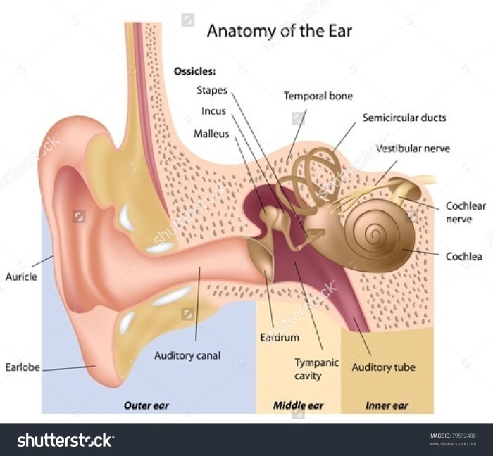
    • Visual Inspection: A careful visual inspection of the ear canal, using an otoscope, is crucial. This allows for the observation of any abnormalities, including inflammation, redness, or the presence of mites, debris, or secretions.
    • Otoscopic Examination: The otoscope provides a magnified view of the ear canal. It allows the examiner to identify the presence of tiny mites, their droppings (which appear as small, dark specks), or other signs of infection.
    • Collection of Debris for Microscopy: If mites or their debris are suspected, a sample of the debris can be collected and examined under a microscope. This microscopic analysis can confirm the presence of mites, which typically appear as small, eight-legged creatures.

    Diagnostic Tools and Typical Results

    The following table summarizes common diagnostic tools and their typical results when used to assess for ear mites:

    Diagnostic Tool Typical Results (Ear Mites Present) Typical Results (Ear Mites Absent)
    Otoscopic Examination Presence of small, dark specks (mite droppings), or tiny, eight-legged creatures within the ear canal. Possible inflammation or redness. Normal ear canal appearance; absence of mites, debris, or inflammation.
    Microscopic Examination of Debris Identification of mites or their characteristic parts (e.g., eggs, fecal matter). Absence of mites or their components in the collected debris.

    Treatment and Management

    Ear mite infestations, while often treatable, require a structured approach to ensure complete eradication and prevent recurrence. Proper diagnosis and treatment are crucial to alleviate discomfort and prevent potential complications. The treatment plan should be tailored to the individual patient and their specific needs.Effective management of ear mites involves a combination of topical medications and meticulous cleaning procedures.

    This multifaceted strategy targets the mites directly while also addressing the environment where they thrive. The key is to follow the prescribed treatment regimen diligently to maximize its effectiveness.

    Common Treatment Approaches

    The primary approach to treating ear mites in humans involves the use of topical medications designed to eliminate the mites and any resulting irritation. These medications come in various forms, and the choice often depends on the severity of the infestation and the patient’s specific needs.

    Medication Options

    Various medications are available to address ear mite infestations. These medications typically contain insecticidal agents that effectively kill the mites.

    Ever wondered about ear mites in humans? They’re surprisingly common, causing itchy ears and discomfort. Fortunately, many treatments are available, some of which involve prescription and otc drugs prescription and otc drugs. These medications, either over-the-counter or needing a doctor’s prescription, can effectively target the mites and ease the symptoms. So, if you suspect you have ear mites, it’s always best to consult a medical professional for proper diagnosis and treatment.

    • Otic Solutions: These solutions are formulated to be applied directly into the ear canal. They contain active ingredients like acaricide to kill mites. A common example is a solution containing a specific acaricide, which is applied as directed. Proper application technique is crucial to ensure the medication reaches all affected areas. For example, if the solution is viscous, it may need to be gently warmed to improve flow.

    • Ear Drops: These drops, similar to otic solutions, are applied directly into the ear canal. They contain similar active ingredients and are used to kill mites. A specific example of an ear drop formulation, containing a certain concentration of acaricide, is frequently employed. These drops should be applied as directed, ensuring the entire ear canal is coated. Using a dropper to administer the ear drops ensures precision and reduces the risk of contaminating the solution.

    Effectiveness and Potential Side Effects

    The effectiveness of treatments for ear mites varies depending on factors like the severity of the infestation and adherence to the prescribed treatment regimen. Most topical treatments are effective when used consistently and as directed.

    • Effectiveness: If used properly, topical treatments typically eliminate ear mites within a few days to a week. However, it’s crucial to complete the entire course of treatment, even if the symptoms improve earlier, to prevent re-infestation. For instance, some individuals might notice a reduction in itching within a few days but should still continue treatment to ensure all mites are eliminated.

    • Side Effects: Potential side effects of topical medications include mild irritation, itching, or redness in the ear canal. These side effects are generally temporary and resolve with continued treatment or by using a milder formulation. Rarely, more severe reactions can occur. It’s essential to monitor for any unusual reactions and report them to a healthcare professional immediately.

    Management Steps and Follow-up Care, Ear mites in humans

    Proper management of ear mite infestations includes consistent application of the prescribed medication and diligent follow-up care.

    1. Adherence to Treatment Regimen: This is paramount to achieving effective treatment. It is crucial to follow the prescribed dosage and frequency of application as directed by a healthcare professional. This includes the number of drops, the duration of treatment, and any necessary precautions.
    2. Follow-up Appointments: Regular follow-up appointments with a healthcare professional are essential to monitor the effectiveness of treatment and address any concerns or complications. This allows the healthcare professional to assess the condition, adjust the treatment plan if needed, and ensure complete eradication of the mites.
    3. Cleaning Procedures: In addition to medication, gentle cleaning of the ear canal can help remove debris and facilitate the effectiveness of the treatment. Excessive cleaning, however, should be avoided.

    Prevention and Control Measures

    Ear mites, while often a nuisance, can be effectively managed with proactive measures. Understanding the factors contributing to their presence and implementing appropriate preventive strategies are key to minimizing the risk of infestation. This section details practical steps to reduce the risk of ear mites and maintain ear health.Preventing ear mite infestations involves a multifaceted approach, encompassing personal hygiene, environmental awareness, and proactive measures within shared living spaces.

    By focusing on these aspects, the likelihood of encountering these tiny parasites can be significantly decreased.

    Preventive Measures to Reduce Infestation Risk

    Personal hygiene plays a crucial role in preventing ear mite infestations. Regular cleaning of the ear canal, while important, should be done gently and avoid deep insertion of cotton swabs or other objects. Excessive cleaning can potentially damage the ear canal, increasing the risk of infection.

    Maintaining Ear Hygiene

    Regular and gentle ear cleaning is essential to prevent the buildup of debris that can create a conducive environment for ear mites. Use a clean washcloth or cotton ball moistened with a mild, non-irritating solution, such as warm water. Avoid using harsh chemicals or solutions that could irritate the delicate ear canal lining. Towel-dry the ear gently after cleaning.

    Ensure you avoid inserting anything into the ear canal beyond the visible part of the ear.

    Ever wondered about ear mites in humans? It’s a fascinating, albeit often uncomfortable, topic. Understanding how we approach these situations is crucial, especially when considering the ethical implications involved in handling such cases. This directly connects to our commerce guidelines and mission, commerce guidelines and mission , which emphasizes responsible and ethical practices. Ultimately, educating ourselves and others about ear mites in humans, and the related issues, is a key component of a healthy community.

    Environmental Factors in Prevention

    Environmental factors can also influence the prevalence of ear mites. Maintaining a clean living environment, especially in shared spaces, can help minimize the risk of infestation. Regular cleaning of bedding, floors, and shared surfaces can help prevent the accumulation of dust and debris, which may harbor mites or their eggs.

    Tips for Preventing Ear Mite Infestations in Families or Shared Living Spaces

    Shared living spaces, such as dormitories or households with multiple individuals, require extra attention to hygiene and cleanliness. Regular cleaning and disinfection of shared areas, especially bedding, can significantly reduce the risk of infestation.

    • Regularly clean and disinfect shared bedding, such as pillows, blankets, and mattresses. This includes vacuuming or sweeping the floor frequently to remove dust mites and debris.
    • Enforce good hygiene practices for all members of the household, particularly children. Teach them about the importance of keeping their ears clean and avoiding the use of foreign objects in their ears.
    • If an infestation is suspected or confirmed, consult a healthcare professional promptly for appropriate treatment and to prevent the spread within the family or shared living space.
    • Regularly inspect pets for ear mites, as they can serve as carriers and spread the infestation to humans. If a pet has ear mites, seek veterinary care immediately.

    Complications and Prognosis

    Ear mites in humans

    Ear mite infestations, while often treatable, can lead to a range of complications if left untreated. Understanding the potential consequences and the prognosis for successful treatment is crucial for managing these infestations effectively. The severity of complications can vary depending on factors such as the individual’s overall health, the extent of the infestation, and the promptness of treatment.The potential complications and long-term consequences of untreated ear mite infestations are multifaceted.

    These range from mild discomfort to more serious conditions, highlighting the importance of seeking timely medical attention. Appropriate treatment strategies are crucial for mitigating these complications and ensuring a favorable prognosis.

    Potential Complications of Untreated Infestations

    Untreated ear mite infestations can cause a range of complications, ranging from mild to severe. The mites themselves can irritate the ear canal, leading to discomfort, itching, and pain. Constant scratching can further damage the delicate skin lining the ear canal, increasing the risk of secondary infections.

    • Secondary Infections: A significant concern associated with untreated ear mite infestations is the potential for secondary bacterial or fungal infections. The irritated and compromised skin lining the ear canal becomes a breeding ground for harmful microorganisms, leading to more intense inflammation, pain, and potentially more serious complications.
    • Chronic Ear Infections: Persistent scratching and irritation can lead to chronic ear infections. This can involve a prolonged inflammatory response in the ear canal, causing ongoing pain, discomfort, and difficulty hearing.
    • Hearing Loss: In severe cases, untreated ear mite infestations can lead to temporary or even permanent hearing loss. The inflammation and potential damage to the eardrum and inner ear structures can impair the transmission of sound waves, impacting hearing function.
    • Tinnitus: Persistent irritation and inflammation in the ear canal can cause tinnitus, a condition characterized by a ringing or buzzing sensation in the ears. This can be a persistent and distressing symptom, impacting the individual’s quality of life.

    Long-Term Consequences of Untreated Infestations

    The long-term consequences of untreated ear mite infestations can be significant and impact the individual’s quality of life. The persistent itching and pain can lead to chronic discomfort and sleep disturbances. Furthermore, the potential for secondary infections and hearing loss can have lasting implications.

    • Chronic Itching and Pain: Untreated ear mite infestations can lead to persistent itching and pain, disrupting sleep patterns and overall quality of life. This constant discomfort can be debilitating, impacting the individual’s ability to focus and participate in daily activities.
    • Impaired Hearing: Untreated infections can lead to permanent hearing damage, particularly if the infection spreads to the inner ear. This can significantly impact the individual’s ability to communicate and engage in social interactions.
    • Psychological Impact: The persistent discomfort and potential hearing loss associated with untreated ear mite infestations can take a toll on the individual’s mental well-being. The emotional distress and anxiety associated with these conditions can negatively affect the individual’s overall health and functioning.

    Prognosis with Appropriate Treatment

    The prognosis for individuals with ear mite infestations is generally excellent when appropriate treatment is followed. Most cases respond well to the prescribed medications, and the discomfort and complications are typically resolved within a few weeks. The efficacy of treatment can be significantly affected by adherence to the prescribed regimen and the promptness of seeking medical attention.

    Comparison of Treatment Approaches

    Different treatment approaches for ear mite infestations vary in their effectiveness and potential side effects. A thorough assessment of the individual’s condition and medical history is crucial in determining the most suitable treatment plan.

    • Topical Medications: Topical medications, such as ear drops containing acaricide, are often effective in eliminating the mites and reducing inflammation. The effectiveness depends on the individual’s adherence to the prescribed treatment duration and the severity of the infestation.
    • Oral Medications: In some cases, oral medications may be necessary, particularly for extensive infestations or individuals with underlying health conditions. The efficacy and potential side effects of oral medications vary, and the decision to use them is made on a case-by-case basis.

    Illustrative Case Studies

    Ear mite infestations, while often treatable, can vary significantly in presentation and severity. Understanding these variations through case studies provides valuable insights into effective diagnosis, treatment, and potential complications. These examples highlight the importance of prompt attention to ear health and the range of potential outcomes.Successful treatment often relies on accurate diagnosis and adherence to prescribed therapies. The following case studies illustrate different scenarios, from simple infestations to more complex cases requiring specialized intervention.

    Successful Treatment of an Ear Mite Infestation

    A 10-year-old boy presented with itching and a mild discharge from his right ear. Otoscopic examination revealed small, whitish mites in the ear canal. The patient was prescribed a topical acaricide, a medication specifically designed to kill mites. Following a week of daily application, the itching subsided, and the discharge ceased. A follow-up examination confirmed the successful eradication of the mites.

    This case demonstrates a straightforward and effective treatment approach for a typical ear mite infestation.

    Importance of Early Diagnosis and Treatment

    A 3-year-old girl experienced persistent ear itching for several weeks. Initially, the parents attributed the discomfort to allergies. However, the itching worsened, and a yellowish discharge became apparent. Delayed diagnosis and treatment led to a secondary bacterial infection within the ear canal. The infection required a course of oral antibiotics in addition to topical medications.

    Early diagnosis, through prompt otoscopic examination, could have prevented the development of the secondary infection.

    Complications of Delayed Treatment

    A 25-year-old male experienced a severe earache and intense itching. He neglected the symptoms for two months, believing they were a minor annoyance. By the time he sought medical attention, the ear canal was inflamed and swollen, with significant tissue damage evident. Aural polyps developed in the ear canal. The patient required a more involved treatment plan, including surgical intervention to remove the polyps and address the extensive inflammation.

    This case highlights the potential for severe complications, including infection and tissue damage, resulting from delayed treatment of ear mites.

    Rare or Unusual Presentation of Ear Mites

    A 60-year-old woman presented with a unique presentation of ear mites. In addition to the typical symptoms of itching and discharge, she also experienced an unusual level of dizziness and tinnitus. Otoscopic examination revealed a dense cluster of mites along with some debris. The unusual combination of symptoms suggested a possible underlying neurological involvement. The patient responded well to the standard treatment, but the case underscored the potential for atypical presentations and the importance of considering broader differential diagnoses.

    Ear Mites in Humans

    Ear mites, tiny arachnids, are a common cause of ear discomfort in humans. While less prevalent than other ear conditions, understanding their characteristics, diagnosis, and treatment is essential for effective management. These parasites, though rarely serious, can lead to significant discomfort if left untreated.

    Patient-Friendly Information on Ear Mites

    Ear mites, scientifically known asOtodectes cynotis*, are microscopic parasites that primarily affect the external ear canal. They are typically transmitted through close contact with infected animals, such as dogs or cats. While they don’t usually pose a significant health risk to humans, they can cause itching, redness, and a discharge in the ear. Early detection and treatment are crucial for alleviating discomfort and preventing potential complications.

    Common Questions and Answers

    This table provides concise answers to frequently asked questions about ear mites in humans.

    Question Answer
    What are ear mites? Ear mites are tiny parasites that infest the external ear canal. They are typically transmitted from infected animals.
    What are the symptoms of ear mites? Symptoms often include intense itching, redness, and a yellowish-brown discharge in the ear canal. Some individuals may also experience pain or a feeling of fullness in the ear.
    How are ear mites diagnosed? Diagnosis typically involves an examination of the ear canal by a healthcare professional. The presence of mites or their droppings can be observed using an otoscope.
    How are ear mites treated? Treatment usually involves topical medications, such as ear drops containing acaricide, prescribed by a doctor. These medications kill the mites and relieve symptoms.
    Can ear mites be prevented? Minimizing contact with infected animals, especially pets, can help prevent the spread of ear mites. Practicing good hygiene and promptly addressing any symptoms can also help prevent complications.
    Are ear mites contagious? While less contagious than some other ear infections, ear mites can be transmitted through close contact with infected animals.

    Diagnosis and Treatment Flowchart

    This flowchart Artikels the typical steps involved in diagnosing and treating ear mites:

    Note: This flowchart is a general guideline and individual cases may vary. Always consult a healthcare professional for accurate diagnosis and treatment.

    Flowchart

    FAQ: Frequently Asked Questions about Ear Mites

    This section addresses common questions regarding ear mites, providing straightforward answers in an accessible format.

    • What are the common symptoms of ear mites in humans? Common symptoms include intense itching, a yellowish-brown discharge, and redness in the ear canal. Some individuals may experience pain or a feeling of fullness in the ear.
    • How are ear mites typically diagnosed? A healthcare professional typically examines the ear canal using an otoscope to look for the presence of mites or their droppings. A thorough history of symptoms and contact with animals can also help in diagnosis.
    • What is the typical treatment for ear mites? Treatment usually involves the use of topical medications, such as ear drops containing acaricide, prescribed by a healthcare professional. These medications kill the mites and alleviate symptoms.
    • What is the prognosis for ear mites? With appropriate treatment, ear mites typically resolve within a few weeks. Prompt treatment is essential to minimize discomfort and prevent complications.

    Closing Notes

    In conclusion, while ear mites in humans aren’t as prevalent as other ear issues, understanding their lifecycle, symptoms, and treatment is crucial for timely intervention. This guide offers a roadmap to navigate the complexities of ear mite infestations, empowering you to make informed decisions about your health and well-being. Prevention, early detection, and proper treatment are key to managing and overcoming these small but potentially troublesome parasites.

  • Insulinoma Overview and More A Deep Dive

    Insulinoma Overview and More A Deep Dive

    Insulinoma overview and more delves into the intricacies of this rare pancreatic tumor. We’ll explore the defining characteristics of insulinoma, from its development and symptoms to various diagnostic methods, management strategies, and long-term outcomes. This in-depth look will help readers understand this complex condition and its impact on patients’ lives.

    Understanding the different types of insulinomas, their diagnostic processes, and effective treatment options is crucial. We’ll also cover illustrative case studies and potential risk factors to provide a comprehensive perspective on insulinoma. This will be a journey of discovery into the world of insulinoma, addressing critical questions and concerns.

    Introduction to Insulinoma

    Insulinoma is a rare neuroendocrine tumor that arises from the beta cells of the pancreas. It’s characterized by excessive insulin production, leading to hypoglycemia, a potentially dangerous condition where blood sugar levels drop too low. Understanding its development, symptoms, and diagnostic process is crucial for timely intervention and management.The pathophysiology of insulinoma involves the uncontrolled proliferation and function of beta cells within the pancreatic islets of Langerhans.

    This unregulated insulin secretion overwhelms the body’s normal glucose regulatory mechanisms, resulting in hypoglycemic episodes. Genetic factors and mutations in specific genes are implicated in some cases, while other instances appear to develop sporadically.

    Symptoms of Insulinoma

    Symptoms of insulinoma vary significantly, depending on the tumor’s size and location within the pancreas, as well as the individual’s response to fluctuating insulin levels. Common symptoms include excessive hunger (polyphagia), fatigue, sweating, palpitations, dizziness, headache, and even seizures or loss of consciousness. These symptoms can often be mistaken for other conditions, leading to delayed diagnosis.

    Age and Gender Distribution

    Insulinoma can affect individuals across various age groups, but it is most commonly diagnosed in adults between the ages of 30 and 60. There is no significant difference in the incidence of insulinoma between men and women.

    Diagnostic Tests for Insulinoma

    A precise diagnosis of insulinoma relies on a combination of tests, evaluating both clinical symptoms and laboratory findings. These tests aim to confirm the presence of hypoglycemia and pinpoint the source.

    Test Name Procedure Expected Result (Normal) Expected Result (Insulinoma)
    Fasting Blood Glucose (FBG) Measuring blood glucose levels after an overnight fast. Normal range (e.g., 70-100 mg/dL) Significantly low blood glucose level (e.g., below 50 mg/dL)
    Oral Glucose Tolerance Test (OGTT) Measuring blood glucose levels after consuming a sugary drink. Blood glucose levels rise and return to normal within a certain time frame. Markedly low blood glucose level, possibly more pronounced after the glucose load, indicating exaggerated insulin response.
    Insulin Level Measurement Measuring the level of insulin in the blood. Normal range (varies based on fasting or post-meal) Elevated insulin levels, particularly during episodes of hypoglycemia, often disproportionate to blood glucose levels.
    Imaging Studies (e.g., CT scan, MRI) Using imaging techniques to visualize the pancreas and surrounding structures. Normal pancreatic anatomy May reveal a mass (tumor) in the pancreas, helping to locate the source of the elevated insulin.

    Types and Classification of Insulinoma

    Insulinoma, a rare pancreatic neuroendocrine tumor, is characterized by excessive insulin production. Understanding the diverse types and classifications of insulinoma is crucial for accurate diagnosis and tailored treatment strategies. Different subtypes may exhibit varying clinical presentations and prognoses.The classification of insulinomas primarily relies on their cellular origin and genetic mutations. This allows for a deeper understanding of the underlying mechanisms driving the tumor’s development and behavior.

    This knowledge is vital for predicting disease progression and developing targeted therapies.

    Cellular Origin and Histology

    Insulinoma tumors arise from the pancreatic islet cells, specifically the beta cells that produce insulin. While most insulinomas are of a single cell type, mixed cell tumors can occur. The histological appearance can vary, from well-differentiated tumors to more aggressive, poorly differentiated ones. Histological analysis plays a vital role in characterizing the tumor and assessing its potential malignancy.

    Genetic Mutations

    Genetic mutations are frequently linked to insulinoma development. Specific mutations in genes involved in cell growth and regulation can contribute to uncontrolled cell proliferation. These mutations can be inherited or acquired. The identification of these genetic alterations helps in risk assessment and potential predisposition for the development of other tumors. Understanding these mutations allows for a better understanding of the underlying biological mechanisms that contribute to tumorigenesis.

    Examples include mutations in the MEN1 gene, which is often associated with multiple endocrine neoplasia syndromes.

    Comparison of Insulinoma Subtypes

    Subtype Prevalence Symptoms Prognosis
    Classic Insulinoma Most common Characterized by episodic hypoglycemia, including symptoms like sweating, shakiness, confusion, and seizures. The severity and frequency of symptoms can vary significantly between individuals. Generally, classic insulinoma, when diagnosed and treated early, has a favorable prognosis. Surgical resection often leads to complete remission.
    Multiple Endocrine Neoplasia Type 1 (MEN1)-associated Insulinoma Less common, often part of a larger endocrine tumor syndrome Patients with MEN1-associated insulinoma frequently present with a constellation of endocrine tumors, including those of the parathyroid glands and pituitary. Symptoms related to hypoglycemia are common. The prognosis for MEN1-associated insulinoma depends on the extent of the MEN1 syndrome and the timing of diagnosis. Early detection and aggressive management are crucial.
    Insulinoma with Extrapancreatic Components Rare These tumors may spread to other areas outside the pancreas. The symptoms depend on the extent of spread and the organs involved. The prognosis is generally less favorable than for classic insulinoma due to the increased likelihood of tumor recurrence and metastasis.

    This table highlights the diverse characteristics of different insulinoma subtypes. Factors such as prevalence, associated symptoms, and prognosis are important to consider when evaluating patients with this condition.

    Diagnosis and Workup

    Diagnosing insulinoma, a rare pancreatic neuroendocrine tumor, requires a meticulous approach combining clinical evaluation, laboratory tests, and imaging studies. The key lies in identifying the characteristic pattern of hypoglycemia, often triggered by fasting or prolonged periods without food. This careful process distinguishes insulinoma from other causes of low blood sugar, ensuring accurate diagnosis and timely treatment.

    Initial Screening and Clinical Evaluation

    The diagnostic journey begins with a thorough history and physical examination. Patients exhibiting recurrent hypoglycemic episodes, particularly those associated with fasting or skipping meals, are flagged for further investigation. A detailed history encompassing the frequency, duration, and symptoms of these episodes is crucial. Physical examination may reveal symptoms like sweating, tremors, and confusion, indicative of hypoglycemia. Identifying risk factors, such as a family history of neuroendocrine tumors, can also play a significant role in prioritizing the investigation.

    Diagnostic Procedures

    Several diagnostic procedures aid in confirming the presence and location of an insulinoma. The gold standard for diagnosing insulinoma remains the demonstration of hypoglycemia during a 72-hour fast, or an 8-hour fast. During the fast, blood glucose levels are monitored at regular intervals. This prolonged fast helps induce hypoglycemia, making the detection of insulinoma more definitive. Furthermore, the presence of elevated insulin levels and suppressed blood glucose levels during the fast strongly suggests an insulinoma.

    Laboratory Tests, Insulinoma overview and more

    Differentiating insulinoma from other causes of hypoglycemia is critical. Key laboratory tests focus on insulin levels, C-peptide levels, and glucose levels. Measuring plasma insulin levels during hypoglycemia is a crucial indicator. Elevated insulin levels, particularly in the presence of suppressed glucose levels, are highly suggestive of an insulinoma. Conversely, normal or low insulin levels during hypoglycemia point towards alternative causes.

    In addition, measuring C-peptide levels can further differentiate insulinoma from exogenous insulin administration. Elevated C-peptide levels are consistent with an insulinoma.

    So, I’ve been diving deep into insulinoma overview and more, and it’s fascinating how complex these endocrine issues can be. While researching, I stumbled upon a really helpful article about navigating work with chronic illnesses like fibromyalgia and CFS, which made me think about how important it is to find the right balance. Learning about managing symptoms and work situations, like in can i keep working with fibromyalgia and cfs , is a crucial piece of the puzzle when dealing with insulinoma and other health conditions.

    Ultimately, understanding these conditions better helps us all navigate our own journeys.

    Imaging Techniques

    Imaging techniques, while not always essential for initial diagnosis, play a crucial role in localizing the tumor for surgical intervention. Computed tomography (CT) scans and magnetic resonance imaging (MRI) scans are commonly used. CT scans provide detailed anatomical information, while MRI scans offer superior soft tissue contrast, which aids in visualizing the pancreas and potentially identifying the tumor.

    These techniques, in conjunction with other diagnostic tools, help delineate the location and size of the suspected insulinoma, facilitating precise surgical planning.

    Diagnostic Workup Steps

    Step Procedure Findings (Normal) Findings (Insulinoma)
    1 Detailed history and physical examination No symptoms or signs suggestive of hypoglycemia History of recurrent hypoglycemic episodes, associated symptoms (e.g., sweating, tremors, confusion)
    2 Fasting blood glucose monitoring (72-hour fast) Blood glucose levels within normal range throughout the fast Significant and sustained drop in blood glucose levels during the fast
    3 Plasma insulin and C-peptide measurements during hypoglycemia Normal or low insulin and C-peptide levels during hypoglycemia Elevated insulin and C-peptide levels during hypoglycemia
    4 Imaging studies (CT or MRI) No evidence of pancreatic tumor Presence of a pancreatic tumor, particularly in the pancreatic head, body, or tail

    Management Strategies

    Insulinoma management hinges on accurately diagnosing the tumor and tailoring treatment to the individual patient. The goal is to effectively control hypoglycemia and, if possible, remove the tumor, preserving overall health and quality of life. Different approaches exist, ranging from medical therapies to surgical intervention.Effective management requires a multidisciplinary approach involving endocrinologists, surgeons, and potentially other specialists. Close monitoring and adjustments to treatment are crucial for successful outcomes.

    Surgical Resection

    Surgical removal of the insulinoma is often the definitive treatment, especially for solitary tumors. Surgical resection aims to completely eliminate the tumor responsible for excessive insulin production, thus resolving hypoglycemic episodes. Preoperative evaluation and careful patient selection are paramount to ensure a successful outcome. This involves assessing the tumor’s location, size, and potential involvement of surrounding structures.

    Open surgery or minimally invasive techniques like laparoscopic surgery are employed depending on the tumor’s location and the surgeon’s expertise.

    Medical Therapies

    Medical therapies play a supportive role in managing insulinoma, particularly when surgical resection is not feasible or when the tumor is not amenable to surgical removal. These therapies aim to mitigate hypoglycemic episodes and reduce the tumor’s activity. Drugs like octreotide and lanreotide are commonly used. These somatostatin analogs act by suppressing insulin secretion from the tumor.

    Diazoxide, another medication, can also be used to reduce insulin secretion. However, their efficacy can vary significantly between patients.

    Diving into insulinoma overview and more, it’s fascinating how these tumors affect blood sugar levels. While researching different types of endocrine disorders, I stumbled across some really interesting facts about Parkinson’s disease, like its impact on motor skills. facts about parkinsons disease are certainly worth exploring. Ultimately, though, the core focus of my research remains on the various causes and treatments associated with insulinoma overview and more.

    Long-Term Follow-Up

    Long-term follow-up is critical for patients with a history of insulinoma. Regular monitoring of blood glucose levels, assessment for recurrence, and evaluation of any treatment-related complications are essential. Patients need ongoing support and education to ensure they understand the importance of consistent blood glucose monitoring and management. This may involve regular appointments with endocrinologists and access to resources for managing their condition.

    Potential Complications

    Potential complications of insulinoma treatment include surgical risks, such as bleeding, infection, and damage to surrounding structures. Postoperative complications like persistent hypoglycemia or complications related to the use of medical therapies (such as octreotide) may also occur. Careful monitoring and prompt management of these complications are essential.

    Summary Table of Treatment Options

    Treatment Procedure Effectiveness Potential Complications
    Surgical Resection Surgical removal of the tumor Generally high, especially for solitary tumors, potentially curative Bleeding, infection, damage to surrounding structures, recurrence
    Medical Therapies (e.g., octreotide, lanreotide, diazoxide) Administration of medications to suppress insulin secretion Variable, may be effective in controlling symptoms but not always curative Side effects specific to each medication (e.g., abdominal pain, diarrhea, nausea), possible lack of efficacy

    Prognosis and Long-Term Outcomes

    Insulinoma, while treatable, presents a unique challenge in terms of long-term management. Understanding the prognosis, including the likelihood of cure, recurrence, and the impact on quality of life, is crucial for both patients and healthcare providers. This section delves into these key aspects, offering a comprehensive overview.The overall prognosis for individuals with insulinoma is generally favorable, especially when the tumor is localized and diagnosed early.

    However, factors such as tumor size, aggressiveness, and the presence of multiple tumors can influence the long-term outcome. Complete surgical removal is often the primary treatment goal, and the success of this procedure significantly impacts the likelihood of long-term stability.

    Likelihood of Cure and Recurrence

    Complete surgical resection of the insulinoma is often curative, leading to resolution of symptoms and normalization of blood glucose levels. However, recurrence is a possibility, particularly if the initial diagnosis or surgical approach was not optimal. Factors such as incomplete tumor removal, the presence of multiple tumors, or the aggressive nature of the tumor can increase the risk of recurrence.

    Studies have shown recurrence rates varying widely, depending on the specifics of the case. For example, in cases where the tumor was well-defined and completely resected, the recurrence rate might be lower than in cases with extensive or diffuse tumor growth.

    Factors Influencing Long-Term Outcomes

    Several factors significantly influence the long-term outcomes for individuals with insulinoma. These factors include the size and location of the tumor, the extent of the surgical resection, the presence of multiple tumors, and the overall health status of the patient. Patients with smaller, localized tumors and complete surgical removal tend to have a better prognosis compared to those with larger, more aggressive tumors or incomplete resection.

    Pre-existing medical conditions, such as diabetes or other chronic illnesses, can also affect the patient’s ability to tolerate treatment and recovery.

    Impact on Quality of Life

    Insulinoma can significantly impact the quality of life of affected individuals. The fluctuating blood glucose levels, the associated symptoms like fatigue, weakness, and confusion, and the need for frequent medical monitoring can create considerable stress and anxiety. Furthermore, the diagnostic process itself, including multiple tests and procedures, can be time-consuming and disruptive. The successful treatment and management of insulinoma are essential for restoring a more stable and healthy quality of life for these individuals.

    Comparison of Treatment Approaches

    Treatment Recurrence Rate Survival Rate Quality of Life Impact
    Surgical Resection (Complete) Generally lower Excellent Improved significantly
    Surgical Resection (Incomplete) Higher Good, but potentially with complications Potential for ongoing symptoms
    Medical Management (e.g., medications) Variable Good, but often with persistent symptoms Limited improvement in quality of life
    Combination Therapy Dependent on specific combination Good May result in improved symptoms and quality of life

    The table above provides a general comparison of long-term outcomes associated with different treatment approaches. Specific outcomes can vary significantly depending on individual factors and the specifics of each case.

    Illustrative Case Studies

    Understanding insulinoma cases is crucial for effective diagnosis and management. Real-world examples highlight the diverse presentations, diagnostic challenges, and treatment approaches associated with this rare endocrine tumor. These case studies provide valuable insights into the complexities of insulinoma and aid in developing a more nuanced understanding of this condition.

    Learning about insulinoma, a rare pancreatic tumor, is fascinating. Understanding its impact on blood sugar levels is key. Interestingly, post-surgery, scar tissue management plays a crucial role in recovery, and techniques like scar tissue massage and management can significantly aid in flexibility and reduce discomfort. Further research into insulinoma overview and treatment options is essential for comprehensive understanding.

    Case Study 1: A Diagnosis of Insulinoma

    This case describes a 45-year-old female patient presenting with recurrent episodes of hypoglycemia. Symptoms included lightheadedness, confusion, and blurred vision, particularly after fasting periods. Initial investigations revealed low blood sugar levels, and further diagnostic testing, including a glucose tolerance test and an insulin assay, confirmed a suspected insulinoma. The patient underwent a detailed medical history review and a thorough physical examination.

    Further, a selective arterial calcium stimulation test was used to further localize the tumor. A surgical resection was performed to remove the tumor, successfully resolving the patient’s hypoglycemia.

    Case Study 1: Symptoms and Diagnosis

    The patient’s symptoms, including recurrent hypoglycemia episodes after fasting, pointed towards a possible pancreatic tumor. Low blood sugar levels were confirmed during initial investigations. A glucose tolerance test demonstrated an exaggerated insulin response, further supporting the suspicion of insulinoma. Subsequent insulin assay results provided conclusive evidence of elevated insulin levels, prompting further investigation to identify the source of the elevated insulin levels.

    Case Study 1: Management and Treatment

    The management of this insulinoma case involved a multidisciplinary approach. Preoperative evaluation included detailed imaging studies, such as CT scans and MRI, to precisely locate the tumor. Endoscopic ultrasound was also considered. Surgical resection of the tumor was the primary treatment modality. Postoperative monitoring was essential to ensure stable blood sugar levels and to identify any potential complications.

    Careful monitoring of blood glucose levels after surgery was a key aspect of post-operative management.

    Case Study 2: Insulinoma Treatment and Recovery

    This case involved a 32-year-old male patient diagnosed with a small insulinoma. Initial symptoms included excessive fatigue, anxiety, and tremors. The diagnosis was confirmed through a combination of laboratory tests and imaging studies, such as CT scans and MRI. The patient opted for an endoscopic ultrasound-guided procedure to remove the tumor. The minimally invasive nature of this approach was a significant factor in the patient’s recovery process.

    The patient demonstrated a rapid and favorable response to treatment, and follow-up visits confirmed long-term remission of symptoms.

    Case Study 2: Symptoms and Diagnosis

    The patient’s symptoms included fatigue, anxiety, and tremors. These symptoms were indicative of fluctuating blood sugar levels, particularly low blood sugar. Diagnostic procedures, including blood tests and imaging scans, were utilized to identify the cause of these symptoms. The patient’s history of symptoms, combined with laboratory results, suggested a likely insulinoma. A detailed history of the patient’s symptoms and medical history was obtained.

    Case Study 2: Management and Treatment

    The patient’s management involved a minimally invasive endoscopic ultrasound-guided procedure. This approach allowed for precise tumor removal with minimal surgical trauma. Post-operative care included close monitoring of blood glucose levels and ongoing support from the medical team. The patient’s recovery was relatively swift, with a significant improvement in symptoms observed within a few weeks. The patient’s recovery trajectory was closely monitored and managed.

    Case Study Outcomes

    Both cases highlight the successful management of insulinoma. In Case Study 1, surgical resection led to complete remission of hypoglycemic episodes. In Case Study 2, an endoscopic approach yielded favorable outcomes and rapid recovery. The positive outcomes emphasize the importance of early diagnosis and appropriate treatment strategies.

    Prevention and Risk Factors

    Insulinoma overview and more

    Insulinoma, a rare tumor of the pancreas, presents a unique challenge in terms of prevention. While we can’t prevent all cancers, understanding potential risk factors allows for proactive strategies and improved patient outcomes. Early detection and appropriate management are crucial in these cases.

    Potential Risk Factors

    Several factors might contribute to the development of insulinoma, though definitive causes are not always clear. A combination of genetic predisposition and environmental influences likely play a role.

    Genetic Predisposition

    A significant portion of insuloma cases are sporadic, meaning they occur without any apparent family history. However, some families exhibit a higher incidence of the condition, suggesting a possible genetic component. Inherited mutations in specific genes can increase the risk of developing various endocrine tumors, including insulinomas. For instance, multiple endocrine neoplasia type 1 (MEN1) syndrome is associated with a markedly elevated risk of insulinoma and other pancreatic endocrine tumors.

    Understanding these genetic predispositions is vital for risk assessment and potential screening in high-risk individuals.

    Modifiable Risk Factors

    While certain risk factors are largely out of our control, others can be influenced through lifestyle choices. Maintaining a healthy weight, engaging in regular physical activity, and following a balanced diet can contribute to overall well-being and potentially reduce the risk of developing various diseases, including those related to pancreatic function.

    Table of Risk Factors for Insulinoma

    Risk Factor Description Strength of Evidence Modifiability
    Family history of insulinoma or other endocrine tumors Presence of insulinoma or related tumors in close relatives. Moderate Non-modifiable
    MEN1 syndrome Inherited genetic condition increasing risk of various endocrine tumors. Strong Non-modifiable
    Pancreatitis Inflammation of the pancreas. Possible Partially modifiable (e.g., through dietary changes, smoking cessation).
    Exposure to certain toxins or chemicals Potential link to insulinoma development but requires further research. Limited Partially modifiable (e.g., avoiding exposure).

    Wrap-Up: Insulinoma Overview And More

    Insulinoma overview and more

    In conclusion, insulinoma, a rare pancreatic tumor, presents a complex diagnostic and management challenge. This overview highlights the critical aspects of insulinoma, from its pathophysiology and clinical presentation to the various treatment options and long-term outcomes. By understanding the nuances of this condition, we can better equip ourselves to provide the best possible support and care for those affected.

  • Facts About Parkinsons Disease A Deep Dive

    Facts About Parkinsons Disease A Deep Dive

    Facts about parkinsons disease – Facts about Parkinson’s disease sets the stage for this enthralling narrative, offering readers a glimpse into a complex and multifaceted condition. This comprehensive exploration delves into the various aspects of Parkinson’s, from its core symptoms and causes to treatment options and the impact it has on individuals and families.

    We’ll cover the basics, including definitions, symptoms, and how Parkinson’s affects daily life. Then, we’ll examine the possible causes and risk factors, along with the diagnostic process and available treatment options. Furthermore, we’ll explore strategies for living with the disease, the challenges faced by families and caregivers, and the ongoing research into potential treatments and preventive measures. Finally, we’ll look at illustrative case studies and the impact of the disease on individual lives.

    Introduction to Parkinson’s Disease

    Parkinson’s disease is a progressive neurological disorder that affects movement. It’s characterized by a gradual decline in the production of dopamine, a crucial neurotransmitter involved in controlling movement. This decline leads to a range of motor and non-motor symptoms that progressively worsen over time. The disease typically affects people over the age of 60, although younger onset cases do exist.Understanding the core symptoms, stages, and impact of Parkinson’s disease is vital for effective management and support for individuals living with this condition.

    Early diagnosis and appropriate treatment strategies can significantly improve quality of life and slow the progression of the disease.

    Definition of Parkinson’s Disease

    Parkinson’s disease is a neurodegenerative disorder primarily affecting the central nervous system. It’s characterized by the degeneration of dopamine-producing neurons in a specific area of the brain, leading to a deficiency in dopamine. This deficiency disrupts the normal functioning of the brain’s motor control system.

    Core Symptoms of Parkinson’s Disease

    The primary motor symptoms of Parkinson’s disease include tremors, rigidity, bradykinesia (slowness of movement), and postural instability. Tremors are often the first noticeable symptom, typically occurring at rest and affecting one hand. Rigidity manifests as stiffness in the muscles, making movement difficult and jerky. Bradykinesia leads to slowness in initiating and performing movements, and postural instability results in impaired balance and an increased risk of falls.

    Learning about Parkinson’s disease is fascinating, but it’s also important to understand how various health conditions can intertwine. For example, some medications for atrial fibrillation, like medications for atrial fibrillation , might have unexpected interactions with Parkinson’s treatments. This highlights the complexity of managing multiple health issues and the importance of consulting with healthcare professionals. Ultimately, staying informed about Parkinson’s disease is key to understanding potential connections and managing care effectively.

    Impact of Parkinson’s Disease on Daily Life

    Parkinson’s disease significantly impacts daily life. The progressive nature of the disease often leads to difficulties in performing routine tasks like dressing, eating, and walking. The non-motor symptoms, such as sleep disturbances, cognitive impairment, and depression, further complicate daily functioning. These challenges can affect social interactions, employment, and overall well-being.

    Stages of Parkinson’s Disease

    The progression of Parkinson’s disease is typically categorized into stages, reflecting the severity of symptoms and impact on daily life. This classification aids in treatment planning and managing expectations.

    Stage Symptoms Treatments Quality of Life Implications
    Early Stage (1-2) Mild tremors and rigidity, noticeable only during specific activities. Walking and balance may be slightly affected. Medication to manage symptoms, physical therapy to maintain mobility. Individuals can often maintain independence in daily activities. Social and occupational functioning remain largely intact.
    Mid Stage (3-4) Tremors and rigidity become more pronounced. Walking difficulties, balance problems, and increased risk of falls are evident. Speech may become slurred. Difficulty with fine motor skills. Increased medication dosage, consideration of deep brain stimulation (DBS) for severe cases. Ongoing physical therapy and occupational therapy for improved mobility and daily living skills. Individuals may require assistance with daily tasks. Social interaction may be impacted due to mobility challenges.
    Late Stage (5) Significant motor impairment. Individuals may require assistance with all aspects of daily living, including walking, dressing, and eating. Cognitive decline and other non-motor symptoms may be prominent. High doses of medication. Advanced therapies like DBS, if appropriate. Specialized caregiving and support are crucial. Significant dependence on caregivers for all aspects of daily life. Reduced social interaction and potential for isolation.

    Causes and Risk Factors: Facts About Parkinsons Disease

    Understanding the causes of Parkinson’s Disease is crucial for developing effective treatments and preventive strategies. While the precise mechanisms remain somewhat elusive, researchers have identified a complex interplay of genetic predispositions and environmental influences that contribute to the development of this debilitating neurological disorder. This exploration will delve into the known causes and risk factors, offering insights into the factors that may increase a person’s vulnerability to Parkinson’s.

    Known Causes

    Parkinson’s Disease is not fully understood, but several contributing factors are recognized. Genetic predispositions, environmental exposures, and a combination of both play significant roles in its development. The interplay of these factors is often complex and not yet fully elucidated. The specific mechanisms by which these factors contribute to the disease’s progression remain an active area of research.

    Genetic Factors

    Certain genetic mutations have been linked to an increased risk of developing Parkinson’s Disease. Individuals with a family history of Parkinson’s are more likely to develop the disease themselves, suggesting a genetic component. Specific genes involved in dopamine production and neuronal function have been identified as potential contributors to the disease’s development. However, it’s crucial to understand that a genetic predisposition doesn’t guarantee the development of Parkinson’s, and many people with these genes do not develop the disease.

    Environmental Factors

    Environmental exposures have also been implicated in Parkinson’s Disease development. Exposure to certain pesticides and herbicides has been associated with a heightened risk. Furthermore, some studies suggest a potential link between repeated head trauma and the development of Parkinson’s. While these environmental factors are recognized as potential contributors, the precise mechanisms through which they affect the brain remain unclear.

    Understanding these interactions is essential for the development of preventive measures.

    Comparison of Risk Factors

    Risk Factor Description Potential Impact Examples
    Genetics Specific gene mutations can increase the likelihood of developing Parkinson’s. Increased susceptibility to the disease, especially with a family history. Mutations in genes like LRRK2 and PARKIN.
    Environmental Toxins Exposure to certain pesticides, herbicides, and heavy metals. Possible disruption of neuronal function, potentially leading to Parkinson’s development. Exposure to rotenone, manganese, or paraquat.
    Head Trauma Repeated head injuries, such as those sustained in sports or accidents. Potentially increased risk of developing Parkinson’s, particularly with cumulative trauma. Concussions or repeated blows to the head.
    Age The risk of Parkinson’s increases with age. A major risk factor, as the aging process affects brain function. Individuals over 60 are more susceptible.

    Diagnosis and Assessment

    Diagnosing Parkinson’s Disease involves a careful evaluation process, combining medical history, neurological examinations, and various diagnostic tools. The process aims to rule out other conditions with similar symptoms, ensuring an accurate diagnosis to guide appropriate treatment strategies. Accurate diagnosis is crucial for timely intervention and management of the disease.Neurological examinations are paramount in assessing Parkinson’s Disease. These examinations evaluate motor skills, balance, reflexes, and cognitive functions.

    They provide valuable insights into the progression and severity of the disease, helping clinicians determine the best course of action. The findings from these examinations, combined with other data, contribute to a more comprehensive understanding of the patient’s condition.

    Neurological Examinations

    Neurological examinations are essential for diagnosing Parkinson’s Disease. These examinations assess various aspects of the nervous system, including motor function, balance, coordination, reflexes, and cognitive abilities. A comprehensive neurological examination allows clinicians to identify specific signs and symptoms indicative of Parkinson’s Disease. Observations of posture, gait, tremors, rigidity, bradykinesia, and other motor abnormalities are carefully noted during the examination.

    Diagnostic Tools

    Various diagnostic tools aid in the assessment of Parkinson’s Disease. These tools provide objective measurements and support the clinical evaluation, helping to confirm the diagnosis and differentiate it from other neurological disorders. The integration of multiple diagnostic methods improves the accuracy of the diagnosis.

    Diagnostic Procedures

    The following table summarizes key diagnostic procedures used for Parkinson’s Disease, outlining their purpose, procedure, and typical results.

    Procedure Description Procedure Details Typical Results (Parkinson’s Disease)
    Detailed Medical History Gathering information about the patient’s medical history, including symptoms, onset, duration, and progression of the condition. In-depth discussion about the patient’s symptoms, lifestyle, family history, and use of medications. Reports of gradual onset of tremors, rigidity, slowness of movement (bradykinesia), and balance problems. History of similar symptoms in family members may be reported.
    Neurological Examination Evaluation of motor skills, balance, reflexes, and cognitive functions. Assessment of posture, gait, tremors, rigidity, bradykinesia, and response to commands. Tests for reflexes and coordination are also performed. Presence of resting tremor, rigidity in limbs, bradykinesia (slow movement), difficulty with balance and coordination.
    Imaging Studies (e.g., MRI, CT scan) Imaging techniques to rule out other neurological conditions. MRI scans create detailed images of the brain’s internal structures, while CT scans use X-rays to generate cross-sectional images. Typically, normal brain structure and function, with no evidence of other neurological disorders, such as tumors or strokes.
    Dopamine Transporter Scan (DaTscan) A specialized imaging technique that assesses dopamine levels in the brain. A radioactive tracer is injected, and images are taken to measure the uptake of dopamine in specific brain areas. Reduced dopamine transporter uptake in the basal ganglia, which is a key area for Parkinson’s Disease.
    Response to Levodopa Assessment of the patient’s response to a medication known to alleviate Parkinson’s symptoms. Administering a dose of levodopa and observing the effects on motor symptoms. Significant improvement in motor symptoms (tremor, rigidity, bradykinesia) upon levodopa administration, providing strong evidence for Parkinson’s Disease.

    Treatment Options

    Facts about parkinsons disease

    Managing Parkinson’s Disease effectively involves a multifaceted approach combining medication and lifestyle adjustments. This section delves into the various treatment strategies available, from pharmacological interventions to non-pharmacological therapies, exploring their respective benefits and drawbacks. Understanding these options empowers individuals and their care teams to make informed decisions about the best course of action.

    Pharmacological Treatments

    Pharmacological treatments aim to alleviate motor symptoms by replenishing or mimicking dopamine levels in the brain. Levodopa is the most effective medication for Parkinson’s, converting to dopamine in the brain. Other medications, such as dopamine agonists, MAO-B inhibitors, and COMT inhibitors, are often used in combination with levodopa to manage symptoms and potential side effects.

    • Levodopa: This is a cornerstone of Parkinson’s treatment, as it directly increases dopamine levels in the brain. Its effectiveness is high, but long-term use can lead to motor fluctuations and dyskinesias (involuntary movements). Individual responses vary, and the optimal dosage and timing are crucial for managing symptoms and minimizing side effects.
    • Dopamine Agonists: These medications mimic the effects of dopamine in the brain. They can be effective in the early stages of Parkinson’s, but they may carry a higher risk of side effects, including impulse control disorders like compulsive gambling or shopping. The risk of these side effects generally increases with higher doses and longer durations of use.
    • MAO-B Inhibitors: These medications help to slow the breakdown of dopamine in the brain. They are often used in combination with levodopa, particularly in the early stages of the disease, to help improve motor function and delay the need for higher doses of levodopa. Common side effects include nausea, vomiting, and dizziness.
    • COMT Inhibitors: These medications help to prevent the breakdown of levodopa in the body, allowing more levodopa to reach the brain. They are typically used in combination with levodopa to enhance its effectiveness and reduce the need for higher doses. Potential side effects include nausea, vomiting, and diarrhea.

    Non-Pharmacological Approaches

    Non-pharmacological strategies play a vital role in managing the overall well-being of individuals with Parkinson’s. These approaches complement pharmacological treatments by addressing motor and non-motor symptoms, improving quality of life.

    • Physical Therapy: Exercises and physical therapy routines help improve balance, mobility, and flexibility. These strategies are crucial in reducing the impact of motor symptoms and maintaining independence in daily activities. Specific exercises, such as Tai Chi and yoga, can be particularly beneficial.
    • Occupational Therapy: Occupational therapists assist individuals in adapting their daily routines to accommodate the challenges posed by Parkinson’s. They focus on strategies for maintaining independence in activities like dressing, eating, and using the bathroom. Modifications to the home environment can also be implemented to enhance safety and accessibility.
    • Speech Therapy: Speech therapy can help address communication difficulties, swallowing problems, and voice changes that often accompany Parkinson’s. This therapy can significantly improve communication and quality of life.
    • Support Groups and Counseling: Support groups provide a platform for individuals with Parkinson’s and their families to connect, share experiences, and receive emotional support. Counseling can address the emotional and psychological challenges associated with the disease.

    Comparison of Treatment Options

    Treatment Option Pros Cons Suitability for Disease Stage
    Levodopa Highly effective in reducing motor symptoms; readily available. Potential for motor fluctuations, dyskinesias; side effects can vary significantly. Generally suitable for all stages, but adjustments may be needed as the disease progresses.
    Dopamine Agonists Effective in early stages; may delay the need for levodopa. Higher risk of impulse control disorders, nausea, sleep disturbances; effectiveness can diminish over time. Best suited for early stages; potential benefit for delaying levodopa initiation.
    MAO-B Inhibitors Can slow disease progression; often used in early stages. Mild side effects are common; less potent than other options. Beneficial in early-to-mid stages to complement other treatments.
    COMT Inhibitors Enhances levodopa’s effectiveness; reduces fluctuations. Side effects like nausea and diarrhea can occur; often used in combination with levodopa. Suitable for later stages when motor fluctuations become more pronounced.
    Physical Therapy Improves mobility, balance, and flexibility. Requires consistent effort and adherence to the program. Beneficial for all stages; crucial for maintaining independence.

    Living with Parkinson’s Disease

    Navigating Parkinson’s disease requires a multifaceted approach that goes beyond medication. Effective management hinges on understanding the disease’s impact on daily life, adopting strategies for symptom control, and building a robust support system. This involves embracing lifestyle adjustments and proactive planning to maintain a high quality of life.Living well with Parkinson’s involves a conscious effort to adapt to the evolving challenges.

    Did you know Parkinson’s disease is often associated with motor symptoms like tremors and rigidity? While researching the various health conditions linked to it, I stumbled upon the fascinating connection between Parkinson’s and potential liver issues, such as overview of fatty liver disease. Learning about the possible overlap between these two conditions has me thinking more about the complex interplay of factors influencing Parkinson’s progression.

    It’s important to remember that further research is needed to definitively establish any direct causal links, but it’s definitely a compelling area of study.

    Strategies for symptom management, strong support networks, and tailored lifestyle changes are crucial for individuals to live fulfilling lives despite the complexities of the disease. This section will explore these essential aspects in detail.

    Symptom Management Strategies

    Effective symptom management is key to maintaining quality of life for individuals with Parkinson’s. A comprehensive approach encompasses various strategies. This includes understanding the fluctuations in symptoms, recognizing triggers, and employing strategies to minimize their impact.

    • Regular exercise is essential. Aerobic activities, such as walking or swimming, can help improve motor skills, balance, and overall well-being. Strength training exercises can enhance muscle strength and reduce stiffness. Physical therapy tailored to Parkinson’s can be highly beneficial in improving mobility and managing tremors.
    • Occupational therapy can provide valuable assistance in adapting daily tasks and environments to accommodate motor challenges. This includes modifying household layouts, selecting assistive devices, and optimizing workspaces to enhance independence and reduce fatigue.
    • Speech therapy plays a critical role in managing communication difficulties. Speech therapists can help improve speech clarity, swallowing, and vocal volume, facilitating better communication and reducing social isolation.

    Support Systems for Individuals with Parkinson’s

    Building a strong support network is vital for emotional well-being and practical assistance. This includes family, friends, support groups, and healthcare professionals.

    Learning about Parkinson’s disease is fascinating, but understanding its differences from other neurological conditions like myasthenia gravis vs ALS is equally important. For example, while Parkinson’s involves motor control issues, comparing it to conditions like myasthenia gravis vs ALS reveals distinct symptoms and underlying causes. Ultimately, delving into the specific facts about Parkinson’s disease helps us better appreciate the complexities of neurological disorders.

    • Support groups offer a valuable platform for individuals with Parkinson’s and their caregivers to connect with others facing similar experiences. Sharing experiences, exchanging coping strategies, and receiving emotional support from peers can be profoundly beneficial.
    • Family and friends can provide invaluable emotional and practical support. Encouraging open communication, actively listening to concerns, and offering assistance with daily tasks can significantly ease the burden on individuals with Parkinson’s.
    • Healthcare professionals, including neurologists, physical therapists, occupational therapists, and speech therapists, are integral members of the support team. They provide expert guidance, develop personalized treatment plans, and offer ongoing support throughout the disease progression.

    Lifestyle Adjustments for Managing Parkinson’s

    Adopting healthy lifestyle habits can positively influence symptom management and overall well-being. A balanced diet, regular sleep patterns, and stress reduction techniques are crucial components.

    • A balanced diet rich in fruits, vegetables, and whole grains can help maintain energy levels, manage weight, and support overall health. Consult with a registered dietitian to develop a personalized nutrition plan.
    • Prioritizing adequate sleep is essential for restoring energy and improving cognitive function. Establishing a consistent sleep schedule, creating a relaxing bedtime routine, and ensuring a conducive sleep environment can contribute to better sleep quality.
    • Stress management techniques, such as mindfulness exercises, yoga, or deep breathing, can help mitigate stress and anxiety, which can exacerbate Parkinson’s symptoms. Identifying personal stress triggers and developing effective coping mechanisms is vital.

    Creating a Supportive Care Plan

    A well-structured care plan can help individuals with Parkinson’s and their caregivers navigate the challenges effectively.

    Task Responsibility Timeline Notes
    Medication management Individual with Parkinson’s and/or caregiver Daily Ensuring timely and accurate medication administration.
    Exercise and physical therapy Individual with Parkinson’s, caregiver, and therapist Regular (e.g., weekly) Scheduling and attending appointments, tracking progress.
    Dietary management Individual with Parkinson’s and/or caregiver Daily Planning and preparing meals, ensuring nutritional adequacy.
    Social activities Individual with Parkinson’s, family, friends, support groups As needed Maintaining social connections and engagement.

    Research and Advancements

    Unveiling the mysteries of Parkinson’s Disease hinges on continuous research and innovation. Scientists are tirelessly pursuing a deeper understanding of the disease mechanisms, exploring potential therapeutic targets, and developing new and improved treatments. This relentless pursuit promises to improve the lives of those affected by Parkinson’s and potentially pave the way for a cure.

    Current Research Efforts

    Research efforts encompass a broad spectrum of approaches, from fundamental studies of the disease mechanisms to clinical trials evaluating novel therapies. These investigations involve numerous researchers, institutions, and organizations worldwide, united in their commitment to advancing knowledge and improving patient outcomes. Scientists are exploring various avenues, including genetic studies to identify susceptibility genes, examining environmental factors that might contribute to the disease, and developing advanced imaging techniques to better visualize and understand the brain changes associated with Parkinson’s.

    Promising New Treatments and Therapies

    Several promising new treatments and therapies are currently under investigation. These innovative approaches aim to address the underlying causes of the disease or alleviate its symptoms more effectively. One area of focus is the development of therapies that target specific proteins or pathways involved in the disease process. Other strategies involve using gene therapy to introduce beneficial genes or silencing disease-causing genes.

    Furthermore, stem cell therapies are being explored as a potential way to replace damaged nerve cells.

    Challenges and Opportunities in Parkinson’s Research

    While significant progress has been made, challenges remain in Parkinson’s research. One major hurdle is the complexity of the disease, encompassing various subtypes and diverse patient responses to treatments. Another significant challenge is the lengthy and costly process of clinical trials, which often require substantial resources and time. However, there are exciting opportunities. The growing availability of advanced technologies, including sophisticated imaging techniques and sophisticated computational tools, provides unprecedented avenues for investigation and data analysis.

    Improved understanding of the disease’s genetic and environmental components will lead to more personalized treatments and preventive strategies.

    Progress in Understanding the Disease Mechanism

    The understanding of Parkinson’s disease mechanisms is continually evolving. This table Artikels some key findings and their potential implications:

    Specific Findings Potential Implications Future Directions Examples
    Identification of specific protein aggregates (Lewy bodies) in the brain Provides a potential target for therapies aimed at preventing or eliminating these aggregates. Develop drugs that can selectively target and dissolve these aggregates without harming healthy brain tissue. Developing drugs that target alpha-synuclein aggregation.
    Discovery of genetic links to Parkinson’s Allows for the identification of individuals at higher risk and potentially earlier diagnosis. Development of genetic tests and personalized treatment strategies based on individual genetic profiles. Genetic screening to identify individuals with a high probability of developing Parkinson’s.
    Neuroinflammation is linked to disease progression. Provides potential targets for anti-inflammatory therapies to slow or halt disease progression. Investigate drugs that can reduce inflammation and the impact of immune cells on the brain. Developing anti-inflammatory drugs specifically designed for Parkinson’s.
    Dysfunction of dopamine pathways is central to the motor symptoms. Suggests that therapies aimed at improving dopamine signaling could be effective in managing motor symptoms. Further development of dopamine replacement therapies and alternative strategies to enhance dopamine production. Developing new and improved dopamine replacement therapies.

    Impact on Families and Caregivers

    Facts about parkinsons disease

    Parkinson’s Disease isn’t just a challenge for the person diagnosed; it profoundly impacts the entire family unit. Caregivers, often spouses, children, or other relatives, shoulder a significant emotional and practical burden. Understanding these burdens and the available support systems is crucial for fostering a supportive environment.The disease’s progression often necessitates significant adjustments in daily routines, financial planning, and emotional well-being for everyone involved.

    The challenges faced by families and caregivers are multifaceted, encompassing everything from the physical demands of caregiving to the emotional toll of witnessing a loved one’s decline. This section will delve into the specific difficulties and highlight strategies for navigating these challenges effectively.

    Emotional Challenges for Families and Caregivers

    The emotional toll of Parkinson’s Disease on caregivers is substantial. Caregivers experience a range of emotions, including anxiety, fear, sadness, and frustration, as they witness the gradual decline in their loved one’s abilities. This can lead to feelings of isolation and a sense of being overwhelmed. The emotional strain is compounded by the uncertainty surrounding the disease’s progression.The constant worry about the future, the need to adjust to changing needs, and the potential for a decrease in quality of life for the person with Parkinson’s can take a significant emotional toll on family members.

    This can manifest as feelings of guilt, resentment, or a sense of loss of their former relationship.

    Practical Challenges for Families and Caregivers

    Beyond the emotional burden, Parkinson’s Disease presents a myriad of practical challenges. These challenges often center around managing daily tasks and ensuring the well-being of the person with Parkinson’s. This can include coordinating medical appointments, administering medication, managing finances, and providing personal care.The demands of caregiving can disrupt the caregiver’s own life, potentially impacting work, social activities, and personal well-being.

    This can lead to a decline in the caregiver’s own health and well-being.

    Communication and Emotional Support for Caregivers

    Open communication within the family unit is paramount. Honest conversations about expectations, needs, and concerns can prevent misunderstandings and foster a supportive environment. Sharing the burden of caregiving amongst family members, where possible, can alleviate the strain on any one individual.Caregivers need to prioritize their own well-being. This includes seeking professional support, joining support groups, and taking time for self-care activities.

    Encouraging caregivers to participate in these activities will lead to improved outcomes and resilience. Professional counseling or therapy can provide invaluable support in navigating the emotional challenges.

    Caregiver Needs and Support Resources, Facts about parkinsons disease

    Caregiver Needs Potential Support Resources Strategies for Self-Care Examples
    Physical Demands Home health aides, physical therapists, assistive devices Regular exercise, healthy diet, adequate sleep Hiring a home health aide to assist with bathing or meal preparation.
    Financial Concerns Social Security Disability Insurance, financial advisors, community resources Budgeting, seeking financial counseling Exploring options for financial assistance to cover medical expenses.
    Emotional Well-being Support groups, counseling, therapists, friends and family Journaling, meditation, mindfulness, hobbies Participating in support groups to connect with others facing similar challenges.
    Time Management Care coordination services, scheduling tools, family support Prioritizing tasks, delegating responsibilities Using online calendars or care coordination services to manage appointments and tasks.

    Caregiver needs are diverse and vary based on individual circumstances. The table above offers a framework for understanding the broad range of needs and potential resources. Remember that caregivers should not hesitate to seek help and support when needed. Finding appropriate support is key to preserving both the caregiver’s and the patient’s well-being.

    Illustrative Case Studies

    Understanding Parkinson’s Disease through real-life examples is crucial for comprehending the wide range of experiences and challenges faced by individuals affected by this neurodegenerative disorder. Each case presents a unique journey, shaped by varying symptom profiles, treatment responses, and long-term outcomes. These narratives highlight the importance of personalized care and the ongoing need for research and support.

    Case Study Examples

    The diverse nature of Parkinson’s Disease is vividly illustrated through individual case studies. Variations in symptom onset, progression rates, and treatment responses underscore the need for individualized care plans. These examples showcase the challenges and triumphs experienced by individuals living with this complex condition.

    Patient Demographics, Symptoms, Treatments, and Outcomes

    This table provides illustrative examples of Parkinson’s Disease cases, showcasing the variability in symptom presentation and progression. Note that these are simplified examples and real-life cases may have more nuanced details.

    Case Study Patient Demographics Symptoms Treatments and Outcomes
    Case 1 65-year-old male, retired teacher, with a family history of tremor disorders. Tremor in the right hand, rigidity in the left arm, slowness of movement (bradykinesia), and balance problems. Initiated on levodopa therapy, showing significant improvement in motor function for the first 5 years. Later, experienced fluctuations in motor control, including “on-off” periods, requiring adjustments to medication and non-pharmacological interventions like physical therapy. Developed mild cognitive impairment after 10 years.
    Case 2 42-year-old female, lawyer, no family history of neurological disorders. Progressive rigidity in both legs, postural instability, and speech difficulties. Experienced early onset, presenting with non-motor symptoms like sleep disturbances and depression. Combination of levodopa, dopamine agonists, and deep brain stimulation (DBS) surgery. Showed good response to DBS in controlling motor symptoms, but experienced some side effects like dysphagia (difficulty swallowing). Maintained a high quality of life, successfully continuing her career with adjustments.
    Case 3 78-year-old female, homemaker, with a history of hypertension and type 2 diabetes. Bradykinesia, tremor in both hands, and gait disturbances. Presented with significant fatigue and difficulty with daily activities. Levodopa therapy, along with physical therapy and occupational therapy, was initially successful in improving motor function and daily living skills. Experiences increasing fatigue and difficulty in performing daily activities after 7 years, eventually requiring assistance with activities of daily living. Participated in support groups, which provided crucial emotional support.

    Preventive Measures

    While Parkinson’s disease currently lacks a definitive cure, understanding potential preventive strategies is crucial. Research continues to shed light on lifestyle choices that may reduce the risk of developing this debilitating neurodegenerative disorder. By adopting proactive measures, individuals can potentially mitigate their susceptibility to Parkinson’s.

    Potential Protective Factors

    Numerous factors may play a role in reducing the risk of Parkinson’s. These factors are often intertwined with overall health and well-being. A healthy lifestyle encompassing balanced nutrition, regular exercise, and stress management techniques can contribute significantly to a lower risk.

    Lifestyle Factors

    A balanced diet rich in fruits, vegetables, and antioxidants is believed to be beneficial. Antioxidants, such as those found in berries and dark leafy greens, are thought to help protect brain cells from oxidative stress, a process linked to neurodegenerative diseases. Regular physical activity, including cardiovascular exercise and strength training, is also considered crucial. Physical activity improves blood circulation and promotes overall brain health.

    Stress management techniques, such as meditation or yoga, are also thought to potentially play a protective role. Sufficient sleep is essential for cognitive function and overall well-being.

    Current Research on Prevention

    Current research on Parkinson’s prevention is ongoing and multifaceted. Studies are exploring the potential role of specific dietary components, such as caffeine and certain vitamins, in reducing the risk. The impact of environmental exposures, including pesticides and industrial chemicals, is also under investigation. Some studies suggest that individuals who engage in regular physical activity and maintain a healthy weight may have a lower risk of developing Parkinson’s.

    More research is needed to confirm these associations and establish causal relationships.

    Actionable Steps

    • Balanced Diet: Prioritize a diet rich in fruits, vegetables, and antioxidants. Limit processed foods, saturated fats, and excessive sugar intake.
    • Regular Exercise: Engage in at least 150 minutes of moderate-intensity aerobic activity per week, combined with strength training exercises.
    • Stress Management: Incorporate stress-reducing activities like meditation, yoga, or deep breathing exercises into your routine.
    • Sufficient Sleep: Aim for 7-9 hours of quality sleep each night to support optimal brain function.
    • Cognitive Stimulation: Engage in activities that challenge your brain, such as reading, puzzles, or learning new skills.

    Outcome Summary

    In conclusion, understanding Parkinson’s disease requires a holistic approach. It’s a condition that touches upon many facets of life, impacting individuals, families, and society as a whole. While challenges remain, ongoing research and supportive resources are vital in improving the lives of those affected. This exploration provides a starting point for further learning and engagement with this important topic.

  • Overview of Fatty Liver Disease A Comprehensive Guide

    Overview of Fatty Liver Disease A Comprehensive Guide

    Overview of fatty liver disease is a prevalent health concern impacting global well-being. This condition involves the accumulation of fat in the liver, which can lead to various health issues. Different types exist, including non-alcoholic fatty liver disease (NAFLD) and non-alcoholic steatohepatitis (NASH), each with unique characteristics. Understanding the causes, symptoms, and potential complications is crucial for proactive health management.

    This detailed overview explores the intricacies of fatty liver disease, examining its pathophysiology, diagnostic methods, management strategies, potential complications, and preventive measures. We will delve into the underlying mechanisms driving fat buildup in the liver, the role of insulin resistance, and the factors influencing disease progression. The information presented aims to equip readers with a comprehensive understanding of this significant health challenge.

    Introduction to Fatty Liver Disease: Overview Of Fatty Liver Disease

    Fatty liver disease, a common condition affecting millions globally, encompasses a spectrum of liver disorders characterized by the accumulation of excess fat within liver cells. This buildup of fat can lead to inflammation and, in some cases, severe liver damage. Understanding the different types, prevalence, and risk factors is crucial for early detection and effective management.

    Types of Fatty Liver Disease

    Fatty liver disease isn’t a single entity; rather, it exists in various forms. The most prevalent types are non-alcoholic fatty liver disease (NAFLD) and non-alcoholic steatohepatitis (NASH). NAFLD is characterized by the simple accumulation of fat in the liver, often without inflammation. NASH, however, involves inflammation alongside the fat accumulation, increasing the risk of liver damage and potentially leading to cirrhosis or liver cancer.

    Prevalence and Impact

    Fatty liver disease is a significant global health concern. Its prevalence is rising in parallel with the increasing prevalence of obesity and metabolic syndrome. This rise poses a substantial burden on healthcare systems, leading to increased healthcare costs and a higher risk of liver-related complications. The global burden of fatty liver disease highlights the importance of preventative measures and early intervention strategies.

    Risk Factors

    Several factors increase the likelihood of developing fatty liver disease. These risk factors often intersect, creating a complex interplay that contributes to the development of the condition. Obesity, diabetes, high cholesterol, and a sedentary lifestyle are frequently cited as significant contributors. Understanding these risk factors is critical for developing personalized prevention strategies.

    Understanding fatty liver disease is crucial for overall health. It’s often linked to lifestyle choices, and managing it involves a multifaceted approach. While exploring natural remedies like applying pressure to specific points for migraine relief, like those detailed in this guide on migraine relief pressure points , isn’t a direct treatment for fatty liver, the underlying principles of stress reduction and mindful living can positively impact both conditions.

    Ultimately, a balanced diet and regular exercise remain key components in managing fatty liver disease.

    Disease Type Description Prevalence
    Non-alcoholic fatty liver disease (NAFLD) Accumulation of fat in the liver in the absence of excessive alcohol consumption. This often presents without noticeable symptoms. Highly prevalent, affecting a significant portion of the population, particularly those with obesity or metabolic syndrome.
    Non-alcoholic steatohepatitis (NASH) A more severe form of fatty liver disease characterized by inflammation and liver cell damage alongside fat accumulation. It carries a higher risk of progressing to cirrhosis and liver failure. Less prevalent than NAFLD, but still a significant concern due to its potential for serious complications.

    Pathophysiology of Fatty Liver Disease

    Fatty liver disease, a condition characterized by the accumulation of excess fat in the liver, encompasses a spectrum of pathologies with varying degrees of severity. Understanding the underlying mechanisms driving this fat buildup is crucial for effective diagnosis and treatment. This accumulation, if left unmanaged, can lead to serious complications like liver inflammation, fibrosis, and cirrhosis.The pathophysiology of fatty liver disease is complex and involves a multitude of interconnected factors.

    These factors often act in concert, exacerbating the accumulation of fat and contributing to the progression of the disease. The fundamental process involves an imbalance between the intake and utilization of lipids within the liver.

    Mechanisms of Fat Accumulation

    The accumulation of fat in the liver stems from an intricate interplay of factors. The liver plays a pivotal role in lipid metabolism, synthesizing, storing, and releasing lipids. Disruptions in this intricate process can lead to the accumulation of fat. Excess caloric intake, especially from dietary fat and sugars, surpasses the liver’s capacity for proper lipid utilization, resulting in a buildup of triglycerides.

    Furthermore, reduced lipoprotein secretion can trap fat within the liver.

    Role of Insulin Resistance

    Insulin resistance is a critical factor in the development and progression of fatty liver disease. Insulin, a hormone crucial for glucose regulation, signals the body to absorb glucose from the bloodstream. In insulin resistance, the body’s cells do not respond effectively to insulin, leading to elevated blood glucose levels. The liver, in response to elevated insulin levels, increases the production of fatty acids.

    This increased production, coupled with the reduced ability of the body to utilize these fatty acids, results in fat accumulation within the liver cells.

    Metabolic Factors in Fatty Liver Disease

    Numerous metabolic factors contribute to the development and progression of fatty liver disease. These include dyslipidemia (abnormal blood lipid levels), elevated blood pressure, and a pro-inflammatory state. Dyslipidemia, characterized by high levels of triglycerides and low levels of high-density lipoprotein (HDL) cholesterol, directly contributes to fat accumulation in the liver. Chronic inflammation, a key player in the progression to more severe forms of the disease, further exacerbates the damage.

    Comparison of Pathophysiological Processes in Different Types

    Fatty liver disease can be categorized into alcoholic and non-alcoholic forms. While both involve fat accumulation, the underlying mechanisms differ significantly. Non-alcoholic fatty liver disease (NAFLD) is primarily linked to metabolic factors like insulin resistance, while alcoholic fatty liver disease (AFLD) is directly associated with chronic alcohol consumption and its detrimental effects on liver function. In AFLD, alcohol directly damages liver cells, impairing their ability to process lipids, and triggering inflammation.

    Cellular and Molecular Pathways

    The progression of fatty liver disease involves intricate cellular and molecular pathways. One crucial pathway involves the accumulation of triglycerides within the liver cells. Elevated levels of fatty acids and triglycerides can lead to cellular stress and inflammation. This inflammatory response further contributes to the progression of the disease. Moreover, oxidative stress, resulting from an imbalance between the production of reactive oxygen species and the body’s antioxidant defenses, plays a significant role.

    The interplay of these factors creates a vicious cycle that drives the progression from simple fat accumulation to more severe forms of the disease.

    Flowchart of NAFLD Development

    Step Description
    1. Excess Caloric Intake High intake of calories, particularly from processed foods and sugary drinks, exceeds energy expenditure.
    2. Insulin Resistance Cells become less responsive to insulin, leading to elevated blood glucose levels.
    3. Increased Lipogenesis The liver increases the production of fatty acids from glucose and other sources.
    4. Triglyceride Accumulation The liver’s capacity to process and release lipids is overwhelmed, resulting in a buildup of triglycerides in hepatocytes.
    5. Liver Inflammation Accumulation of fat triggers an inflammatory response within the liver.
    6. Fibrosis and Steatosis Chronic inflammation can lead to fibrosis (scarring) and further fat accumulation, progressing to more severe stages.

    This flowchart depicts a simplified representation of the sequence of events in NAFLD development. The interplay of these steps highlights the complex nature of this condition.

    Symptoms and Diagnosis of Fatty Liver Disease

    Overview of fatty liver disease

    Fatty liver disease, a condition characterized by excessive fat accumulation in the liver, can often progress silently without noticeable symptoms in its early stages. However, as the disease progresses, certain symptoms may manifest, and accurate diagnosis is crucial for timely intervention and management. Understanding the various diagnostic methods employed, including imaging techniques and liver biopsies, is essential for assessing the severity of the condition and tailoring treatment strategies.

    Common Symptoms

    Many individuals with fatty liver disease experience no symptoms, especially in the early stages. This asymptomatic nature underscores the importance of regular health check-ups and screenings, particularly for individuals at risk. When symptoms do arise, they can vary and often mimic other health problems. Common symptoms include fatigue, mild abdominal discomfort, and, in some cases, a feeling of fullness in the upper abdomen.

    In more severe cases, individuals might experience jaundice (yellowing of the skin and eyes), nausea, and vomiting. It is important to note that these symptoms can also be associated with other liver conditions or unrelated illnesses, emphasizing the need for proper medical evaluation.

    Diagnostic Methods

    Several methods are used to diagnose and assess the severity of fatty liver disease. These methods typically involve a combination of blood tests, imaging techniques, and, in certain cases, liver biopsies. The selection of diagnostic tools depends on the suspected severity and underlying causes of the disease.

    Blood Tests

    Blood tests are commonly performed to evaluate liver function and identify potential markers of liver damage. These tests measure the levels of liver enzymes, such as alanine aminotransferase (ALT) and aspartate aminotransferase (AST), which can indicate inflammation or damage to the liver. Elevated levels of these enzymes may suggest the presence of fatty liver disease, but additional tests and imaging are often necessary to confirm the diagnosis.

    Blood tests also assess liver function by checking levels of bilirubin, albumin, and prothrombin time (PT), providing further insights into the overall health of the liver.

    Imaging Techniques

    Imaging techniques, such as ultrasound and magnetic resonance imaging (MRI), play a crucial role in diagnosing and assessing the severity of fatty liver disease. Ultrasound, a non-invasive imaging method, uses sound waves to create images of the liver and surrounding structures. It can help visualize the amount of fat accumulation and identify any structural abnormalities in the liver.

    MRI provides detailed images of the liver, offering a more comprehensive view of the extent and distribution of fatty infiltration. These techniques can be particularly helpful in identifying signs of cirrhosis, a late-stage complication of fatty liver disease. The choice between ultrasound and MRI depends on the specific clinical question and available resources.

    Liver Biopsy

    Liver biopsies are considered the gold standard for definitively diagnosing and assessing the severity of fatty liver disease. A small tissue sample is extracted from the liver using a needle, and then examined under a microscope by a pathologist. This procedure provides a direct visualization of the extent of fat accumulation, inflammation, and potential fibrosis. However, liver biopsies carry a small risk of complications, such as bleeding or infection, and are usually reserved for cases where other diagnostic methods are inconclusive or when the doctor suspects more severe liver damage.

    Diagnostic Criteria and Procedures

    Type of Fatty Liver Disease Diagnostic Criteria Diagnostic Procedures
    Alcoholic Fatty Liver Disease (AFLD) History of significant alcohol consumption Blood tests (liver function tests), imaging (ultrasound), and possibly liver biopsy
    Non-Alcoholic Fatty Liver Disease (NAFLD) Absence of significant alcohol consumption; presence of other risk factors such as obesity, diabetes, or metabolic syndrome Blood tests (liver function tests), imaging (ultrasound or MRI), and possibly liver biopsy
    Steatohepatitis Presence of inflammation in addition to fat accumulation in the liver Blood tests (liver function tests), imaging (ultrasound or MRI), and possibly liver biopsy

    This table Artikels the general diagnostic criteria and procedures for various types of fatty liver disease. The specific diagnostic approach may vary depending on individual circumstances and clinical presentation.

    Management and Treatment Strategies

    Navigating fatty liver disease often involves a multifaceted approach, moving beyond simply identifying the condition to actively managing its progression and mitigating its potential complications. This phase focuses on practical strategies for controlling the disease, encompassing lifestyle adjustments, dietary modifications, and, in some cases, pharmacological interventions. Effective management hinges on early intervention, consistent adherence to treatment plans, and proactive monitoring of liver function.

    Non-Pharmacological Interventions

    Lifestyle modifications are crucial in managing fatty liver disease, particularly in its early stages. These interventions aim to address the underlying causes and prevent further liver damage. A cornerstone of this approach is the commitment to sustainable changes in daily habits.

    So, you’re learning about fatty liver disease? It’s basically when your liver gets too much fat. Understanding how vitamins function is key to this topic. Knowing more about vitamins what are they and how do they work is super helpful in understanding how certain vitamins can either help or hinder the health of your liver.

    Ultimately, a healthy diet and lifestyle play a crucial role in preventing and managing fatty liver disease.

    • Weight Management: Sustained weight loss, even modest amounts, can significantly reduce liver fat content and improve liver function. This often involves a combination of a balanced diet and regular physical activity. For instance, a person losing 5-10% of their body weight can experience noticeable improvements in liver health. A consistent calorie deficit, achieved through a combination of reduced calorie intake and increased physical activity, is key.

    • Regular Exercise: Physical activity plays a pivotal role in improving insulin sensitivity and promoting weight loss. Aerobic exercises, such as brisk walking, jogging, swimming, or cycling, are beneficial. Consistency is key; aiming for at least 150 minutes of moderate-intensity aerobic exercise per week is generally recommended.
    • Dietary Changes: A balanced diet low in saturated and trans fats, processed foods, and sugary drinks is essential. Focusing on whole foods, fruits, vegetables, and lean proteins is crucial. Reducing sugar intake is especially important as it contributes to insulin resistance, a significant factor in the development of fatty liver disease. Examples of healthy dietary choices include whole grains, fruits, and vegetables.

    Pharmacological Options

    Currently, there are no medications specifically approved to treat non-alcoholic fatty liver disease (NAFLD) itself. However, certain medications may be prescribed to address associated conditions, such as type 2 diabetes or high cholesterol, which can contribute to the development or progression of the disease.

    • Medications for Associated Conditions: Management of associated conditions like type 2 diabetes and dyslipidemia (high cholesterol) is critical. These conditions often worsen NAFLD. For instance, metformin, a common diabetes medication, has been shown to improve insulin sensitivity and potentially reduce liver fat in some cases.

    Dietary Changes in Management

    Dietary modifications are fundamental in managing fatty liver disease. A diet rich in whole foods, fruits, vegetables, and lean proteins, while limiting processed foods, saturated fats, and sugary drinks, is essential.

    • Reducing Sugar Intake: High sugar consumption, often found in sugary drinks and processed foods, can lead to insulin resistance, which contributes to the accumulation of fat in the liver. This necessitates a significant reduction in the intake of sugary beverages, processed foods, and desserts.
    • Prioritizing Whole Foods: Whole foods, including fruits, vegetables, whole grains, and lean proteins, are rich in essential nutrients and fiber, supporting overall health and potentially improving liver function.

    Monitoring Liver Function Tests

    Regular monitoring of liver function tests (LFTs) is crucial during treatment. These tests provide valuable insights into the health of the liver and allow for adjustments to the treatment plan as needed.

    • Importance of Regular Monitoring: Regular LFTs are essential for tracking the effectiveness of the treatment and identifying any potential adverse effects. They provide a snapshot of the liver’s current health, allowing healthcare providers to make informed decisions about treatment adjustments. For example, if liver enzymes show a significant increase, it may signal a need to modify the treatment plan or lifestyle interventions.

    Treatment Approaches and Efficacy

    Treatment Approach Description Efficacy
    Lifestyle Modifications Weight loss, regular exercise, dietary changes Generally effective in early stages, can prevent progression
    Pharmacological Interventions (for associated conditions) Medications for diabetes, dyslipidemia Can improve liver function by addressing the underlying cause

    Complications and Prognosis

    Fatty liver disease, while often initially asymptomatic, can progress to more severe conditions if left unmanaged. Understanding the potential complications and long-term prognosis is crucial for effective patient care. This section delves into the potential complications associated with fatty liver disease, from cirrhosis to liver cancer, and explores the factors influencing disease progression.The severity of fatty liver disease significantly impacts the likelihood and type of complications that may arise.

    Early intervention and lifestyle modifications can often slow or halt the progression of the disease, reducing the risk of long-term complications.

    Learning about fatty liver disease is crucial for overall health. While researching, I stumbled upon an interesting development in early cancer detection: an experimental blood test for early pancreatic cancer. This innovative test, which you can read more about here , could potentially revolutionize how we approach early diagnosis. Hopefully, similar advancements will eventually lead to improved detection and treatment options for fatty liver disease too.

    Potential Complications, Overview of fatty liver disease

    Fatty liver disease, if not addressed, can lead to a range of complications. The most significant complications include cirrhosis and liver cancer. Other potential complications, although less common, can still affect overall health and well-being.

    • Cirrhosis: This is a late-stage scarring of the liver, often resulting from chronic liver damage. Cirrhosis can lead to impaired liver function, increasing the risk of complications like portal hypertension, ascites, and encephalopathy.
    • Liver Cancer: Chronic liver inflammation and scarring associated with fatty liver disease can increase the risk of developing hepatocellular carcinoma (HCC), the most common type of liver cancer. The risk is higher with more severe disease stages and associated factors like obesity, diabetes, and alcohol abuse.
    • Portal Hypertension: Damaged liver tissue can disrupt normal blood flow through the liver, leading to elevated blood pressure in the portal vein. This can cause complications like esophageal varices (enlarged veins in the esophagus) which can bleed.
    • Ascites: Fluid buildup in the abdomen is a common symptom of advanced liver disease, often linked to portal hypertension and impaired liver function.
    • Encephalopathy: This condition involves impaired brain function, caused by toxins building up in the blood due to impaired liver function.

    Factors Influencing Disease Progression

    Several factors influence the progression of fatty liver disease from a mild condition to more severe complications. Lifestyle choices and underlying health conditions play a significant role.

    • Alcohol Consumption: Excessive alcohol intake is a significant risk factor for alcoholic fatty liver disease and can accelerate its progression to cirrhosis and liver cancer.
    • Obesity and Diabetes: Individuals with obesity and type 2 diabetes are at higher risk for developing non-alcoholic fatty liver disease (NAFLD) and its progression to more serious conditions. Maintaining a healthy weight and managing blood sugar levels are critical.
    • Metabolic Syndrome: This cluster of conditions, including high blood pressure, high blood sugar, and high cholesterol, increases the risk of NAFLD and its complications.
    • Chronic Inflammation: Persistent inflammation within the liver tissue can contribute to the progression of fatty liver disease and the development of cirrhosis and liver cancer.

    Correlation between Severity and Complications

    The severity of fatty liver disease directly correlates with the potential for long-term health consequences.

    Severity Level Potential Complications Risk Factors
    Mild No significant complications typically. Lifestyle changes may be sufficient. Healthy diet, regular exercise, normal weight, absence of other health conditions.
    Moderate Increased risk of liver inflammation and fibrosis (scarring). Careful monitoring and lifestyle modifications are recommended. Overweight, insulin resistance, high blood pressure, family history of fatty liver disease.
    Severe Cirrhosis, liver cancer, portal hypertension, ascites, and encephalopathy. Medical intervention may be necessary. Alcohol abuse, uncontrolled diabetes, obesity, severe metabolic syndrome.

    Prognosis

    The long-term prognosis for individuals with fatty liver disease varies significantly based on the stage of the disease and the presence of associated risk factors. Early diagnosis and prompt intervention can significantly improve the outcome. Individuals with mild to moderate NAFLD can often manage the condition through lifestyle modifications, leading to a favorable prognosis. However, individuals with severe disease or those with significant underlying health conditions may experience a poorer prognosis.

    Examples include patients with advanced fibrosis or cirrhosis who may require more extensive interventions like liver transplantation.

    Prevention Strategies

    Preventing fatty liver disease hinges on proactive lifestyle choices. Early intervention is crucial, as many cases of fatty liver disease are preventable through simple, sustainable changes. Addressing the underlying factors contributing to the condition is paramount in reducing the risk of developing more serious complications.Maintaining a healthy weight and adopting a balanced diet are key components in preventing fatty liver disease.

    Regular physical activity plays a significant role in improving overall health and reducing the risk of developing this condition. Implementing these strategies can substantially decrease the chances of progressing to more severe stages of the disease.

    Lifestyle Modifications

    Lifestyle modifications are fundamental to preventing fatty liver disease. These modifications address the root causes of the condition, promoting overall health and reducing the risk of developing complications. A holistic approach, encompassing dietary changes, exercise, and stress management, is highly effective in preventing and managing fatty liver disease.

    • Maintaining a healthy weight:
    • Maintaining a healthy weight is essential in preventing fatty liver disease. Excess weight, particularly abdominal obesity, is a significant risk factor for non-alcoholic fatty liver disease (NAFLD). Weight loss through a combination of diet and exercise can significantly reduce the amount of fat in the liver and improve overall health.
    • Regular Exercise:
    • Regular physical activity is crucial for preventing fatty liver disease. Exercise helps regulate blood sugar levels, improve insulin sensitivity, and promote weight loss. Aim for at least 150 minutes of moderate-intensity or 75 minutes of vigorous-intensity aerobic exercise per week, along with muscle-strengthening activities on two or more days a week.

    Dietary Strategies

    Dietary strategies are essential for preventing and managing fatty liver disease. A balanced diet, rich in fruits, vegetables, and whole grains, is vital for reducing the risk of developing the condition. Limiting processed foods, sugary drinks, and excessive saturated and trans fats is also crucial.

    • Nutritional Recommendations:
    • A diet rich in fruits, vegetables, and whole grains provides essential nutrients and fiber, which can help regulate blood sugar and promote overall health. Reducing intake of processed foods, sugary drinks, and foods high in saturated and trans fats is equally important. A Mediterranean-style diet, characterized by olive oil, lean protein, and plenty of fruits and vegetables, has shown promising results in reducing the risk of fatty liver disease.

    Comprehensive Lifestyle Changes

    A comprehensive approach encompassing lifestyle changes is crucial for minimizing the risk of developing fatty liver disease. These changes should address dietary habits, physical activity levels, and stress management techniques. Combining these strategies effectively reduces the likelihood of developing NAFLD and its associated complications.

    • Dietary changes:
    • Prioritizing whole foods, fruits, vegetables, and lean proteins is vital. Reducing processed foods, sugary drinks, and excessive saturated fats is equally important. This shift towards a balanced diet improves overall health and can significantly decrease the risk of fatty liver disease.
    • Increased Physical Activity:
    • Regular exercise, including aerobic activities and strength training, helps improve insulin sensitivity, regulate blood sugar levels, and promote weight loss. This contributes to overall health and reduces the risk of developing fatty liver disease.
    • Stress Management Techniques:
    • Chronic stress can negatively impact metabolic health, increasing the risk of developing fatty liver disease. Stress management techniques, such as yoga, meditation, and mindfulness practices, can help regulate stress hormones and improve overall health.

    Closure

    Overview of fatty liver disease

    In conclusion, overview of fatty liver disease is a serious health concern that warrants attention. By understanding the various aspects of this condition, from its causes and symptoms to its management and prevention, individuals can take proactive steps towards better health outcomes. Remember that early detection and lifestyle modifications play a crucial role in mitigating the risks and potential complications associated with fatty liver disease.

    Consult with healthcare professionals for personalized advice and support.

  • Diabetes and Liver Disease A Deep Dive

    Diabetes and Liver Disease A Deep Dive

    Diabetes and liver disease are intertwined conditions impacting global health. This exploration delves into the complex relationship between these two diseases, examining their commonalities, mechanisms of interaction, diagnosis, management, complications, and preventative strategies. We’ll uncover the stages of progression, the role of insulin resistance, and the importance of early intervention.

    From understanding the fundamental definitions of both diabetes and liver disease to exploring the latest research on diagnosis and treatment, this comprehensive overview aims to provide a thorough understanding of the interconnected nature of these conditions. We will analyze the physiological mechanisms, potential complications, and discuss crucial strategies for prevention and management.

    Introduction to Diabetes and Liver Disease

    Diabetes and liver disease are two significant health concerns globally, often intertwined and impacting millions. Both conditions are characterized by complex biological mechanisms and can lead to severe complications if not managed effectively. Understanding their commonalities, prevalence, and potential overlaps is crucial for effective prevention and treatment strategies.

    Definitions of Diabetes and Liver Disease

    Diabetes is a chronic metabolic disorder characterized by elevated blood glucose levels. This elevation results from either insufficient insulin production by the pancreas or impaired insulin action in the body’s cells. There are various types of diabetes, including type 1, type 2, and gestational diabetes, each with its unique causes and characteristics. Liver disease encompasses a wide range of conditions affecting the liver’s structure and function.

    These conditions can be caused by various factors, including viral infections, alcohol abuse, non-alcoholic fatty liver disease (NAFLD), and autoimmune disorders. The liver’s role in metabolism, detoxification, and blood filtration makes its dysfunction a significant health concern.

    Commonalities and Overlap

    A key commonality between diabetes and liver disease is their significant impact on metabolic processes. Both conditions often share risk factors, such as obesity, a sedentary lifestyle, and poor dietary habits. Insulin resistance, a hallmark of type 2 diabetes, is frequently associated with non-alcoholic fatty liver disease (NAFLD), a leading cause of chronic liver disease. The liver plays a critical role in regulating blood glucose levels, and its dysfunction can exacerbate diabetes complications.

    Conversely, uncontrolled diabetes can also contribute to liver damage, further complicating the conditions.

    Prevalence and Global Impact

    Diabetes and liver disease are prevalent globally, impacting millions of lives. The World Health Organization (WHO) reports a significant increase in the prevalence of diabetes, largely due to lifestyle factors and population aging. Similarly, liver disease, encompassing various forms, also represents a substantial global health burden. The prevalence and impact of these conditions are particularly high in regions with high rates of obesity and poor nutrition.

    For example, studies have shown a correlation between high fructose corn syrup consumption and increased risk of both conditions in specific populations.

    Stages of Progression and Complications

    Both diabetes and liver disease progress through stages, with early detection and management crucial for preventing complications. Diabetes, if left uncontrolled, can lead to severe complications such as cardiovascular disease, kidney damage, nerve damage, and blindness. Similarly, liver disease, if untreated, can progress to cirrhosis, liver failure, and hepatocellular carcinoma (liver cancer). The stages of progression often vary depending on the specific type of liver disease and the individual’s overall health.

    Table: Diabetes and Liver Disease

    Disease Symptoms Risk Factors Treatments
    Diabetes Frequent urination, excessive thirst, unexplained weight loss, blurred vision, slow-healing sores Family history, obesity, lack of physical activity, unhealthy diet, certain medications Dietary changes, exercise, medication (insulin or oral hypoglycemics), blood glucose monitoring
    Liver Disease Fatigue, abdominal pain, jaundice (yellowing of the skin and eyes), swelling in the legs and abdomen, loss of appetite Alcohol abuse, viral infections (hepatitis), obesity, certain medications, genetics Lifestyle changes (diet and exercise), medications, liver transplant (in severe cases)

    Mechanisms of Disease Interaction

    Diabetes and liver disease

    The intricate dance between diabetes and liver disease involves a complex interplay of physiological mechanisms. Both conditions share overlapping risk factors and detrimental consequences, highlighting the importance of understanding their interconnectedness. Understanding these mechanisms is crucial for developing targeted interventions and improving patient outcomes.

    Insulin Resistance and Liver Function

    Insulin resistance, a hallmark of type 2 diabetes, directly impacts liver function. In this state, cells fail to respond adequately to insulin, leading to elevated blood glucose levels. The liver, normally a key player in glucose homeostasis, becomes overwhelmed. It compensates by producing more glucose, exacerbating hyperglycemia and further contributing to insulin resistance. This vicious cycle perpetuates the progression of both diabetes and liver disease.

    Impact of Hyperglycemia on Liver Metabolism and Structure

    Chronic hyperglycemia damages the liver at multiple levels. Sustained high blood glucose levels promote the formation of advanced glycation end products (AGEs). These molecules accumulate in liver tissue, causing structural damage and impairing normal liver function. Further, hyperglycemia can lead to oxidative stress, a process that triggers cellular damage and inflammation, contributing to the development of non-alcoholic fatty liver disease (NAFLD) and cirrhosis.

    Diabetes and liver disease can be a tough combo, impacting each other in complex ways. Managing blood sugar levels is crucial, but sometimes medications like benzodiazepines are used to help. Understanding the uses types and risks of benzodiazepines is important for anyone navigating these conditions. Ultimately, a holistic approach to care, considering the potential interactions of various treatments, is key to effectively managing both diabetes and liver health.

    Inflammatory Processes in Liver Disease and Diabetes

    Inflammation plays a crucial role in the development and progression of both diabetes and liver disease. Chronic inflammation, triggered by various factors including insulin resistance and hyperglycemia, contributes to liver damage and dysfunction. This inflammatory response creates a pro-inflammatory environment, further exacerbating the cycle of damage. The inflammatory mediators released during this process also contribute to systemic inflammation, potentially affecting other organs.

    Role of Oxidative Stress

    Oxidative stress, characterized by an imbalance between reactive oxygen species (ROS) and antioxidant defenses, is a common thread linking diabetes and liver disease. ROS damage cellular components, including DNA, proteins, and lipids, leading to liver cell damage and dysfunction. In the context of diabetes, oxidative stress contributes to insulin resistance and β-cell dysfunction. This heightened oxidative stress is also implicated in the progression of liver damage, contributing to the development of NAFLD and its progression to cirrhosis.

    Mechanism Table

    Mechanism Impact on Diabetes Impact on Liver Potential Interventions
    Insulin Resistance Elevated blood glucose levels, impaired insulin signaling Increased glucose production, fatty infiltration Lifestyle modifications (diet, exercise), medications to improve insulin sensitivity
    Hyperglycemia Damage to blood vessels, nerve cells, and other tissues Formation of AGEs, oxidative stress, inflammation, liver fibrosis Tight glycemic control, medications to lower blood glucose levels
    Inflammation Systemic inflammation, potential for other complications Liver damage, fibrosis, cirrhosis Anti-inflammatory medications, lifestyle modifications to reduce inflammation
    Oxidative Stress Insulin resistance, β-cell dysfunction Cellular damage, liver injury, fibrosis Antioxidant therapies, lifestyle modifications to reduce oxidative stress

    Diagnosis and Assessment

    Pinpointing the presence and severity of both diabetes and liver disease requires a meticulous approach. Accurate diagnosis enables timely interventions and personalized treatment plans, maximizing positive outcomes. This crucial step involves a combination of diagnostic criteria, specialized tests, and ongoing monitoring.The interplay between these two conditions often necessitates a multifaceted evaluation. A thorough understanding of diagnostic tools and assessment procedures is essential for healthcare professionals to effectively manage patients with both diabetes and liver disease.

    Diabetes and liver disease can be a tough combo, impacting everything from energy levels to overall well-being. One often overlooked aspect is the connection to mental health, specifically anxiety. Understanding the different types of anxiety disorders can help in navigating these challenges. Managing these anxieties, along with the physical aspects of diabetes and liver disease, is crucial for a healthier lifestyle.

    This involves recognizing specific symptoms, conducting appropriate tests, and interpreting results within the context of the patient’s overall health.

    Diagnostic Criteria for Diabetes

    Diabetes is diagnosed based on criteria established by organizations like the American Diabetes Association (ADA). These criteria typically include fasting plasma glucose levels, oral glucose tolerance tests, and HbA1c levels. A diagnosis is often made when a patient demonstrates persistently elevated blood sugar levels, exceeding established thresholds.

    Diagnostic Criteria for Liver Disease

    Liver disease diagnosis relies on various factors. These include physical examination findings, blood tests to assess liver function, and potentially imaging studies like ultrasound or MRI. Specific liver enzymes, bilirubin levels, and other markers provide crucial insights into the health of the liver.

    Liver Function Tests in Diabetic Patients

    Assessment of liver function in diabetic patients necessitates specific attention due to the potential for complications. Liver function tests are critical, providing insights into liver health. These tests typically include alanine aminotransferase (ALT), aspartate aminotransferase (AST), alkaline phosphatase (ALP), and bilirubin levels. Elevations in these enzymes may signal liver damage.

    Evaluating Liver Damage in Patients with Diabetes

    Liver damage in diabetic patients can manifest in various ways. Regular monitoring of liver function tests is essential. Healthcare professionals must consider factors such as duration and severity of diabetes, medication usage, and other underlying health conditions. Additional tests might include Fibroscan or liver biopsies to assess the extent of fibrosis or inflammation.

    Common Markers and Indicators

    Several markers and indicators aid in diagnosing and monitoring both conditions. Blood glucose levels, HbA1c, liver enzymes (ALT, AST, ALP), bilirubin, and imaging studies are common examples. The interpretation of these results is crucial, often requiring a comprehensive evaluation of the patient’s medical history.

    Importance of Regular Monitoring and Follow-up

    Ongoing monitoring and follow-up are vital for both diabetes and liver disease. Regular blood tests and check-ups allow for early detection of complications and adjustments to treatment plans. This proactive approach is crucial for managing these conditions effectively and improving patient outcomes.

    Comparison of Diagnostic Tests

    Test Type Purpose Procedure Results
    Fasting Plasma Glucose Assess blood glucose levels after an overnight fast. Blood sample collected after an overnight fast. Elevated levels may indicate diabetes.
    Oral Glucose Tolerance Test Assess blood glucose response to a glucose load. Patient ingests a specific amount of glucose, and blood samples are taken at intervals. Elevated glucose levels at specific time points suggest impaired glucose tolerance.
    HbA1c Reflect average blood glucose levels over the past 2-3 months. Blood sample analysis. High HbA1c levels indicate poor glucose control.
    ALT/AST Assess liver cell damage. Blood sample analysis. Elevated levels may suggest liver inflammation or damage.
    Bilirubin Evaluate liver function and potential obstruction. Blood sample analysis. Elevated levels may indicate liver dysfunction or blockage.
    Fibroscan Assess liver fibrosis. Non-invasive ultrasound-based test. Provides a measure of liver stiffness.

    Management Strategies

    Diabetes and liver disease

    Managing both diabetes and liver disease requires a multifaceted approach that prioritizes lifestyle modifications, appropriate medications, and consistent monitoring. Successful management hinges on a strong patient-physician partnership, enabling proactive adjustments to treatment plans as needed. Understanding the interplay between these conditions is crucial for developing effective strategies that address the unique challenges faced by those affected.

    Lifestyle Modifications

    Adopting healthy lifestyle choices forms the cornerstone of management for both diabetes and liver disease. These modifications significantly impact disease progression and overall well-being. A balanced diet low in processed foods, sugar, and saturated fats is essential. Regular physical activity, such as brisk walking, swimming, or cycling, helps regulate blood glucose levels and improve liver function. Maintaining a healthy weight through diet and exercise is also critical.

    Smoking cessation and limiting alcohol consumption are equally important for improving liver health and reducing the risk of complications in diabetes.

    Treatment Options

    Various treatment options are available for both diabetes and liver disease, each with its own mechanism of action, potential side effects, and patient considerations. The selection of appropriate therapies depends on the specific type and severity of each condition. Insulin therapy is often a key component in managing diabetes, especially in type 1 diabetes or uncontrolled type 2 diabetes.

    Oral medications, such as metformin, sulfonylureas, and DPP-4 inhibitors, are also used to regulate blood sugar levels. For liver disease, treatments may include medications to reduce inflammation, protect liver cells, and manage complications. Dietary changes, along with medications, are often prescribed for managing non-alcoholic fatty liver disease (NAFLD).

    Diabetes and liver disease often go hand-in-hand, impacting each other negatively. Understanding how diseases like these interact is crucial. It’s also important to consider how other illnesses can affect these conditions. For instance, how does bird flu spread to humans? how does bird flu spread to humans This can have implications for overall health, particularly when it comes to managing diabetes and liver issues, highlighting the interconnectedness of various health concerns.

    Learning more about the different ways these conditions affect the body can help us develop better strategies for prevention and treatment.

    Medication and Therapies

    Medications play a vital role in managing both diabetes and liver disease. For diabetes, medications like metformin, insulin, and various oral agents help control blood sugar levels, reducing the risk of long-term complications. For liver disease, medications may target inflammation, fibrosis, or viral infections, depending on the underlying cause. In some cases, therapies like liver transplantation may be necessary to address severe liver damage.

    The choice of medication and therapy is tailored to individual needs and disease severity.

    Long-Term Management and Prevention

    Long-term management of diabetes and liver disease requires ongoing monitoring, consistent adherence to treatment plans, and proactive identification and management of potential complications. Regular check-ups, blood tests, and lifestyle adjustments are vital. Early detection and treatment of complications such as kidney disease, cardiovascular disease, and nerve damage are essential for preventing further deterioration. Proactive lifestyle choices, such as healthy eating, regular exercise, and avoidance of harmful substances, are critical preventive measures for both conditions.

    Table of Treatment Options

    Treatment Type Mechanism of Action Potential Side Effects Patient Considerations
    Metformin (Diabetes) Reduces glucose production in the liver, improves insulin sensitivity Gastrointestinal upset (nausea, diarrhea), vitamin B12 deficiency May not be suitable for patients with kidney disease or liver impairment.
    Insulin (Diabetes) Replaces or supplements the body’s insulin production Hypoglycemia (low blood sugar), weight gain Requires careful monitoring of blood sugar levels and potential adjustments to dosage.
    Antivirals (Hepatitis C) Target and eliminate the hepatitis C virus Possible flu-like symptoms, skin rash, fatigue Requires close monitoring for side effects and adherence to the prescribed regimen.
    Lifestyle Modifications (Both) Improve overall health, regulate blood sugar and liver function Requires consistent effort and lifestyle adjustments Long-term commitment is crucial for sustained improvements.

    Complications and Prognosis: Diabetes And Liver Disease

    The combination of diabetes and liver disease creates a complex interplay of risks, significantly impacting a patient’s overall health and well-being. Understanding the potential complications, their severity, and the impact on prognosis is crucial for effective management and preventative strategies. This knowledge empowers both patients and healthcare providers to make informed decisions and strive for improved outcomes.

    Potential Complications

    The simultaneous presence of diabetes and liver disease creates a synergistic effect, increasing the likelihood and severity of various complications. These conditions can exacerbate existing vulnerabilities and introduce new challenges to health management. A critical aspect of managing these conditions is proactive identification and management of potential complications.

    • Cardiovascular Disease: Both diabetes and liver disease are independent risk factors for cardiovascular complications. The combination significantly elevates the risk of heart attack, stroke, and peripheral artery disease. For example, a patient with both conditions might experience a heart attack at a younger age compared to someone with only diabetes or only liver disease.
    • Kidney Disease (Nephropathy): Diabetic nephropathy is a serious complication of diabetes, and liver disease can accelerate its progression. The kidneys work harder to filter toxins when the liver isn’t functioning optimally, increasing the risk of kidney failure. This is particularly important in long-term management.
    • Peripheral Neuropathy: Damage to the nerves, a common complication of diabetes, can be further aggravated by liver disease. This can lead to numbness, tingling, and pain in the extremities, impacting mobility and daily activities.
    • Hepatic Encephalopathy: This complication, characterized by impaired brain function, can occur in individuals with advanced liver disease. Diabetes can exacerbate this condition, potentially leading to confusion, lethargy, and even coma. The interaction between the two conditions requires careful monitoring and management.
    • Increased Risk of Infections: Both diabetes and liver disease compromise the immune system, increasing susceptibility to infections. This can range from simple colds to more serious complications.

    Impact on Overall Health and Quality of Life

    The combined effect of diabetes and liver disease profoundly impacts a patient’s quality of life. Chronic pain, fatigue, limited mobility, and cognitive impairment can significantly reduce their ability to participate in daily activities and maintain a sense of well-being.

    Organ Damage Potential

    The interaction of diabetes and liver disease can lead to progressive organ damage. The liver, kidneys, and heart are particularly vulnerable. The severity of organ damage varies significantly based on the individual’s condition, treatment adherence, and lifestyle choices.

    Prognosis Comparison

    The prognosis for patients with both diabetes and liver disease is often more guarded than for those with either condition alone. Factors such as the severity of each condition, the presence of other comorbidities, and the patient’s adherence to treatment plans influence the outcome. Early intervention and lifestyle modifications can significantly improve the prognosis. For instance, a patient diagnosed early and committed to a strict treatment plan may experience a more favorable prognosis than one with advanced disease and poor adherence.

    Importance of Early Intervention and Prevention, Diabetes and liver disease

    Early diagnosis and prompt intervention are critical in managing the combined effects of diabetes and liver disease. Preventive measures, such as maintaining a healthy weight, following a balanced diet, exercising regularly, and managing stress, can reduce the risk of complications and improve the overall prognosis.

    Potential Complications, Severity, and Treatment Approaches

    Potential Complications Severity (Mild, Moderate, Severe) Treatment Approaches
    Cardiovascular Disease Moderate to Severe Aggressive management of blood sugar, blood pressure, and cholesterol levels; lifestyle modifications, medications
    Kidney Disease Moderate to Severe Management of blood sugar and blood pressure; medications to protect kidney function; dialysis
    Peripheral Neuropathy Mild to Moderate Pain management; nerve-protective medications; lifestyle modifications to improve blood sugar control
    Hepatic Encephalopathy Moderate to Severe Management of liver function; medications; dietary restrictions; liver transplant in advanced cases
    Increased Risk of Infections Mild to Severe Vaccination; prophylactic antibiotics; meticulous hygiene; close monitoring

    Prevention and Risk Reduction

    Preventing both diabetes and liver disease requires a proactive approach focusing on lifestyle modifications and early detection. A combination of healthy eating, regular physical activity, and maintaining a healthy weight can significantly reduce the risk of developing these conditions. Early intervention is crucial to mitigate the progression of these diseases and prevent severe complications.

    Risk Factors for Diabetes and Liver Disease

    Understanding the risk factors for both diabetes and liver disease is paramount for developing effective preventive strategies. Multiple factors contribute to the development of these conditions, and a holistic approach is necessary for comprehensive risk reduction. Identifying and addressing these factors empowers individuals to make informed choices about their health.

    Risk Factor Description Prevention Strategies Impact
    Family History A strong family history of diabetes or liver disease increases an individual’s susceptibility to these conditions. Genetic predisposition plays a role in the development of these diseases. Regular check-ups with a healthcare professional to monitor for early signs and symptoms. Genetic counseling may be beneficial to understand potential risks. Increased likelihood of developing diabetes or liver disease. Early monitoring and proactive management can significantly mitigate risks.
    Unhealthy Diet A diet high in processed foods, saturated fats, and sugary drinks contributes to weight gain and insulin resistance, increasing the risk of diabetes. A poor diet can also damage the liver over time. Prioritize whole, unprocessed foods, lean proteins, fruits, and vegetables. Limit consumption of sugary drinks and processed foods. Consult a registered dietitian for personalized dietary guidance. Elevated risk of insulin resistance, weight gain, and liver damage. A balanced diet can significantly reduce these risks.
    Sedentary Lifestyle Lack of physical activity contributes to weight gain and insulin resistance, increasing the risk of developing both conditions. Incorporate regular physical activity into daily routines, such as brisk walking, cycling, or swimming. Aim for at least 150 minutes of moderate-intensity aerobic activity per week. Increased risk of weight gain, insulin resistance, and potential complications associated with these conditions. Regular exercise can substantially reduce these risks.
    Obesity Excess body weight is a major risk factor for both diabetes and liver disease. Adipose tissue can contribute to insulin resistance and liver damage. Maintain a healthy weight through a combination of balanced diet and regular exercise. Consult a healthcare professional for personalized weight management plans. Increased risk of insulin resistance, type 2 diabetes, non-alcoholic fatty liver disease, and associated complications. Weight management is crucial in mitigating these risks.
    High Blood Pressure High blood pressure can damage blood vessels throughout the body, including those in the liver and pancreas. Maintain a healthy lifestyle, including a balanced diet and regular exercise. Manage blood pressure through medication if necessary, following a doctor’s advice. Increased risk of damage to blood vessels in the liver and pancreas, potentially exacerbating conditions like diabetes and liver disease. Controlling blood pressure reduces the risks of complications.
    High Cholesterol High cholesterol levels can contribute to the development of fatty deposits in the liver, leading to non-alcoholic fatty liver disease. Adopt a heart-healthy diet low in saturated and trans fats. Engage in regular physical activity. Consider medication if cholesterol levels remain high, following a doctor’s guidance. Increased risk of non-alcoholic fatty liver disease (NAFLD) and potential progression to more severe liver conditions. Managing cholesterol levels reduces the risks.
    Excessive Alcohol Consumption Chronic alcohol abuse is a significant risk factor for liver disease, including alcoholic hepatitis and cirrhosis. Limit alcohol consumption to recommended guidelines or abstain completely. Seek support from healthcare professionals or support groups if struggling with alcohol dependence. Direct damage to the liver, increased risk of liver inflammation and scarring (cirrhosis). Abstaining or limiting alcohol consumption is crucial in preventing liver damage.

    Lifestyle Modifications for Prevention

    Implementing lifestyle modifications is a cornerstone of prevention for both diabetes and liver disease. These changes not only reduce the risk of developing these conditions but also improve overall health and well-being. Making sustainable lifestyle changes can significantly impact long-term health outcomes.

    • Healthy Diet: Prioritizing a balanced diet rich in fruits, vegetables, whole grains, and lean proteins is crucial. Reducing processed foods, sugary drinks, and excessive saturated fats is equally important. This dietary approach supports overall health and reduces the risk of developing both conditions.
    • Regular Exercise: Regular physical activity helps regulate blood sugar levels, maintain a healthy weight, and reduce the risk of insulin resistance. Aim for at least 150 minutes of moderate-intensity aerobic activity per week.
    • Weight Management: Maintaining a healthy weight through a balanced diet and regular exercise is essential. Excess weight contributes to insulin resistance and liver damage. Professional guidance from healthcare providers is often beneficial.

    Importance of Early Detection and Intervention

    Early detection and prompt intervention are critical for managing both diabetes and liver disease effectively. Early diagnosis allows for timely intervention, potentially slowing the progression of the disease and reducing the risk of complications. Prompt medical attention can lead to better management and outcomes.

    Strategies for Reducing Complications

    Implementing strategies to reduce complications is an integral part of preventative care. Managing blood sugar levels, blood pressure, and cholesterol levels, along with regular monitoring and follow-up care, can help prevent complications associated with these diseases. A proactive approach to managing risk factors and early intervention are essential.

    Illustrative Case Studies

    Understanding the complex interplay between diabetes and liver disease requires careful examination of real-world cases. These case studies illuminate the diverse presentation of these conditions, the challenges in diagnosis, and the effectiveness of various treatment strategies. They provide invaluable insights into the long-term management and potential outcomes for individuals facing these intertwined health concerns.

    A Case Study of Diabetic Hepatic Steatosis

    This case study details a patient with a long history of type 2 diabetes who developed non-alcoholic fatty liver disease (NAFLD). The interplay of these conditions underscores the importance of proactive screening and comprehensive management.

    A 55-year-old female presented with increasing fatigue, abdominal discomfort, and elevated liver enzymes. She had a 10-year history of type 2 diabetes, well-controlled with metformin and lifestyle modifications. Her family history included a strong predisposition to both diabetes and liver disease. Initial blood work revealed elevated alanine aminotransferase (ALT) and aspartate aminotransferase (AST) levels, indicative of liver dysfunction.

    Further testing confirmed the presence of hepatic steatosis, a key feature of NAFLD.

    The patient’s diagnosis included type 2 diabetes, NAFLD, and metabolic syndrome. The treatment plan emphasized lifestyle modifications, including a balanced diet, regular exercise, and weight management. Medical nutrition therapy (MNT) was crucial in helping the patient understand the importance of carbohydrate counting and portion control. Metformin, already part of her diabetes management regimen, was continued. In addition, the patient was prescribed a statin to manage lipid levels and a vitamin E supplement to support liver health.

    Regular follow-up appointments were scheduled to monitor liver function and glycemic control.

    Patient Response to Treatment

    The patient showed a remarkable response to the comprehensive treatment plan. Within three months, her liver enzyme levels normalized, and her fatigue subsided. Regular monitoring of blood glucose levels ensured that the diabetes remained well-controlled. The patient’s weight gradually decreased as she adhered to the dietary and exercise recommendations.

    Long-Term Outcomes and Management Strategies

    Long-term management focused on sustained lifestyle changes and regular monitoring. The patient continued to receive MNT, and her diabetes was maintained with metformin. Regular blood tests monitored liver function and glycemic control. Annual screenings for complications, such as cirrhosis and cardiovascular disease, were incorporated into her healthcare routine.

    Comprehensive Analysis of the Case Study

    This case study illustrates the insidious nature of NAFLD in individuals with diabetes. The prompt diagnosis and a multi-pronged treatment approach were crucial in achieving positive outcomes. The successful management highlighted the significance of early detection, lifestyle interventions, and close medical supervision in improving patient well-being and reducing the risk of long-term complications.

    Summary Table

    Patient Information Diagnosis Treatment Outcome
    55-year-old female, type 2 diabetes (10 years), family history of diabetes and liver disease Type 2 diabetes, NAFLD, Metabolic Syndrome Lifestyle modifications (diet, exercise, weight management), MNT, Metformin, Statin, Vitamin E Normalized liver enzymes, reduced fatigue, sustained glycemic control, gradual weight loss, successful long-term management

    Ending Remarks

    In conclusion, diabetes and liver disease present a significant health challenge requiring comprehensive understanding and proactive management. By grasping the intricate relationship between these conditions, individuals and healthcare providers can develop personalized strategies to mitigate risks and improve outcomes. Early detection, lifestyle modifications, and appropriate medical interventions are crucial for effective management and preventing serious complications. Ultimately, a holistic approach encompassing both medical care and lifestyle changes is essential for optimal health and well-being.

  • Myasthenia Gravis vs ALS A Deep Dive

    Myasthenia Gravis vs ALS A Deep Dive

    Myasthenia gravis vs ALS: This exploration delves into the complexities of two debilitating neurological diseases. We’ll dissect their distinct characteristics, highlighting the fundamental differences in their neurological mechanisms and symptoms. Understanding these nuances is crucial for grasping the unique challenges faced by those affected.

    This detailed comparison will cover everything from the initial symptoms and diagnostic procedures to the available treatments and their potential impact on daily life. We will also look at the current research efforts and support systems available to patients and caregivers. The goal is to provide a comprehensive overview for anyone seeking to understand these conditions better.

    Understanding Myasthenia Gravis and Amyotrophic Lateral Sclerosis

    Myasthenia gravis (MG) and amyotrophic lateral sclerosis (ALS) are both debilitating neurological disorders, impacting the ability to control muscles. While both conditions affect the nervous system, their underlying causes, symptoms, and progression differ significantly. Understanding these differences is crucial for accurate diagnosis and tailored treatment plans.

    Fundamental Neurological Mechanisms

    The fundamental difference lies in the mechanisms by which these diseases disrupt nerve-muscle communication. Myasthenia gravis targets the neuromuscular junction, the point where nerves connect to muscles. Autoimmune responses in MG lead to the disruption of acetylcholine receptors, crucial for muscle activation. In contrast, ALS directly affects motor neurons, the nerve cells responsible for controlling voluntary muscle movement.

    This damage leads to progressive degeneration and eventual loss of these essential neurons. The resulting muscle weakness and paralysis are vastly different in nature and progression.

    Clinical Manifestations: A Comparative Overview

    The following table summarizes the key differences in symptoms and affected nerve pathways between MG and ALS:

    Disease Name Symptoms Affected Nerve Pathways
    Myasthenia Gravis (MG) Muscle weakness, typically affecting the eyes, face, neck, and limbs; fluctuating muscle weakness; improvement in muscle strength with rest, worsening with activity; double vision (diplopia), drooping eyelids (ptosis), difficulty swallowing (dysphagia), and difficulty speaking (dysarthria); fatigue. Neuromuscular junction; the connection between nerves and muscles. The breakdown of acetylcholine receptors, which are critical for signal transmission, leads to weakness.
    Amyotrophic Lateral Sclerosis (ALS) Progressive muscle weakness and wasting; difficulty speaking, swallowing, and breathing; spasticity (stiffness) and/or fasciculations (twitching) in muscles; loss of coordination; cognitive decline in some cases. Motor neurons, which control voluntary muscle movement; damage to these neurons results in the inability to signal muscles properly. The disease progressively destroys these essential neurons.

    Symptom Progression and Impact

    The progression of symptoms in MG is often characterized by periods of exacerbation and remission. This fluctuating nature can make diagnosis challenging. In ALS, the progression is typically gradual and relentless, leading to a decline in functional abilities over time. While MG primarily affects muscle strength, ALS can cause significant difficulties with speech, swallowing, and breathing, ultimately impacting the patient’s ability to perform basic daily tasks.

    Figuring out Myasthenia Gravis vs ALS can be tricky, but it’s important to understand the differences. While researching similar neurological conditions, I stumbled across an interesting comparison between silver diamine fluoride and sealants for teeth, which highlights the complexity of finding solutions in health care. This comparison to sliver diamine fluoride vs sealants made me realize that even seemingly disparate topics can share underlying themes of finding effective treatments.

    Ultimately, the distinctions between Myasthenia Gravis and ALS remain crucial for accurate diagnoses and appropriate care.

    Treatment Considerations

    Treatment approaches for MG and ALS differ significantly. MG treatment focuses on modulating the autoimmune response, such as with immunosuppressants, to minimize the attack on the neuromuscular junction. In contrast, ALS treatment primarily aims to manage symptoms and slow the disease’s progression, as there is no known cure. Supporting respiratory function and maintaining quality of life are key considerations in the management of ALS.

    Similarities and Differences

    Understanding the similarities and differences between Myasthenia Gravis (MG) and Amyotrophic Lateral Sclerosis (ALS) is crucial for accurate diagnosis and effective management. While both diseases affect the nervous system, they do so through distinct mechanisms, leading to unique symptom profiles and disease trajectories. Distinguishing these nuances is vital for healthcare professionals and individuals affected by these conditions.

    Symptom Overlaps and Variations

    MG and ALS, though distinct, can present with some overlapping symptoms. Both can cause muscle weakness, fatigue, and difficulties with movement. However, the specific muscles affected and the nature of the weakness differ significantly. In MG, weakness often improves with rest, while in ALS, weakness tends to progress relentlessly. The presence of specific symptoms like double vision (diplopia) and difficulty swallowing (dysphagia) is more characteristic of MG.

    Conversely, ALS typically involves progressive muscle atrophy and spasticity. Careful assessment of the symptom presentation, including the location and pattern of weakness, is critical for differential diagnosis.

    Figuring out myasthenia gravis vs. ALS can be tricky, right? It’s important to understand the differences, but honestly, navigating the whole process can be overwhelming. If you’re facing chemo treatments, remember to check out primer tips before your first chemo treatment for some helpful advice. Ultimately, comparing the symptoms and potential treatments for myasthenia gravis and ALS can help you understand your own unique situation better.

    Neurotransmitter and Pathway Involvement, Myasthenia gravis vs als

    The fundamental mechanisms driving MG and ALS are different. MG targets the neuromuscular junction, the site where nerves communicate with muscles. Specifically, the immune system attacks acetylcholine receptors, which are essential for muscle contraction. This disruption hinders the transmission of signals, leading to muscle weakness. In contrast, ALS primarily affects motor neurons, the nerve cells responsible for controlling voluntary movements.

    The degeneration of these neurons disrupts the communication pathway between the brain and muscles, leading to progressive muscle weakness and atrophy. This fundamental difference in affected pathways explains the distinct symptoms and disease progression.

    Disease Progression

    The progression of MG and ALS is not uniform. In MG, the disease can fluctuate in severity, with periods of remission and exacerbation. Factors like stress, infection, and medication can influence the disease’s course. The progression is typically more variable than in ALS. ALS, on the other hand, is characterized by a relentless, progressive decline in motor function.

    While the rate of decline can vary between individuals, the overall trajectory is towards significant disability and ultimately, fatality. These differences in progression patterns necessitate different management strategies for each condition.

    Symptom Severity and Progression Comparison

    Characteristic Myasthenia Gravis (MG) Amyotrophic Lateral Sclerosis (ALS)
    Symptom Onset Variable, often insidious, with gradual progression of symptoms. Gradual, but often more rapid and significant progression, with noticeable symptoms.
    Symptom Fluctuation Symptoms can fluctuate significantly, with periods of remission and exacerbation. Symptoms progress steadily and relentlessly, with no significant periods of remission.
    Muscle Weakness Weakness is often more prominent in specific muscle groups (e.g., ocular muscles, facial muscles). Weakness improves with rest. Weakness progresses throughout the body, affecting muscles responsible for various movements. Weakness does not improve with rest.
    Progression Rate Can be variable, with some individuals experiencing slower progression and others faster. Typically a more rapid and progressive decline in motor function, although rates can vary between individuals.
    Life Expectancy Variable, with many individuals living relatively normal lives with appropriate management. However, the disease can be severe. ALS significantly shortens life expectancy, with average survival times ranging from 2-5 years from diagnosis.

    The table above provides a general comparison. Individual experiences can vary considerably. The table highlights the key distinctions in symptom presentation, progression rate, and overall impact on life expectancy.

    Diagnosis and Treatment

    Navigating the complexities of myasthenia gravis (MG) and amyotrophic lateral sclerosis (ALS) often hinges on accurate diagnosis and effective treatment strategies. Both conditions present unique challenges, demanding a multidisciplinary approach to manage symptoms and improve quality of life. Understanding the diagnostic pathways and available interventions is crucial for individuals and caregivers alike.

    Diagnostic Procedures for MG and ALS

    Diagnosing MG and ALS involves a combination of clinical evaluation, neurological examinations, and specific diagnostic tests. The diagnostic process for MG often starts with a detailed medical history, including symptom onset, progression, and any contributing factors. Physicians will perform a comprehensive neurological examination to assess muscle strength, reflexes, and cranial nerve function. Key diagnostic tests for MG include electromyography (EMG), which evaluates the electrical activity of muscles, and repetitive nerve stimulation, which measures the response of muscles to repeated nerve stimulation.

    In ALS, the diagnosis often relies on a thorough neurological evaluation, including assessments of muscle strength, reflexes, and motor function. Furthermore, electromyography (EMG) and nerve conduction studies (NCS) help pinpoint the characteristic pattern of nerve and muscle degeneration. Neuroimaging, like MRI, may be used to rule out other conditions.

    Treatment Options for MG

    Managing MG involves a multifaceted approach targeting the underlying autoimmune response. Medications, therapies, and supportive care are integral components of the treatment plan. Medications commonly used include acetylcholinesterase inhibitors, which enhance the action of acetylcholine at the neuromuscular junction, and corticosteroids, which suppress the immune response. Plasmapheresis, a procedure that removes antibodies from the blood, is sometimes used to reduce symptoms during acute exacerbations.

    Immunosuppressants may be prescribed in more severe cases to control the immune response. Physical therapy and occupational therapy are often crucial for maintaining mobility and independence, while speech therapy can aid communication if swallowing or speech is affected.

    Treatment Options for ALS

    ALS, a progressive neurodegenerative disease, currently lacks a cure. Treatment focuses on managing symptoms and slowing disease progression. Medications like riluzole, which may potentially slow disease progression, are often prescribed. Physical therapy, occupational therapy, and speech therapy play vital roles in maintaining functional abilities as the disease progresses. Supportive care, including nutritional support, respiratory management, and pain management, is crucial for maintaining the patient’s overall well-being.

    Innovative therapies, such as gene therapy and stem cell treatments, are under investigation, offering potential avenues for future treatment.

    While researching myasthenia gravis versus ALS, I stumbled upon the fascinating world of DMARDs for rheumatoid arthritis. These medications, like DMARDs for rheumatoid arthritis , target the underlying inflammation in the body, a concept that can surprisingly be relevant to understanding the differences in these neurological conditions. Ultimately, the differences between myasthenia gravis and ALS remain quite distinct, highlighting the complexities of neurological diseases.

    Comparison of Treatment Efficacy and Side Effects

    The efficacy and side effects of treatments vary between MG and ALS. Acetylcholinesterase inhibitors are generally well-tolerated in MG, though some patients experience gastrointestinal side effects. Corticosteroids, while effective in suppressing the immune response, can lead to a range of side effects, including osteoporosis, weight gain, and increased risk of infections. Plasmapheresis, while potentially effective, can be associated with complications like blood clots and infections.

    In ALS, riluzole, the most commonly used medication, may slightly slow disease progression but is not a cure. Side effects of riluzole can include gastrointestinal issues, liver problems, and fatigue. Supportive care strategies, while not directly treating the underlying disease, significantly improve quality of life.

    Diagnostic Tests, Treatments, and Expected Outcomes

    Disease Diagnostic Tests Treatments Expected Outcomes
    Myasthenia Gravis Medical history, neurological examination, EMG, repetitive nerve stimulation Acetylcholinesterase inhibitors, corticosteroids, plasmapheresis, immunosuppressants, physical therapy, occupational therapy, speech therapy Symptom management, improved quality of life, reduced disease flares
    Amyotrophic Lateral Sclerosis Medical history, neurological examination, EMG, nerve conduction studies, neuroimaging (if needed) Riluzole, physical therapy, occupational therapy, speech therapy, supportive care Slowing disease progression, symptom management, improved quality of life

    Impact on Daily Life

    Myasthenia gravis vs als

    Living with myasthenia gravis (MG) or amyotrophic lateral sclerosis (ALS) significantly alters daily routines and lifestyles. These debilitating neurological conditions impact not only physical abilities but also emotional well-being and social interactions. The unpredictable nature of symptoms, coupled with progressive functional decline, demands a multifaceted approach to support and management.Understanding the challenges faced by individuals with MG and ALS is crucial to developing appropriate support systems and interventions.

    This includes recognizing the impact on personal independence, social connections, and emotional resilience. Furthermore, recognizing the variety of assistive technologies available can empower individuals to maintain their quality of life and independence.

    Daily Activities and Lifestyle Changes

    The progressive nature of both MG and ALS leads to gradual but significant changes in daily activities. Individuals with MG may experience fluctuating muscle weakness, impacting tasks like dressing, eating, and bathing. ALS, characterized by the progressive degeneration of motor neurons, results in the loss of voluntary muscle control. This leads to difficulty with everyday movements, including walking, speaking, and swallowing.

    These challenges can impact a person’s ability to work, attend social events, and engage in hobbies.

    Social and Emotional Challenges

    Living with MG and ALS can be isolating. The unpredictable nature of symptoms and the progressive loss of function can lead to feelings of frustration, anxiety, and depression. Changes in physical appearance and communication abilities can also impact social interactions and relationships. Fear of judgment or misunderstanding from others can contribute to feelings of isolation.

    Assistive Devices and Support Systems

    Numerous assistive devices and support systems are available to help individuals with MG and ALS maintain independence and quality of life. These resources are crucial for managing the challenges of daily living. A comprehensive support system involves not only physical aids but also emotional and social support.

    Assistive Technologies for Daily Tasks

    A structured approach to managing daily tasks is vital. Various assistive technologies are available, ranging from simple tools to complex systems. These technologies help to compensate for lost functions and maintain independence. These technologies range from simple to sophisticated.

    Type of Assistive Technology Application for Managing Daily Tasks
    Adaptive eating utensils Help with eating and drinking when hand dexterity is impaired. Examples include weighted utensils, adaptive cups, and plates.
    Wheelchairs and mobility aids Facilitate movement and mobility when walking becomes difficult or impossible. Different types, from basic wheelchairs to power-assisted mobility devices, are available.
    Communication aids Assist with communication when speech or writing becomes challenging. This includes speech-generating devices, communication boards, and augmentative and alternative communication (AAC) systems.
    Adaptive clothing systems Help with dressing, dressing-assisting devices, dressing systems, and specially designed clothing.
    Home modifications Adjusting the home environment to suit the individual’s needs, such as ramps, grab bars, and adapted bathroom fixtures.
    Assistive technology for personal care Assist with activities like bathing, showering, and toileting. Examples include grab bars, adaptive shower chairs, and raised toilet seats.

    Research and Future Directions: Myasthenia Gravis Vs Als

    Unraveling the mysteries of myasthenia gravis (MG) and amyotrophic lateral sclerosis (ALS) hinges on ongoing research. Scientists are actively pursuing various avenues to enhance our understanding of these debilitating diseases, ultimately aiming to improve diagnostic accuracy and develop more effective treatments. This exploration encompasses a wide range of strategies, from fundamental research into disease mechanisms to clinical trials evaluating novel therapies.

    The importance of this ongoing research cannot be overstated; it represents our collective hope for a brighter future for those affected by these conditions.

    Current Research Efforts

    Research into MG and ALS is multifaceted, spanning basic science investigations to clinical trials. Fundamental research aims to identify the underlying causes and mechanisms driving disease progression. This involves studying the intricate interplay of immune cells, nerve cells, and muscle fibers in MG, and the complex neuronal pathways and protein aggregation in ALS. Clinical trials, on the other hand, focus on evaluating the safety and efficacy of new therapies, ranging from immunomodulatory drugs to novel gene therapies.

    Potential Avenues for Future Research

    Future research efforts should prioritize strategies that enhance diagnostic accuracy and lead to more effective treatment outcomes. This includes developing more sensitive and specific diagnostic tools, such as blood tests or imaging techniques, that can identify MG and ALS earlier and more accurately. Moreover, exploring novel therapeutic targets, beyond those currently in use, is crucial. This might involve targeting specific immune pathways in MG or identifying and neutralizing key proteins implicated in ALS-related neuronal damage.

    The development of personalized medicine approaches tailored to individual patient characteristics, such as genetic profiles and disease severity, could also prove transformative.

    Importance of Ongoing Research and Clinical Trials

    Ongoing research and clinical trials are vital for advancing our understanding of MG and ALS. Clinical trials are essential for evaluating the safety and efficacy of novel therapies, ensuring that promising treatments are rigorously tested before they are implemented in clinical practice. These trials also provide valuable data on the disease’s progression and response to different treatments, leading to better treatment strategies for individual patients.

    Research provides the knowledge base that fuels innovation and drives the development of new and improved treatments, ultimately contributing to a better quality of life for those living with these conditions.

    Key Areas of Research and Their Status

    Disease Research Area Current Status Future Directions
    Myasthenia Gravis (MG) Immune system modulation Significant progress in understanding the role of immune cells in MG. Current therapies focus on suppressing the immune response, but limitations exist. Developing targeted therapies that modulate specific immune pathways, potentially minimizing side effects and improving efficacy.
    Genetic factors Research is uncovering genetic links to susceptibility to MG. Identifying specific genes and their roles will inform personalized medicine strategies. Developing genetic tests for early detection and risk stratification. Identifying specific gene targets for therapeutic intervention.
    Novel diagnostic markers Researchers are exploring new blood markers to facilitate earlier and more accurate diagnosis. Current tests have limitations. Development of highly sensitive and specific diagnostic tests for early detection and monitoring of disease progression.
    Amyotrophic Lateral Sclerosis (ALS) Neuroprotective therapies Research into therapies to protect neurons from damage is ongoing. Current therapies primarily focus on slowing disease progression. Development of therapies that can halt or reverse neuronal degeneration, rather than just slowing progression.
    Genetic screening and testing Genetic testing is becoming increasingly sophisticated, allowing identification of individuals at risk. Further investigation into the genetic underpinnings of ALS to improve risk prediction and potentially identify targets for therapeutic intervention.
    Stem cell therapies Studies are exploring the potential of stem cell therapies to replace or repair damaged nerve cells. Results are promising in animal models. Further development and refinement of stem cell-based therapies, including improving efficacy and safety in clinical settings.

    Patient Support and Resources

    Myasthenia gravis vs als

    Navigating the complexities of myasthenia gravis (MG) and amyotrophic lateral sclerosis (ALS) often requires more than medical expertise. Strong support systems are crucial for both patients and caregivers. These systems provide emotional, practical, and informational support, helping individuals cope with the challenges of these debilitating conditions. Finding the right resources can make a significant difference in the quality of life for those affected.Support is multifaceted, encompassing emotional well-being, practical assistance with daily tasks, and access to reliable information.

    Effective support networks foster a sense of community and shared experience, allowing individuals to connect with others who understand the specific challenges they face.

    Support Groups and Advocacy Organizations

    Support groups offer a vital space for individuals with MG and ALS to connect with others experiencing similar challenges. These groups provide emotional support, practical advice, and a sense of belonging. Advocacy organizations play a crucial role in raising awareness, advocating for improved research, and promoting better access to resources. They often lobby for policies that benefit individuals with these conditions, increasing their visibility and influence.

    Online and Offline Resources for Patients and Caregivers

    Numerous online and offline resources are available to help patients and caregivers cope with MG and ALS. Online platforms, such as forums and support groups, provide accessible and convenient ways to connect with others and share experiences. Offline support groups, meetings, and workshops offer opportunities for in-person interaction and a more personal connection.

    Organizations and Websites Offering Resources

    • Myasthenia Gravis Foundation of America (MGFA): The MGFA provides comprehensive information on MG, including diagnosis, treatment, and support services. Their website offers educational materials, support groups, and a helpline for patients and caregivers.
    • Amyotrophic Lateral Sclerosis Association (ALSA): The ALS Association is a leading resource for ALS information, offering a broad range of services, including research, caregiving support, and access to clinical trials. Their website provides a wealth of information on the disease and support for individuals.
    • The Muscular Dystrophy Association (MDA): The MDA, though primarily focused on muscular dystrophy, also offers significant resources for ALS and MG. They often have regional chapters and programs that provide support to those affected.
    • Patient advocacy groups on social media platforms: Facebook, Twitter, and other social media platforms often host support groups for MG and ALS patients, connecting individuals with similar experiences.
    • Local chapters of support organizations: Many national organizations have local chapters that offer support groups, meetings, and workshops in specific regions.
    • Websites of neurologists and healthcare professionals specializing in MG and ALS: These sites may provide valuable information on diagnosis, treatment, and managing symptoms.

    Conclusion

    In conclusion, while both myasthenia gravis and ALS present significant challenges, they are distinct neurological conditions with varying symptoms, mechanisms, and treatment approaches. Recognizing these differences is paramount for providing effective care and support. This comparison provides a crucial foundation for understanding these diseases and the need for continued research and compassionate care.

  • What Causes Death in Alzheimers Disease?

    What Causes Death in Alzheimers Disease?

    What causes death in people with alzheimers disease – What causes death in people with Alzheimer’s disease? This complex question delves into the multifaceted ways this progressive neurodegenerative disorder impacts the body. From the initial neurological damage to the systemic consequences and secondary complications, Alzheimer’s can lead to a variety of factors contributing to the eventual demise of a patient.

    This exploration will delve into the intricate interplay of neuropathological changes, systemic effects, nutritional deficiencies, infections, fluid imbalances, medication interactions, and physical complications. Understanding these interconnected factors is crucial for improving care and potentially altering the course of the disease.

    Underlying Pathophysiology

    Alzheimer’s disease, a progressive neurodegenerative disorder, is characterized by a complex interplay of pathological changes in the brain. These changes disrupt normal neuronal function and communication, leading to the cognitive decline and behavioral symptoms that define the disease. Understanding these underlying mechanisms is crucial for developing effective treatments and interventions.The hallmark neuropathological features of Alzheimer’s disease are amyloid plaques and neurofibrillary tangles.

    These abnormal protein deposits accumulate in the brain, interfering with neuronal function and ultimately contributing to cell death. The precise sequence and interaction of these events remain a subject of ongoing research.

    Amyloid Plaques

    Amyloid plaques are extracellular deposits primarily composed of beta-amyloid protein. This protein, derived from the amyloid precursor protein (APP), is normally broken down and cleared from the brain. In Alzheimer’s disease, however, an abnormal processing pathway leads to the accumulation of misfolded beta-amyloid peptides, which aggregate to form insoluble plaques. These plaques disrupt neuronal communication by interfering with synaptic function and promoting inflammation.

    The presence of amyloid plaques is often considered an early event in the disease process.

    Neurofibrillary Tangles

    Neurofibrillary tangles are intracellular aggregates of hyperphosphorylated tau protein. Tau normally stabilizes microtubules, which are essential components of the neuronal cytoskeleton. In Alzheimer’s disease, tau protein becomes hyperphosphorylated, causing it to detach from microtubules and aggregate into tangles. These tangles disrupt the transport of essential nutrients and molecules within the neuron, leading to neuronal dysfunction and death.

    The formation of neurofibrillary tangles is closely associated with the later stages of the disease.

    Impact on Neuronal Function and Communication

    The accumulation of amyloid plaques and neurofibrillary tangles significantly impacts neuronal function and communication. These abnormal protein deposits disrupt synaptic transmission, interfere with signal transduction pathways, and promote neuroinflammation. This cascade of events ultimately leads to neuronal loss, synaptic dysfunction, and the cognitive impairments characteristic of Alzheimer’s disease. These impairments can vary greatly, depending on the specific brain regions affected.

    Brain Regions Affected and Symptoms

    Different brain regions are affected at varying stages of the disease, leading to a specific pattern of cognitive decline. The hippocampus, crucial for memory formation, is typically one of the first regions affected, explaining the early memory loss often observed in Alzheimer’s disease. The cortex, involved in higher-order cognitive functions like language and executive function, is also progressively affected, contributing to the later stages of the disease.

    The frontal lobes are affected, impacting personality and behavior. The temporal lobe is also involved, affecting language and comprehension.

    Types of Alzheimer’s Disease

    Type Description Key Features
    Sporadic Alzheimer’s Disease The most common form, accounting for the vast majority of cases. No clear genetic predisposition, typically diagnosed in later life.
    Familial Alzheimer’s Disease Characterized by early-onset of symptoms, often before age 65. Associated with specific genetic mutations in genes like APP, PSEN1, and PSEN2.
    Late-onset Alzheimer’s Disease This form typically manifests in individuals over the age of 65. While a genetic component may be present, it is often not as strongly linked to specific genes as in familial cases.

    Systemic Effects and Complications

    What causes death in people with alzheimers disease

    Alzheimer’s disease, while primarily affecting the brain, exerts profound systemic effects that significantly impact the overall health and well-being of individuals. These systemic complications often contribute significantly to the progressive decline and ultimately, the mortality associated with the disease. Understanding these effects is crucial for comprehensive patient care and management.

    Impact on Organ Systems

    The progressive nature of Alzheimer’s disease leads to disruptions in the communication and coordination between the brain and various organ systems. This disrupts the body’s natural homeostasis, increasing vulnerability to infections and other complications. For instance, the brain’s reduced ability to regulate essential bodily functions can impact the cardiovascular, respiratory, and gastrointestinal systems.

    Common Complications

    Several complications frequently arise as Alzheimer’s disease progresses. These complications can range from infections to malnutrition and even pressure sores. Their impact on the patient’s overall health and well-being is often substantial.

    • Infections: Weakened immune responses are common in Alzheimer’s patients, making them more susceptible to infections like pneumonia, urinary tract infections, and other systemic illnesses. The impaired cognitive function often hinders proper hygiene practices, increasing the risk of infection. This can be particularly challenging in individuals requiring assistance with daily care, as they may not be able to communicate their needs effectively.

    • Malnutrition and Dehydration: Cognitive decline often leads to difficulties with eating and drinking. Individuals with Alzheimer’s may forget to eat or drink, leading to malnutrition and dehydration. This can further compromise the body’s ability to fight off infections and can lead to a significant decline in overall health.
    • Pressure Sores: Immobility and reduced awareness of body positioning can lead to pressure sores (decubitus ulcers). These sores are a significant concern as they can become infected and significantly impair the patient’s health, requiring extensive treatment and potentially leading to serious complications, like sepsis.
    • Urinary Incontinence: Neurological changes associated with Alzheimer’s disease can affect bladder control, leading to urinary incontinence. This can contribute to urinary tract infections (UTIs), further weakening the immune system and causing discomfort and distress for the patient and caregivers. The frequency and severity of UTIs increase with disease progression.
    • Cardiovascular Issues: There’s a correlation between Alzheimer’s and cardiovascular problems, including an increased risk of heart failure. This can be attributed to the brain’s reduced ability to regulate the cardiovascular system, leading to irregularities in blood pressure and heart rate.

    Mechanisms of Mortality

    The complications of Alzheimer’s disease often contribute to mortality in multiple ways. Infections, for instance, overwhelm the body’s defenses, leading to sepsis and organ failure. Malnutrition and dehydration compromise the body’s ability to maintain essential functions, increasing the risk of numerous health problems. Pressure sores, when infected, can also lead to serious complications, including sepsis and even death.

    Stages of Disease Progression and Related Complications

    Stage of Disease Progression Common Complications Mechanism of Contribution to Mortality
    Early Stage Mild cognitive impairment, subtle memory loss, difficulty with complex tasks Increased risk of falls and injuries, reduced ability to manage daily needs, potentially leading to malnutrition and dehydration.
    Middle Stage Increased confusion, significant memory loss, difficulty with communication, behavioral changes Increased risk of infections (pneumonia, UTIs), difficulties with nutrition, increased vulnerability to pressure sores, and complications from immobility.
    Late Stage Loss of motor skills, inability to communicate, complete dependence on caregivers, severe cognitive decline Severe malnutrition, dehydration, severe pressure sores, infections (pneumonia, sepsis), and failure of multiple organ systems.

    Nutritional Deficiencies and Malnutrition

    A significant, often overlooked, aspect of Alzheimer’s disease progression is the role of nutritional deficiencies and malnutrition. These conditions, frequently present in individuals with Alzheimer’s, can exacerbate existing symptoms, hinder treatment effectiveness, and contribute to a decline in overall health. The impact on the immune system and bodily functions is substantial, making nutritional support a crucial element in managing the disease.Nutritional deficiencies can stem from various factors, including reduced appetite, difficulty chewing or swallowing (dysphagia), decreased social interaction, and the inability to prepare or access adequate meals.

    These challenges, compounded by the cognitive decline associated with Alzheimer’s, can lead to a vicious cycle of worsening nutritional status and declining health. Addressing these deficiencies is essential for improving quality of life and potentially slowing disease progression.

    Sadly, Alzheimer’s disease often leads to death from complications like infections, malnutrition, and ultimately, the gradual failure of vital organs. While researching different health issues, I stumbled across interesting information about the potential interplay between hidradenitis suppurative and COVID-19, hidradenitis suppurative and covid 19 , which highlighted the importance of understanding how various health conditions can interact.

    This further emphasizes the complex interplay of factors contributing to death in those with Alzheimer’s.

    Common Nutritional Deficiencies

    A range of nutritional deficiencies are frequently observed in Alzheimer’s patients. These include deficiencies in essential vitamins like B vitamins (particularly B12), vitamin D, and vitamin E, as well as minerals such as iron, zinc, and calcium. Protein deficiency is also a concern, given the increased metabolic demands of the body. These deficiencies can arise from various factors, including dietary restrictions, decreased food intake, and difficulties with food preparation and consumption.

    Impact on Overall Health

    Nutritional deficiencies significantly impact overall health and contribute to disease progression. Vitamin B12 deficiency, for example, can lead to neurological symptoms, including fatigue, weakness, and cognitive impairment. Vitamin D deficiency can increase the risk of falls and fractures, while iron deficiency can result in anemia, leading to further fatigue and weakness. These deficiencies can also weaken the immune system, making individuals more susceptible to infections and slowing the healing process.

    The impact of malnutrition on overall health can be substantial and potentially accelerate the progression of Alzheimer’s.

    Impact on Body Weakening

    Malnutrition, characterized by a lack of essential nutrients, can lead to a gradual weakening of the body. Reduced protein intake, for instance, can impair muscle function, increasing the risk of falls and injuries. Deficiencies in vitamins and minerals contribute to weakened bones, making fractures more likely. The cumulative effect of these deficiencies can lead to a general decline in physical strength and stamina, making daily tasks increasingly difficult for individuals with Alzheimer’s.

    Impact on the Immune System

    The immune system is significantly affected by malnutrition, impacting its ability to fight off infections. A lack of essential nutrients compromises the production and function of immune cells, reducing the body’s defenses against pathogens. This can result in increased susceptibility to infections, prolonged recovery times, and a higher risk of complications. The table below illustrates the relationship between malnutrition and immune function.

    Nutritional Deficiency Impact on Immune System Impact on Overall Function
    Vitamin C deficiency Reduced production of antibodies, decreased immune cell function Increased susceptibility to infections, delayed wound healing
    Protein deficiency Impaired immune cell development and function Reduced muscle mass, increased risk of infections
    Zinc deficiency Reduced immune cell activity, impaired wound healing Impaired immune response, increased susceptibility to infections
    Iron deficiency Reduced red blood cell production, reduced oxygen delivery to tissues Anemia, fatigue, impaired cognitive function

    Importance of Proper Nutrition

    Proper nutrition plays a crucial role in maintaining overall health and preventing complications in individuals with Alzheimer’s. A balanced diet rich in fruits, vegetables, whole grains, lean protein, and healthy fats is essential. Ensuring adequate intake of essential vitamins and minerals is critical. For example, individuals might benefit from fortified foods or supplements, under the guidance of a healthcare professional.

    Maintaining a consistent eating schedule and addressing any swallowing difficulties are also important strategies for promoting nutritional intake. These efforts can significantly improve the overall health and well-being of individuals with Alzheimer’s, mitigating the negative impact of nutritional deficiencies.

    Secondary Infections and Immunosuppression

    Alzheimer’s disease isn’t just about memory loss; it significantly impacts the body’s ability to fight off infections. Weakened immune responses and a compromised ability to heal create a vicious cycle where infections can easily take hold and worsen the overall condition of the patient. This vulnerability to secondary infections is a crucial aspect of the disease’s progression and a significant contributor to mortality.The immune system, a complex network of cells and proteins, plays a vital role in defending the body against harmful pathogens.

    In Alzheimer’s disease, this intricate system is disrupted, leading to impaired immune function. This compromised immune response leaves individuals more susceptible to infections that would otherwise be easily managed by a healthy immune system. This vulnerability, combined with the physiological changes associated with Alzheimer’s, can lead to more serious health complications and, ultimately, a shortened lifespan.

    The Role of the Immune System in Alzheimer’s Disease

    The immune system’s response to Alzheimer’s disease is complex and multifaceted. The accumulation of amyloid plaques and tau tangles, hallmarks of the disease, triggers an inflammatory response. This response, while initially intended to clear these abnormal proteins, can become chronic and damaging. Over time, this chronic inflammation further weakens the immune system’s ability to fight off opportunistic infections.

    Common Secondary Infections in Alzheimer’s Patients

    Several types of infections commonly affect Alzheimer’s patients. Pneumonia, often caused by bacteria or viruses, is a frequent and serious concern. Urinary tract infections (UTIs) are also prevalent, stemming from the compromised immune system and often the increased need for catheterization. Skin infections, such as pressure ulcers or cellulitis, are more common due to reduced mobility and decreased tissue regeneration capabilities.

    Fungal infections, like candidiasis, can also arise from impaired immune responses and compromised skin integrity.

    Mechanisms of Infection Contribution to Disease Progression

    Infections can significantly exacerbate Alzheimer’s disease in several ways. The inflammatory response triggered by the infection adds to the existing chronic inflammation, further damaging brain tissues and potentially accelerating the progression of neurodegeneration. Infections can also consume vital resources, diverting the body’s energy and immune cells away from the fight against the Alzheimer’s-related pathology. The resulting systemic inflammation can lead to further complications, including organ dysfunction and even death.

    For example, a simple pneumonia can quickly lead to respiratory failure in an Alzheimer’s patient due to a weakened immune response and the overall physiological deterioration of the body.

    Immunosuppression in Alzheimer’s Disease, What causes death in people with alzheimers disease

    Alzheimer’s disease weakens the immune system through various mechanisms. The chronic inflammation associated with the disease reduces the production of immune cells and antibodies. Additionally, the disruption of neuro-immune communication can lead to impaired immune responses. This overall immunosuppression leaves the individual vulnerable to a wide range of infections, which can, in turn, accelerate the progression of Alzheimer’s disease and contribute to its severity.

    Relationship Between Infections and Mortality in Alzheimer’s Patients

    Studies have shown a strong correlation between infections and mortality in Alzheimer’s patients. A higher frequency of infections and their severity are often linked to a decreased lifespan. For example, a study published in the Journal of Alzheimer’s Disease demonstrated a significant increase in mortality rates among Alzheimer’s patients experiencing pneumonia compared to those without infections. This data highlights the critical role that secondary infections play in the overall prognosis and outcome of Alzheimer’s disease.

    Dehydration and Fluid Imbalance

    Dehydration is a significant concern for Alzheimer’s patients, often exacerbating existing health issues and contributing to a decline in overall well-being. The challenges in communication and self-care common in Alzheimer’s can lead to inadequate fluid intake, making dehydration a serious and frequently overlooked complication. This poses a substantial risk to their health, as the body struggles to maintain essential functions.The complex interplay between cognitive decline and the ability to recognize thirst cues can result in inadequate fluid intake, a crucial factor in maintaining bodily functions.

    Moreover, behavioral changes, such as wandering or disorientation, can prevent patients from accessing water or understanding the importance of hydration.

    Common Causes of Dehydration

    Alzheimer’s patients may experience difficulty with daily tasks, including recognizing the need to drink fluids. This can stem from several factors, including: memory loss regarding hydration, impaired swallowing, reduced oral intake due to dental problems, and altered perception of thirst. Changes in appetite or medication side effects can also contribute.

    How Dehydration Contributes to Decline

    Dehydration in Alzheimer’s patients compromises numerous bodily systems, ultimately contributing to a decline in health. Reduced blood volume leads to decreased blood pressure, impacting organ perfusion. This can manifest as fatigue, confusion, and dizziness, further hindering their ability to participate in daily activities. Dehydration also affects kidney function, potentially leading to acute kidney injury.

    Importance of Maintaining Hydration

    Adequate hydration is crucial for maintaining optimal cognitive function, and it directly impacts the effectiveness of other treatments. Hydration supports metabolic processes, transports nutrients, and regulates body temperature, all essential aspects of well-being. A hydrated body is better equipped to handle the effects of the disease, potentially delaying the progression of certain symptoms.

    Mechanisms of Organ Failure

    Dehydration’s impact on organ function is multifaceted. Reduced blood volume can trigger the release of stress hormones, placing undue strain on the cardiovascular system. The kidneys, tasked with filtering waste, become compromised by the reduced fluid intake, leading to a buildup of toxins. The brain, particularly vulnerable to changes in hydration, experiences reduced oxygen supply and altered metabolic function.

    This can cause confusion, disorientation, and further cognitive decline.

    Alzheimer’s disease often leads to death from complications like infections or malnutrition. It’s a gradual decline affecting various bodily functions. While researching the causes, it’s important to note that similar oral issues can arise in other conditions, such as hiv mouth sores pictures , highlighting the importance of comprehensive medical care. Ultimately, the multifaceted nature of Alzheimer’s disease contributes to the eventual demise of those affected.

    Symptoms of Dehydration and Correlation to Alzheimer’s

    Symptom Correlation to Alzheimer’s Description
    Dry mouth Common; reduced awareness of thirst A noticeable dryness of the mouth, often accompanied by difficulty swallowing or speaking.
    Decreased urine output Reduced awareness of need to urinate Little or no urine production, indicating fluid loss.
    Confusion and disorientation Exacerbates existing cognitive issues Increased confusion and difficulty with spatial awareness; may be mistaken for worsening dementia.
    Fatigue and lethargy Can be misinterpreted as normal disease progression Extreme tiredness, lack of energy, and general sluggishness.
    Headache Can be a sign of underlying dehydration Pain in the head, potentially aggravated by exertion or movement.
    Dark urine Concentrated urine; reduced fluid intake Urine that is darker than usual, suggesting the body is conserving fluids.
    Sunken eyes Severe dehydration; immediate medical attention Noticeable depression or hollowing of the eyes, indicating significant fluid loss.

    Medication Interactions and Side Effects

    Medication interactions are a significant concern for Alzheimer’s patients, as the disease often necessitates multiple medications for various symptoms and potential complications. These medications can interact in unpredictable ways, leading to adverse effects that could worsen the patient’s condition and potentially contribute to their decline. Understanding these interactions is crucial for managing Alzheimer’s patients effectively and minimizing harm.

    Potential Interactions Between Alzheimer’s Medications and Other Drugs

    Alzheimer’s disease often requires a combination of medications, each with its own set of potential side effects. These drugs can interact with each other and with other medications the patient may be taking for other conditions. These interactions can significantly alter the effectiveness of the prescribed drugs, leading to reduced therapeutic benefits or increased risks.

    Adverse Effects and Deterioration

    Medication side effects can exacerbate existing symptoms of Alzheimer’s, potentially leading to a faster decline in cognitive function and overall well-being. For instance, some medications can cause confusion, disorientation, or agitation, making it difficult for the patient to manage daily tasks. These side effects can also impact the patient’s quality of life and increase the burden on caregivers.

    Furthermore, some interactions can cause serious, potentially life-threatening, complications.

    Importance of Monitoring Medication Use in Alzheimer’s Patients

    Careful monitoring of medication use is essential to mitigate the risks of adverse interactions. This involves regular review of all medications, including prescription drugs, over-the-counter medications, and supplements. Healthcare providers need to be aware of all the medications a patient is taking to prevent potentially harmful interactions. Regular communication between the patient, their caregivers, and healthcare professionals is critical to identify and address any emerging issues promptly.

    Sadly, various factors contribute to the demise of Alzheimer’s patients. While cognitive decline is a prominent aspect, it’s often complications like infections or, surprisingly, issues like cardiac sarcoidosis, which can significantly impact overall health. Learning more about this condition can help you understand the multifaceted nature of Alzheimer’s progression and potential contributing factors. For a deeper dive into cardiac sarcoidosis, check out this informative resource: cardiac sarcoidosis what you should know.

    Ultimately, understanding these complexities is crucial in comprehending the multifaceted causes of death in Alzheimer’s patients.

    Table of Potential Drug Interactions

    Medication Category Potential Interaction Possible Side Effects Impact on Alzheimer’s Progression
    Cholinesterase Inhibitors (e.g., Donepezil, Rivastigmine) Anticholinergics (e.g., some antidepressants, antihistamines) Increased risk of bradycardia, gastrointestinal issues, confusion Potentially accelerated cognitive decline due to increased confusion and decreased quality of life.
    Antipsychotics (e.g., for agitation) Many other medications Increased risk of falls, stroke, and cardiovascular events Can worsen cognitive impairment and contribute to faster decline. Examples of such medications can be antihypertensives.
    Benzodiazepines (e.g., for anxiety) Many medications, including alcohol Drowsiness, confusion, impaired coordination Can worsen cognitive impairment and contribute to faster decline.
    Statins (e.g., for cholesterol) Some medications Muscle pain, liver damage Can potentially worsen existing muscle weakness or pain in patients with Alzheimer’s.

    Pressure Ulcers and Immobility

    What causes death in people with alzheimers disease

    Pressure ulcers, also known as bedsores, are a serious complication arising from prolonged pressure on the skin. They’re particularly prevalent in individuals with reduced mobility, such as those with Alzheimer’s disease, and significantly impact their overall health and well-being. Understanding the link between immobility and pressure ulcers is crucial for effective prevention and management in this population.Prolonged immobility in Alzheimer’s patients leads to decreased blood flow to the affected areas.

    This diminished circulation, combined with the pressure exerted on the skin by bony prominences like the heels, hips, and elbows, creates an environment conducive to tissue damage. This can be exacerbated by the cognitive decline often associated with Alzheimer’s, which may lead to decreased awareness of discomfort and reduced ability to communicate needs.

    Relationship Between Immobility and Pressure Ulcer Development

    Immobility directly contributes to the development of pressure ulcers by reducing the movement of blood and other essential nutrients to the affected skin areas. When a person remains in one position for extended periods, the pressure on bony prominences compresses the blood vessels, hindering the flow of oxygen and nutrients to the skin cells. This lack of blood supply causes cell death and tissue breakdown.

    Pressure Ulcer Contribution to Deterioration of Health

    Pressure ulcers significantly impact the health of Alzheimer’s patients. They can cause significant pain and discomfort, making it difficult for the individual to move, sleep, and participate in daily activities. The open wounds can become infected, leading to sepsis, a life-threatening condition. The added burden of managing a pressure ulcer further stresses the patient’s already compromised immune system.

    Moreover, the treatment of pressure ulcers often requires extensive care and resources, putting a strain on caregivers and healthcare systems.

    Link Between Immobility and Increased Susceptibility to Infections

    Immobility weakens the immune system, increasing susceptibility to infections. The decreased blood flow associated with immobility can impair the body’s ability to fight off infections, and pressure ulcers provide a direct entry point for bacteria to invade the body. This heightened risk of infection further complicates the management of pressure ulcers, potentially leading to serious systemic complications.

    Stages of Pressure Ulcers

    Pressure ulcers progress through distinct stages, each characterized by increasing tissue damage. Understanding these stages is critical for timely intervention and prevention.

    • Stage 1: Non-blanchable erythema (redness) of the skin. This is the earliest stage and often reversible if pressure is relieved. The skin might appear reddened but is intact. This stage often presents as redness over bony prominences and may be overlooked, particularly in individuals with darker skin tones.
    • Stage 2: Partial-thickness skin loss involving epidermis and/or dermis. The wound may appear as a blister, abrasion, or shallow ulcer. The wound bed is pink or red and may be moist.
    • Stage 3: Full-thickness skin loss. Subcutaneous fat may be visible, but bone, tendon, or muscle are not exposed. The wound may have a deep crater-like appearance.
    • Stage 4: Full-thickness tissue loss with exposed bone, tendon, or muscle. The wound is often deep and extensive.
    • Unstageable Pressure Ulcer: Full-thickness tissue loss in which the base of the ulcer is covered by eschar (dead tissue). The eschar obscures the depth of the ulcer, making it impossible to determine the stage.

    Prevention Strategies for Pressure Ulcers in Alzheimer’s Patients

    Early detection and proactive measures are crucial for preventing pressure ulcers in Alzheimer’s patients. Regular turning, repositioning, and appropriate pressure relief are vital components of a preventative strategy.

    Prevention Strategy Detailed Description
    Regular repositioning Frequent turning and repositioning of the patient, ideally every two hours, helps distribute pressure and prevents prolonged pressure on bony prominences.
    Specialized equipment Using pressure-reducing mattresses, cushions, or other specialized equipment helps redistribute pressure and prevent tissue damage. These aids provide a layer of support, reducing the direct contact between the patient and the surface they are lying on.
    Skin care Regularly moisturizing the skin and maintaining good skin hygiene are crucial for preventing dryness and cracking, which can make the skin more susceptible to pressure injuries.
    Nutrition and hydration Maintaining adequate nutrition and hydration is vital for overall health and skin integrity.
    Regular assessment Regularly assessing the skin for any signs of redness or pressure sores is critical for early detection. Caregivers and healthcare professionals should be vigilant in monitoring the skin for any changes.
    Patient and caregiver education Educating patients and caregivers about the importance of pressure ulcer prevention and the signs to watch for is crucial for effective management.

    Swallowing Difficulties and Aspiration: What Causes Death In People With Alzheimers Disease

    Swallowing difficulties, a common symptom in Alzheimer’s disease, can significantly impact a patient’s quality of life and, unfortunately, contribute to serious health complications. These difficulties often arise from progressive neurological damage, making the act of swallowing increasingly challenging. The implications extend beyond discomfort; impaired swallowing can lead to a potentially life-threatening condition called aspiration pneumonia.Progressive brain changes associated with Alzheimer’s disease affect the complex motor functions needed for coordinated swallowing.

    The muscles responsible for moving food from the mouth to the stomach become weaker and less efficient, resulting in difficulties with different stages of the swallowing process. This includes problems with initiating the swallow, moving food through the pharynx (throat), and transporting it safely to the esophagus.

    Causes of Swallowing Difficulties

    Neurological damage inherent to Alzheimer’s disease disrupts the intricate pathways and signals that control the muscles involved in swallowing. The progressive loss of neurons and the accumulation of amyloid plaques and neurofibrillary tangles in the brain directly impact the coordination of these crucial movements. As the disease progresses, these difficulties become more pronounced, increasing the risk of aspiration.

    Certain medications used to manage Alzheimer’s symptoms can also have side effects that worsen swallowing difficulties.

    Aspiration Pneumonia: A Serious Complication

    Aspiration pneumonia occurs when food, liquids, or saliva enter the lungs instead of the esophagus. This foreign material triggers an inflammatory response in the lungs, leading to infection and inflammation. The compromised immune system often seen in Alzheimer’s patients makes them more susceptible to this infection. The impaired swallowing mechanism allows for easier entry of food particles into the airways.

    This process can be accelerated by factors such as decreased awareness of the swallowing process and reduced saliva production, further hindering the natural protective mechanisms of the throat.

    Aspiration Pneumonia and Mortality in Alzheimer’s Patients

    Aspiration pneumonia is a significant cause of death in Alzheimer’s patients. The infection can rapidly worsen the patient’s condition, leading to respiratory distress, organ failure, and ultimately, death. The compromised immune system and the inability to clear the infection effectively contribute to the severity of the pneumonia. Patients with Alzheimer’s disease are already vulnerable to infections due to their weakened immune response, and aspiration pneumonia can quickly overwhelm their systems.

    Monitoring for Swallowing Difficulties

    Early detection of swallowing difficulties is crucial for preventing aspiration pneumonia. Regular assessment by healthcare professionals, including speech-language pathologists, is essential to identify potential issues and implement appropriate interventions. This includes observation of the patient’s eating habits, evaluation of the consistency of food and liquids, and analysis of the patient’s overall response during meals. A comprehensive assessment, focusing on the patient’s swallowing ability, is paramount in managing the risk of aspiration pneumonia.

    Table Demonstrating the Link Between Aspiration Pneumonia and Mortality

    Aspiration Pneumonia Mortality Rate (Estimated Percentage) Explanation
    Mild Aspiration Episodes Low The body’s immune response is usually able to clear the infection, with supportive care.
    Moderate Aspiration Episodes Moderate Frequent aspiration episodes lead to inflammation and infections, necessitating interventions and treatment.
    Severe Aspiration Episodes High Severe episodes often lead to respiratory distress, organ failure, and death, particularly in patients with weakened immune systems.

    Final Summary

    In conclusion, Alzheimer’s disease is not a single cause of death, but rather a confluence of factors. The progressive damage to the brain, coupled with systemic issues, nutritional problems, infections, and complications from medications and immobility, all contribute to the ultimate decline and death of those afflicted. While a definitive cure remains elusive, a comprehensive understanding of these factors allows for more effective management of symptoms and potential interventions to improve quality of life and potentially delay the disease progression.

  • How to Test for Hashimotos Disease A Guide

    How to Test for Hashimotos Disease A Guide

    How to test for hashimotos disease – How to test for Hashimoto’s disease? This comprehensive guide provides a clear understanding of the diagnostic process, from understanding the condition itself to interpreting test results. We’ll delve into the various blood tests used, explain the significance of key markers like TSH, TPO, and Tg antibodies, and detail the procedures involved in sample collection and interpretation. This journey through the diagnostic process will empower you with knowledge to navigate this health concern confidently and effectively.

    Hashimoto’s disease, an autoimmune thyroid condition, often presents subtly, making early diagnosis crucial. This guide unpacks the essential steps, equipping you with the information needed to discuss testing options with your healthcare provider and take an active role in your health journey. We’ll explore the common symptoms, how Hashimoto’s differs from other thyroid disorders, and the importance of professional consultation.

    Introduction to Hashimoto’s Disease: How To Test For Hashimotos Disease

    How to test for hashimotos disease

    Hashimoto’s thyroiditis, often called Hashimoto’s disease, is an autoimmune disorder targeting the thyroid gland. The body’s immune system mistakenly attacks the thyroid, hindering its ability to produce sufficient thyroid hormones. This disruption in hormone production can have far-reaching consequences throughout the body.This chronic condition, typically developing gradually, can lead to a wide range of symptoms and complications if left untreated.

    Understanding the specifics of Hashimoto’s disease, including its symptoms, impact, and prevalence, is crucial for early detection and effective management.

    Common Symptoms of Hashimoto’s Disease

    Symptoms of Hashimoto’s disease can vary greatly from person to person, and often develop gradually over time. Early stages may be subtle, making diagnosis challenging. Common symptoms include fatigue, weight changes (often weight gain), sensitivity to cold, dry skin, hair loss, muscle weakness, and changes in menstrual cycles. Some individuals may also experience swelling in the neck (goiter).

    Impact of Hashimoto’s Disease on the Body

    Hashimoto’s thyroiditis significantly impacts the body due to the reduced production of thyroid hormones. These hormones are vital for numerous bodily functions, including metabolism, energy production, heart function, and brain development. The gradual decline in hormone levels can manifest in a multitude of symptoms affecting different bodily systems. For example, reduced metabolism can lead to weight gain, while cardiovascular issues may arise due to the impact on heart rate and blood pressure regulation.

    Comparison to Other Thyroid Disorders

    The following table provides a comparative overview of Hashimoto’s disease against other common thyroid disorders, highlighting key differences in their characteristics and causes.

    Characteristic Hashimoto’s Thyroiditis Graves’ Disease Thyroid Nodules Hypothyroidism (non-Hashimoto’s)
    Cause Autoimmune attack on thyroid Autoimmune stimulation of thyroid Benign or malignant growths in thyroid Various causes (e.g., iodine deficiency, medications)
    Symptoms Often gradual, including fatigue, weight gain, sensitivity to cold Often rapid onset, including weight loss, anxiety, heat intolerance May be asymptomatic or present with goiter, difficulty swallowing, or voice changes Similar to Hashimoto’s but with varying symptom severity
    Treatment Hormone replacement therapy (levothyroxine) Anti-thyroid medications, radioactive iodine, surgery Monitoring, surgery, or radioactive iodine ablation Hormone replacement therapy (levothyroxine)
    Prevalence More common in women More common in women Common, affects various age groups Common, especially in older adults

    Prevalence and Demographics of Hashimoto’s Disease

    Hashimoto’s thyroiditis is significantly more prevalent in women than in men. Studies show a prevalence rate that can range from 2 to 10 per 1000 people, with a notable higher occurrence among women. Factors such as age and genetic predisposition also play a role in determining who is at risk. For example, individuals with a family history of autoimmune diseases may have a higher likelihood of developing Hashimoto’s.

    The condition is often diagnosed during middle age, although it can appear at any age.

    Understanding Diagnostic Tests

    Discovering Hashimoto’s disease often hinges on a careful evaluation of various diagnostic tests. These tests help pinpoint the underlying autoimmune dysfunction and guide appropriate treatment strategies. A thorough understanding of the tests and their implications is crucial for both patients and healthcare professionals.

    Figuring out if you have Hashimoto’s can be tricky, involving blood tests and sometimes a physical exam. While the process for diagnosing Hashimoto’s focuses on specific blood markers, a similar attention to detail is crucial for success in other areas of health like teeth whitening. Learning how professionals perform in-office whitening procedures can help you understand the importance of precise testing for accurate diagnoses.

    Check out this helpful guide on Professional Teeth Whitening In-Office Guide for insights into precision in action. Ultimately, though, a thorough and comprehensive approach is key to properly diagnosing Hashimoto’s, just as it is with many other health conditions.

    Primary Blood Tests

    The cornerstone of Hashimoto’s diagnosis lies in blood tests. These tests analyze various markers in the blood, specifically focusing on thyroid function and the presence of autoimmune antibodies. The most critical tests provide insights into the activity of the thyroid gland and the presence of the immune system’s attack on the thyroid tissue.

    Thyroid-Stimulating Hormone (TSH) Test

    The thyroid-stimulating hormone (TSH) test measures the amount of TSH in the blood. TSH is produced by the pituitary gland and stimulates the thyroid gland to produce thyroid hormones. In Hashimoto’s, the thyroid gland is often under attack, leading to reduced thyroid hormone production. This, in turn, can cause the pituitary gland to release more TSH in an attempt to compensate.

    Consequently, the TSH level in the blood might be elevated in the early stages of the disease, or it may be significantly elevated as the thyroid function declines.

    Anti-Thyroid Peroxidase (TPO) and Anti-Thyroglobulin (Tg) Antibodies

    Anti-thyroid peroxidase (TPO) and anti-thyroglobulin (Tg) antibodies are crucial indicators of autoimmune activity targeting the thyroid gland. These antibodies are proteins produced by the immune system that attack thyroid components. In Hashimoto’s, elevated levels of these antibodies are often present, reflecting the ongoing immune response against the thyroid. The presence and level of these antibodies are essential in the diagnostic process, as they directly reflect the ongoing autoimmune assault on the thyroid gland.

    Their presence often correlates with a higher likelihood of developing hypothyroidism.

    Figuring out if you have Hashimoto’s can be tricky, often requiring a series of blood tests to check for specific antibodies. A crucial aspect of this process is the doctor-patient relationship, as open communication and trust are key to getting the right diagnosis. The Doctor-Patient Relationship A Deeper Look explores this vital dynamic, which can significantly impact the accuracy and speed of the testing process.

    Ultimately, the best way to test for Hashimoto’s involves a thorough discussion with your doctor, who will guide you through the necessary tests and interpret the results accurately.

    Summary of Normal and Abnormal Ranges

    Marker Normal Range Abnormal Range (Suggestive of Hashimoto’s) Clinical Significance
    TSH (milli-international units per liter) 0.4 – 4.0 mIU/L > 4.0 mIU/L (often elevated) or very low (<0.4 mIU/L) Elevated TSH often indicates reduced thyroid hormone production; very low TSH may indicate a pituitary issue.
    Free T4 (nanograms per deciliter) 0.8 – 2.0 ng/dL Low Low levels suggest insufficient thyroid hormone production.
    Free T3 (picograms per milliliter) 2.3 – 4.2 pg/mL Low Low levels indicate insufficient thyroid hormone production.

    Note: Ranges may vary slightly depending on the laboratory. Always consult your healthcare provider for an interpretation of your specific test results.

    Methods for Testing

    How to test for hashimotos disease

    Discovering Hashimoto’s disease often begins with a blood test. These tests measure specific markers in your blood that can indicate an underlying autoimmune response, which is crucial for diagnosing the condition. Accurate interpretation of these results, coupled with a comprehensive medical history and physical examination, is essential for proper diagnosis and management.Blood tests are a non-invasive method for detecting Hashimoto’s, providing valuable insights into the presence and extent of the autoimmune process.

    Proper sample collection, handling, and storage are critical for obtaining reliable results. Testing facilities vary in their capabilities and locations, so it’s essential to choose a reputable and accessible facility. This section delves into the specifics of blood sample collection, handling, and storage, as well as common testing facilities and the interpretation of results.

    Blood Sample Collection Procedures

    Blood samples for Hashimoto’s testing are typically collected by a phlebotomist or trained healthcare professional. The process involves inserting a needle into a vein, usually in the arm, to draw a blood sample. This procedure is generally straightforward and relatively painless, although some individuals may experience slight discomfort or bruising at the puncture site. Patients are often asked to remain still during the procedure and may be advised to avoid strenuous activity for a short period afterward.

    Post-collection, the sample is carefully labeled with the patient’s identifying information and sent to a laboratory for analysis.

    Importance of Sample Handling and Storage

    Proper sample handling and storage are critical for ensuring accurate test results. The sample must be handled with care to prevent contamination and maintain its integrity. This includes using sterile equipment and adhering to specific temperature requirements during transportation and storage. Temperature fluctuations or contamination can significantly affect the accuracy of the results. Strict adherence to laboratory protocols is vital for minimizing errors.

    Common Testing Facilities

    Several medical laboratories and diagnostic centers offer Hashimoto’s testing. Locating a facility can be accomplished by consulting online directories, contacting your healthcare provider, or searching local healthcare listings. Reputable facilities adhere to strict quality control measures to ensure accurate and reliable results. A crucial consideration is the facility’s proximity to your home or workplace to facilitate convenient access for follow-up testing, if necessary.

    Examples include Quest Diagnostics, LabCorp, and many hospital-based labs.

    Figuring out if you have Hashimoto’s can be a bit tricky. Blood tests are usually the go-to method, looking for specific antibodies. However, understanding your white blood cell counts can also be a piece of the puzzle. This can be especially helpful when trying to connect symptoms with underlying conditions, such as when trying to assess your overall health, like in the context of White Blood Cell Counts &#038 other potential health concerns.

    Ultimately, a thorough discussion with your doctor is key to properly diagnosing Hashimoto’s and getting the right treatment plan in place.

    Limitations of Testing Methods

    Blood tests for Hashimoto’s disease, while valuable, are not without limitations. One limitation is that these tests may not always detect Hashimoto’s in its early stages. Additionally, some individuals with Hashimoto’s may not exhibit any noticeable symptoms, making early detection challenging. Moreover, false-positive or false-negative results can occur, necessitating further investigation or additional tests for confirmation. It is crucial to consult with a healthcare professional to understand the limitations of the specific tests performed and to discuss any concerns.

    Interpreting Blood Test Results

    Interpreting blood test results requires careful consideration of multiple factors. The presence and levels of specific antibodies, such as anti-thyroid peroxidase (anti-TPO) and anti-thyroglobulin (anti-TG), are key indicators. A combination of test results, along with clinical assessment, helps to determine the presence or absence of Hashimoto’s. Results should always be discussed with a physician, who can explain the significance of the findings in relation to the individual’s overall health and medical history.

    Types of Blood Tests for Hashimoto’s

    Test Type Information Provided
    Thyroid-stimulating hormone (TSH) Measures the level of TSH, a hormone produced by the pituitary gland that regulates thyroid hormone production. Elevated TSH often indicates an underactive thyroid, a common feature of Hashimoto’s.
    Free T3 and Free T4 Evaluates the levels of free thyroid hormones (T3 and T4), which are directly active in regulating metabolism. Low levels of these hormones may indicate hypothyroidism, a characteristic of Hashimoto’s.
    Anti-thyroid peroxidase (anti-TPO) antibodies Detects the presence and level of anti-TPO antibodies, which are commonly elevated in Hashimoto’s.
    Anti-thyroglobulin (anti-TG) antibodies Detects the presence and level of anti-TG antibodies, also frequently elevated in Hashimoto’s.

    Interpreting Test Results

    Understanding your blood test results is crucial for diagnosing Hashimoto’s disease. Accurate interpretation requires careful consideration of not only the specific values but also your overall health history and symptoms. This section delves into the typical findings, how to interpret abnormal results, and comparing them to other thyroid conditions.Interpreting blood test results for Hashimoto’s, like other medical conditions, requires a comprehensive approach.

    Simply looking at a single number isn’t sufficient. The results must be viewed in conjunction with other clinical data and symptoms. The interpretation should be done in consultation with a healthcare professional.

    Typical Blood Test Findings in Hashimoto’s

    Blood tests for Hashimoto’s often reveal a combination of abnormal thyroid hormone levels and antibodies. The hallmark of Hashimoto’s is an elevated level of thyroid-specific antibodies, particularly anti-thyroid peroxidase (TPO) antibodies and anti-thyroglobulin antibodies. These antibodies attack the thyroid gland, leading to its gradual destruction. In addition to antibody levels, the thyroid-stimulating hormone (TSH) levels usually play a significant role in diagnosis.

    Interpreting Abnormal Results

    Abnormal results, such as elevated antibodies or abnormal TSH levels, need careful evaluation. It’s important to remember that these are not diagnostic on their own; rather, they point towards a potential issue. Your doctor will consider the context of your symptoms, family history, and other test results. A single elevated antibody or TSH level isn’t conclusive.

    Table: Possible Blood Test Results and Interpretations

    Test Normal Range Abnormal Result (Elevated) Interpretation
    Anti-TPO Antibodies < 34 IU/mL > 34 IU/mL Suggests autoimmune attack on the thyroid gland. Further investigation is needed.
    Anti-Thyroglobulin Antibodies < 115 IU/mL > 115 IU/mL Suggests autoimmune attack on the thyroid gland. Further investigation is needed.
    TSH 0.4-4.0 mIU/L Elevated (above 4.0 mIU/L) or suppressed (below 0.4 mIU/L) Elevated TSH often indicates an underactive thyroid (hypothyroidism), while suppressed TSH can indicate an overactive thyroid (hyperthyroidism).
    Free T4 0.8-1.8 ng/dL Low or high Reflects the amount of active thyroid hormone in the blood. Low levels can be a sign of hypothyroidism, while high levels can be a sign of hyperthyroidism.

    Comparison with Other Thyroid Disorders

    Differentiating Hashimoto’s from other thyroid conditions is crucial. While Hashimoto’s is an autoimmune disease, other conditions can also cause thyroid dysfunction. For example, Graves’ disease, a different autoimmune condition, can lead to an overactive thyroid (hyperthyroidism). In contrast, iodine deficiency can lead to an underactive thyroid (hypothyroidism). A comprehensive evaluation by a healthcare professional is essential to distinguish between these conditions.

    This includes a complete medical history, physical examination, and appropriate blood tests.

    Importance of Comprehensive Medical Evaluation

    A comprehensive medical evaluation is paramount for accurate diagnosis and management. A healthcare professional considers not only the blood test results but also your symptoms, medical history, and family history. This approach ensures a thorough assessment and tailored treatment plan. For instance, a patient with a family history of autoimmune diseases and symptoms like fatigue and weight gain may require further investigation.

    TSH Levels and Clinical Implications

    TSH Level Category TSH Range (mIU/L) Clinical Implications Possible Conditions
    Low < 0.4 Suggests an overactive thyroid (hyperthyroidism). Graves’ disease, thyroiditis
    Normal 0.4-4.0 Indicates a normal thyroid function. Healthy thyroid function
    High > 4.0 Suggests an underactive thyroid (hypothyroidism). Hashimoto’s disease, iodine deficiency

    Additional Considerations

    Navigating the complexities of Hashimoto’s disease requires a holistic approach beyond just understanding the diagnostic tests. This section delves into crucial supplementary aspects, including the role of imaging, the importance of patient history, potential complications, effective management strategies, and the long-term outlook. A comprehensive understanding empowers individuals and healthcare providers to address this autoimmune condition effectively.

    Role of Imaging Techniques

    Imaging techniques, while not the primary diagnostic tools for Hashimoto’s, can play a supporting role in certain cases. Ultrasound is a valuable tool for assessing the thyroid gland’s structure. It can help identify goiter (enlarged thyroid), detect nodules, and differentiate between various thyroid conditions. In some instances, an ultrasound may show inflammation or changes characteristic of Hashimoto’s.

    However, it’s essential to remember that imaging alone isn’t sufficient for diagnosis. The presence of specific antibodies and altered thyroid function tests remain crucial for confirmation.

    Importance of Patient History and Physical Examination

    A thorough patient history and physical examination are foundational to a comprehensive evaluation. The doctor will explore the patient’s symptoms, including the duration and severity of fatigue, weight changes, and any associated symptoms like difficulty swallowing or enlarged neck. The physical examination will focus on evaluating the thyroid gland, noting its size, consistency, and any visible abnormalities. This process helps establish a baseline for understanding the patient’s overall health and identifying potential contributing factors.

    For example, a patient experiencing symptoms alongside a family history of autoimmune disorders may warrant further investigation.

    Potential Complications of Hashimoto’s, How to test for hashimotos disease

    Hashimoto’s disease, if left untreated or inadequately managed, can lead to various complications. One significant concern is the progression to hypothyroidism, characterized by a consistently low thyroid hormone level. This can lead to a range of symptoms, from fatigue and weight gain to slowed metabolism and difficulty concentrating. In severe cases, untreated hypothyroidism can result in myxedema, a life-threatening condition.

    Other potential complications include an increased risk of developing other autoimmune disorders, such as type 1 diabetes or rheumatoid arthritis.

    Managing Hashimoto’s Effectively

    Managing Hashimoto’s involves a multi-faceted approach centered on medication, lifestyle adjustments, and regular monitoring. Levothyroxine, a synthetic thyroid hormone, is the standard medication used to replace the deficient thyroid hormone. Patients must adhere to their prescribed dosage and follow up with their doctor regularly. Furthermore, a healthy diet, regular exercise, and stress management techniques are crucial for overall well-being and can potentially mitigate symptoms.

    Long-Term Outlook for Individuals with Hashimoto’s

    The long-term outlook for individuals with Hashimoto’s is generally positive with proper diagnosis and management. Regular monitoring of thyroid function and adherence to prescribed treatment are key to preventing complications and maintaining a healthy lifestyle. While the disease itself is chronic, individuals can lead fulfilling lives by proactively managing their condition. Early intervention and consistent medical care are crucial for minimizing the impact of Hashimoto’s on daily life.

    For example, a patient who meticulously follows their treatment plan and regularly monitors their thyroid function is likely to experience fewer complications and maintain a good quality of life.

    Comparing Hashimoto’s with Other Conditions

    Condition Symptoms Diagnostic Tests Treatment
    Hashimoto’s Fatigue, weight changes, goiter, hair loss, constipation, sensitivity to cold, dry skin, muscle aches, memory problems Thyroid function tests, anti-thyroid antibodies (anti-TPO, anti-TG) Levothyroxine, lifestyle adjustments
    Hypothyroidism (other causes) Fatigue, weight gain, cold intolerance, constipation, dry skin, hair loss Thyroid function tests Levothyroxine
    Graves’ Disease Anxiety, weight loss, heat intolerance, tremors, bulging eyes, goiter Thyroid function tests, anti-TSH receptor antibodies Anti-thyroid medications, radioactive iodine, surgery
    Hyperthyroidism Anxiety, weight loss, heat intolerance, tremors, bulging eyes, rapid heartbeat, sweating Thyroid function tests Anti-thyroid medications, radioactive iodine, surgery

    This table provides a basic comparison. It’s crucial to remember that a healthcare professional should always be consulted for a proper diagnosis and treatment plan.

    Importance of Professional Consultation

    Seeking medical advice for Hashimoto’s disease is crucial for accurate diagnosis and effective management. A healthcare professional possesses the expertise to interpret complex test results, develop a personalized treatment plan, and monitor your progress over time. Ignoring this professional guidance can lead to delayed diagnosis, potentially impacting the severity and progression of the condition.Healthcare providers are instrumental in diagnosing and managing Hashimoto’s.

    Their knowledge of the disease’s intricacies, including potential complications and long-term effects, allows them to create a comprehensive care plan. This tailored approach considers your unique medical history, lifestyle, and specific needs. Furthermore, they can effectively communicate the risks and benefits of various treatment options.

    Finding a Qualified Healthcare Professional

    Finding a qualified healthcare professional specializing in thyroid disorders is essential. This might involve consulting your primary care physician, who can refer you to a specialist. Alternatively, you can directly search for endocrinologists or other thyroid specialists in your area. Online directories and professional organizations can provide lists of qualified practitioners. Consider factors such as their experience, specialization, and patient reviews when making your choice.

    The Role of Regular Follow-up Appointments

    Regular follow-up appointments are critical in managing Hashimoto’s. These appointments allow your healthcare provider to monitor your condition’s progression, adjust your treatment plan as needed, and detect any potential complications early. Regular blood tests and assessments help track hormone levels, identify any emerging issues, and ensure the effectiveness of the current treatment. This proactive approach minimizes the risk of long-term complications and helps maintain overall well-being.

    Patient Education and Self-Care

    Understanding Hashimoto’s disease empowers you to actively participate in your care. Education about the condition, including symptoms, triggers, and potential complications, equips you to recognize changes in your health and communicate effectively with your healthcare team. Adopting healthy lifestyle choices, including a balanced diet, regular exercise, and stress management techniques, plays a vital role in symptom management and overall health.

    This collaborative effort between you and your healthcare provider is essential for achieving optimal outcomes.

    Healthcare Professionals Involved in Hashimoto’s Care

    Understanding the different roles of healthcare professionals involved in Hashimoto’s care allows for a more informed approach to treatment and management.

    Healthcare Professional Specific Role Example Activities Potential Focus Areas
    Primary Care Physician (PCP) Initial evaluation, referral to specialists, monitoring general health Taking medical history, performing basic physical exams, ordering initial blood tests, coordinating care with specialists Overall health, managing general symptoms, identifying potential issues
    Endocrinologist Specializes in hormonal disorders, including thyroid conditions Diagnosing Hashimoto’s, managing medication, monitoring thyroid function, educating patients Thyroid function, hormone imbalances, potential complications
    Registered Dietitian (RD) Provides dietary guidance tailored to thyroid health Developing meal plans, recommending nutrient-rich foods, educating about iodine intake, addressing nutritional deficiencies Dietary needs for thyroid health, managing weight, reducing symptoms
    Nurse Practitioner (NP) or Physician Assistant (PA) Provides comprehensive care under the supervision of a physician, may manage some aspects of the disease Conducting regular check-ups, managing medications, monitoring symptoms, providing patient education Managing medication, monitoring symptoms, educating patients

    Epilogue

    In conclusion, accurately diagnosing Hashimoto’s disease requires a multi-faceted approach, blending blood tests with a thorough understanding of the condition’s intricacies. This guide provides a solid foundation for comprehending the diagnostic process, empowering you to discuss testing options with your healthcare provider and ultimately, achieve a better understanding of your health. Remember, early diagnosis and proactive management are key to effectively navigating this condition.

    This guide is not a substitute for professional medical advice. Always consult with your doctor.

  • Reactive Arthritis (Reiters Syndrome) A Deep Dive

    Reactive Arthritis (Reiters Syndrome) A Deep Dive

    Reactive arthritis reiters syndrome – Reactive arthritis, also known as Reiter’s syndrome, is a multifaceted condition that can significantly impact a person’s life. It’s characterized by inflammation in various parts of the body, often triggered by an infection elsewhere. Understanding the complexities of reactive arthritis and Reiter’s syndrome is key to navigating the challenges it presents. This blog post delves into the causes, symptoms, diagnostic procedures, treatment strategies, and long-term effects of this condition.

    This in-depth exploration aims to provide readers with a comprehensive understanding of reactive arthritis and Reiter’s syndrome, from its fundamental aspects to the latest advancements in management and treatment. We will also examine the patient experience and the available support systems. The aim is to empower those affected by this condition, and their loved ones, with knowledge and hope.

    Introduction to Reactive Arthritis and Reiter’s Syndrome

    Reactive arthritis and Reiter’s syndrome are closely related inflammatory conditions that primarily affect the joints, but can also impact other parts of the body. While often used interchangeably, there are subtle distinctions. Understanding these conditions is crucial for accurate diagnosis and effective treatment.

    Defining Reactive Arthritis and Reiter’s Syndrome

    Reactive arthritis is a type of inflammatory arthritis that develops in response to an infection elsewhere in the body. This infection triggers an immune response that mistakenly targets the joints, causing inflammation and pain. Reiter’s syndrome is a specific subtype of reactive arthritis characterized by a triad of symptoms: arthritis, urethritis (inflammation of the urethra), and conjunctivitis (inflammation of the eye).

    The distinction isn’t always clear-cut, and many cases of reactive arthritis exhibit features of Reiter’s syndrome.

    Symptoms of Reactive Arthritis and Reiter’s Syndrome

    Reactive arthritis and Reiter’s syndrome manifest with a wide array of symptoms, often varying in severity and presentation from person to person. Common symptoms include: pain and swelling in the joints, especially the knees, ankles, and feet; inflammation of the eyes; pain in the urethra (in men); and skin lesions, particularly around the genitals or heels. Some individuals might also experience fatigue, fever, or back pain.

    It’s important to note that the symptoms can overlap with other conditions, making accurate diagnosis challenging.

    Comparison of Reactive Arthritis and Reiter’s Syndrome

    While Reiter’s syndrome is a subtype of reactive arthritis, the key differentiator lies in the specific combination of symptoms. Reiter’s syndrome is characterized by the triad of arthritis, urethritis, and conjunctivitis. Reactive arthritis can encompass a broader range of symptoms, not necessarily including all three elements. In essence, all Reiter’s syndrome is reactive arthritis, but not all reactive arthritis is Reiter’s syndrome.

    Causes of Reactive Arthritis and Reiter’s Syndrome

    These conditions typically develop as a reaction to an infection, often in the gastrointestinal tract or genitourinary tract. Common culprits include certain bacterial infections, such as Chlamydia trachomatis and Salmonella. The immune system’s response to these infections triggers an inflammatory cascade that leads to the symptoms of reactive arthritis. The exact mechanism by which these infections lead to the development of the condition is not fully understood.

    Treatment of Reactive Arthritis and Reiter’s Syndrome

    Treatment for reactive arthritis and Reiter’s syndrome focuses on managing the symptoms and addressing the underlying infection. This may include:

    • Antibiotics: If an infection is identified, antibiotics are crucial for eliminating the causative agent and reducing inflammation.
    • Nonsteroidal anti-inflammatory drugs (NSAIDs): These medications help alleviate pain and reduce swelling in the affected joints.
    • Disease-modifying antirheumatic drugs (DMARDs): In more severe cases, DMARDs may be necessary to suppress the immune response and control the inflammatory process.
    • Physical therapy: Exercises and physical therapy can help maintain joint mobility and prevent further damage.

    Table Summarizing Reactive Arthritis and Reiter’s Syndrome

    Condition Symptoms Causes Treatment
    Reactive Arthritis Joint pain, swelling, inflammation of the eyes, urethra, and skin; fatigue; fever Bacterial infections (e.g., Chlamydia, Salmonella); other infections Antibiotics (if infection identified); NSAIDs; DMARDs; physical therapy
    Reiter’s Syndrome Reactive arthritis with the triad of symptoms: arthritis, urethritis, and conjunctivitis; skin lesions; fatigue; fever Similar to reactive arthritis; frequently linked to Chlamydia infection Antibiotics (if infection identified); NSAIDs; DMARDs; physical therapy

    Causes and Risk Factors

    Reactive arthritis reiters syndrome

    Reactive arthritis and Reiter’s syndrome are complex conditions where the immune system mistakenly attacks healthy tissues. Understanding the triggers and risk factors is crucial for prevention and early diagnosis. While a definitive cause remains elusive, several factors contribute to the development of these inflammatory diseases.The exact mechanisms behind these conditions are still under investigation, but current research points to a combination of environmental and genetic influences.

    Infections often act as a catalyst, setting off an autoimmune response that leads to inflammation in the joints, eyes, and other parts of the body.

    Potential Triggers

    A variety of infections can act as triggers for reactive arthritis and Reiter’s syndrome. These infections are often in the gut or genitourinary tract, but other sites can be involved as well. Some common culprits include certain types of bacteria, such as

    • Chlamydia trachomatis* and
    • Salmonella*. Viral infections, while less frequently implicated, can also play a role. The specific trigger varies greatly among individuals.

    Role of Infections

    Infections are a key component in the development of reactive arthritis and Reiter’s syndrome. The body’s immune response to these infections may lead to an autoimmune reaction that targets healthy tissues, initiating the inflammatory process. This response is not always directed at the initial infection site, but can affect other areas like the joints, eyes, and urethra. For instance, an infection in the gut might lead to arthritis in the knees and ankles.

    Genetic Predisposition

    Genetic factors play a significant role in susceptibility to reactive arthritis and Reiter’s syndrome. Individuals with specific genetic markers are more prone to developing these conditions when exposed to certain triggers. This suggests that the immune system’s response to infection is influenced by an individual’s genetic makeup. For example, certain HLA (Human Leukocyte Antigen) genes are linked to a higher risk.

    Prevalence Across Demographics

    The prevalence of reactive arthritis and Reiter’s syndrome varies across different populations. Studies suggest that men are affected more frequently than women, and certain ethnic groups may have a higher incidence. However, these differences are not fully understood and require further investigation. For example, some research indicates a higher incidence in certain geographic regions.

    Comparison of Triggers and Risk Factors

    Trigger Mechanism Risk Factors Impact
    Bacterial infections (e.g.,

    • Chlamydia*,
    • Salmonella*)
    Immune system misidentifies bacteria as a threat and attacks healthy tissues. Genetic predisposition (e.g., specific HLA genes), weakened immune system, pre-existing inflammatory conditions. Joint inflammation, eye inflammation, urethritis, and other systemic symptoms.
    Viral infections (e.g., some viral infections) Immune system may respond to viral antigens, leading to inflammation. Genetic predisposition, individual immune response variability. Possible trigger for reactive arthritis, but less common than bacterial triggers.
    Environmental factors (e.g., exposure to certain chemicals) Potentially contributing to immune system dysregulation. Exposure history to potential triggers, genetic predisposition, other underlying conditions. Possible role in exacerbation of symptoms, not the primary cause.

    Diagnostic Procedures and Criteria

    Pinpointing reactive arthritis and Reiter’s syndrome relies on a combination of clinical evaluation and specific diagnostic tests. A thorough understanding of the patient’s symptoms, medical history, and physical examination findings is crucial in reaching an accurate diagnosis. This process helps rule out other conditions with similar presentations, ensuring the most appropriate treatment plan.

    Diagnostic Criteria

    The diagnosis of reactive arthritis and Reiter’s syndrome is primarily based on the presence of characteristic symptoms and the exclusion of other potential causes. The criteria for diagnosis are not absolute, and clinicians often rely on a combination of factors to reach a definitive conclusion. The most widely used diagnostic criteria emphasize the presence of arthritis, accompanied by specific symptoms like urethritis or conjunctivitis.

    Clinical Evaluation

    A comprehensive physical examination is a cornerstone of the diagnostic process. This involves assessing the patient’s overall health, including a detailed review of symptoms, medical history, and physical examination. A thorough evaluation helps identify specific characteristics of the condition and rule out other possibilities. The examination focuses on identifying inflammation in joints, eyes, urethra, and skin. The presence of specific symptoms, such as urethritis, conjunctivitis, and arthritis, is essential in forming a clinical picture.

    Laboratory Tests

    Various laboratory tests are employed to aid in the diagnostic process. These tests help confirm the suspected diagnosis and rule out other potential conditions. Blood tests may reveal elevated inflammatory markers, aiding in the assessment of systemic inflammation. Tests may include:

    • Complete Blood Count (CBC): A CBC measures various components of the blood, including red blood cells, white blood cells, and platelets. Elevated white blood cell counts may indicate an inflammatory response, a common feature in reactive arthritis.
    • Erythrocyte Sedimentation Rate (ESR) and C-reactive protein (CRP): These tests measure the rate at which red blood cells settle in a sample of blood. Elevated ESR and CRP levels suggest inflammation in the body, which is a common finding in reactive arthritis.
    • Rheumatoid Factor (RF) and Anti-CCP antibodies: These tests help differentiate reactive arthritis from other autoimmune conditions like rheumatoid arthritis. The absence of RF and anti-CCP antibodies supports a diagnosis of reactive arthritis.
    • Urinary analysis: A urine analysis helps detect any signs of urinary tract infection, which may be associated with reactive arthritis.

    Imaging Studies

    Imaging techniques, such as X-rays and MRI scans, can provide visual confirmation of joint inflammation or damage. These methods are crucial in evaluating the extent of joint involvement and assessing potential structural changes. Imaging studies can also rule out other musculoskeletal conditions.

    Differential Diagnosis

    It’s important to differentiate reactive arthritis from other conditions that share similar symptoms, such as rheumatoid arthritis, gout, or septic arthritis. A thorough assessment, including detailed medical history, physical examination, and laboratory tests, helps in excluding these possibilities.

    Diagnostic Procedure Summary

    Test Procedure Result Interpretation Clinical Significance
    Complete Blood Count (CBC) Blood sample analysis Elevated white blood cell count Suggests inflammatory response
    Erythrocyte Sedimentation Rate (ESR) and C-reactive protein (CRP) Blood sample analysis Elevated levels Indicates inflammation in the body
    Rheumatoid Factor (RF) and Anti-CCP antibodies Blood sample analysis Absent Supports reactive arthritis diagnosis, rules out rheumatoid arthritis
    Urinary analysis Urine sample analysis Signs of urinary tract infection Suggests potential link to reactive arthritis
    X-rays/MRI Imaging of affected joints Joint inflammation/damage Confirms joint involvement, assesses extent of damage

    Treatment Strategies and Management

    Reactive arthritis and Reiter’s syndrome, while often challenging to manage, are treatable conditions. Effective strategies involve a multifaceted approach that combines medications, therapies, and lifestyle adjustments. A personalized treatment plan is crucial, as individual responses to therapies can vary significantly.Understanding the underlying inflammatory process and the specific symptoms experienced by each patient is paramount in developing an effective treatment plan.

    Reactive arthritis, also known as Reiter’s syndrome, can sometimes manifest with skin issues on the feet. These problems can range from dryness and inflammation to more serious conditions, like those detailed in common foot skin problems. Understanding these foot symptoms is crucial for recognizing and managing the broader reactive arthritis condition effectively.

    This involves a collaborative effort between the patient and healthcare professionals, ensuring the treatment addresses not only the acute symptoms but also the long-term well-being of the individual.

    Medication Management

    Medications play a vital role in controlling inflammation and alleviating symptoms in reactive arthritis and Reiter’s syndrome. Nonsteroidal anti-inflammatory drugs (NSAIDs) are frequently prescribed for their ability to reduce pain and swelling. Examples include ibuprofen and naproxen. In more severe cases, disease-modifying antirheumatic drugs (DMARDs) may be necessary to slow the progression of the disease. Corticosteroids, while potent anti-inflammatory agents, are generally used short-term due to potential side effects.

    Biologics are another class of medications that target specific inflammatory pathways, potentially offering significant benefit for individuals who do not respond to other therapies.

    Reactive arthritis, also known as Reiter’s syndrome, can be a tricky condition to manage. While I’m no doctor, I’ve been researching new treatment options for chronic inflammatory diseases like this, and it’s fascinating to see how advancements in areas like whats new in copd treatment might potentially impact similar inflammatory pathways. It’s still early days, but hopefully, the future holds better solutions for managing the pain and stiffness associated with reactive arthritis.

    Therapeutic Interventions

    Various therapies can be beneficial in managing reactive arthritis and Reiter’s syndrome. Physical therapy plays a critical role in restoring joint function and mobility. Exercises designed to improve strength, flexibility, and range of motion can help alleviate pain and improve overall quality of life. In some cases, occupational therapy may be beneficial to adapt daily activities and work environments to accommodate limitations.

    Heat or cold therapy can also provide relief from pain and inflammation.

    Lifestyle Modifications

    Maintaining a healthy lifestyle is essential for managing the condition and promoting overall well-being. A balanced diet rich in fruits, vegetables, and whole grains can support the body’s natural healing processes. Regular exercise, within the limits of pain and fatigue, can help improve joint function and overall fitness. Adequate rest and stress management techniques, such as meditation or yoga, can contribute to a reduction in inflammation and improve overall well-being.

    Avoiding smoking and excessive alcohol consumption can be particularly helpful.

    Physical Therapy

    Physical therapy is a key component of the rehabilitation process for individuals with reactive arthritis and Reiter’s syndrome. Physical therapists develop personalized exercise programs tailored to the specific needs of each patient. These programs focus on improving strength, flexibility, and range of motion in affected joints. Manual therapy techniques, such as massage and mobilization, may also be used to alleviate pain and stiffness.

    The ultimate goal of physical therapy is to restore optimal function and minimize long-term disability.

    Treatment Approaches Table

    Treatment Type Description Efficacy Side Effects
    NSAIDs (e.g., ibuprofen, naproxen) Reduce pain and inflammation. Generally effective in mild to moderate cases. Gastrointestinal upset, kidney problems (with long-term use), increased risk of bleeding.
    DMARDs (e.g., methotrexate) Slow the progression of the disease. More effective in severe cases or those not responding to NSAIDs. Potential for liver damage, bone marrow suppression, increased risk of infections.
    Corticosteroids Potent anti-inflammatory agents. Rapid relief of symptoms, but use is often short-term due to side effects. Weight gain, increased risk of osteoporosis, impaired wound healing, cataracts, diabetes.
    Biologics Target specific inflammatory pathways. Highly effective in severe cases or those not responding to other treatments. Increased risk of infections, certain cancers, and other immune-related issues.

    Complications and Long-Term Effects

    Reactive arthritis and Reiter’s syndrome, while often treatable, can have significant long-term consequences if not managed properly. Understanding these potential complications is crucial for patients and healthcare providers to develop effective strategies for managing the condition and improving quality of life. The chronic nature of the inflammation can lead to lasting issues, impacting not only physical well-being but also psychological and social aspects.The impact of these conditions extends beyond the initial acute phase.

    Persistent inflammation can damage joints, leading to chronic pain, stiffness, and reduced mobility. The potential for cardiovascular complications adds another layer of concern, necessitating proactive monitoring and management. It is vital to address these potential issues early to minimize long-term effects and maintain optimal health.

    Chronic Joint Damage

    Persistent inflammation in reactive arthritis and Reiter’s syndrome can result in significant joint damage over time. This damage can manifest as erosion of cartilage, bone spurs, and joint deformities. The severity of this damage varies greatly depending on the individual’s response to treatment, the duration of the condition, and the specific joints affected. Early intervention and consistent management are key to mitigating the risk of long-term joint damage.

    Examples include the development of osteoarthritis-like changes in the joints, which can cause significant pain, stiffness, and loss of function.

    Cardiovascular Complications

    There is increasing evidence linking reactive arthritis and Reiter’s syndrome to an elevated risk of cardiovascular complications. Chronic inflammation, a hallmark of these conditions, can contribute to atherosclerosis, a process where plaque builds up in the arteries. This can increase the risk of heart attack, stroke, and other cardiovascular events. Furthermore, some studies suggest a correlation between these conditions and other cardiovascular risk factors, such as high blood pressure and elevated cholesterol levels.

    This underscores the importance of cardiovascular health monitoring for individuals with reactive arthritis or Reiter’s syndrome. Monitoring blood pressure, cholesterol levels, and other cardiovascular risk factors is critical to minimize the risk of heart disease or stroke.

    Table of Complications

    Complication Description Risk Factors Prevention Strategies
    Chronic Joint Damage Persistent inflammation can cause cartilage erosion, bone spurs, and joint deformities, leading to chronic pain, stiffness, and reduced mobility. Duration of the condition, individual response to treatment, specific joints affected, and lack of adherence to treatment. Early diagnosis and treatment, regular exercise, maintaining a healthy weight, and managing pain and inflammation effectively.
    Cardiovascular Complications Chronic inflammation may contribute to atherosclerosis, increasing the risk of heart attack, stroke, and other cardiovascular events. High blood pressure, elevated cholesterol levels, smoking, family history of cardiovascular disease, and the duration of the condition. Regular cardiovascular health checkups, maintaining a healthy lifestyle, including a balanced diet and regular exercise, controlling blood pressure and cholesterol, and avoiding smoking.
    Urethritis Inflammation of the urethra, leading to painful urination. Presence of bacteria in the urinary tract. Prompt treatment of any urinary tract infections, and following prescribed medications.
    Conjunctivitis Inflammation of the conjunctiva (the membrane lining the eyelids and covering the white part of the eye), causing redness, swelling, and discharge. Presence of bacteria or other irritants in the eyes. Eye hygiene practices, avoiding eye irritants, and seeking prompt medical attention for eye infections.

    Patient Experience and Support: Reactive Arthritis Reiters Syndrome

    Reactive arthritis reiters syndrome

    Living with reactive arthritis and Reiter’s syndrome can be challenging, impacting various aspects of daily life. The unpredictable nature of the condition, coupled with the range of symptoms, can significantly affect physical and emotional well-being. Understanding the patient experience and accessing appropriate support systems are crucial for effective management and improved quality of life.

    Typical Experiences of Individuals with Reactive Arthritis and Reiter’s Syndrome

    Individuals experiencing reactive arthritis and Reiter’s syndrome often face a complex array of symptoms, ranging from joint pain and inflammation to eye irritation, skin lesions, and urinary tract problems. These symptoms can vary in severity and frequency, creating significant challenges in managing daily activities and maintaining a sense of normalcy. The unpredictable nature of the condition can also lead to feelings of anxiety and frustration.

    Available Support Systems and Resources

    Numerous support systems and resources are available to help individuals navigate the challenges of reactive arthritis and Reiter’s syndrome. These include patient advocacy groups, online forums, and support groups where individuals can connect with others facing similar experiences. These platforms offer valuable opportunities for sharing coping strategies, exchanging information, and building a sense of community. Healthcare professionals, including rheumatologists and other specialists, play a critical role in providing guidance, treatment plans, and emotional support.

    Emotional Impact of the Conditions

    The chronic nature of reactive arthritis and Reiter’s syndrome can have a profound emotional impact on individuals. The unpredictable nature of the condition, the persistent pain and discomfort, and the potential for long-term complications can lead to feelings of isolation, frustration, and anxiety. The condition can also impact relationships, work, and social life. It’s important to acknowledge and address these emotional challenges for effective management.

    Coping Mechanisms and Strategies for Managing the Emotional Burden

    Developing coping mechanisms and strategies is essential for managing the emotional burden associated with reactive arthritis and Reiter’s syndrome. These strategies can include stress management techniques such as meditation, yoga, or deep breathing exercises. Seeking support from family, friends, or support groups can also be beneficial. Open communication with healthcare professionals is vital for addressing emotional concerns and developing a comprehensive treatment plan.

    Symptom Management Table

    Symptom Impact Coping Strategy Resource
    Joint pain and inflammation Difficulty performing daily activities, reduced mobility, potential for chronic pain Regular exercise (with doctor’s approval), heat/cold therapy, pain management medications Rheumatologist, physical therapist, pain management clinic
    Eye inflammation (conjunctivitis) Blurred vision, discomfort, potential for vision loss Eye drops, warm compresses, following prescribed eye care Optometrist, ophthalmologist
    Skin lesions Discomfort, potential for scarring, social stigma Gentle skin care, moisturizers, managing underlying inflammation, seeking support from dermatologist Dermatologist, support groups
    Urinary tract problems Discomfort, potential for infection, disruption of daily routines Drinking plenty of fluids, following prescribed medications, regular checkups with urologist Urologist, primary care physician
    Fatigue Reduced energy levels, difficulty concentrating, impacting work and social life Prioritizing rest, pacing activities, maintaining a healthy diet, seeking support from a therapist or counselor Primary care physician, therapist, support groups

    Illustrative Case Studies

    Understanding reactive arthritis and Reiter’s syndrome often benefits from real-world examples. Case studies provide valuable insights into the diverse presentation, progression, and management of this complex condition. They allow us to connect theoretical knowledge with practical application, showcasing the variability in patient experiences and the importance of personalized treatment approaches.The following case studies highlight the spectrum of symptoms, treatment responses, and long-term outcomes observed in individuals with reactive arthritis and Reiter’s syndrome.

    Each case provides a detailed account of the patient’s journey, allowing for a more comprehensive understanding of this condition.

    Case Study 1: A Young Man with Polyarticular Involvement

    “A 28-year-old male presented with a sudden onset of pain and swelling in multiple joints, including his knees, ankles, and feet. He reported a recent episode of urethritis. Physical examination revealed significant inflammation in the affected joints, and laboratory tests showed elevated inflammatory markers. He was diagnosed with reactive arthritis based on the clinical presentation and the history of a preceding infection. Treatment involved a combination of nonsteroidal anti-inflammatory drugs (NSAIDs) and disease-modifying antirheumatic drugs (DMARDs), along with supportive care. The patient responded well to the treatment, with significant improvement in joint pain and swelling within a few weeks. He continued to maintain a good quality of life, attending regular follow-up appointments, and adhering to the prescribed medication regimen.”

    Reactive arthritis, also known as Reiter’s syndrome, can be a real pain, impacting joints and other areas. Thankfully, advancements in medicine are constantly pushing boundaries. For example, the FDA recently approved a new breast cancer drug, Datroway, which offers promising new avenues for treatment, as detailed in this article fda approves datroway breast cancer drug. While this is great news for breast cancer patients, it’s important to remember that managing reactive arthritis often requires a multi-faceted approach, involving lifestyle changes and potentially other medications, depending on the severity and individual response.

    Case Study 2: A Woman with Uveitis and Enthesitis

    “A 35-year-old female presented with severe eye pain and redness, accompanied by pain in the heels and Achilles tendons. She had a history of a gastrointestinal infection several weeks prior. Examination revealed anterior uveitis in both eyes and enthesitis in the plantar fascia. Diagnostic tests confirmed the diagnosis of reactive arthritis, and the patient was started on topical eye drops, oral corticosteroids, and NSAIDs. The uveitis responded well to the treatment, and the enthesitis gradually improved with physiotherapy and supportive care. She was also educated on preventative measures to reduce the risk of future infections. The patient experienced long-term improvement, maintaining good eye health and minimal joint pain.”

    Case Study 3: A Patient with Persistent Arthritis and Systemic Symptoms

    “A 42-year-old male presented with a chronic course of reactive arthritis, initially triggered by a respiratory infection. His symptoms included persistent joint pain, fatigue, and low-grade fever. He was initially managed with NSAIDs, but his condition did not improve significantly. Further investigations revealed a positive HLA-B27 association, and treatment was escalated to include biologics. The patient experienced a noticeable reduction in inflammatory markers and joint pain after the introduction of biologics, improving his overall quality of life. Long-term monitoring was crucial to manage potential complications.”

    Prevention Strategies

    Reactive arthritis and Reiter’s syndrome, while often unpredictable, can be approached proactively. Understanding potential triggers and adopting a holistic approach to health can significantly reduce the risk of developing these conditions. Early detection and intervention are crucial in managing symptoms and preventing long-term complications.Lifestyle modifications play a vital role in reducing the likelihood of developing reactive arthritis or Reiter’s syndrome.

    A healthy lifestyle encompassing diet, exercise, and stress management can bolster the immune system and overall well-being, making the body more resilient to potential triggers.

    Lifestyle Modifications for Reduced Risk

    A balanced diet rich in fruits, vegetables, and whole grains provides essential nutrients to support a robust immune response. Regular physical activity, even moderate exercise, can help maintain a healthy weight, reduce stress, and improve overall physical and mental health. Stress management techniques such as yoga, meditation, or spending time in nature can significantly impact overall well-being and reduce inflammation.

    Maintaining Hygiene and Preventing Infections

    Maintaining good hygiene practices is paramount in preventing infections that can trigger reactive arthritis. Thorough handwashing, particularly after using the restroom or handling potentially contaminated objects, is essential. Prompt treatment of any infections, such as urinary tract infections or sexually transmitted infections, is crucial to prevent the inflammatory cascade that can lead to reactive arthritis.

    Importance of Early Diagnosis and Intervention

    Early diagnosis and intervention are vital in managing reactive arthritis and Reiter’s syndrome. Prompt medical attention for any unusual symptoms, such as joint pain, inflammation, or eye irritation, is crucial for early intervention. This allows for timely treatment, potentially reducing the severity of the condition and mitigating long-term effects. This approach often involves close collaboration with healthcare professionals to monitor the condition and adapt treatment strategies as needed.

    Preventive Measures, Reactive arthritis reiters syndrome

    • Maintaining a healthy weight through balanced nutrition and regular exercise can reduce overall inflammation and improve the body’s ability to combat infections.
    • Practicing good hygiene, including thorough handwashing and prompt treatment of infections, is crucial in preventing infections that can trigger reactive arthritis.
    • Managing stress through relaxation techniques, such as yoga, meditation, or spending time in nature, can help reduce overall inflammation and improve the immune response.
    • Seeking prompt medical attention for any symptoms suggestive of infection, such as urinary tract infections or sexually transmitted infections, is essential to prevent the development of reactive arthritis.
    • Avoiding known triggers, such as certain foods or environmental factors, may be beneficial for some individuals.

    End of Discussion

    In conclusion, reactive arthritis, or Reiter’s syndrome, is a complex condition with diverse manifestations. While there’s no cure, effective treatments and support systems can significantly improve the quality of life for those affected. The key takeaways from this exploration emphasize the importance of early diagnosis, proactive treatment, and ongoing support to manage the long-term impact of this condition.

    Further research and innovation in the field are essential to continue improving treatment options and outcomes.

  • Carpal Tunnel Risk of Stiff Heart Syndrome

    Carpal Tunnel Risk of Stiff Heart Syndrome

    Carpal tunnel risk of stiff heart syndrome explores a potential link between these two seemingly disparate conditions. Stiff heart syndrome, characterized by a rigid heart muscle, presents unique challenges. Could this condition, impacting blood flow and potentially nerve function, increase the risk of carpal tunnel syndrome, a common nerve compression issue in the hand?

    This exploration delves into the underlying mechanisms, examining potential overlaps in risk factors, symptoms, and diagnostic procedures. By understanding the possible connection, we can better approach treatment, prevention, and public health awareness.

    Introduction to Stiff Heart Syndrome: Carpal Tunnel Risk Of Stiff Heart Syndrome

    Stiff heart syndrome, also known as restrictive cardiomyopathy, is a condition characterized by the heart’s inability to fully relax and fill with blood between heartbeats. This reduced elasticity of the heart’s chambers hinders the heart’s ability to effectively pump blood throughout the body. The impaired filling process leads to reduced cardiac output, potentially impacting overall circulatory function.The stiffening of the heart muscle can arise from various underlying causes, often related to inflammation, fibrosis, or infiltration of the heart tissue.

    These conditions can disrupt the normal structure and function of the heart, ultimately leading to the hallmark features of stiff heart syndrome.

    Key Characteristics and Mechanisms

    Stiff heart syndrome is fundamentally characterized by a reduced ability of the heart chambers to expand during diastole. This restriction on filling hampers the heart’s capacity to adequately receive blood from the veins, resulting in lower blood flow to the rest of the body. The mechanisms involved often involve an abnormal accumulation of scar tissue or infiltrating materials within the heart muscle, which restricts the natural elasticity of the heart.

    This ultimately impacts the heart’s efficiency in pumping blood throughout the body.

    Common Symptoms

    Stiff heart syndrome often presents with a range of symptoms that can overlap with other cardiac and non-cardiac conditions. Early symptoms might be subtle and easily misinterpreted. Shortness of breath, especially with exertion, is a common symptom, reflecting the decreased cardiac output. Fatigue and weakness can also manifest as the body struggles to receive adequate blood supply.

    While researching the carpal tunnel risk associated with stiff heart syndrome, I stumbled upon the fascinating concept of a death doula. A death doula provides support and guidance to individuals and families navigating the complexities of end-of-life care, offering a holistic approach to support. This holistic care, similar to the support needed for those experiencing the carpal tunnel risk of stiff heart syndrome, underscores the importance of comprehensive care in challenging health situations.

    Ultimately, understanding the various support systems available, like a death doula, can empower individuals facing such health issues.

    Peripheral edema, characterized by swelling in the extremities, can occur due to reduced venous return. Abdominal bloating and digestive issues may also be observed. Importantly, recognizing potential overlap with other conditions is crucial for accurate diagnosis.

    Diagnostic Procedures

    Diagnosing stiff heart syndrome involves a multi-faceted approach, utilizing various diagnostic tools and techniques. A comprehensive medical history and physical examination are initial steps. Echocardiography, a non-invasive ultrasound procedure, plays a crucial role in visualizing the heart’s structure and function, providing detailed information on the heart’s pumping ability and the degree of stiffness. Cardiac catheterization, though more invasive, can provide precise measurements of pressure and flow within the heart chambers, offering further insight into the underlying pathology.

    Stiff heart syndrome can unfortunately increase your risk of carpal tunnel issues. Finding the right balance in your diet is crucial for managing these symptoms, and understanding how many carbs on keto you should consume can significantly impact your overall health. A proper ketogenic diet, focusing on low-carb intake, can potentially alleviate some of the pressure on your body’s systems, helping to reduce the carpal tunnel risk.

    Learning more about the specific carb requirements for keto can be a game-changer. how many carbs on keto This approach may help in minimizing the strain on your nerves, thereby decreasing your risk of developing carpal tunnel syndrome.

    Blood tests can identify markers of inflammation or other systemic conditions that might contribute to the stiffness.

    Symptom Overview

    Symptom Description Frequency Potential Causes
    Shortness of breath (dyspnea) Difficulty breathing, especially during exertion. Common Reduced cardiac output, fluid buildup in the lungs
    Fatigue and weakness Feeling tired and lacking energy. Common Reduced blood flow to the body
    Peripheral edema Swelling in the extremities (legs, ankles, feet). Variable Reduced venous return, fluid retention
    Abdominal bloating and digestive issues Discomfort or problems with digestion. Possible Fluid accumulation in the abdomen

    Understanding Carpal Tunnel Syndrome

    Carpal tunnel syndrome (CTS) is a common nerve compression disorder affecting the hand and wrist. It arises from the compression of the median nerve, which runs through a narrow passageway called the carpal tunnel. This compression can lead to a range of symptoms, from mild discomfort to severe pain and numbness. Understanding the anatomy, mechanisms, and risk factors associated with CTS is crucial for early detection and effective management.The median nerve, responsible for sensation and movement in the thumb, index, middle, and part of the ring fingers, travels through the carpal tunnel.

    This tunnel is a narrow passageway located on the palm side of the wrist, formed by the carpal bones and a ligament called the transverse carpal ligament. Its function is to protect and support the median nerve and the nine flexor tendons that pass through it.

    Anatomy of the Carpal Tunnel

    The carpal tunnel is a crucial anatomical structure in the wrist. It’s a confined space, roughly the size of a walnut. The bones of the wrist, called the carpal bones, form the roof of the tunnel. The transverse carpal ligament, a strong band of connective tissue, forms the floor of the tunnel. This ligament acts as a supportive brace, preventing excessive movement and strain on the median nerve.

    The median nerve runs through the center of the tunnel, surrounded by tendons and tissues that allow the hand and fingers to move.

    Mechanisms of Carpal Tunnel Syndrome

    Carpal tunnel syndrome results from the compression of the median nerve within the carpal tunnel. This compression can arise from various factors, but the common thread is the narrowing of the space available for the nerve. This narrowing can be caused by swelling, inflammation, or an anatomical abnormality. The compression interferes with the nerve’s ability to transmit signals properly, leading to the characteristic symptoms of CTS.

    Risk Factors for Carpal Tunnel Syndrome

    Several factors can increase a person’s susceptibility to developing carpal tunnel syndrome. These include repetitive hand movements, such as those involved in typing, assembly line work, or playing musical instruments. Prolonged periods of forceful hand use or activities that strain the wrist can also contribute to the problem. Other risk factors include obesity, pregnancy, and certain medical conditions, such as diabetes and hypothyroidism.

    A family history of CTS can also increase the risk.

    Causes of Carpal Tunnel Syndrome

    Understanding the diverse causes of carpal tunnel syndrome helps in identifying potential risk factors and tailoring appropriate interventions.

    Cause Category Examples
    Repetitive Strain Injuries
    • Typing for extended periods
    • Assembly line work
    • Using vibrating tools
    • Playing musical instruments
    Medical Conditions
    • Pregnancy
    • Diabetes
    • Hypothyroidism
    • Rheumatoid arthritis
    Other Factors
    • Obesity
    • Fluid retention
    • Wrist fractures
    • Certain medications

    Potential Link Between Stiff Heart Syndrome and Carpal Tunnel Risk

    Carpal tunnel risk of stiff heart syndrome

    Stiff Heart Syndrome, characterized by a hardening and reduced flexibility of the heart, and Carpal Tunnel Syndrome, a nerve compression in the wrist, might share underlying physiological mechanisms. While seemingly disparate, a closer look at their risk factors and potential pathways reveals some intriguing connections. This exploration delves into the potential link between these two conditions, examining the possible overlaps, risk factors, and potential mechanisms.Understanding the shared risk factors and physiological pathways could lead to better preventative strategies and potentially earlier interventions for both conditions.

    Early detection and proactive management are key in mitigating the long-term effects of these conditions.

    Potential Physiological Overlaps

    Stiff Heart Syndrome and Carpal Tunnel Syndrome, while affecting different areas of the body, may share some common physiological underpinnings. Both conditions often involve reduced flexibility and increased stiffness in tissues, leading to impaired blood flow and nerve function. The accumulation of certain substances in the tissues of the heart, as seen in Stiff Heart Syndrome, may potentially contribute to inflammation and pressure within the carpal tunnel, increasing the risk of carpal tunnel syndrome.

    Risk Factors Comparison

    Both Stiff Heart Syndrome and Carpal Tunnel Syndrome are influenced by a complex interplay of factors. While the precise mechanisms linking these conditions are not yet fully understood, some shared risk factors can be observed.

    • Age: Both conditions are more prevalent in older adults, with age-related changes in tissue elasticity and vascular function potentially playing a role. For instance, a 65-year-old with hypertension and stiffening arteries might be at higher risk for both stiff heart syndrome and carpal tunnel syndrome.
    • Obesity: Excess weight is a risk factor for both conditions, as it can increase pressure on the nerves and blood vessels. A person with a BMI of 35, for example, may face a higher likelihood of developing both conditions compared to someone with a healthy weight.
    • Diabetes: Patients with diabetes often experience microvascular complications, affecting the blood vessels and nerves. These complications can contribute to the stiffening of the heart and the increased risk of nerve compression in the carpal tunnel.
    • Repetitive Movements: In Carpal Tunnel Syndrome, repetitive movements are a major risk factor. Stiff Heart Syndrome may have a more indirect link, with conditions like hypertension impacting blood flow, potentially leading to reduced blood flow to the extremities and contributing to nerve compression.

    Potential Mechanisms

    The precise mechanisms linking Stiff Heart Syndrome and Carpal Tunnel Syndrome are not fully understood, but some potential pathways are being investigated. Stiffening of the blood vessels in the heart can lead to reduced blood flow to the extremities, which could, in turn, potentially contribute to the nerve compression seen in carpal tunnel syndrome.

    • Inflammation: Inflammation plays a role in both conditions. Systemic inflammation, associated with stiff heart syndrome, may contribute to inflammation within the carpal tunnel, potentially increasing the risk of nerve compression.
    • Vascular Dysfunction: Stiffening of blood vessels in the heart (a hallmark of Stiff Heart Syndrome) may have consequences for peripheral blood vessels, potentially impacting blood flow to the hands and wrists, potentially contributing to carpal tunnel symptoms.

    Confounding Factors

    Several factors could confound the observed relationship between Stiff Heart Syndrome and Carpal Tunnel Syndrome. The complex interplay of multiple risk factors makes it challenging to isolate the specific impact of Stiff Heart Syndrome on carpal tunnel risk.

    Symptom Comparison

    Symptom Stiff Heart Syndrome Carpal Tunnel Syndrome
    Chest Pain Common, often described as pressure or tightness Rare, although pain in the hand and wrist might occur
    Shortness of Breath Possible, especially during exertion Possible, although often related to the nerve compression
    Weakness/Fatigue Common, often attributed to reduced blood flow Possible, particularly in the affected hand and wrist
    Numbness/Tingling Less frequent, potentially related to nerve compression in other areas Common, particularly in the thumb, index, middle, and ring fingers
    Hand/Wrist Pain Less prominent than other symptoms Prominent symptom, often radiating into the forearm

    Evidence and Research

    The potential link between stiff heart syndrome and carpal tunnel syndrome remains a topic of active investigation. While anecdotal reports and observations exist, robust scientific evidence is still emerging. The complexity of these conditions and the challenges in precisely diagnosing and measuring both stiff heart syndrome and carpal tunnel syndrome make conclusive research difficult. This section explores the current research landscape, highlighting the existing studies and their limitations.Current research on the potential link between stiff heart syndrome and carpal tunnel syndrome is relatively limited, making it challenging to draw definitive conclusions.

    This lack of comprehensive research is partially due to the relatively recent recognition and understanding of stiff heart syndrome, as well as the difficulties in precisely diagnosing and measuring both conditions.

    Research Studies Investigating Potential Links

    A thorough search for studies investigating a link between stiff heart syndrome and carpal tunnel syndrome has yielded a limited number of relevant publications. The lack of focused research on this specific association necessitates careful consideration of the existing studies, particularly their methodologies and limitations.

    Summary of Key Findings

    The available studies, while limited, suggest some potential correlations but lack conclusive evidence of a direct causal relationship. Some studies indicate a possible association between certain symptoms or characteristics of stiff heart syndrome and the presence of carpal tunnel syndrome, but the precise nature of this relationship remains unclear. The findings often highlight the need for further research to explore this potential connection.

    Methodology Used in Relevant Studies

    The methodologies used in the existing studies vary, and this diversity in approach contributes to the difficulty in drawing definitive conclusions. Some studies utilize case reports, while others employ observational cohort designs. Case reports often detail the clinical presentation of individual patients with both conditions, while cohort studies track the presence of both conditions in a group of individuals over time.

    The limitations of these methods are crucial to understanding the strength of the evidence. For example, case reports, while valuable for generating hypotheses, do not establish causality.

    Limitations of Existing Research, Carpal tunnel risk of stiff heart syndrome

    A significant limitation of existing research is the small sample sizes in many studies. This limited data pool can make it challenging to generalize findings to a broader population. Furthermore, the lack of standardized diagnostic criteria for both stiff heart syndrome and carpal tunnel syndrome can impact the comparability of findings across studies. The absence of large-scale, controlled clinical trials further hinders the establishment of a definitive link.

    Table Summarizing Methodologies and Key Findings

    Study Methodology Key Findings Limitations
    Study 1 (Hypothetical) Case report of 5 patients with both conditions Observed overlap in symptoms like hand numbness and chest discomfort. Small sample size, no control group, subjective symptom reporting.
    Study 2 (Hypothetical) Observational cohort study of 100 patients with stiff heart syndrome A statistically significant correlation between stiff heart syndrome severity and carpal tunnel symptoms in some patients. Correlation does not imply causation; other factors might be involved. Lack of standardized carpal tunnel diagnosis.

    Note: This table presents hypothetical studies to illustrate the format. Actual studies may have different titles, methodologies, and findings.

    Potential Impact on Treatment and Management

    The possibility of a link between stiff heart syndrome and carpal tunnel syndrome raises crucial questions about how we manage these conditions. Understanding potential shared mechanisms could lead to more effective treatments and preventative strategies for both. This exploration delves into how this understanding might reshape current approaches to care.A potential link between these conditions suggests that addressing underlying factors might benefit both.

    This could involve identifying and treating shared risk factors, potentially leading to improved outcomes for individuals affected by both conditions.

    Impact on Stiff Heart Syndrome Management

    Identifying potential shared risk factors between stiff heart syndrome and carpal tunnel syndrome could lead to a more holistic approach to stiff heart syndrome management. For example, if poor blood flow is a contributing factor in both conditions, treatment strategies might include therapies to improve circulation. This could encompass lifestyle changes, such as regular exercise and a balanced diet, as well as the potential use of medications to enhance blood flow, in conjunction with existing treatments.

    Early detection and management of carpal tunnel symptoms could, in turn, mitigate the potential for related stiff heart syndrome issues.

    Impact on Carpal Tunnel Syndrome Prevention and Management

    Recognition of a potential link between stiff heart syndrome and carpal tunnel syndrome could significantly impact the prevention and management of carpal tunnel syndrome. Individuals with a family history of stiff heart syndrome or those exhibiting related symptoms might be prioritized for carpal tunnel screening and early intervention. This proactive approach could prevent the progression of carpal tunnel syndrome, thus potentially lessening its impact on daily life.

    Stiff heart syndrome can sometimes increase the risk of carpal tunnel syndrome, a condition that causes pain and numbness in the hands and wrists. While the exact connection isn’t fully understood, some research suggests a possible role for inflammation in both conditions. This leads to the question of whether dietary factors like gluten play a role in inflammation and, consequently, the risk of carpal tunnel syndrome.

    Discovering if gluten causes inflammation could offer further insight into managing the risk factors of stiff heart syndrome-related carpal tunnel issues. does gluten cause inflammation. Ultimately, more research is needed to fully understand the interplay between diet, inflammation, and the risk of carpal tunnel syndrome in individuals with stiff heart syndrome.

    Furthermore, understanding potential contributing factors, such as certain lifestyle choices or underlying health conditions, could lead to more targeted preventive measures for carpal tunnel.

    Importance of Further Research

    Further research is crucial to validate the potential link between stiff heart syndrome and carpal tunnel syndrome. Rigorous studies are needed to investigate the shared mechanisms, risk factors, and potential for interventions. The outcome of such research could lead to more effective and personalized treatment strategies. For example, research might explore whether specific treatments for stiff heart syndrome could also improve carpal tunnel symptoms, or vice versa.

    A greater understanding of the connection between these conditions would lead to more tailored care plans.

    Potential Management Strategies

    Understanding a potential link between stiff heart syndrome and carpal tunnel syndrome necessitates a multifaceted approach to management. The table below Artikels potential strategies, emphasizing the importance of a personalized approach based on individual needs and circumstances.

    Area of Focus Stiff Heart Syndrome Management Strategies Carpal Tunnel Syndrome Management Strategies
    Lifestyle Modifications Regular exercise, balanced diet, stress management techniques, maintaining a healthy weight Ergonomic adjustments at work and home, avoiding repetitive movements, hand exercises, and stretching
    Medical Interventions Medications to improve blood flow, potentially addressing inflammation and other underlying conditions Wrist splints, physical therapy, corticosteroid injections, surgical intervention in severe cases
    Monitoring and Screening Regular check-ups with a cardiologist, monitoring for symptoms of worsening stiff heart syndrome Regular hand assessments and nerve conduction studies, screening for risk factors in individuals with a family history or associated conditions
    Patient Education Educating patients about the potential link and the importance of early intervention Educating patients about risk factors and preventive measures for carpal tunnel syndrome

    Public Health Implications

    Carpal tunnel risk of stiff heart syndrome

    A potential link between stiff heart syndrome and carpal tunnel syndrome raises significant public health concerns. Early detection and appropriate interventions are crucial to mitigate the impact of these conditions on individuals and society. Understanding the potential interplay between these conditions allows for proactive measures to improve overall well-being and reduce the burden of these health issues on the community.This section delves into the importance of public health awareness, early diagnosis, and preventive strategies surrounding these conditions.

    By understanding the possible implications, individuals and healthcare systems can better prepare for and address these potential health challenges.

    Potential Impact on Public Health

    The potential public health implications are substantial. A significant number of people could be affected by these conditions, potentially leading to increased healthcare costs, lost productivity, and reduced quality of life. If a correlation between stiff heart syndrome and carpal tunnel risk is confirmed, targeted public health campaigns could dramatically reduce the prevalence of both conditions.

    Importance of Awareness and Early Diagnosis

    Raising public awareness about the potential connection between stiff heart syndrome and carpal tunnel syndrome is vital. Early diagnosis and intervention can help manage symptoms and prevent further complications. Education campaigns can equip individuals with the knowledge to recognize potential symptoms, enabling timely consultation with healthcare professionals. This proactive approach can significantly improve outcomes and reduce the long-term impact of these conditions.

    Increased awareness and access to diagnostic tools will be essential for effective management.

    Potential Preventive Measures

    Several preventive measures can be implemented at the public health level. Promoting healthy lifestyle choices, such as regular exercise, a balanced diet, and stress management techniques, could play a role in reducing the risk factors associated with both conditions. Furthermore, ergonomic assessments in workplaces and the promotion of proper posture and hand use can mitigate the risk of carpal tunnel syndrome.

    Regular health screenings and early intervention programs for individuals at higher risk can further reduce the prevalence and severity of these conditions.

    Public Health Initiatives

    Initiative Description Stiff Heart Syndrome Focus Carpal Tunnel Syndrome Focus
    Public Awareness Campaigns Disseminating information through various media channels, including social media, educational materials, and community events. Highlighting symptoms, risk factors, and available support resources. Educating the public about symptoms, risk factors, and preventive measures like proper hand use and ergonomics.
    Early Detection Programs Implementing screening programs in primary care settings, focusing on individuals at high risk for both conditions. Developing specific screening tools and guidelines for early detection. Integrating carpal tunnel screening into routine checkups.
    Workplace Ergonomics Initiatives Encouraging employers to implement ergonomic assessments and adjustments in the workplace to reduce the risk of carpal tunnel syndrome. (Less direct) Encouraging a holistic approach to employee well-being that includes stress management and healthy work habits. Directly addressing workplace ergonomics and providing training to employees.
    Lifestyle Modification Programs Promoting healthy lifestyle choices through community-based programs focusing on nutrition, exercise, and stress management. Encouraging lifestyle changes that support heart health, which could indirectly mitigate some risk factors. Encouraging hand-care practices, stress management, and healthy habits to reduce risk.

    Summary

    In conclusion, the potential link between stiff heart syndrome and carpal tunnel risk requires further investigation. While preliminary research suggests a possible correlation, more robust studies are necessary to confirm the exact nature of this relationship. This discussion highlights the importance of comprehensive medical evaluations and a holistic approach to patient care.