Category: Health Conditions

  • Numbness on One Side of Body A Comprehensive Guide

    Numbness on One Side of Body A Comprehensive Guide

    Numbness on one side of the body can be a concerning symptom, potentially signaling an underlying medical condition. This comprehensive guide explores the various causes, symptoms, diagnostic processes, and treatment options for this often perplexing issue. We’ll delve into the neurological pathways, anatomical locations, and potential complications to provide a thorough understanding of this medical concern.

    Understanding the different types of numbness, ranging from mild tingling to profound loss of sensation, is crucial. We’ll also examine how associated symptoms like pain, weakness, and changes in sensation can help pinpoint the potential cause. This exploration aims to equip readers with knowledge to navigate this potentially serious health issue.

    Causes of Numbness

    Numbness on one side of the body can be a concerning symptom, potentially indicating an underlying medical issue. While often a temporary condition, persistent or worsening numbness warrants immediate medical attention to determine the root cause and initiate appropriate treatment. This exploration delves into the various medical conditions that can lead to this symptom, highlighting the neurological pathways and anatomical structures involved.Understanding the potential causes of unilateral numbness is crucial for timely diagnosis and effective management.

    It’s important to remember that this information is for educational purposes only and should not be considered a substitute for professional medical advice.

    Potential Medical Conditions

    A range of medical conditions can cause numbness on one side of the body. These conditions can affect different neurological pathways and structures, leading to localized symptoms. The specific symptoms and their severity can vary significantly depending on the underlying cause.

    • Stroke: Ischemic stroke, caused by a blocked artery, or hemorrhagic stroke, resulting from a ruptured blood vessel, can disrupt blood flow to specific brain regions. This interruption of blood supply can lead to the loss of function in areas controlled by those regions, including the sensation of numbness. For example, a stroke affecting the right side of the brain can cause numbness on the left side of the body.

    • Multiple Sclerosis (MS): This chronic autoimmune disease affects the myelin sheath, the protective covering around nerve fibers. Degradation of this sheath can disrupt nerve signals, potentially causing numbness and other neurological symptoms, like muscle weakness or vision problems. MS symptoms can vary widely in their manifestation and intensity.
    • Brain Tumors: The growth of a tumor within the brain can compress or damage surrounding tissues and nerves. This compression can lead to numbness in the area served by the affected nerves. The location of the tumor, its size, and growth rate significantly influence the resulting symptoms.
    • Cervical Spondylosis: This condition involves degenerative changes in the cervical spine, often causing the formation of bone spurs or herniated discs. These changes can compress nerves in the neck, leading to numbness in the arms, hands, and even parts of the face. The extent of compression determines the extent of the numbness.
    • Trauma to the Head or Neck: Physical injuries to the head or neck, like concussions or fractures, can damage or compress nerves, resulting in numbness. The severity of the injury correlates with the severity of the resulting symptoms.

    Neurological Pathways and Structures, Numbness on one side of the body

    Numbness on one side of the body often arises from damage or dysfunction in the brain, spinal cord, or peripheral nerves. The specific pathways affected determine the precise location of the numbness.

    • Spinal Cord: The spinal cord is a vital pathway for transmitting sensory information from the body to the brain. Damage to the spinal cord, such as from a spinal cord injury, can interrupt these signals, leading to numbness below the level of injury. For example, damage to the thoracic spinal cord can lead to numbness in the lower extremities.

    • Peripheral Nerves: Peripheral nerves transmit sensory information from the extremities to the spinal cord. Conditions like carpal tunnel syndrome or nerve entrapment can compress peripheral nerves, causing numbness and pain in the affected area. For instance, carpal tunnel syndrome, which compresses the median nerve, can lead to numbness in the thumb, index, middle, and ring fingers.
    • Brain Regions: Specific regions in the brain are responsible for processing sensory information from different parts of the body. Damage to these areas can cause numbness on the opposite side of the body. For example, damage to the right parietal lobe can cause numbness on the left side of the body.

    Anatomical Locations

    The precise location of numbness can provide clues about the potential source of the problem.

    • Brain Stem: Damage to the brainstem, a vital region connecting the brain to the spinal cord, can disrupt sensory signals, causing numbness. This often manifests as a widespread or global numbness rather than being localized to one side.
    • Cervical Spine: Issues in the cervical spine can affect nerves exiting from the spinal cord in the neck region, causing numbness in the shoulder, arm, and hand. This is common in conditions like cervical spondylosis.
    • Brachial Plexus: This network of nerves in the shoulder can be injured by trauma, leading to numbness and weakness in the arm. This is commonly seen in sports injuries or accidents.

    Detailed Table of Conditions

    Condition Symptoms (besides numbness) Possible Risk Factors Treatment Options
    Stroke Weakness, speech difficulties, vision problems High blood pressure, high cholesterol, smoking Medication to dissolve blood clots, rehabilitation
    Multiple Sclerosis Muscle weakness, fatigue, vision problems Genetics, environmental factors Disease-modifying therapies, symptom management
    Brain Tumors Headaches, seizures, personality changes Genetics, exposure to certain chemicals Surgery, radiation therapy, chemotherapy
    Cervical Spondylosis Neck pain, stiffness, headaches Age, repetitive neck movements Physical therapy, medication, surgery (in severe cases)

    Symptoms and Associated Factors

    Numbness on one side of the body can manifest in a variety of ways, and understanding these nuances is crucial for accurate diagnosis and appropriate treatment. This section delves into the different presentations of this symptom, including intensity, duration, and accompanying sensations, to highlight the importance of detailed symptom reporting. Recognizing patterns and associated factors can significantly aid in identifying the potential underlying causes.The experience of numbness is highly subjective, but a detailed description of the symptoms, including their location, extent, and duration, can provide vital information to healthcare professionals.

    This detailed description is important to pinpoint the potential causes and guide the diagnostic process. Factors such as the presence of other symptoms, such as pain, tingling, or weakness, can also provide valuable insights.

    Experiencing numbness on one side of your body can be a concerning symptom. It’s crucial to understand that this isn’t something to ignore, and consulting a doctor is always the best first step. While exploring potential causes, it’s interesting to consider dietary supplements like fish oil and how long they remain in your system. For example, knowing how long does fish oil stay in your system might offer some insight, but remember that this is just one potential factor.

    Ultimately, proper medical evaluation is essential for determining the underlying cause of the numbness.

    Varied Presentations of Numbness

    Different individuals experience numbness with varying intensities, durations, and patterns. Some might feel a mild, fleeting sensation, while others experience a profound, persistent loss of feeling. The duration can range from momentary to lasting for days or even weeks. Furthermore, the pattern of the numbness might be constant, intermittent, or follow a specific sequence, such as spreading from a central point.

    Recognizing these variations is critical in assessing the potential causes. For example, a sudden, intense numbness, often associated with a stroke, is markedly different from the gradual onset of numbness that might accompany a nerve compression.

    Accompanying Symptoms

    Numbness is often accompanied by other sensations. Tingling, often described as “pins and needles,” is frequently reported alongside numbness. Pain, ranging from mild discomfort to severe throbbing, can also be present. Weakness or a loss of muscle strength on the affected side of the body is another possible associated symptom. Changes in sensation, such as a loss of temperature or touch perception, can also occur.

    Experiencing numbness on one side of your body can be a concerning symptom. It’s important to understand that this could stem from various factors, and it’s always best to consult a doctor. One potential dietary element that might play a role is magnesium intake, and knowing how long magnesium citrate stays in your system might be relevant to your overall health and well-being.

    For more information on how long magnesium citrate stays in your system , consider this helpful resource. Ultimately, if the numbness persists or worsens, don’t hesitate to seek professional medical advice.

    These additional symptoms, when considered in conjunction with the numbness, provide a more complete picture of the condition. For example, numbness accompanied by severe pain and weakness might suggest a spinal cord injury, whereas numbness with tingling and intermittent pain could point to a nerve compression.

    Location and Extent of Numbness

    The specific location and extent of the numbness can be highly suggestive of the underlying cause. For instance, numbness confined to the hand might indicate carpal tunnel syndrome, while numbness extending down the arm and into the hand might point to a more widespread nerve issue. Numbness in the face, combined with drooping on one side, could suggest a stroke.

    The extent of the numbness, whether it affects a small area or a larger region, can also help in differentiating potential causes. A carefully mapped area of numbness can provide vital information for the diagnosis. For instance, a precise area of numbness in the leg, combined with pain, might indicate a herniated disc.

    Comparison of Numbness Types

    Type of Numbness Potential Origins Associated Symptoms Location
    Sudden, severe numbness (e.g., stroke) Vascular issues, stroke, trauma Weakness, slurred speech, facial droop, vision changes Often widespread, may affect one side of the body
    Gradual numbness (e.g., nerve compression) Nerve entrapment, pinched nerves, tumors Tingling, pain, weakness, altered sensation May start in a localized area and spread
    Numbness related to diabetes Peripheral neuropathy Tingling, pain, loss of sensation, foot ulcers Often affects the extremities (feet, hands)

    This table provides a simplified comparison of different types of numbness. It is important to remember that this is not an exhaustive list, and many other factors can contribute to the development of numbness. Furthermore, the presence of multiple symptoms and the interplay of various factors often lead to a more complex picture. Always consult with a healthcare professional for accurate diagnosis and treatment.

    Diagnostic Considerations

    Pinpointing the exact cause of one-sided numbness requires a methodical approach that combines a thorough patient history with a series of diagnostic tests. This process aims to differentiate between various potential neurological and systemic conditions. A comprehensive understanding of the patient’s symptoms, medical history, and lifestyle factors is crucial in directing the diagnostic pathway and ultimately leading to an accurate diagnosis.The diagnostic process for one-sided numbness involves a systematic evaluation, beginning with a detailed history and physical examination.

    This initial assessment helps narrow down the possibilities and prioritize further investigations. Imaging studies, neurological examinations, and blood tests play vital roles in identifying the underlying cause. The choice of specific tests is tailored to the suspected etiology, reflecting the clinician’s judgment based on the gathered information.

    Medical History

    The patient’s medical history provides valuable context for understanding the potential cause of the numbness. Factors such as prior neurological conditions, recent illnesses, medications, and family history of neurological disorders can significantly influence the diagnostic process. For example, a patient with a history of stroke is more likely to have a cerebrovascular cause for their numbness than a patient with no such history.

    Similarly, recent infections or exposure to toxins might suggest an infectious or environmental etiology. Allergies and other pre-existing conditions also play a role in determining the potential causes.

    Neurological Examination

    A neurological examination assesses the function of the nervous system. This involves evaluating reflexes, muscle strength, sensation, coordination, and balance on both sides of the body. Specific neurological deficits, such as weakness or altered reflexes on the affected side, can point to the involvement of specific nerve pathways. For instance, a diminished or absent corneal reflex on the affected side could indicate a lesion in the trigeminal nerve.

    The results of the neurological examination guide the choice of further investigations.

    Imaging Studies

    Imaging techniques, such as MRI and CT scans, provide detailed images of the brain and spinal cord. MRI scans are often preferred for visualizing soft tissues like the brain and spinal cord due to their high resolution. CT scans, on the other hand, are useful for detecting bony structures and identifying possible fractures or abnormalities in the skull.

    The choice between MRI and CT depends on the suspected pathology and the clinical context. For example, if a patient presents with sudden onset numbness and suspected stroke, a CT scan is often prioritized to rule out bleeding in the brain. In contrast, if the numbness is associated with chronic pain, an MRI might be more informative.

    Blood Tests

    Blood tests help evaluate various bodily functions and detect underlying systemic conditions that might be contributing to the numbness. These tests may include complete blood counts, metabolic panels, and specific markers for infections or autoimmune diseases. For example, elevated levels of certain inflammatory markers could suggest an autoimmune process, while abnormal blood cell counts might indicate an underlying hematological disorder.

    The results of blood tests can provide clues about systemic causes of numbness.

    Diagnostic Tests Summary

    Diagnostic Test Purpose Information Provided
    MRI Scan Visualizes soft tissues (brain, spinal cord) Structures, lesions, inflammation
    CT Scan Detects bony structures and possible fractures Bone abnormalities, hemorrhages
    Neurological Examination Evaluates nervous system function Reflexes, muscle strength, sensation, coordination
    Blood Tests Assesses various bodily functions Infections, autoimmune disorders, metabolic abnormalities

    Potential Treatments and Management

    One-sided numbness can stem from a variety of underlying conditions, necessitating tailored treatment strategies. Effective management hinges on accurate diagnosis and prompt intervention, addressing the root cause rather than just the symptom. This section explores potential treatments for different causes of this discomfort, emphasizing the importance of a personalized approach.

    Treatment Approaches Based on Underlying Conditions

    Various treatment options are available, ranging from lifestyle modifications to medical interventions. The most suitable approach depends critically on the specific cause of the numbness. For example, if the numbness is due to a nerve compression, physical therapy and medications might be prescribed, whereas a vascular issue could require more intensive treatments, such as angioplasty or medication to improve blood flow.

    Pharmacological Treatments

    Medications play a significant role in managing the symptoms and underlying causes of one-sided numbness. These can include pain relievers, anti-inflammatory drugs, and medications to address specific conditions, such as nerve damage or vascular problems. The selection of medications and their dosages is crucial and should be determined by a healthcare professional after a thorough assessment. For instance, anti-seizure medications might be beneficial in cases of nerve compression or certain neurological disorders.

    Non-Pharmacological Treatments

    Non-pharmacological interventions are often a key component of a comprehensive treatment plan. These can include lifestyle adjustments, physical therapy, and occupational therapy. For instance, gentle stretching exercises and posture correction can alleviate pressure on nerves, while ergonomic adjustments in the workplace can reduce the risk of repetitive strain injuries.

    Surgical Interventions

    In some cases, surgical intervention may be necessary to address the underlying cause of the numbness. This is particularly true for conditions like nerve entrapment or vascular blockages. The decision to pursue surgery is made on a case-by-case basis, considering the patient’s overall health, the severity of the condition, and the potential benefits and risks of the procedure.

    For example, surgical decompression of a compressed nerve might restore sensation and function in a patient experiencing numbness due to a herniated disc.

    Complementary and Alternative Therapies

    Complementary and alternative therapies, such as acupuncture, massage therapy, and mindfulness exercises, may also be considered as adjunctive treatments. While these therapies may not be a primary treatment for the underlying condition, they can be helpful in managing pain, reducing stress, and improving overall well-being. These therapies can be beneficial in conjunction with other treatments.

    Importance of Timely Intervention and Proper Management

    Early diagnosis and prompt treatment are critical in mitigating the long-term effects of one-sided numbness. Delaying treatment can lead to permanent nerve damage, muscle atrophy, or other complications. The patient’s commitment to the treatment plan is also vital for successful outcomes. This includes regular follow-up appointments, adherence to medication regimens, and lifestyle adjustments as recommended by the healthcare professional.

    Treatment Options Table

    Treatment Option Effectiveness Potential Side Effects
    Pain relievers Generally effective for managing mild to moderate pain Potential for gastrointestinal upset, allergic reactions
    Anti-inflammatory drugs Can reduce inflammation associated with some conditions Potential for stomach ulcers, kidney problems
    Physical therapy Effective in improving nerve function and reducing pain in some cases Muscle soreness, temporary discomfort
    Surgical interventions Potentially curative for specific conditions Surgical risks (infection, bleeding, etc.)
    Complementary therapies May provide adjunctive support Generally low, but potential interactions with medications

    Lifestyle and Preventive Measures

    One-sided numbness can stem from various underlying causes, some linked to lifestyle choices. Understanding these connections is crucial for preventative measures and managing potential risks. Modifying certain habits can significantly reduce the likelihood of developing conditions that lead to this discomfort. This section delves into actionable strategies to mitigate risk.

    Dietary Considerations

    A balanced diet plays a vital role in overall health and can influence the risk of conditions associated with numbness. Consuming a diet rich in essential nutrients supports healthy nerve function and vascular health, both critical for preventing numbness.

    • Nutrient-Rich Foods: Incorporating foods rich in vitamins (like B vitamins, vitamin D, and E) and minerals (like magnesium and potassium) is crucial. These nutrients support nerve function and blood circulation, reducing the risk of nerve damage and blood vessel issues. Examples include leafy greens, fruits, whole grains, and lean proteins.
    • Hydration: Adequate hydration is essential for optimal bodily functions, including nerve function. Dehydration can lead to nerve dysfunction and exacerbate existing conditions. Maintaining a regular intake of water throughout the day is crucial.
    • Reducing Processed Foods: Diets high in processed foods, sugar, and saturated fats can contribute to inflammation and vascular problems, potentially increasing the risk of nerve damage. Limiting these types of foods can have a positive impact on overall health and reduce the risk of developing conditions associated with numbness.

    Physical Activity and Posture

    Regular physical activity promotes healthy blood circulation and strengthens the body’s supporting structures. Poor posture and prolonged periods of inactivity can contribute to nerve compression and exacerbate numbness.

    • Exercise Routine: Engaging in regular exercise, including both cardiovascular and strength training, can significantly improve blood circulation and reduce the risk of nerve compression. Finding activities you enjoy, like brisk walking, swimming, or cycling, makes it easier to maintain a consistent routine.
    • Postural Awareness: Maintaining good posture, whether sitting, standing, or sleeping, is vital. Proper posture reduces stress on nerves and blood vessels, minimizing the risk of compression and associated discomfort. Ergonomic adjustments at work or while using electronic devices can be very helpful.
    • Stress Management: Chronic stress can negatively impact the nervous system, potentially contributing to nerve damage and numbness. Implementing stress-reducing techniques, such as meditation, yoga, or deep breathing exercises, can be beneficial.

    Stress Management Techniques

    Chronic stress can significantly impact the nervous system, potentially exacerbating or triggering numbness. Implementing stress management strategies is crucial for reducing risk.

    • Mindfulness and Meditation: Practicing mindfulness and meditation techniques can help regulate the nervous system, reduce stress hormones, and improve overall well-being. These practices can positively impact nerve function and reduce the likelihood of numbness.
    • Relaxation Techniques: Employing relaxation techniques like progressive muscle relaxation, deep breathing exercises, or guided imagery can help manage stress and anxiety, thereby minimizing potential negative impacts on nerve function.
    • Prioritizing Sleep: Adequate sleep is essential for the body to repair and restore itself. Getting sufficient sleep allows the nervous system to function optimally, reducing the risk of numbness or exacerbating existing conditions.

    Environmental Factors

    Certain environmental factors can contribute to or exacerbate one-sided numbness.

    Experiencing numbness on one side of your body can be a concerning symptom. It’s important to remember that maintaining a healthy lifestyle in your 30s, like those outlined in 30s lifestyle changes for longevity , can contribute to overall well-being and potentially reduce the risk of various health issues. So, if you’re noticing this, scheduling a check-up with your doctor is crucial for a proper diagnosis and personalized care plan.

    • Exposure to Toxins: Exposure to certain toxins or chemicals in the environment can negatively impact nerve function. Reducing exposure to these toxins can help minimize risk. This includes minimizing exposure to heavy metals, pesticides, and other harmful substances.
    • Temperature Extremes: Exposure to extreme temperatures can constrict blood vessels, potentially affecting nerve function and leading to numbness. Taking precautions to avoid extreme heat or cold, particularly in prolonged periods, can be helpful.

    Illustrative Case Studies

    One-sided numbness can be a perplexing symptom, demanding a careful diagnostic approach. Understanding the diverse causes and the nuances of each patient’s experience is crucial for effective treatment. These case studies illustrate the complexity of these situations and highlight the importance of individualized care.The following case studies depict patients with varying presentations of one-sided numbness. Each case emphasizes the need for a comprehensive evaluation, considering both the patient’s medical history and the specific neurological examination findings.

    The diagnostic process, including imaging and other tests, and the ultimate treatment strategies, are described. This approach underscores the critical role of personalized care in managing such conditions.

    Case Study 1: Cervical Spondylosis

    This patient, a 65-year-old woman, presented with progressive numbness in her left arm and hand. The numbness started subtly, gradually worsening over several months. She also reported neck pain and stiffness. The patient’s medical history included osteoarthritis and hypertension. A neurological examination revealed decreased sensation in the left arm, along with some weakness.

    Imaging studies, including X-rays and MRI of the cervical spine, showed signs of cervical spondylosis, specifically narrowing of the spinal canal at the C5-C6 level. Treatment focused on pain management with anti-inflammatory medications and physical therapy exercises to improve posture and neck mobility. The patient experienced significant symptom improvement after several months of treatment. This case demonstrates how age-related degenerative changes in the spine can cause neurological symptoms.

    Case Study 2: Stroke

    A 58-year-old man presented with sudden onset of numbness on his right side, including the face, arm, and leg. He also experienced slurred speech and difficulty with balance. His medical history included hypertension, diabetes, and a family history of stroke. A rapid neurological assessment confirmed the presence of hemiparesis (weakness on one side of the body) and sensory deficits.

    An immediate CT scan ruled out intracranial hemorrhage and showed evidence of an ischemic stroke. The patient received intravenous thrombolysis, a clot-busting treatment, within the critical timeframe. Rehabilitation therapy was crucial in restoring his functional abilities. The patient showed marked improvement in motor and sensory functions. This case highlights the critical importance of recognizing stroke symptoms and the urgent need for prompt medical intervention.

    Case Study 3: Multiple Sclerosis

    A 30-year-old woman experienced intermittent episodes of numbness and tingling in her right leg, followed by similar symptoms in her left arm. The symptoms waxed and waned over several months. Her medical history was unremarkable. A neurological examination revealed fluctuating sensory deficits, and the patient reported visual disturbances. An MRI of the brain and spinal cord showed characteristic demyelinating lesions, supporting a diagnosis of multiple sclerosis.

    Treatment involved disease-modifying therapies (DMTs) to reduce the frequency and severity of relapses. Regular follow-up visits and adjustments to treatment strategies were essential to manage the patient’s condition effectively. This case illustrates the diagnostic challenge of fluctuating neurological symptoms and the importance of considering less common conditions.

    Illustrative Anatomical Diagrams: Numbness On One Side Of The Body

    Numbness on one side of the body

    Understanding the sensory pathways and potential sites of damage is crucial for comprehending numbness on one side of the body. This section will delve into the intricate anatomical structures involved, focusing on the affected side and illustrating the pathways transmitting sensory information to the brain. Visualizing these structures and their relationships is key to understanding the underlying causes of the numbness.

    Sensory Pathways: A Detailed Overview

    The sensory system is a complex network that relays information from the body to the brain. Sensory neurons, specialized cells, detect stimuli like touch, temperature, and pain. These signals are then transmitted along specific pathways to the brain for processing and interpretation. These pathways are composed of various anatomical structures, and damage or dysfunction in any part of this complex network can result in numbness.

    Peripheral Nerves and their Branches

    Peripheral nerves are bundles of axons that extend from the central nervous system (spinal cord and brain) to the rest of the body. These nerves carry sensory information from the skin, muscles, and other tissues to the spinal cord. These nerves branch extensively to reach every part of the body. Damage to a specific nerve or its branches can lead to numbness in the corresponding area.

    For instance, damage to the ulnar nerve in the arm can result in numbness and tingling in the ring and little fingers. Illustrative diagrams should show the intricate branching pattern of peripheral nerves in the affected area, highlighting the potential locations for injury or compression.

    Spinal Cord and Sensory Tracts

    Once sensory information reaches the spinal cord, it travels along specific sensory tracts. These tracts relay the information to higher centers in the brain. The dorsal columns, for example, carry information about touch and proprioception (body position). Damage to these tracts within the spinal cord can lead to numbness, affecting the areas innervated by the damaged nerves. The diagrams should depict the spinal cord’s cross-section, emphasizing the locations of the sensory tracts and their connections to the peripheral nerves.

    Consider highlighting potential compression sites within the spinal canal.

    Brain Stem and Sensory Relay Centers

    The brainstem acts as a crucial relay station for sensory information. Various nuclei within the brainstem process and further transmit the sensory signals to the thalamus. The thalamus acts as a central processing hub, directing sensory input to the appropriate cortical areas in the brain. Diagrams should showcase the location and structure of the relevant brainstem nuclei and their connections to the thalamus.

    Highlight potential sites of damage within the brainstem that might cause numbness on one side of the body.

    Cerebral Cortex: Processing Sensory Information

    Finally, the sensory information reaches the cerebral cortex, where it is processed and interpreted. The somatosensory cortex, located in the parietal lobe, is responsible for processing tactile information. Illustrative diagrams should show the location of the somatosensory cortex and its relationship to other cortical areas involved in sensory perception. The diagrams should visually represent how the sensory input from the affected side of the body is processed in the cortex.

    Importance of Immediate Medical Attention

    Numbness on one side of the body

    One-sided numbness can be a worrisome symptom, and delaying medical attention can have serious consequences. Understanding the potential complications and the urgency of prompt diagnosis is critical for effective management and preventing long-term issues. Ignoring this symptom can lead to irreversible damage, especially if the underlying cause involves the nervous system.Prompt medical intervention is crucial because the longer you wait, the more difficult it becomes to pinpoint the exact cause and implement the most effective treatment strategy.

    This delay can also allow the condition to worsen, potentially causing more significant problems and impacting your quality of life.

    Potential Consequences of Delayed Treatment

    Delaying treatment for one-sided numbness can allow underlying conditions to progress, potentially leading to irreversible damage. The specific consequences vary depending on the underlying cause, but some common outcomes include further nerve damage, loss of function, and long-term disability. Prompt diagnosis and intervention are essential to prevent these detrimental outcomes.

    Potential Complications of Delayed Diagnosis and Treatment

    Potential Complication Description Impact
    Further Nerve Damage Prolonged pressure or injury to nerves can lead to further deterioration of nerve function, potentially resulting in permanent loss of sensation, movement, or both. Progressive loss of function, pain, and disability.
    Stroke If the numbness is caused by a stroke, delaying treatment can limit the effectiveness of clot-busting medications, potentially increasing the extent of brain damage and resulting in long-term disabilities, such as speech impairment, paralysis, or cognitive deficits. Severe disability, potentially life-threatening.
    Spinal Cord Injury Delayed diagnosis of a spinal cord injury can lead to irreversible paralysis and loss of function in the affected areas, significantly impacting mobility and daily activities. Permanent paralysis, loss of bladder and bowel control, and other severe complications.
    Multiple Sclerosis Progression In cases where numbness is a symptom of Multiple Sclerosis, delayed treatment can allow the disease to progress further, potentially worsening neurological symptoms and leading to a more severe and debilitating condition. Progressive worsening of neurological symptoms, including muscle weakness, vision problems, and cognitive impairment.
    Peripheral Neuropathy Delayed diagnosis of peripheral neuropathy can lead to further nerve damage and more widespread symptoms. This can lead to chronic pain, muscle weakness, and decreased mobility. Chronic pain, loss of function, and potentially difficulty with daily tasks.

    It’s important to emphasize that these are potential complications, and not every case will result in all of these consequences. However, the risk of complications significantly increases with delay.

    Wrap-Up

    In conclusion, numbness on one side of the body warrants immediate medical attention. Early diagnosis and appropriate treatment are paramount to preventing potential complications and restoring function. Remember, this guide provides valuable information but does not substitute professional medical advice. Always consult a healthcare provider for proper diagnosis and personalized treatment plans.

  • Bacterial Vaginosis in Men A Deep Dive

    Bacterial Vaginosis in Men A Deep Dive

    Bacterial vaginosis in men, while surprisingly rare, is a condition that warrants attention. Understanding this often overlooked issue requires exploring the physiological differences between male and female genital tracts, and how these differences impact susceptibility to infection. This blog post will delve into the intricacies of bacterial vaginosis in men, from its pathophysiology to potential symptoms, diagnosis, and management.

    While bacterial vaginosis (BV) is predominantly associated with women, it’s crucial to acknowledge its potential presence in men. This exploration will examine the unique microbial communities involved, potential risk factors, and the importance of accurate diagnosis and treatment. We’ll also discuss the similarities and differences in symptoms between men and women, and the crucial role of prevention in reducing the risk.

    Introduction to Bacterial Vaginosis in Males

    Bacterial vaginosis (BV) is a common vaginal infection in women, characterized by an imbalance in the normal vaginal bacteria. It’s typically caused by an overgrowth of certain bacteria, displacing the beneficial bacteria that normally maintain a healthy vaginal environment. This imbalance can lead to various symptoms, ranging from mild discomfort to more severe issues. Understanding BV in women is crucial for accurate diagnosis and treatment, which is often facilitated by recognizing its typical presentation.

    Typical Presentation of BV in Women

    Bacterial vaginosis in women typically presents with a thin, grayish-white vaginal discharge, often described as fishy smelling, especially after intercourse. This discharge may be accompanied by itching, burning, or discomfort during urination. Crucially, these symptoms can sometimes overlap with other vaginal conditions, such as yeast infections or trichomoniasis. Distinguishing BV from these conditions often requires careful examination and diagnostic testing by a healthcare professional.

    Physiological Differences Between Male and Female Genital Tracts

    The male and female genital tracts differ significantly in their physiology, which contributes to the rarity of BV in men. Women have a naturally acidic vaginal environment, a crucial factor in maintaining a healthy balance of vaginal flora. This acidic pH inhibits the overgrowth of harmful bacteria. The male urethra, on the other hand, has a more neutral pH and a different bacterial community.

    This difference in environment makes the male genital tract less susceptible to the bacterial imbalance that characterizes BV.

    Rarity of BV in Men

    Bacterial vaginosis is exceptionally rare in men. The physiological differences between the male and female genital tracts, as previously mentioned, play a pivotal role. The unique environment of the male urethra, with its different bacterial flora and pH balance, makes it less hospitable to the bacteria associated with BV. Additionally, the limited research on BV in men highlights the rarity of this condition.

    Comparison of Symptoms in Men and Women

    Characteristic Women Men
    Discharge Thin, grayish-white, fishy smelling Potentially present, but often minimal and less noticeable
    Odor Often described as fishy Potentially present, but often less pronounced or absent
    Itching/Burning Possible, ranging from mild to severe Less likely, but possible in some cases
    Pain during urination Possible Possible, but less frequent
    Pain during intercourse Possible Possible, but less common

    This table summarizes the common symptoms of BV in women and men. Note that these symptoms can vary, and the absence of symptoms does not rule out the condition in either sex. It’s important to remember that the symptoms in men may be subtle and easily overlooked.

    Pathophysiology of Bacterial Vaginosis in Males: Bacterial Vaginosis In Men

    Bacterial vaginosis (BV) is a common condition primarily affecting women, characterized by an imbalance in the vaginal microbiome. However, the condition can also occur in men, albeit less frequently. Understanding the pathophysiology of BV in males is crucial for accurate diagnosis and effective treatment. This involves exploring the microbial communities, potential contributing factors, and mechanisms of inflammation.

    Microbial Communities Associated with BV in Men and Women

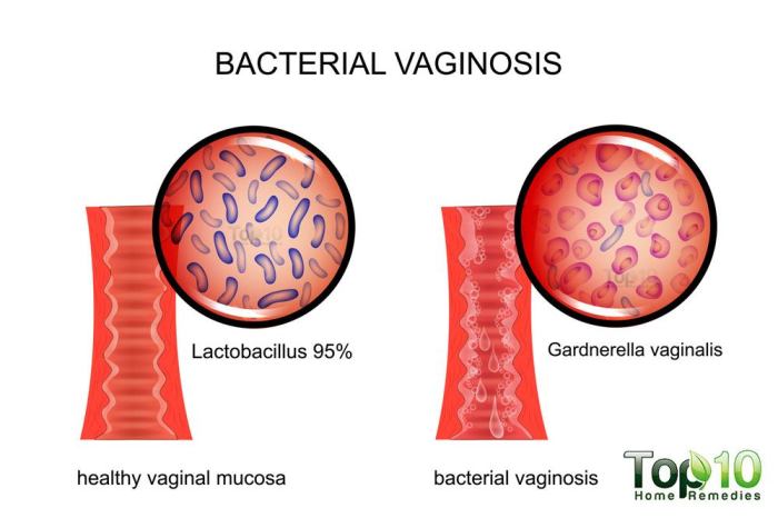
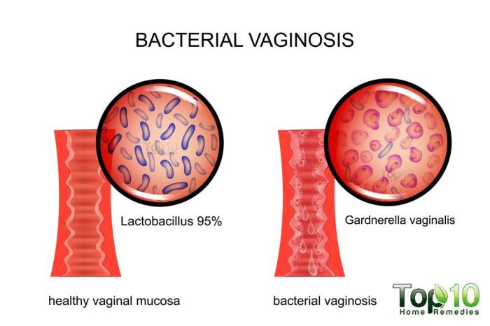
    The vaginal and urethral environments of both men and women harbor complex microbial communities. In healthy individuals, these communities are dominated by beneficial bacteria, which help maintain a balanced ecosystem. However, in cases of BV, an overgrowth of certain anaerobic bacteria disrupts this balance. Key differences exist between the microbial composition in healthy women and men. In women, a healthy vaginal environment usually contains Lactobacillus species, while in men, the urethral microbiome is more diverse, with a variety of facultative and anaerobic bacteria coexisting.

    This difference in normal flora is a critical factor in considering the specific bacterial species implicated in BV in each sex.

    Comparison of Bacterial Species Involved in BV in Both Sexes

    While the specific bacterial species implicated in BV can vary, some commonalities exist. Gardnerella vaginalis is often identified in both women and men with BV. However, other bacteria, like Mobiluncus species and various anaerobic bacteria, may also contribute to the condition in men. This implies that the causative agents may overlap but not be identical, reflecting the differences in the normal flora of the female and male genital tracts.

    Factors Contributing to BV in Men

    Several factors may predispose men to developing BV. Lifestyle choices, such as poor hygiene practices, can disrupt the delicate balance of the urethral microbiome. Sexual activity plays a significant role, potentially leading to the introduction of bacteria from a partner with BV. The use of certain personal care products or exposure to chemicals may also contribute to dysbiosis.

    Role of Sexual Activity in BV Transmission

    Sexual activity is a potential avenue for the transmission of BV-associated bacteria in men. If a partner has BV, the exchange of fluids during sexual intercourse can introduce the problematic bacteria into the male genital tract. The frequency and nature of sexual activity, as well as the presence of other sexually transmitted infections, can influence the risk.

    Mechanisms of Inflammation and Infection

    The overgrowth of certain bacteria in the male genital tract can trigger an inflammatory response. The bacteria may produce toxins or other substances that irritate the tissues, leading to discomfort and potential infection. This response varies based on the specific bacteria involved and the individual’s immune system.

    Factors Influencing the Onset and Progression of BV in Men

    Factor Description
    Hygiene Poor hygiene practices can disrupt the balance of the urethral microbiome.
    Sexual Activity Exposure to BV-associated bacteria from a partner can lead to infection.
    Lifestyle Factors Certain lifestyle choices (e.g., use of harsh soaps) may contribute to dysbiosis.
    Pre-existing Conditions Conditions that compromise the immune system can increase susceptibility.
    Urethral Microbiome A pre-existing imbalance in the urethral microbiome can make an individual more vulnerable.

    Symptoms and Diagnosis of Bacterial Vaginosis in Males

    Bacterial vaginosis (BV) is a common vaginal infection in women, but it can also affect men, albeit less frequently. Understanding the symptoms and diagnostic processes in men is crucial for early detection and appropriate treatment. Misdiagnosis is a significant concern due to the relative rarity of BV in males, and it’s essential to differentiate it from other, more common conditions.

    Hey everyone, talking about bacterial vaginosis in men today. It’s a less common condition, but it can still affect guys. While there’s no direct link between BV in men and treating dandruff, some natural remedies like using coconut oil for dandruff efficacy and how to use it are gaining popularity. coconut oil for dandruff efficacy and how to use it is an interesting option for some skin concerns.

    Regardless of its efficacy on dandruff, it’s important to remember that a doctor should be consulted for any health concerns, including bacterial vaginosis in men.

    Common Symptoms in Males

    Male BV often presents with a range of symptoms, some of which may be subtle or easily overlooked. Symptoms may not always be directly related to the genitals, potentially causing confusion with other issues. A thin, grayish-white discharge from the penis is a possible indicator. This discharge may have a fishy odor, particularly after sexual activity. Some men may experience irritation or burning sensations around the penis or groin area.

    It’s important to note that these symptoms can be quite mild and vary significantly in severity. Furthermore, some men may experience no noticeable symptoms at all, making early detection challenging.

    Potential for Misdiagnosis

    The relatively low prevalence of BV in men often leads to misdiagnosis or delayed diagnosis. Physicians might initially suspect other more common conditions, such as urinary tract infections (UTIs) or sexually transmitted infections (STIs). The lack of clear, distinctive symptoms can contribute to the challenge of accurate diagnosis. Misinterpreting the symptoms as part of a routine infection can further hinder timely diagnosis.

    Conditions Mimicking BV in Males

    Several conditions can mimic the symptoms of BV in men, requiring careful differentiation by healthcare professionals. Urinary tract infections (UTIs) frequently exhibit similar symptoms, such as burning during urination and discharge. Certain sexually transmitted infections (STIs), like gonorrhea or chlamydia, can also present with similar discharge characteristics. Furthermore, some skin conditions or allergies can also cause irritation and discharge, making a correct diagnosis even more challenging.

    Therefore, a comprehensive medical history and physical examination are crucial for differentiating BV from other potential causes.

    Diagnostic Procedures

    Diagnosing BV in men involves a multifaceted approach that considers clinical presentation, medical history, and appropriate laboratory tests. A physical examination of the penis and surrounding area is essential to evaluate for any visible signs of inflammation or discharge. Furthermore, a detailed medical history, including sexual history, helps to rule out other potential causes. Laboratory tests are critical in confirming the diagnosis.

    Laboratory Tests and Methods

    A crucial aspect of diagnosing BV in men is the use of laboratory tests to identify the specific microorganisms associated with the condition. Microscopic examination of the discharge can reveal the presence of clue cells, which are characteristic of BV. Specific culture methods may be employed to isolate and identify the specific bacteria involved. Furthermore, a Gram stain can aid in differentiating BV from other infections based on the types of bacteria present.

    The use of specific tests for other conditions, like STIs, is often necessary to rule them out.

    Table of Diagnostic Methods

    Diagnostic Method Sensitivity Specificity Description
    Microscopic Examination High Moderate Visual inspection of discharge for clue cells.
    Gram Stain High Moderate Identifies bacterial types present in the sample.
    Culture Moderate High Growth and identification of specific bacteria.
    Nucleic Acid Amplification Tests (NAATs) High High Identifies specific bacteria through genetic material.

    Note: Sensitivity and specificity values can vary depending on the specific laboratory and the method used.

    Management and Treatment of Bacterial Vaginosis in Males

    Bacterial vaginosis (BV) in men, though less common than in women, requires appropriate management to alleviate symptoms and prevent complications. Effective treatment hinges on accurate diagnosis and adherence to prescribed regimens. The underlying causes and contributing factors should be considered alongside the treatment plan to ensure long-term well-being.Understanding the nuances of BV treatment in men is crucial for successful management.

    Similar to women, the goal is to restore the natural vaginal microbiome balance. However, the specific approach may differ based on individual circumstances.

    Hey everyone! So, bacterial vaginosis, while more common in women, can sometimes affect men too. It’s important to get it checked out if you’re experiencing symptoms. Speaking of health, have you considered incorporating some flavorful spices into your diet for weight loss? Exploring options like ginger, turmeric, and cinnamon might help you reach your goals! spices for weight loss can be a delicious and potentially effective part of a balanced approach to wellness.

    Regardless, if you’re a man experiencing any unusual discharge or discomfort, it’s always best to consult a healthcare professional for proper diagnosis and treatment.

    Treatment Options for BV in Males

    Treatment for BV in men primarily focuses on eliminating the overgrowth of anaerobic bacteria, often employing antibiotics. Similar to women, the choice of antibiotic and dosage depends on factors such as the severity of the infection, the patient’s overall health, and any potential drug interactions.

    Common Antibiotics Used to Treat BV in Males

    A variety of antibiotics are used to treat BV in men. Metronidazole is frequently prescribed due to its effectiveness in combating the causative bacteria. Tinidazole is another common option, often preferred for a single-dose administration. These antibiotics are typically well-tolerated, but potential side effects exist and should be considered.

    Importance of Appropriate Antibiotic Selection and Dosage

    Proper antibiotic selection is paramount for effective treatment. Choosing the right antibiotic, and the appropriate dosage, is crucial to minimize the risk of antibiotic resistance. A healthcare provider should tailor the treatment plan to the individual patient, taking into account factors like age, existing health conditions, and potential drug interactions.

    Importance of Following Treatment to Prevent Recurrence

    Adherence to the prescribed treatment regimen is essential to prevent recurrence of BV. Incomplete treatment can allow the causative bacteria to persist, potentially leading to a return of symptoms. Patients should complete the full course of antibiotics as directed by their healthcare provider, even if symptoms subside before the course is finished. This ensures that the infection is eradicated completely.

    Table of Treatment Options

    Treatment Option Potential Side Effects Efficacy in Managing BV
    Metronidazole Nausea, vomiting, headache, metallic taste, and potentially a disulfiram-like reaction with alcohol Generally effective, but adherence to the full course is crucial
    Tinidazole Nausea, vomiting, headache, metallic taste Generally effective, often with a single-dose regimen
    Clindamycin Diarrhea, abdominal pain, nausea, and vaginal irritation in women Can be effective, but requires careful monitoring due to potential side effects

    The efficacy of treatment depends on factors such as the individual’s response to the antibiotic and their adherence to the prescribed regimen. Regular follow-up appointments with the healthcare provider are vital to monitor progress and adjust the treatment plan as needed.

    Prevention and Public Health Implications

    Bacterial vaginosis (BV) in men, while less common than in women, can still cause discomfort and potentially impact sexual health. Understanding preventive measures and public health implications is crucial for promoting overall well-being and reducing the spread of this condition. Effective prevention strategies require a multifaceted approach encompassing safe sexual practices, hygiene, and public health initiatives.Preventing BV in men involves similar principles to preventing it in women, but with some nuances.

    Effective prevention often relies on responsible sexual behaviors, good hygiene practices, and awareness campaigns. This section delves into these aspects, highlighting the importance of early detection and treatment for both individual and community health.

    Preventive Measures for Reducing BV Risk

    Understanding the risk factors and implementing preventative measures is crucial for reducing the incidence of BV in men. These strategies are designed to minimize exposure to potential causative agents and promote a healthy vaginal environment.

    While bacterial vaginosis is primarily associated with women, it can affect men too. Understanding the potential complications of this condition is crucial, and it’s important to note that certain underlying health conditions, like internal shingles risks and complications here , can increase the risk of complications. Ultimately, men experiencing symptoms should consult a healthcare professional for accurate diagnosis and treatment.

    • Safe Sexual Practices: Consistent and correct condom use significantly reduces the risk of transmitting BV. Using condoms during all sexual activities, including vaginal, anal, and oral sex, helps prevent the exchange of vaginal fluids and reduces exposure to potential pathogens. Open communication with partners about sexual health and practices is also essential.
    • Hygiene Practices: Maintaining good hygiene, including regular cleansing of the genital area with mild soap and water, can help prevent the overgrowth of bacteria. Avoiding harsh soaps, douches, or scented products that can disrupt the natural vaginal flora is recommended. Proper hygiene practices, when coupled with safe sexual practices, can contribute significantly to minimizing the risk of BV.
    • Early Detection and Treatment: Regular check-ups with a healthcare provider are vital. Early detection and treatment of BV can prevent potential complications and transmission to partners. Prompt medical intervention can ensure the best possible outcomes and minimize the impact of the infection.

    Role of Education and Awareness Campaigns

    Public awareness campaigns play a critical role in educating individuals about BV prevention and management. These campaigns aim to dispel myths, promote responsible sexual behavior, and emphasize the importance of seeking timely medical attention.

    • Dissemination of Information: Educational materials, including brochures, websites, and social media campaigns, can effectively disseminate information about BV, its causes, symptoms, and prevention strategies. These resources can reach diverse audiences and empower individuals with knowledge to make informed decisions about their sexual health.
    • Addressing Misconceptions: Awareness campaigns can address misconceptions about BV, emphasizing the importance of seeking medical advice for any unusual symptoms. This can help reduce stigma associated with the condition and encourage open communication about sexual health.

    Comparison of Prevention Strategies for BV in Men and Women

    While the underlying principles of BV prevention are similar for both men and women, some strategies may differ based on anatomical and physiological differences.

    • Focus on Safe Sexual Practices: Condom use is critical for both men and women to reduce transmission risks. However, the specific details of safe sexual practices may vary based on the types of sexual activity.
    • Emphasis on Hygiene: Maintaining good genital hygiene is important for both genders. However, the specific products and techniques used might vary based on individual needs and preferences.

    Importance of Early Diagnosis and Treatment

    Prompt diagnosis and treatment of BV are essential for preventing complications and reducing the risk of transmission.

    • Minimizing Complications: Early intervention can prevent the development of more serious conditions. Untreated BV can lead to complications such as pelvic inflammatory disease (PID) in women. In men, it might lead to inflammation of the urethra or other related conditions.
    • Reducing Transmission: Prompt treatment prevents the spread of BV to sexual partners. This is crucial for maintaining sexual health and preventing the cycle of infection.

    Preventive Measures Table

    Preventive Measure Effectiveness Ease of Implementation
    Consistent and correct condom use High Moderate
    Regular genital hygiene with mild soap and water Moderate High
    Open communication about sexual health with partners High High
    Regular check-ups with a healthcare provider High Moderate

    Illustrative Cases

    Bacterial vaginosis in men

    Understanding bacterial vaginosis (BV) in men requires careful consideration of potential presentations and diagnostic approaches. This section provides a hypothetical case study to illustrate the process of identifying and managing BV in males. While a formal diagnosis can only be made by a healthcare professional, this case study will help readers understand the potential symptoms, diagnostic considerations, and treatment plans involved.

    Patient Presentation

    A 28-year-old male presents to his primary care physician complaining of a thin, grayish-white discharge from his penis, accompanied by a slight burning sensation during urination. He reports no fever or other systemic symptoms. He denies any recent sexual activity or changes in hygiene practices. The patient’s medical history is unremarkable, and he denies any allergies. These symptoms suggest a possible infection, but further investigation is needed to determine the specific cause.

    Diagnostic Approach

    Given the patient’s presentation, the physician will first conduct a thorough history and physical examination. A focused history should inquire about potential risk factors for sexually transmitted infections (STIs) and other possible causes of urethritis. The physical examination will include a visual inspection of the penis and urethral meatus for any abnormalities or discharge. A sample of the discharge will be collected for laboratory analysis, including a Gram stain and wet mount microscopy.

    These analyses will aid in identifying the presence of clue cells, a hallmark of bacterial vaginosis.

    Differential Diagnoses, Bacterial vaginosis in men

    Several conditions can present with similar symptoms to bacterial vaginosis in males. Differential diagnoses that should be considered include:

    • Urinary tract infection (UTI): UTIs can cause urethral discharge and dysuria. The absence of systemic symptoms, such as fever, might make BV a more likely diagnosis.
    • Sexually transmitted infections (STIs): STIs like gonorrhea and chlamydia can also cause urethritis. A comprehensive STI panel is often necessary to rule out these possibilities.
    • Non-infectious urethritis: Allergic reactions or other inflammatory conditions can also cause urethral discharge and discomfort.

    Treatment Plan

    The treatment plan will be guided by the laboratory results. If bacterial vaginosis is confirmed, treatment will likely involve a course of oral or topical antibiotics. The choice of antibiotic and duration of treatment will be determined by the physician based on the specific causative agents and the patient’s response to therapy. Patient education about the importance of completing the full course of antibiotics is crucial to prevent recurrence.

    Patient Progression

    Time Point Action Findings Diagnosis Treatment
    Initial Visit History and physical exam, discharge sample collection Thin, grayish-white discharge, mild dysuria Suspected BV Gram stain, wet mount, STI panel ordered
    2-3 Days Laboratory results Positive for clue cells on wet mount, negative for other STIs Bacterial Vaginosis Confirmed Metronidazole 500 mg twice daily for 7 days prescribed
    1 Week Follow-up visit Patient reports improvement in symptoms BV Resolved Treatment completed

    Outcome Summary

    Bacterial vaginosis in men

    In conclusion, bacterial vaginosis in men, though rare, presents a complex interplay of microbial factors, lifestyle considerations, and potential for misdiagnosis. This in-depth look at the topic highlights the need for greater awareness and understanding among healthcare professionals. Early diagnosis and appropriate treatment are key to managing the condition and preventing potential complications. Further research is crucial to better understand the nuances of BV in men and to improve diagnostic and treatment protocols.

  • Pulmonary Embolism How Long Before It Kills You?

    Pulmonary Embolism How Long Before It Kills You?

    How long before a pulmonary embolism kills you? This question, while grim, is crucial for understanding the swift and often unpredictable nature of this potentially fatal condition. Factors like the embolus size, location, pre-existing health issues, and the speed of treatment can dramatically affect the outcome. This exploration delves into the factors that influence the timeline, the progression of symptoms, medical interventions, and even preventative strategies.

    A pulmonary embolism occurs when a blood clot travels to the lungs, blocking blood flow. The severity of this blockage directly impacts the time until death. This article will delve into the complex interplay of factors that determine the potential outcome, offering a comprehensive understanding of this life-threatening condition.

    Factors Affecting Time to Death from Pulmonary Embolism

    Pulmonary embolism (PE) is a life-threatening condition where a blood clot blocks the pulmonary arteries, the blood vessels in the lungs. The time it takes for a PE to lead to death varies significantly, depending on several critical factors. Understanding these factors is crucial for both diagnosis and treatment, as swift action can be life-saving.The speed at which a pulmonary embolism progresses to death is influenced by a complex interplay of factors.

    The severity of the obstruction in the pulmonary arteries, pre-existing conditions, and the promptness and efficacy of treatment all contribute to the ultimate outcome. A large, centrally located embolism can cause rapid and devastating effects, while a smaller, peripheral embolism might not cause immediate harm. Ultimately, the time it takes for death to occur is highly variable.

    Size and Location of the Embolus

    The size and location of the blood clot (embolus) significantly impact the time to death. Larger emboli, obstructing a larger portion of the pulmonary artery, lead to more significant and rapid reductions in blood flow to the lungs, resulting in decreased oxygenation and increased pressure in the circulatory system. This, in turn, can quickly cause severe complications. Peripheral emboli, on the other hand, might not cause immediate life-threatening problems.

    However, even small emboli in critical locations can have a severe impact.

    Pre-existing Medical Conditions, How long before a pulmonary embolism kills you

    Pre-existing conditions significantly influence the outcome of a pulmonary embolism. Individuals with pre-existing heart disease, such as coronary artery disease or heart failure, are at higher risk of rapid deterioration due to PE. Similarly, individuals with chronic lung diseases like emphysema or chronic bronchitis may have diminished respiratory reserve, making them more vulnerable to the effects of decreased lung perfusion.

    These conditions can make the body less able to compensate for the reduced blood flow caused by the embolism.

    Treatment Interventions

    Treatment interventions play a critical role in determining the time to death from a pulmonary embolism. Anticoagulants, such as heparin and warfarin, work by preventing the blood clot from growing larger and further obstructing the arteries. Thrombolytics, such as tissue plasminogen activator (tPA), are used to dissolve the existing blood clot, thereby restoring blood flow to the lungs.

    Pulmonary embolism is a serious condition, and unfortunately, the time it takes to be fatal varies greatly. It depends on factors like the size of the clot and your overall health. While some people might not survive a pulmonary embolism for very long, others have a longer battle. Knowing what foods pack more protein than an egg can be a great way to improve your health, which is obviously important when facing such a serious health issue.

    Foods with more protein than an egg can help you build up strength. Ultimately, it’s crucial to seek medical attention immediately if you suspect you might be experiencing a pulmonary embolism. The quicker you get help, the better your chances for a positive outcome.

    Prompt and effective administration of these treatments can significantly improve survival time. Studies have shown that early intervention with appropriate treatments can reduce the risk of fatal outcomes.

    Factors Affecting Time to Death from Pulmonary Embolism

    Factor Description Impact on Time to Death
    Size of Embolus The larger the embolus, the greater the obstruction of the pulmonary arteries. Larger emboli typically lead to a shorter time to death.
    Location of Embolus Central emboli, obstructing major arteries, are more likely to cause rapid deterioration than peripheral emboli. Central emboli often result in a quicker time to death.
    Pre-existing Conditions Conditions such as heart disease, lung disease, and other underlying health issues can exacerbate the effects of PE. Pre-existing conditions can shorten the time to death from PE.
    Treatment Interventions Prompt and effective administration of anticoagulants and thrombolytics can improve survival time. Early and appropriate treatment can significantly extend survival time.

    Symptoms and Progression

    Understanding the progression of pulmonary embolism (PE) symptoms is crucial for timely diagnosis and treatment. While the time until death varies significantly, a pattern of escalating symptoms often precedes the fatal outcome. Recognizing these signs can help healthcare providers intervene effectively and potentially save lives.

    Common Symptoms of Pulmonary Embolism

    Early symptoms of a pulmonary embolism can be subtle and easily mistaken for other conditions. Chest pain, shortness of breath, and coughing are common, but their severity and presentation can vary greatly. Initial symptoms might be mild and intermittent, increasing in intensity over time.

    Progression of Symptoms Leading to Death

    The sequence of symptoms in a fatal PE often begins with a sudden onset of shortness of breath. This initial symptom is frequently accompanied by chest pain, which can range from a sharp, stabbing sensation to a dull ache. As the embolism grows or more emboli lodge, shortness of breath intensifies, and the patient may experience rapid, irregular heartbeats (tachycardia).

    Coughing, often with blood-tinged sputum, might also occur. The severity and frequency of these symptoms generally increase as the blockage progresses. Critical stages include the onset of severe, unrelenting shortness of breath, cyanosis (bluish discoloration of the skin), and loss of consciousness, indicating a significant reduction in oxygen delivery to the body’s tissues.

    Severity and Time to Death Correlation

    The severity of symptoms correlates directly with the size and location of the embolus. Larger emboli, or those obstructing major pulmonary arteries, lead to more severe and rapid symptom progression. Conversely, smaller emboli might cause milder, less rapidly escalating symptoms. In some cases, the time between symptom onset and death can be relatively short, measured in hours.

    In others, the progression may be more gradual, spanning days. The specific timeline depends on the individual’s overall health, the size of the embolism, and the effectiveness of treatment.

    Table: Progression of Symptoms from Onset to Death

    Symptom Description Time of Appearance (approximate) Severity
    Shortness of Breath Difficulty breathing, initial feeling of tightness or pressure in the chest Within minutes to hours of clot formation Mild to moderate
    Chest Pain Sharp, stabbing, or dull ache in the chest, often worsened by deep breaths Concurrent with shortness of breath Moderate to severe
    Cough Dry cough or cough with blood-tinged sputum Hours to days after initial symptoms Mild to severe
    Tachycardia Rapid, irregular heartbeat Early in the progression Moderate to severe
    Cyanosis Bluish discoloration of the skin, lips, and fingertips As oxygen levels decrease Severe
    Loss of Consciousness Inability to respond to stimuli Indicates severe oxygen deprivation Severe, typically fatal

    Note: The times listed are approximations and can vary considerably based on individual circumstances.

    Medical Interventions and Their Effectiveness

    Pulmonary embolism (PE) is a serious condition requiring swift and effective medical intervention. The choice of treatment and its timely administration can significantly impact the outcome, potentially determining the difference between life and death. Understanding the various interventions and their respective effectiveness is crucial for both patients and healthcare providers.Effective treatment for pulmonary embolism hinges on a multifaceted approach, recognizing that no single intervention guarantees a complete resolution.

    The goal is to stabilize the patient, prevent further clot formation, and restore normal blood flow as quickly as possible. This often involves a combination of therapies, tailored to the severity of the embolism and the patient’s overall health.

    Wondering how long a pulmonary embolism takes to become deadly? The truth is, it varies greatly. Factors like the size and location of the clot play a huge role. While some people might experience symptoms for a while, others face a sudden and severe decline. If you’re experiencing symptoms like chest pain or shortness of breath, it’s crucial to seek immediate medical attention.

    Knowing why your thumb is tingling could also be a sign of something more serious, so checking out this resource on why is my thumb tingling might be helpful, but it’s not a substitute for medical advice. Ultimately, timely intervention is key in potentially preventing a fatal outcome from a pulmonary embolism.

    Anticoagulants

    Anticoagulants, or blood thinners, are a cornerstone of PE treatment. They work by preventing the formation of new blood clots and helping to dissolve existing ones. This is crucial for preventing further emboli and reducing the risk of recurrent PE. Different anticoagulants have varying onset times and durations of action. Some, like heparin, act quickly, while others, like warfarin, take several days to reach full effect.

    The selection of an anticoagulant is dependent on the specific circumstances of each patient.

    Thrombolytics

    Thrombolytics, sometimes referred to as clot busters, are potent medications that actively dissolve blood clots. They are typically reserved for patients with severe PE who are at high risk of death. Their rapid action, while potentially life-saving, also comes with a higher risk of complications like bleeding. The speed of action of thrombolytics varies depending on the specific medication and route of administration, ranging from a few hours to a few days.

    Surgical Procedures

    In cases where other interventions are insufficient or contraindicated, surgical procedures, such as embolectomy, may be necessary. Embolectomy involves physically removing the embolus, restoring blood flow to the lungs. This method is typically employed for massive or life-threatening PE where rapid clot removal is essential. Surgical procedures are generally associated with the highest risk among the options, but can offer the quickest resolution for severe emboli.

    Comparison of Interventions

    Intervention Mechanism Speed of Action Effectiveness
    Anticoagulants Prevent clot formation and promote clot breakdown Variable (hours to days) High for prevention and long-term management; may not be fast enough for acute, severe cases
    Thrombolytics Dissolve existing clots Hours High for severe PE; carries higher risk of bleeding
    Surgical Embolectomy Direct removal of the embolus Rapid (minutes to hours) Highest for massive PE; highest risk

    The table above provides a general comparison of the interventions. It’s important to note that the “speed of action” and “effectiveness” are relative and depend on the individual patient’s condition and the severity of the PE. A healthcare professional will carefully consider the patient’s overall health and the specifics of the situation to select the most appropriate and effective treatment plan.

    Case Studies and Examples: How Long Before A Pulmonary Embolism Kills You

    Understanding the variability in time to death from pulmonary embolism (PE) requires analyzing specific case studies. These case studies offer valuable insights into the factors influencing the severity and progression of the disease. By examining individual cases, we can gain a deeper appreciation for the complex interplay of factors that determine the outcome of PE.Analyzing case studies allows us to identify patterns and potential risk factors associated with different PE presentations.

    Pulmonary embolism’s a serious condition, and the time it takes to be fatal varies greatly. Factors like the size and location of the clot play a crucial role. While some cases are immediately life-threatening, others might give you a bit more time to seek medical attention. It’s crucial to remember that a completely unrelated procedure, like a colonic, what is a colonic , doesn’t have any bearing on the timeframe of a pulmonary embolism’s effects.

    Ultimately, prompt medical intervention is key to improving your chances of survival in a pulmonary embolism situation.

    This approach is crucial in refining our understanding of PE, ultimately improving diagnostic and therapeutic strategies for patients. Detailed case studies provide a nuanced view of the disease, which is essential for personalized medicine and effective patient care.

    Variability in Time to Death from Pulmonary Embolism

    Significant variations in the time from PE onset to death are observed across different cases. Factors such as the size and location of the embolus, pre-existing conditions, and the effectiveness of treatment interventions all play a critical role in determining the timeframe.

    Case Study 1: Rapid Progression

    A 65-year-old female patient with a history of deep vein thrombosis (DVT) presented with sudden onset of shortness of breath and chest pain. The patient reported experiencing increasing difficulty breathing, along with sharp chest pain. The embolus, determined to be large and lodged in the main pulmonary artery, caused significant obstruction of blood flow. Immediate treatment, including anticoagulation therapy and thrombolytic therapy, was initiated.

    Despite aggressive intervention, the patient experienced a rapid decline in respiratory function. Symptoms worsened over a period of 2 hours, culminating in cardiac arrest and death within 4 hours of symptom onset.

    Case Study 2: Delayed Progression

    A 42-year-old male patient, a marathon runner with no significant pre-existing conditions, experienced sudden onset of shortness of breath and mild chest discomfort after a strenuous workout. The patient’s embolus was smaller and located in a branch of the pulmonary artery, causing a less severe obstruction. Treatment, consisting of anticoagulation therapy, was initiated promptly. The patient experienced a gradual decline in respiratory function, with symptoms worsening over 12 hours.

    The patient’s condition stabilized, and he was discharged after 3 days of intensive care monitoring. The patient remained stable for a few months.

    Case Study 3: Impact of Pre-existing Conditions

    A 78-year-old male patient with chronic obstructive pulmonary disease (COPD) and heart failure presented with worsening dyspnea and cough. The patient had a history of recurrent DVT. The embolus, while relatively small, caused significant hemodynamic compromise due to the patient’s pre-existing conditions. The patient experienced progressive shortness of breath and increasing cyanosis. Treatment, including supplemental oxygen and anticoagulation, was initiated promptly.

    However, due to the patient’s already compromised respiratory and cardiac function, the patient’s condition deteriorated over 24 hours, ultimately leading to death within 36 hours of symptom onset.

    Characteristics of the Cases

    The presented case studies highlight the significant variations in time to death from PE. The size and location of the embolus, pre-existing medical conditions, and the promptness and efficacy of treatment all contribute to the diverse outcomes.

    Prevention Strategies and Public Awareness

    How long before a pulmonary embolism kills you

    Pulmonary embolism (PE) is a serious condition, but its risk can be significantly reduced through proactive measures and public awareness. Understanding the factors that contribute to PE, combined with knowledge of prevention strategies, empowers individuals to make informed choices that safeguard their health. Early detection through recognizing symptoms is crucial for timely intervention, potentially saving lives.Public awareness campaigns are essential to highlight the signs and symptoms of PE, enabling individuals to seek medical attention promptly.

    Education about preventative measures, combined with knowledge of risk factors, allows individuals to take proactive steps towards minimizing their chances of developing PE. This knowledge is particularly valuable in high-risk populations, where the likelihood of PE is higher.

    Preventative Measures to Reduce PE Risk

    Several lifestyle changes and medical interventions can significantly reduce the risk of developing a pulmonary embolism. These strategies target the underlying factors that increase the likelihood of blood clots forming.

    • Maintaining a Healthy Weight: Obesity is a significant risk factor for blood clots. Maintaining a healthy weight through a balanced diet and regular exercise reduces the risk of developing blood clots in the veins, a primary cause of PE. For example, a study published in the Journal of the American Medical Association showed a strong correlation between obesity and increased risk of venous thromboembolism, a condition that includes PE.

    • Regular Exercise: Physical inactivity contributes to blood clot formation. Regular exercise promotes blood circulation, reducing the risk of blood clots. Walking, swimming, or cycling are effective ways to increase physical activity and lower the risk of PE. For instance, air marshals, who are often required to sit for extended periods, are at higher risk of PE. Implementing exercise routines can mitigate this risk.

    • Proper Hydration: Dehydration can lead to increased blood viscosity, making it more likely for blood clots to form. Maintaining adequate hydration by drinking enough water throughout the day can help prevent blood clots and thus reduce the risk of PE.
    • Avoiding Prolonged Periods of Immobility: Sitting for extended periods, especially during air travel or long car rides, increases the risk of blood clots. Getting up and moving around every hour can significantly decrease the likelihood of blood clots forming. For example, flight attendants and long-haul truck drivers can mitigate the risk of PE by taking regular breaks and moving around.
    • Smoking Cessation: Smoking is a well-established risk factor for blood clots. Quitting smoking significantly reduces the risk of PE, along with numerous other health benefits. For instance, former smokers have a lower likelihood of developing PE compared to current smokers.

    Public Awareness Campaigns and Education

    Effective public awareness campaigns play a vital role in reducing PE-related deaths. Educating the public about the signs and symptoms of PE empowers individuals to seek medical attention promptly, increasing the chances of successful treatment.

    • Public Service Announcements (PSAs): PSAs should provide clear, concise information about the warning signs of PE, including shortness of breath, chest pain, and unexplained coughing. PSAs should emphasize the importance of seeking immediate medical attention if these symptoms are experienced.
    • Educational Materials: Educational materials, such as brochures and pamphlets, can be distributed in public places like hospitals, airports, and doctor’s offices. These materials should provide detailed information about the risk factors, symptoms, and preventative measures for PE.
    • Partnerships with Healthcare Professionals: Collaboration with healthcare professionals can ensure accurate and up-to-date information is disseminated to the public. Healthcare professionals can provide accurate information regarding PE prevention strategies and highlight the importance of early diagnosis.
    • Social Media Campaigns: Utilizing social media platforms to spread awareness about PE can reach a wider audience. Social media campaigns can include engaging content, such as infographics, videos, and interactive quizzes, to educate the public about PE.

    Visual Representation of the Process

    Understanding the progression of a pulmonary embolism (PE) is crucial for both prevention and treatment. A PE occurs when a blood clot, often originating in the deep veins of the legs (deep vein thrombosis or DVT), travels to the lungs, blocking blood flow. This blockage leads to a cascade of events that can rapidly compromise the body’s ability to oxygenate its tissues.

    The severity and speed of the process vary greatly, influenced by factors like the size of the clot and the individual’s overall health.

    Anatomical Structures and Processes

    The pulmonary circulation, a critical part of the respiratory system, plays a central role in oxygenating the blood. Deoxygenated blood returns to the heart through the vena cava, then passes through the right atrium and ventricle. From the right ventricle, the blood is pumped into the pulmonary artery, which branches into smaller vessels within the lungs. The tiny air sacs (alveoli) in the lungs facilitate the exchange of oxygen and carbon dioxide.

    A PE disrupts this process by obstructing the pulmonary arteries, hindering the flow of oxygenated blood throughout the body.

    Effects on Blood Flow and Oxygen Delivery

    The blockage caused by a PE directly impacts the blood flow to the lungs and subsequently to the rest of the body. The immediate effect is reduced blood flow through the affected pulmonary arteries, leading to reduced oxygen uptake in the lungs. This decreased oxygen delivery to the tissues can rapidly cause cellular damage and organ dysfunction. A large PE can cause a significant drop in blood pressure, as the heart struggles to pump blood against the obstructed vessels.

    Flow Chart of the Process

    • A blood clot forms in a deep vein (e.g., leg). This clot can travel to the lungs.
    • The clot travels through the bloodstream to the pulmonary arteries.
    • The clot lodges in a pulmonary artery, partially or completely blocking the flow of blood through that vessel.
    • Reduced blood flow to the affected lung areas occurs.
    • The heart struggles to pump blood effectively, potentially leading to a decrease in blood pressure.
    • The body’s tissues receive less oxygen, leading to damage and potential organ failure.
    • If the clot is large or multiple clots exist, the body may not be able to compensate, leading to rapid deterioration and potentially death.

    Circulatory System and Pulmonary Embolism

    Imagine the circulatory system as a network of roads. The pulmonary arteries are like major highways carrying oxygen-poor blood to the lungs. A PE is like a large truck blocking a portion of these highways. This blockage hinders the smooth flow of blood, leading to a backup in the system. The heart works harder to compensate, but the blockage can eventually lead to a cascade of effects, potentially causing heart failure and death.

    A visual representation would show the normal flow of blood through the heart and lungs, contrasted with the obstructed flow in the case of a PE. The diagram would highlight the pulmonary arteries and veins, emphasizing the point of blockage and the resulting reduced blood flow to the tissues.

    Example Case

    A 45-year-old woman with a history of sedentary lifestyle and recent surgery experiences sudden chest pain, shortness of breath, and lightheadedness. A blood clot is identified in her pulmonary artery, resulting in a significant blockage. The patient receives immediate medical intervention, including clot-busting medication and anticoagulants. While she recovers, the example highlights the rapid progression of PE and the importance of prompt diagnosis and treatment.

    Final Wrap-Up

    How long before a pulmonary embolism kills you

    Ultimately, the time from a pulmonary embolism to death is highly variable. Understanding the influencing factors, from the size of the clot to pre-existing conditions, is essential. Swift medical intervention plays a critical role in potentially extending survival. By learning about prevention and recognizing the symptoms, individuals can better protect themselves and those around them from this serious threat.

    Knowledge is power, and this discussion provides valuable insights into this often-overlooked condition.

  • Lung Pain Symptoms, Causes, and Diagnosis

    Lung Pain Symptoms, Causes, and Diagnosis

    Lung pain symptoms causes and diagnosis – Lung pain symptoms, causes, and diagnosis is a crucial topic to understand. It’s important to differentiate between lung pain and chest pain, and to recognize the various symptoms that may accompany it. This comprehensive guide explores the common and less common symptoms, delves into potential causes ranging from infections to lifestyle factors, and explains the diagnostic process, from medical history to imaging tests.

    Understanding these factors is key to effectively managing and preventing lung pain.

    This in-depth look at lung pain symptoms, causes, and diagnosis will provide a clear picture of the complexities involved, offering insights into how symptoms manifest, what potential underlying causes might be at play, and the various methods used for diagnosis. We’ll examine everything from common infections to underlying diseases, and explore how lifestyle factors can contribute to lung pain.

    Introduction to Lung Pain

    Lung pain, while often perceived as chest pain, is a distinct sensation originating from the structures within the chest that surround the lungs. Understanding this difference is crucial for accurate diagnosis and appropriate treatment. It’s not always straightforward to pinpoint the exact source of the discomfort, as various conditions can manifest as lung pain. Accurate identification of the cause is vital for effective management.Unlike chest pain, which can stem from the heart, ribs, muscles, or other structures in the chest cavity, lung pain is primarily associated with issues affecting the lungs themselves.

    This distinction helps differentiate between potentially serious cardiac problems and respiratory conditions. Common causes of lung pain often include infections, injuries, and underlying lung diseases.

    Common Causes of Lung Pain

    Lung pain can arise from a variety of causes, with some being more common than others. Infections, such as pneumonia or bronchitis, are frequent culprits. These infections inflame the lung tissue, leading to pain. Other potential causes include injuries to the chest wall, rib fractures, or conditions affecting the pleura, the membrane surrounding the lungs. Underlying lung diseases, like asthma or emphysema, can also cause persistent or intermittent pain.

    Additionally, certain medical procedures or treatments can sometimes trigger discomfort.

    Symptoms Associated with Lung Pain

    Recognizing the symptoms accompanying lung pain is essential for proper assessment. These symptoms can vary in intensity and character, reflecting the underlying cause.

    Symptom Description Possible Cause Severity Level
    Sharp, stabbing pain Sudden, intense pain, often worsened by breathing or coughing. Pleurisy, pneumothorax, rib fractures Moderate to Severe
    Aching, dull pain Persistent, less intense pain that may be present for days or weeks. Pneumonia, bronchitis, lung cancer (in later stages) Mild to Moderate
    Pain that worsens with deep breaths Discomfort that intensifies when inhaling deeply or exhaling forcefully. Pleurisy, pneumonia, lung tumors Moderate to Severe
    Pain radiating to the shoulder or back Discomfort that extends beyond the chest to the shoulder or back region. Pleurisy, lung tumors, rib inflammation Moderate to Severe
    Pain accompanied by coughing Discomfort associated with the act of coughing or producing phlegm. Pneumonia, bronchitis, lung infections Mild to Moderate

    Symptoms of Lung Pain: Lung Pain Symptoms Causes And Diagnosis

    Experiencing pain in your chest, particularly around the lungs, can be a concerning symptom. It’s crucial to understand that lung pain itself is often a secondary symptom, meaning the pain originates from another underlying condition. Recognizing the accompanying symptoms is vital for proper diagnosis and treatment. These symptoms can vary significantly, depending on the cause, ranging from mild discomfort to severe distress.Understanding the range of symptoms associated with lung pain is essential for prompt medical attention.

    The following table Artikels common symptoms, their descriptions, potential causes, and associated conditions. This information is intended for educational purposes only and should not be considered a substitute for professional medical advice.

    Common Symptoms of Lung Pain

    Symptom Description Potential Cause Associated Conditions
    Breathlessness (Dyspnea) Difficulty breathing, feeling short of breath, or experiencing labored breathing. This can range from mild shortness of breath to severe gasping for air. Lung conditions (e.g., pneumonia, asthma), heart conditions (e.g., congestive heart failure), anxiety. Asthma, bronchitis, pulmonary embolism, pneumonia, anxiety disorders
    Cough A persistent irritation in the throat, often accompanied by the expulsion of mucus or phlegm. Coughs can vary in severity, from a dry tickle to a forceful expulsion of fluids. Upper respiratory infections, bronchitis, pneumonia, lung cancer, or foreign objects lodged in the airways. Common colds, flu, bronchitis, pneumonia, lung cancer, whooping cough
    Chest Pain Discomfort or pain localized to the chest area, which may radiate to other areas. This can range from sharp, stabbing pain to a dull, aching sensation. Muscle strain, heart attack, pleurisy, lung infection. Pleurisy, pericarditis, musculoskeletal pain
    Wheezing A whistling or squeaking sound during breathing, typically associated with narrowing of the airways. Asthma, bronchitis, or other respiratory illnesses. Asthma, bronchitis, COPD
    Fever Elevated body temperature, often indicative of an infection or inflammatory response. Pneumonia, bronchitis, influenza. Pneumonia, bronchitis, influenza

    Less Common but Potentially Significant Symptoms

    A range of less common symptoms can also accompany lung pain, each carrying the potential to indicate a more serious underlying condition. These include:

    • Hemoptysis (coughing up blood): This is a serious symptom that warrants immediate medical attention. It could indicate infections, tumors, or blood clots in the lungs. For instance, a patient experiencing hemoptysis with other symptoms like chest pain and shortness of breath should seek immediate medical evaluation.
    • Cyanosis (bluish discoloration of the skin): This symptom often signifies a severe oxygen deficiency in the blood, potentially indicating a critical lung condition. A patient exhibiting cyanosis may also show signs of labored breathing or other respiratory distress.
    • Sputum changes (color, consistency): Noticeable changes in the color or consistency of sputum (mucus) can be an indicator of infection or other respiratory issues. A change in sputum color from clear to yellow or green, for example, could suggest a bacterial infection.
    • Loss of appetite and unexplained weight loss: These symptoms can accompany various underlying medical conditions, including lung cancer, which should not be ignored.
    • Fatigue and weakness: Persistent fatigue and weakness can sometimes indicate a more significant respiratory issue or a broader systemic condition.

    Symptom Severity and Underlying Causes

    The severity of symptoms can provide valuable clues about the potential cause of lung pain. For example, sudden, sharp, stabbing pain, often accompanied by difficulty breathing, might suggest a pneumothorax (collapsed lung) or pleurisy. Conversely, a gradual onset of dull, aching pain, along with a persistent cough, might indicate bronchitis or a less serious respiratory infection.

    Manifestations of Lung Pain

    Lung pain can manifest in various ways, including sharp, stabbing sensations, dull aches, or a persistent, throbbing discomfort. The location and character of the pain can offer important clues to the underlying cause. For instance, pain that worsens with deep breathing or coughing is frequently associated with pleurisy, whereas pain that is more constant and less localized might indicate a broader respiratory condition.

    Causes of Lung Pain

    Lung pain symptoms causes and diagnosis

    Lung pain, a distressing symptom, can stem from a multitude of underlying causes. Pinpointing the exact source is crucial for effective treatment. Understanding the potential culprits, from infections to pre-existing conditions, empowers individuals to seek timely medical attention. This section delves into the various medical conditions associated with lung pain.Identifying the root cause of lung pain is vital for appropriate diagnosis and treatment.

    Factors such as the location, intensity, and duration of the pain, along with accompanying symptoms, help physicians narrow down potential causes. Furthermore, lifestyle choices and pre-existing medical conditions can significantly influence the likelihood and severity of lung pain.

    Medical Conditions Causing Lung Pain

    A variety of medical conditions can lead to lung pain. Infections, injuries, and underlying diseases are common contributors. Careful consideration of these possibilities assists in accurate diagnosis and treatment planning.

    Cause Description Symptoms Potential Complications
    Pneumonia Inflammation of the lung tissue, often caused by bacterial or viral infection. Sharp, stabbing chest pain, cough with phlegm, fever, chills, shortness of breath. Lung abscess, pleurisy, respiratory failure, sepsis.
    Pleurisy Inflammation of the lining surrounding the lungs (pleura). Sharp, stabbing chest pain, worsened by breathing, coughing, or sneezing, shortness of breath. Fluid buildup in the pleural space, respiratory distress.
    Lung Cancer Uncontrolled growth of abnormal cells in the lung tissue. Persistent cough, blood in sputum, chest pain, weight loss, shortness of breath. Spread of cancer to other parts of the body, respiratory failure, severe pain.
    Collapsed Lung (pneumothorax) Air leaks into the space between the lung and chest wall, causing the lung to collapse. Sudden, sharp chest pain, shortness of breath, rapid heartbeat, chest tightness. Respiratory distress, lung damage, recurrence.
    Pulmonary Embolism Blood clot that travels to the lungs, blocking blood flow. Sudden chest pain, shortness of breath, rapid heartbeat, coughing up blood, lightheadedness. Lung damage, heart failure, death.

    Lifestyle Factors and Lung Pain

    Certain lifestyle choices can contribute to lung pain. Smoking and exposure to pollutants can irritate and damage lung tissue, increasing the risk of various respiratory issues.Smoking is a significant risk factor for lung pain. Exposure to air pollution can also contribute to lung problems, potentially leading to pain. Chronic exposure to irritants can cause inflammation and damage to the lung tissues, making them more susceptible to pain.

    Pre-existing Conditions and Lung Pain

    Pre-existing conditions can significantly increase the likelihood of experiencing lung pain. Individuals with conditions like asthma, cystic fibrosis, or emphysema are more prone to respiratory issues, including pain.For example, individuals with asthma might experience worsening symptoms and pain during an asthma attack. Cystic fibrosis patients may experience lung infections, leading to inflammation and pain. Similarly, individuals with emphysema may have difficulty breathing, which can cause chest discomfort and pain.

    Comparison of Causes

    Different causes of lung pain can vary significantly in their potential impact on health. While some, like a collapsed lung, might be a sudden, acute problem, others, like lung cancer, may be a chronic issue with potentially severe long-term consequences.Pneumonia, a common cause, can lead to respiratory complications and systemic infections. In contrast, pleurisy, an inflammation of the lining around the lungs, can cause sharp, stabbing pain that can be debilitating.

    Ultimately, accurate diagnosis is essential for effective treatment and management of the condition.

    Diagnosis of Lung Pain

    Pinpointing the cause of lung pain requires a meticulous approach. A thorough evaluation considers various factors, from the patient’s medical history to the results of diagnostic tests. This process aims to distinguish between potentially serious conditions and less severe issues, guiding treatment decisions.

    Diagnostic Process for Lung Pain

    The diagnostic process for lung pain involves a systematic investigation. A healthcare professional will first gather a comprehensive medical history, noting the characteristics of the pain, its duration, and any associated symptoms. This initial assessment forms the cornerstone of the diagnostic journey, highlighting potential contributing factors. Subsequent steps include physical examination, imaging studies, and laboratory tests to confirm or rule out suspected conditions.

    Understanding lung pain symptoms, causes, and diagnosis is crucial for prompt treatment. While the exact source of the discomfort might not always be immediately apparent, various factors can contribute, ranging from infections to underlying health conditions. Sometimes, unexpected connections exist, such as dietary choices impacting overall health. For instance, are you curious about foods that might influence estrogen levels?

    Exploring options like Foods with Estrogen Eat or Avoid? could shed light on potential dietary influences on your well-being. Ultimately, a proper diagnosis of lung pain requires a comprehensive evaluation by a medical professional.

    The specific tests performed depend on the individual’s presentation and the doctor’s suspicion of underlying causes.

    Diagnostic Tests for Lung Pain

    A variety of tests aid in diagnosing the source of lung pain. These tests provide valuable insights into the patient’s condition, assisting in the formulation of an accurate diagnosis. A combination of approaches is often necessary to achieve a comprehensive understanding.

    Diagnostic Test Procedure Purpose Potential Findings
    Chest X-Ray A low-dose X-ray of the chest area. To visualize the structures of the lungs, heart, and major blood vessels, identifying potential abnormalities like pneumonia, pneumothorax, or tumors. Abnormal shadows, infiltrates, fluid accumulation, or enlarged heart structures.
    Computed Tomography (CT) Scan A detailed cross-sectional imaging technique using X-rays. Provides detailed images of the lungs and surrounding structures, allowing for precise identification of lesions, masses, or other abnormalities, which might be missed by a standard X-ray. Tumors, nodules, cysts, fluid collections, and pulmonary emboli.
    Bronchoscopy A procedure where a thin, flexible tube with a camera is inserted through the nose or mouth to examine the airways. Allows direct visualization of the airways and lung tissues. Useful for diagnosing infections, tumors, or foreign bodies in the airways. Inflammation, narrowing of airways, bleeding, or abnormal tissue growths.
    Pulmonary Function Tests (PFTs) Measures the volume and capacity of the lungs, evaluating how well the lungs are functioning. Evaluates the mechanics of breathing and the function of the lungs, assessing airflow and gas exchange. Reduced lung capacity, restricted airflow, or impaired gas exchange, suggestive of conditions like asthma or chronic obstructive pulmonary disease (COPD).
    Blood Tests Various blood tests, such as complete blood count (CBC), arterial blood gases (ABGs), and inflammatory markers, are conducted. Assess the overall health status, detect infections, evaluate oxygen levels in the blood, and identify inflammation, potentially related to lung conditions. Elevated white blood cell count, abnormal oxygen levels, or elevated inflammatory markers, indicative of infection or inflammation.

    Medical History in Lung Pain Diagnosis

    A detailed medical history plays a critical role in the diagnostic process. Information about past illnesses, current medications, allergies, and lifestyle factors, such as smoking habits, provides valuable context for understanding the possible causes of lung pain. This crucial data helps narrow down the potential diagnoses and guide the selection of appropriate diagnostic tests.

    Comprehensive Diagnostic Evaluation

    A comprehensive diagnostic evaluation for lung pain involves a sequential approach:

    • Initial patient assessment, including gathering a complete medical history and conducting a physical examination, focusing on the characteristics of the pain, duration, and associated symptoms.
    • Selection of appropriate diagnostic tests based on the initial assessment. This selection is tailored to the specific patient presentation and suspected conditions.
    • Interpretation of the results of the chosen diagnostic tests to identify any abnormalities or deviations from normal ranges.
    • Integration of all collected information, including medical history, physical examination findings, and test results, to form a comprehensive understanding of the patient’s condition.

    Management and Treatment of Lung Pain

    Lung pain symptoms causes and diagnosis

    Managing lung pain requires a multifaceted approach, focusing on identifying and addressing the underlying cause. Effective treatment hinges on accurate diagnosis, and appropriate interventions can significantly improve quality of life for those experiencing this discomfort. The severity of treatment varies greatly depending on the root cause, from simple over-the-counter medications for mild cases to more complex interventions for serious conditions.Effective management of lung pain involves a combination of medical interventions, lifestyle adjustments, and, importantly, prompt medical attention.

    Ignoring persistent or worsening lung pain can delay diagnosis and potentially lead to more severe complications. Understanding the different treatment options available is crucial for making informed decisions about one’s health.

    Treatment Options for Specific Causes

    Various treatment options are available, tailored to the specific cause of lung pain. For instance, if the pain stems from a respiratory infection, supportive care and medications to manage symptoms are crucial. If the cause is a more serious condition, such as a tumor, a multidisciplinary approach may be necessary.

    Understanding lung pain symptoms, causes, and diagnosis can be tricky, but often, environmental factors play a significant role. For example, dust and allergens can trigger respiratory issues, and a deeper dive into the effectiveness of air purifiers can be really helpful in this regard. Do Air Purifiers Help With Dust? A Deep Dive explores this crucial topic, which in turn can offer valuable insights into managing lung pain symptoms, especially if they’re linked to environmental triggers.

    Ultimately, pinpointing the specific cause of lung pain requires careful medical evaluation.

    Importance of Seeking Medical Attention

    Seeking prompt medical attention for lung pain is paramount. Delaying treatment can potentially worsen the condition and lead to complications. A healthcare professional can accurately diagnose the cause of the pain and recommend the most appropriate course of action. This includes assessing the severity of the pain, its location, and any associated symptoms to determine the underlying cause and implement a personalized treatment plan.

    Examples of Medications Used to Manage Symptoms

    Several medications can be used to manage lung pain symptoms. Examples include over-the-counter pain relievers such as ibuprofen or acetaminophen, which can help alleviate mild to moderate pain. For more severe pain or specific conditions, stronger pain medications may be prescribed. Additionally, medications to treat underlying conditions, such as antibiotics for infections or anti-inflammatory drugs for inflammatory conditions, may be necessary.

    Comparison of Treatment Approaches

    Different treatment approaches vary significantly depending on the cause of the lung pain. For instance, a viral infection may require symptomatic relief and rest, while a collapsed lung may require immediate medical intervention, potentially involving procedures like chest tubes. A careful evaluation of the specific cause is essential for determining the most effective and appropriate treatment plan.

    Treatment Options Table

    Treatment Description Effectiveness Potential Side Effects
    Over-the-counter pain relievers (e.g., ibuprofen, acetaminophen) These medications help reduce pain and inflammation. Generally effective for mild to moderate pain. Potential for stomach upset, allergic reactions in some individuals.
    Antibiotics Used to treat bacterial infections. Effective for bacterial pneumonia or other infections. Potential for allergic reactions, antibiotic-resistant infections if not used correctly.
    Bronchodilators Medications that open up the airways. Effective for conditions like asthma or chronic obstructive pulmonary disease (COPD) causing lung pain. Potential for nervousness, tremors, or irregular heartbeats in some individuals.
    Corticosteroids Powerful anti-inflammatory medications. Effective for inflammatory lung conditions. Potential for increased risk of infections, osteoporosis, and high blood pressure.
    Oxygen therapy Provides supplemental oxygen to improve breathing. Essential for patients with low oxygen levels. Potential for dryness in the nasal passages.

    Prevention of Lung Pain

    Lung pain, while often a symptom of a more significant underlying condition, can be mitigated by proactive lifestyle choices and preventative measures. Understanding the factors that contribute to lung pain allows us to take steps to reduce the risk and promote overall lung health. This proactive approach focuses on both lifestyle adjustments and regular medical attention to ensure early detection and intervention.

    Lifestyle Choices for Reduced Lung Pain Risk

    Adopting healthy lifestyle habits is crucial for reducing the risk of various respiratory issues, including those that can cause lung pain. A balanced diet rich in fruits, vegetables, and whole grains provides essential nutrients for optimal lung function. Regular physical activity strengthens the respiratory system, improving its efficiency. Smoking cessation is paramount, as smoking significantly damages lung tissue and increases the risk of numerous respiratory problems.

    Limiting exposure to secondhand smoke is also vital for protecting oneself from these harmful effects. Avoiding excessive exposure to environmental irritants, such as air pollution and certain chemicals, helps minimize lung irritation and potential pain.

    Importance of Regular Check-ups, Lung pain symptoms causes and diagnosis

    Regular check-ups with a healthcare professional are essential for early detection of potential lung problems. These visits allow for routine screenings, enabling early intervention if any abnormalities or conditions are identified. Early detection can significantly improve treatment outcomes and reduce the severity of potential lung pain. Prompt diagnosis and treatment can prevent the progression of underlying conditions and their associated symptoms.

    Environmental Factors Affecting Lung Pain Risk

    Environmental factors significantly influence lung health and can increase the risk of lung pain. Air pollution, whether from industrial emissions or traffic, can irritate the lungs and trigger pain or discomfort. Exposure to certain occupational hazards, such as chemicals or dust, can also contribute to respiratory problems and subsequent lung pain. Secondhand smoke, a significant environmental risk factor, damages lung tissue and increases the likelihood of developing respiratory conditions.

    Individuals living in areas with high levels of air pollution or exposure to industrial hazards are at increased risk of lung pain.

    Understanding lung pain symptoms, causes, and diagnosis is crucial for your well-being. Sometimes, underlying health issues can lead to chest pain, which can be a sign of something more serious. Knowing about minimum essential coverage can also be important when seeking medical attention, as it plays a role in accessing appropriate care. For example, What is Minimum Essential Coverage and Why Does It Matter?

    helps you understand the implications of having the right health insurance. Ultimately, a proper diagnosis requires careful evaluation of your symptoms and medical history to rule out any potential complications related to lung pain.

    Preventative Measures to Reduce Lung Pain Risk

    Taking proactive steps to minimize exposure to harmful factors and maintain good respiratory health is essential. These measures encompass a broad spectrum of actions, from lifestyle choices to environmental awareness. Prioritizing a healthy lifestyle, regular check-ups, and environmental awareness are all crucial elements in preventing lung pain.

    Preventative Measures Table

    Prevention Method Description Effectiveness Benefits
    Smoking Cessation Quitting smoking completely eliminates the harmful effects of cigarette smoke on lung tissue. High. Studies consistently demonstrate that quitting smoking significantly reduces the risk of respiratory problems. Reduced risk of lung cancer, chronic bronchitis, emphysema, and other respiratory illnesses. Improved lung function and overall health.
    Balanced Diet Consuming a diet rich in fruits, vegetables, and whole grains provides essential nutrients for lung health and overall well-being. Moderate. A healthy diet supports immune function and overall lung health. Improved immune function, reduced inflammation, and enhanced respiratory system efficiency.
    Regular Exercise Engaging in regular physical activity strengthens the respiratory system and improves lung capacity. Moderate to High. Studies show that exercise can improve lung function and reduce the risk of respiratory conditions. Improved cardiovascular health, increased lung capacity, and reduced risk of respiratory problems.
    Environmental Awareness Reducing exposure to air pollution, secondhand smoke, and occupational hazards minimizes lung irritation and potential pain. Moderate. Minimizing exposure to harmful environmental factors reduces the risk of respiratory problems. Reduced risk of lung irritation and inflammation, improved respiratory health, and a healthier living environment.
    Regular Check-ups Routine medical check-ups allow for early detection of potential lung issues. High. Early detection and intervention improve treatment outcomes and reduce the severity of respiratory problems. Early diagnosis and prompt treatment of lung conditions, improved quality of life, and potentially avoiding severe lung conditions.

    Illustrations and Visual Aids

    Visual aids are crucial for understanding complex medical conditions like lung pain. Illustrations and diagrams can help clarify the structure of a healthy lung, the changes in a diseased lung, and diagnostic procedures. They also help connect abstract concepts to tangible representations, making learning and comprehension easier.

    Healthy Lung Structure

    A healthy lung is a complex structure composed of millions of tiny air sacs called alveoli. These alveoli are clustered together in grape-like bunches. Surrounding the alveoli is a network of capillaries, tiny blood vessels that facilitate the exchange of oxygen and carbon dioxide. The bronchi, branching airways, carry air to and from the alveoli. A healthy lung is a vibrant, elastic tissue that allows for efficient gas exchange.

    The color is a light, pinkish-gray. The structure is smooth and even, with no visible masses or abnormalities.

    Diseased Lung Structure

    A diseased lung displays various changes depending on the cause. For example, in chronic obstructive pulmonary disease (COPD), the alveoli lose their elasticity and may become damaged or enlarged. This results in a decreased surface area for gas exchange, leading to shortness of breath and difficulty breathing. The airways may also be inflamed and narrowed, further hindering airflow.

    The lung tissue might appear darker or mottled, with areas of consolidation or scarring. In pneumonia, the lung tissue may appear inflamed and filled with fluid. Cancer can manifest as a mass or tumor, disrupting the normal structure and function of the lung. The specific appearance depends on the type of lung disease and its stage.

    Lung Biopsy Procedure

    A lung biopsy is a procedure to obtain a tissue sample from the lung for examination under a microscope. The procedure typically involves inserting a needle or a surgical tool through the chest wall to collect the sample. A CT scan is usually performed beforehand to guide the procedure and ensure the sample is taken from the correct area.

    The procedure requires careful precision to avoid complications. The image would show a needle or tool inserted into the lung area, with surrounding structures clearly labeled. The position of the needle or tool would be precisely indicated, along with the intended area for sample collection.

    CT Scan Showing Lung Abnormalities

    A CT scan of the lungs can reveal abnormalities such as nodules, masses, or infiltrates. The scan creates cross-sectional images of the lungs, allowing for detailed visualization of internal structures. The images can be analyzed to identify potential problems, such as pneumonia, lung cancer, or other conditions. The CT scan would display cross-sectional slices of the lung, with any abnormalities highlighted with different shades or colors.

    These abnormalities would be labeled and described, indicating their potential cause and clinical significance.

    Table of Illustrations

    Image Type Description Purpose Related Information
    Healthy Lung A healthy lung appears as a light pinkish-gray, smooth, and even tissue. Alveoli are clearly visible as small, grape-like clusters. To illustrate normal lung structure and function for comparison with diseased lungs. Normal anatomy, gas exchange, respiration
    Diseased Lung (COPD) A diseased lung (COPD) exhibits a darker, mottled appearance with areas of consolidation or scarring. The alveoli may appear enlarged or damaged. To illustrate structural changes in the lungs due to disease. COPD, Emphysema, Asthma
    Lung Biopsy Procedure A needle or surgical tool is shown inserted into the lung area, guided by a CT scan. The surrounding structures are clearly labeled. To demonstrate the process of lung biopsy. Diagnostic procedures, tissue sampling, pathology
    CT Scan Showing Lung Abnormalities Cross-sectional images of the lung, highlighting nodules, masses, or infiltrates with different shades or colors. These abnormalities are labeled. To illustrate how abnormalities are visualized and diagnosed using CT scans. CT scans, lung cancer, pneumonia, other lung diseases
    Diagram of Alveoli A detailed illustration of the alveoli, showcasing their intricate structure and their relationship with capillaries. To demonstrate the gas exchange process at the microscopic level. Gas exchange, respiration, alveoli structure, capillaries

    Final Review

    In conclusion, navigating the complexities of lung pain requires a multifaceted approach. From understanding the diverse symptoms and potential causes to comprehending the diagnostic process and treatment options, this exploration provides a roadmap for individuals seeking to understand and manage this often-challenging condition. Remember, early diagnosis and proactive management are crucial for positive outcomes. By understanding the interconnectedness of symptoms, causes, and diagnosis, individuals can better equip themselves to address potential lung pain issues.