Category: Health & Wellness

  • Scabs on Scalp Causes, Diagnosis, and Treatment

    Scabs on Scalp Causes, Diagnosis, and Treatment

    Scabs on scalp causes diagnosis and treatment is a crucial guide for understanding the various factors contributing to scalp issues. This comprehensive exploration dives deep into the different types of scalp scabs, potential causes ranging from skin conditions to infections, and effective diagnostic methods. We’ll also cover a spectrum of treatment options, from topical remedies to antifungal medications, and even explore some helpful home remedies.

    Finally, we’ll touch on prevention strategies and provide detailed descriptions of various types of scalp scabs.

    From identifying the telltale signs of dry, crusty, or oozing scabs to pinpointing the underlying cause, this guide empowers you with the knowledge to address your scalp concerns effectively. Whether you’re dealing with a fungal infection, seborrheic dermatitis, or another potential cause, this guide provides a detailed roadmap for understanding and managing your scalp health.

    Introduction to Scalp Scabs

    Scabs on scalp causes diagnosis and treatment

    Scalp scabs, those unsightly patches of hardened skin on your scalp, can be a source of concern and discomfort. They can range from minor irritations to more serious underlying conditions. Understanding the different types of scalp scabs, their appearances, and common locations can be helpful in determining the cause and appropriate course of action. This section will delve into the specifics of scalp scabs, enabling you to better recognize and address them.

    Types of Scalp Scabs

    Scalp scabs manifest in various forms, each reflecting a possible cause. Dry, crusty, and oozing scabs represent distinct conditions, and their appearance provides clues to potential diagnoses. Understanding the visual characteristics of each type can aid in self-assessment and prompt appropriate medical attention when necessary.

    Common Locations of Scalp Scabs

    Scalp scabs aren’t confined to specific areas. They can appear anywhere on the scalp, though certain regions may be more prone to irritation or injury, leading to scab formation. Common areas include the crown, the sides, and the hairline, where hair density or scalp exposure might be different. Sometimes, scabs can be clustered, or they may appear as a single, isolated lesion.

    Dealing with scabs on your scalp can be frustrating, but understanding the potential causes and how to diagnose them is key to effective treatment. Sometimes, these scabs might be a symptom of a more serious issue, like an infection. Knowing if there’s a link to other health concerns like HIV transmission through casual contact is crucial.

    But, in most cases, scabs on the scalp are easily treatable with a combination of good hygiene and potentially some over-the-counter remedies. Proper diagnosis and following a suitable treatment plan are essential to restoring scalp health.

    Visual Characteristics of Scalp Scabs

    Identifying the type of scab is crucial for understanding its potential cause. The appearance can vary significantly, influenced by the underlying condition. The consistency, color, and size of the scab offer valuable insights into the nature of the problem.

    Type of Scalp Scab Visual Characteristics
    Dry These scabs appear as thin, flaky, or papery patches. They might be light beige or a similar shade to the surrounding skin. Often, they are accompanied by dryness and itching.
    Crusty Crusty scabs are thicker and rougher than dry scabs. They are often more raised and may appear as hardened, rough patches of various shades, from light brown to dark brown, or even black. The consistency is more solid than dry scabs.
    Oozing Oozing scabs are characterized by a moist or wet appearance. They may have a yellowish or whitish discharge, often accompanied by redness and inflammation. The area surrounding the scab might be swollen or tender to the touch.

    Potential Causes of Scalp Scabs: Scabs On Scalp Causes Diagnosis And Treatment

    Scalp scabs, a common skin issue, can have various underlying causes. Understanding these causes is crucial for proper diagnosis and effective treatment. They range from relatively benign conditions to more serious ones, requiring prompt medical attention. This section will delve into the common reasons behind these skin imperfections.A multitude of factors can contribute to the development of scalp scabs.

    From infections to allergic reactions, and even underlying skin conditions, a careful examination is needed to identify the root cause. Knowing these causes can guide appropriate treatment strategies and prevent potential complications.

    Common Skin Conditions

    Various skin conditions can manifest as scalp scabs. These conditions often involve inflammation and irritation, leading to the formation of crusty or scaly patches. Identifying the specific condition is important for tailoring the right treatment.

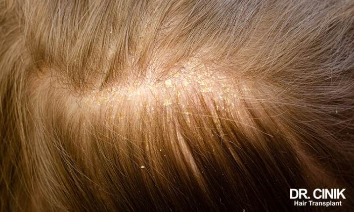
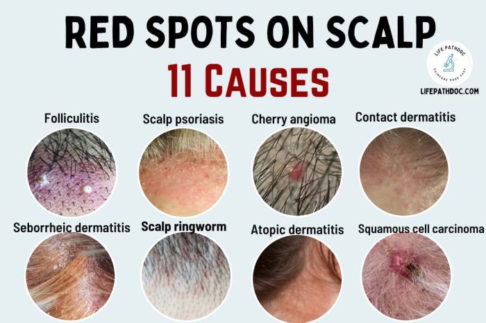
    • Seborrheic dermatitis: This chronic inflammatory skin condition is a frequent culprit behind scalp scabs. It’s characterized by greasy, yellowish scales and often causes significant discomfort.
    • Psoriasis: Another chronic autoimmune condition, psoriasis can lead to inflamed patches on the scalp, accompanied by scaling and sometimes scabs. The appearance can vary significantly between individuals.
    • Eczema: Atopic dermatitis, or eczema, can also affect the scalp, resulting in dry, itchy skin, and potentially scabs. This condition often involves a strong genetic component.

    Fungal Infections and Scalp Scabs

    Fungal infections, particularly tinea capitis, are significant contributors to scalp scabs. These infections are often characterized by inflammation, scaling, and sometimes, the formation of pustules and crusts. Different types of fungi can cause these infections.

    • Tinea capitis: This fungal infection is commonly referred to as ringworm. It’s important to note that while the name suggests a circular pattern, the appearance can be diverse. Tinea capitis can cause patchy hair loss, and the affected areas can be inflamed and have scaling or crusting. The infection is contagious and requires appropriate antifungal treatment.
    • Malassezia furfur: This yeast can cause dandruff, but in some cases, it can lead to more severe inflammation and scabs, particularly in individuals with underlying skin conditions or weakened immune systems.

    Seborrheic Dermatitis and Scalp Scabs

    Seborrheic dermatitis is closely linked to scalp scabs. This chronic inflammatory condition causes greasy, yellowish scales and often presents as patches of red, inflamed skin. It’s important to recognize the connection to prevent misdiagnosis and to provide effective treatment.

    Seborrheic dermatitis can affect various areas of the body, including the scalp, eyebrows, and face, often manifesting as greasy scales and redness.

    Comparison of Fungal Infections

    Different fungal infections can lead to similar symptoms, making accurate diagnosis crucial. Understanding the distinctions can guide treatment strategies.

    Scalp scabs can be a real nuisance, and figuring out the cause is key to effective treatment. Sometimes, they’re linked to underlying skin conditions or even dietary factors like excessive sodium intake. Eating a balanced diet, including foods high in sodium, can contribute to inflammation and potentially exacerbate the issue. Consulting a dermatologist is crucial for an accurate diagnosis and personalized treatment plan, addressing any underlying causes, including possible allergic reactions or infections.

    Identifying the root cause is vital for successful treatment of scalp scabs. foods high in sodium should be consumed in moderation, as excess sodium can be problematic for various health concerns. A proper diagnosis from a healthcare professional is always recommended.

    • Comparison of Different Types of Tinea Capitis: While all types of tinea capitis involve fungal infection, the appearance and severity can differ. Some types may present with more pronounced inflammation, scaling, or hair loss than others. Proper identification is critical for effective antifungal therapy.

    Table of Scalp Conditions and Scabs

    The following table summarizes the appearance and causes of various scalp conditions that can lead to scabs:

    Condition Appearance Causes
    Seborrheic Dermatitis Greasy, yellowish scales, red patches Yeast overgrowth, inflammatory response
    Tinea Capitis (Ringworm) Scaly patches, hair loss, sometimes pustules Fungal infection (various types)
    Psoriasis Thick, silvery scales, red patches Autoimmune response
    Eczema Dry, itchy skin, inflamed patches Allergic reaction, environmental factors

    Diagnosing Scalp Scabs

    Pinpointing the cause of scalp scabs is crucial for effective treatment. A thorough diagnostic approach involves a multifaceted evaluation considering various factors, from the patient’s medical history to physical examination findings. This process aims to differentiate between benign and potentially more serious underlying conditions.Accurate diagnosis is essential to tailor the treatment plan to the specific cause, avoiding ineffective or even harmful interventions.

    A clear understanding of the diverse conditions that can manifest as scalp scabs is vital to ensure the most appropriate course of action.

    Diagnostic Methods for Scalp Scabs

    A comprehensive diagnostic process for scalp scabs begins with a detailed evaluation of the affected area. Physical examination allows for visual assessment of the scabs, their distribution, and any associated symptoms, such as redness, inflammation, or itching. The consistency and appearance of the scabs themselves—whether they are dry, crusted, or oozing—can provide clues about the underlying cause. This assessment is complemented by a careful review of the patient’s medical history.

    • Physical examination, including visual inspection of the scalp, and palpation to detect any underlying lumps or tenderness.
    • Skin biopsy, a procedure involving the removal of a small skin sample for microscopic examination, can identify specific skin conditions, like fungal infections or certain inflammatory diseases. This method is crucial for diagnosing conditions not easily identified by visual inspection alone.
    • Scraping and microscopic examination of the affected area to detect fungal elements or parasites, a simple procedure that can help in diagnosing certain types of infections.
    • Blood tests, particularly in cases where systemic conditions might be suspected, to rule out underlying medical issues contributing to the scalp scabs. For example, a complete blood count (CBC) can identify signs of anemia or infection.
    • Allergy testing, if an allergic reaction is suspected as a possible cause, can identify potential allergens triggering the scalp condition.

    Role of Medical History in Diagnosing Scalp Scabs

    A detailed medical history provides valuable context for interpreting the physical findings. Knowing about any pre-existing skin conditions, recent illnesses, medications, or exposures to irritants or allergens can significantly aid in narrowing down the potential causes. For instance, a history of eczema might suggest a predisposition to certain types of scalp inflammation. Likewise, a recent use of a new hair product or a known allergy to certain substances might be crucial clues.

    Furthermore, a history of stress or other factors that might affect immune function can provide a broader perspective on the problem.

    Differentiating Scalp Conditions Causing Scabs

    Differentiating between various scalp conditions is essential. Seborrheic dermatitis, for example, often presents with yellowish, greasy scales, while psoriasis shows silvery, flaky patches. Fungal infections like tinea capitis might present with pustules or inflamed areas. Understanding these differences is critical for choosing the right treatment. This meticulous process ensures the chosen treatment addresses the specific underlying condition, minimizing the risk of ineffective or inappropriate interventions.

    Importance of Ruling Out Other Possible Causes

    It is essential to rule out other possible causes of scalp scabs. These could include:

    • Infections (bacterial, viral, fungal).
    • Autoimmune diseases.
    • Allergies.
    • Nutritional deficiencies.
    • Medications or other external irritants.

    Diagnostic Steps for Scalp Scabs

    Diagnostic Step Potential Tests Purpose
    Initial Evaluation Physical examination, medical history Identify the nature of the scabs, associated symptoms, and potential contributing factors.
    Further Evaluation (if needed) Skin biopsy, fungal culture, blood tests, allergy testing Confirm suspected diagnoses, rule out other conditions, and identify underlying causes.

    Treatment Options for Scalp Scabs

    Scalp scabs, while often a minor nuisance, can be indicative of underlying skin conditions requiring specific treatment. Proper diagnosis is crucial to selecting the most effective approach. Identifying the cause allows for targeted interventions, leading to faster healing and preventing recurrence.Effective treatment for scalp scabs hinges on addressing the root cause. A dermatologist or primary care physician can accurately assess the situation, determine the specific condition, and recommend the most suitable course of action.

    This personalized approach ensures optimal results and minimizes potential complications.

    Common Topical Treatments

    Topical treatments are frequently used for various scalp conditions causing scabs. Their effectiveness depends heavily on the underlying cause. For example, topical corticosteroids can be helpful for inflammatory conditions like seborrheic dermatitis, while antifungal creams target fungal infections. The selection of topical treatment will be determined by the cause identified by the doctor.

    Antifungal Medications

    Antifungal medications, both topical and oral, are crucial in treating fungal infections that manifest as scalp scabs. These medications work by inhibiting the growth of fungal organisms, thereby controlling the infection and promoting healing. Examples include clotrimazole, ketoconazole, and miconazole, which are available in various formulations for topical application. Oral antifungals are often prescribed for more extensive or systemic infections.

    So, you’ve got those pesky scabs on your scalp? Figuring out the cause and best treatment is key. Sometimes, a simple fungal infection is the culprit, other times it could be a more serious issue. Knowing whether your diet might play a role is also important. Eating a healthy diet, including plenty of nutritious foods like nuts, can be beneficial for overall health, and you might want to explore if are nuts good for you in the context of your scalp health.

    Ultimately, a visit to a dermatologist is crucial for proper diagnosis and a personalized treatment plan to get rid of those pesky scabs.

    Home Remedies

    Many home remedies can provide temporary relief from minor scalp scabs. However, these remedies are not substitutes for professional medical advice, especially when dealing with persistent or worsening conditions.

    Home Remedy Potential Benefits Important Considerations
    Aloe vera gel Soothes irritation, promotes healing May not be effective for all types of scalp scabs
    Tea tree oil Anti-inflammatory, antimicrobial properties Potentially irritating to sensitive skin; dilute before application
    Apple cider vinegar rinse May help with fungal infections Can be drying; use cautiously and dilute with water
    Coconut oil Moisturizing properties May not be effective for all types of scalp scabs; can clog pores if used excessively

    Comparison of Treatment Approaches

    The effectiveness and potential side effects of various treatment approaches differ significantly. Topical corticosteroids, for instance, can effectively reduce inflammation but may cause thinning of the skin with prolonged use. Antifungal medications are generally safe but may cause allergic reactions in some individuals. Home remedies, while generally safe, may not be effective for severe or persistent conditions.

    Consulting a healthcare professional is essential for a proper diagnosis and personalized treatment plan.

    Step-by-Step Home Treatment Guide

    Important Note: This guide is for informational purposes only and should not be considered a substitute for professional medical advice. If your scalp scabs are persistent or worsening, consult a dermatologist or primary care physician immediately.

    1. Cleanse the affected area gently with a mild, fragrance-free cleanser.
    2. Apply a thin layer of the recommended topical treatment (if prescribed by a doctor).
    3. Avoid scratching or picking at the scabs.
    4. Maintain good hygiene by regularly washing hair and using a gentle shampoo.
    5. Monitor the condition regularly for any changes or worsening symptoms. If symptoms persist or worsen, seek medical attention.

    Prevention of Scalp Scabs

    Preventing scalp scabs involves understanding the factors that contribute to their formation and implementing proactive measures to minimize the risk. A proactive approach to scalp health is crucial for overall well-being, as consistent scalp irritation can lead to discomfort and potentially more serious conditions.Scalp scabs are often a symptom of underlying issues, and preventing them hinges on addressing these root causes.

    This involves a multifaceted approach focusing on hygiene, product selection, and overall scalp health. By taking preventive steps, individuals can significantly reduce the likelihood of experiencing scalp scabs.

    Identifying Contributing Factors

    Scalp scabs can arise from various factors, including dryness, infections, and even certain medical conditions. Understanding these factors is key to preventing future occurrences. Dryness, for example, can be caused by environmental conditions or a lack of hydration. Fungal infections or bacterial infestations can also lead to scabs. Furthermore, certain medical conditions like eczema or psoriasis can manifest as scalp scabs.

    Good Hygiene Practices

    Maintaining good hygiene is fundamental to preventing scalp scabs. Regular, gentle cleansing with a mild shampoo is crucial. Over-washing, however, can strip the scalp of its natural oils, leading to dryness and irritation. Furthermore, ensuring the scalp is completely dry after washing is important to avoid moisture-related issues.

    Avoiding Harsh Chemicals and Products

    Harsh chemicals and products can irritate the scalp, increasing the risk of scabs. Avoid using shampoos or hair products containing strong fragrances, dyes, or sulfates. Opt for products specifically formulated for sensitive scalps or those free of harsh chemicals. These products often contain gentler cleansers and moisturizers, reducing the risk of irritation.

    Scalp Care Tips, Scabs on scalp causes diagnosis and treatment

    Implementing consistent scalp care practices is vital in preventing scab formation. These measures can significantly contribute to a healthy scalp.

    • Gentle Cleansing: Use a mild, sulfate-free shampoo to cleanse the scalp without stripping natural oils. Avoid harsh scrubbing motions, which can irritate the scalp.
    • Proper Drying: Ensure the scalp is completely dry after washing to prevent moisture-related issues. Use a soft towel to gently pat the scalp dry, avoiding vigorous rubbing.
    • Regular Checks: Inspect your scalp regularly for any signs of irritation, redness, or unusual scaling. Early detection allows for prompt treatment.
    • Healthy Diet: A balanced diet rich in nutrients can support overall health, including scalp health. Sufficient intake of vitamins and minerals is crucial.
    • Stress Management: Chronic stress can affect the scalp’s health. Practicing stress-reducing techniques like meditation or yoga can help maintain a healthy scalp.
    • Hair Product Selection: Choose hair products carefully, prioritizing those free of harsh chemicals and fragrances. Look for products specifically designed for sensitive scalps or those with natural ingredients.
    • Avoid Over-Styling: Excessive heat styling, chemical treatments, and other damaging hair practices can contribute to scalp irritation and scab formation.
    • Regular Medical Checkups: If scalp issues persist, consult a dermatologist or healthcare professional for a proper diagnosis and treatment plan. This is especially important for persistent scabs or associated symptoms.

    Illustrations of Scalp Scabs

    Scabs on scalp causes diagnosis and treatment

    Visualizing scalp scabs is crucial for accurate diagnosis. Understanding the various appearances, locations, and stages of development helps distinguish between different underlying causes. This section provides detailed descriptions of common types of scalp scabs.Different types of scalp scabs exhibit varying characteristics. These characteristics can offer clues about the potential cause. Color, texture, size, and location patterns on the scalp provide valuable diagnostic information.

    Observing how scabs evolve over time can further aid in the assessment.

    Types of Scalp Scabs

    Scalp scabs can manifest in diverse forms, reflecting different underlying conditions. Careful observation of these visual cues can be a vital step in diagnosis.

    • Small, Crusty Scabs: These scabs are typically small, dry, and flaky, resembling tiny, light-brown or tan-colored flakes. They might be scattered across the scalp or clustered in specific areas. These scabs often appear in conditions like seborrheic dermatitis, a common scalp inflammation, and are commonly seen in the scalp’s oily areas, like the forehead, sides, and the hairline. The texture is rough and easily removable.

      The scabs may look like dandruff but are thicker and more firmly attached to the scalp.

    • Larger, Thickened Scabs: These scabs are larger, and raised, with a rough or uneven surface. Their color can vary from light brown to dark brown or even black. The thickness suggests a longer duration of the underlying condition. These larger scabs often occur in conditions like psoriasis or fungal infections, which may present with patches of inflamed skin and thick, adherent scabs.

      The location might be concentrated on the scalp’s edges or in particular areas.

    • Fluid-Filled Blisters with Scabs: In cases of infection or skin conditions involving blisters, scabs can form over the dried-up fluid in the blister. These scabs might be moist or slightly sticky initially, with a yellowish or whitish color. As the blisters heal, the scabs often dry and become darker, and the texture can vary from slightly sticky to firmly attached to the skin.

      They can appear on any part of the scalp and vary in size, sometimes forming large, crusted areas.

    • Ring-Shaped or Patchy Scabs: These scabs can form in a circular or ring-like pattern, often indicative of fungal infections or specific skin conditions. The scabs may be raised or flat, and their color might vary from light red to dark brown or even black, depending on the infection and the stage of development. The ring shape can be a diagnostic indicator, as well as the appearance of scaling or inflammation around the scabs.

    Stages of Scalp Scab Development

    Understanding the stages of scab development can provide valuable clues to the underlying cause.

    • Initial Stage: Initially, the scalp may show redness or inflammation. Small, pinpoint lesions or blisters may appear, which gradually develop into crusty or fluid-filled scabs. The scabs are usually small and may be difficult to distinguish from the surrounding skin initially.
    • Maturation Stage: As the scab matures, it thickens, and its color might deepen. The texture becomes more coarse, and the scab’s size may increase. This stage can take several days to several weeks, depending on the underlying condition.
    • Healing Stage: The scab’s color usually lightens, and the texture becomes smoother. The scab starts to shrink and eventually falls off, leaving behind the healed skin. This process can take several weeks or months, depending on the extent of the damage and the healing process.

    A Scalp with Scabs: Visual Cues

    A detailed description of a scalp with scabs is essential for diagnosis.

    • Description 1: A patient presents with a scalp exhibiting numerous small, crusty scabs, predominantly located on the forehead and hairline. The scabs are a light brown color, and the scalp shows mild redness around the affected areas. The scabs are easily removable. This description suggests a potential case of seborrheic dermatitis or dandruff.
    • Description 2: A patient presents with a scalp displaying large, thick, raised scabs. The scabs are dark brown and concentrated around the temples and crown. The affected areas show scaling and inflammation. This description suggests a possible case of psoriasis or a fungal infection.
    • Description 3: A patient presents with a scalp showing ring-shaped scabs with inflamed skin around the edges. The scabs are a dark reddish-brown color. The rings are approximately 2-3 cm in diameter and are scattered throughout the scalp. This description suggests a possible fungal infection or a specific skin condition requiring further evaluation.

    Conclusive Thoughts

    In conclusion, addressing scalp scabs requires a multifaceted approach that considers the potential causes, accurate diagnosis, and tailored treatment options. This comprehensive guide has explored the various aspects of scalp scabs, from their different types and appearances to the effective methods of diagnosis and treatment. We’ve also emphasized the importance of prevention through good hygiene and avoiding harsh chemicals.

    By understanding the causes, diagnoses, and treatment options presented here, you’re well-equipped to take control of your scalp health and well-being.

  • Withdrawal Types Symptoms Treatment A Comprehensive Guide

    Withdrawal Types Symptoms Treatment A Comprehensive Guide

    Withdrawal types symptoms treatment is a critical aspect of healthcare, encompassing various types of withdrawal, their associated symptoms, and effective treatment approaches. This guide explores substance withdrawal, medication withdrawal, and the common factors contributing to their development. We’ll delve into the diverse array of physical and psychological symptoms, their severity and duration, and different treatment options available. The information presented will cover a range of factors influencing withdrawal, prevention strategies, and case studies showcasing successful management.

    Understanding the nuances of different withdrawal types, from alcohol and opioid withdrawal to antidepressant and anti-anxiety medication withdrawal, is essential for effective management. This comprehensive guide provides a detailed overview of each type, highlighting the specific symptoms and treatment strategies.

    Introduction to Withdrawal Types

    Withdrawal symptoms are a complex set of physical and psychological reactions that occur when a person stops or significantly reduces their intake of a substance or medication to which they have become physically or psychologically dependent. These symptoms can vary greatly depending on the specific substance or medication, the individual’s history of use, and other factors like overall health.

    Understanding the different types of withdrawal and the common factors contributing to them is crucial for effective treatment and management.The development of withdrawal symptoms is often tied to the body’s adaptation to the presence of a substance or medication. Over time, the body adjusts its physiological processes to accommodate the substance, and when it is removed, the body experiences a period of readjustment.

    This readjustment can manifest as a range of unpleasant symptoms, often requiring medical attention and support.

    Overview of Withdrawal Types

    Withdrawal can occur from various substances and medications. This section provides a general overview of common withdrawal types, including substance withdrawal and medication withdrawal.

    Substance Withdrawal

    Substance withdrawal encompasses the physical and psychological symptoms that arise from stopping or reducing the use of substances like alcohol, opioids, cocaine, benzodiazepines, and nicotine. These substances have a significant impact on the brain and body, leading to physiological adaptations that trigger withdrawal symptoms upon cessation.

    Medication Withdrawal

    Medication withdrawal encompasses the symptoms that arise when a person discontinues or reduces the intake of prescribed medications, including antidepressants, anti-anxiety medications, and pain relievers. These medications can also induce physiological changes, and their abrupt cessation can cause withdrawal reactions.

    Comparison of Withdrawal Types

    Withdrawal Type Examples Brief Description of Symptoms
    Substance Withdrawal Alcohol, Opioids, Cocaine, Benzodiazepines, Nicotine Symptoms can include tremors, anxiety, insomnia, nausea, vomiting, seizures, and in severe cases, delirium tremens (DTs) in alcohol withdrawal. The specific symptoms depend on the substance.
    Medication Withdrawal Antidepressants, Anti-anxiety medications (e.g., benzodiazepines), Pain relievers (e.g., opioids) Symptoms can include irritability, anxiety, difficulty sleeping, nausea, and sometimes rebound symptoms that are more intense than the original condition. The specific symptoms depend on the medication.

    Symptoms of Withdrawal: Withdrawal Types Symptoms Treatment

    Withdrawal from substances, whether physical or psychological, presents a complex array of symptoms that vary significantly based on the substance, individual factors, and the duration and intensity of use. Understanding these symptoms is crucial for effective treatment and support. Recognizing the specific manifestations allows healthcare professionals to tailor interventions to individual needs and facilitate a smoother recovery process.

    Physical Symptoms of Withdrawal

    The physical manifestations of withdrawal can range from mild discomfort to severe medical emergencies. The severity and duration of these symptoms are influenced by factors like the substance, the individual’s tolerance, and the frequency and duration of use. Understanding these symptoms is vital for early detection and appropriate medical intervention.

    • Tremors: Unintentional shaking or quivering, often affecting the hands, arms, or legs. These tremors can range from subtle to quite pronounced and may be accompanied by anxiety or other physical symptoms. Examples include hand tremors during alcohol withdrawal or muscle twitching from stimulant withdrawal.
    • Nausea and Vomiting: A common response to withdrawal, often triggered by the body’s attempt to adjust to the absence of the substance. The severity can range from mild discomfort to severe and prolonged episodes requiring medical attention. For instance, opioid withdrawal can lead to intense nausea and vomiting, demanding immediate medical intervention.
    • Headaches: Often a result of the body’s attempt to re-establish balance and homeostasis. They can range from mild to severe and may be accompanied by other physical symptoms. Examples include intense headaches during opioid withdrawal or throbbing headaches during alcohol withdrawal.
    • Sweating: Excessive sweating is a common response to withdrawal, often a result of the body’s attempt to regulate temperature. This can range from slight perspiration to heavy sweating. Withdrawal from stimulants can result in significant sweating episodes.
    • Insomnia: Difficulty falling asleep or staying asleep, often a consequence of disrupted sleep patterns during substance use. This can be associated with a wide range of other symptoms, including anxiety and mood swings. Examples include insomnia related to benzodiazepine withdrawal or difficulty sleeping after prolonged stimulant use.

    Psychological Symptoms of Withdrawal

    Psychological symptoms of withdrawal can significantly impact an individual’s mental health and well-being. The intensity and duration of these symptoms vary based on individual factors and the substance involved. Addressing these psychological aspects is equally important as managing physical symptoms for a complete recovery.

    • Anxiety: A feeling of worry, nervousness, or unease, often accompanied by physical symptoms such as rapid heartbeat or sweating. Anxiety can range from mild to severe and can significantly interfere with daily activities. This is often seen in alcohol or benzodiazepine withdrawal.
    • Irritability: A heightened sensitivity to frustration or annoyance. This can manifest as anger, aggression, or mood swings. It is often a prominent symptom during withdrawal from various substances.
    • Depression: A persistent feeling of sadness, hopelessness, or loss of interest in activities. Withdrawal from certain substances, such as opioids, can trigger or exacerbate depressive symptoms.
    • Hallucinations: Sensory experiences that seem real but are not, such as seeing or hearing things that are not there. These can be a serious concern during severe withdrawal, particularly in cases of alcohol or stimulant withdrawal.
    • Paranoia: An unreasonable distrust or suspicion of others. This can manifest as feelings of being threatened or persecuted and can significantly impair judgment. This can be present in various types of withdrawal, including alcohol or stimulant withdrawal.

    Stages of Withdrawal and Associated Symptoms

    Understanding the stages of withdrawal helps healthcare professionals and individuals alike to anticipate and manage the symptoms effectively.

    Stage Symptoms (Physical) Symptoms (Psychological)
    Early Tremors, anxiety, insomnia, nausea Anxiety, irritability, restlessness, difficulty concentrating
    Moderate Increased severity of physical symptoms, potentially including seizures or hallucinations, profuse sweating, vomiting Increased anxiety, paranoia, severe mood swings, panic attacks
    Late Symptoms may begin to stabilize, but potential for long-term health complications remains. Physical exhaustion. Potential for long-term mental health issues, including depression, PTSD, or cognitive impairments.

    Treatment Approaches for Withdrawal

    Navigating withdrawal from substances is a complex journey, often requiring a multifaceted approach. Effective treatment plans consider individual needs and the specific substance involved, aiming to alleviate symptoms, reduce cravings, and support long-term recovery. The right combination of therapies and medications can significantly improve the chances of successful withdrawal management.Addressing withdrawal symptoms isn’t a one-size-fits-all endeavor. Each individual’s experience and response to treatment varies.

    Treatment approaches must be tailored to the specific type of substance, the severity of withdrawal, and the individual’s overall health and well-being. This personalization is crucial for optimal outcomes.

    Medication Management

    Pharmacological interventions are often a crucial component of withdrawal management. Medications can help manage physical symptoms, reduce cravings, and stabilize the individual’s physiological state. They work by targeting specific neurochemical pathways affected by the substance of abuse. This targeted approach aims to alleviate discomfort and prevent potential complications.Commonly prescribed medications for withdrawal include benzodiazepines for alcohol withdrawal, and specific medications for opioid withdrawal.

    Benzodiazepines, for instance, work by enhancing the effects of GABA, a neurotransmitter that reduces neuronal excitability. This mechanism helps calm the central nervous system and mitigate anxiety and seizures, which are common in alcohol withdrawal. Opioid antagonists like naltrexone are effective in managing opioid withdrawal by blocking the effects of opioids in the brain.

    Therapy Approaches

    Alongside medication, various therapies can play a vital role in supporting recovery. These therapies address the underlying psychological and emotional factors that contribute to substance use and withdrawal. This holistic approach acknowledges the complex interplay between physical, mental, and emotional well-being.Cognitive Behavioral Therapy (CBT) is a widely used therapy that helps individuals identify and modify negative thought patterns and behaviors associated with substance use.

    Understanding withdrawal symptoms and treatment options can be crucial, especially for those seeking affordable care. Fortunately, resources like the Affordable Care Act can provide valuable information on accessing healthcare services, including those dealing with withdrawal symptoms. Affordable care act what you should know offers details on how to navigate the system and find the right support for managing withdrawal effectively.

    This knowledge is essential for anyone needing help with withdrawal-related health concerns.

    This therapy aims to equip individuals with coping strategies to manage cravings and relapse risks. Support groups, like Alcoholics Anonymous or Narcotics Anonymous, offer peer support and encouragement in a shared recovery journey. These groups provide a sense of community and shared experience, offering crucial emotional support and practical advice.

    Support Groups

    Support groups offer a crucial social component to recovery. The shared experience and mutual support provided by these groups can significantly impact an individual’s journey. These groups often offer practical advice, emotional support, and a sense of belonging.The support network offered by support groups can prove vital in the recovery process. The sense of camaraderie and understanding can be powerful, particularly in challenging times.

    Support groups offer a space for sharing experiences, learning from others, and gaining motivation to maintain abstinence.

    Treatment Comparison

    Treatment Option Pros Cons
    Medication Can effectively manage acute withdrawal symptoms, reduce cravings, and prevent complications. Can help stabilize physiological state. May have side effects, require careful monitoring, and may not address underlying psychological issues. Medication alone may not be sufficient for long-term recovery.
    Therapy Addresses underlying psychological issues, helps develop coping mechanisms, and promotes long-term behavioral changes. Can be time-consuming, may not be as effective in managing acute symptoms, and may not be suitable for everyone.
    Support Groups Provide peer support, encouragement, and a sense of community. Can offer practical advice and emotional support. Effectiveness depends on individual engagement and support from group members. May not be a primary treatment modality for all cases.

    Factors Influencing Withdrawal

    Understanding withdrawal symptoms goes beyond simply identifying the substances involved. A multitude of factors influence the severity and duration of these distressing experiences, making personalized treatment crucial. These factors range from individual differences in physiology and genetics to the type of substance and the length of its use.The experience of withdrawal is highly variable, and its intensity isn’t solely determined by the substance itself.

    Individual responses play a significant role, meaning two people using the same substance for the same duration could have vastly different withdrawal experiences. This variability necessitates tailored treatment approaches that consider the unique circumstances of each individual.

    Individual Differences and Genetics

    Individual differences significantly influence the experience of withdrawal. Genetics play a critical role in how our bodies metabolize substances and respond to their absence. Variations in genes related to enzyme activity, neurotransmitter function, and stress response can impact the severity and duration of withdrawal symptoms. Furthermore, individual psychological factors, such as coping mechanisms, pre-existing mental health conditions, and social support systems, can significantly moderate the withdrawal experience.

    Substance/Medication Type and Duration of Use

    The specific substance or medication being withdrawn from profoundly affects the nature and intensity of withdrawal symptoms. Different substances exert varying effects on the brain and body, leading to distinct withdrawal profiles. For example, opioid withdrawal is often characterized by intense physical discomfort, while benzodiazepine withdrawal can present with anxiety and seizures. Moreover, the duration of use plays a critical role.

    Longer periods of use often lead to more severe and prolonged withdrawal symptoms due to the body’s adaptation to the substance. The brain and body adapt to the presence of the substance, and when it’s removed, the body experiences a compensatory response.

    Table: Factors Influencing Withdrawal Severity

    Factor Impact Explanation
    Individual Tolerance Increased tolerance can lessen initial withdrawal symptoms but can result in more intense withdrawal when cessation occurs. Individuals who have used a substance for a long time may have developed a tolerance, meaning they need higher doses to achieve the same effect. This can initially mask the severity of withdrawal symptoms. However, when use ceases, the body’s adaptation can lead to more pronounced and challenging withdrawal experiences.
    Pre-existing Conditions Pre-existing conditions like anxiety disorders or depression can exacerbate withdrawal symptoms. Individuals with pre-existing conditions like anxiety disorders, depression, or other mental health issues may experience a more challenging withdrawal process. The underlying conditions can amplify the emotional distress and physical discomfort associated with withdrawal. For example, an individual with a pre-existing anxiety disorder might experience significantly heightened anxiety during alcohol withdrawal.
    Duration of Use Longer periods of use generally lead to more severe and prolonged withdrawal. Prolonged substance use can lead to more pronounced withdrawal symptoms due to the body’s physiological adaptations to the presence of the substance. The brain and body compensate for the substance’s effects, and its absence can trigger a significant physiological response.
    Method of Use Method of use (e.g., intravenous vs. oral) can affect the speed and intensity of withdrawal. The route of administration influences how quickly the substance enters the bloodstream and how rapidly the body responds to its absence. Intravenous use, for example, often leads to faster onset and more intense withdrawal compared to oral use.
    Social Support Strong social support can mitigate the severity of withdrawal symptoms. Individuals with robust social support systems often find it easier to manage the challenges of withdrawal. The emotional and practical assistance provided by family and friends can significantly reduce stress and promote a smoother recovery.

    Prevention and Management Strategies

    Withdrawal types symptoms treatment

    Preventing withdrawal symptoms is often more effective than treating them. Proactive measures, such as gradual tapering of substances, can significantly reduce the intensity and duration of withdrawal. Understanding the potential for withdrawal and developing strategies for managing it beforehand can greatly improve the experience and outcomes for individuals.Managing withdrawal symptoms effectively involves a multifaceted approach. This includes recognizing the signs and symptoms, creating a supportive environment, and having a plan for managing potential complications.

    It is crucial to understand that withdrawal management is a process that requires patience and persistence.

    Strategies for Preventing Withdrawal, Withdrawal types symptoms treatment

    Proactive strategies are crucial in mitigating the severity of withdrawal symptoms. Gradual reduction in substance or medication use is a cornerstone of prevention. This method, known as tapering, allows the body to adjust gradually, minimizing discomfort. Tapering involves a slow and controlled decrease in dosage, often under medical supervision. Examples include reducing the dose of opioids or benzodiazepines by a small percentage each day or week.

    Dealing with withdrawal symptoms can be tough, but understanding the different types is key to effective treatment. For example, some withdrawal symptoms can be surprisingly similar to other health issues, like an ingrown toenail. Learning about the specifics of ingrown toenails, from their causes to treatment options, can help in differentiating symptoms and aid in seeking the correct help.

    Ingrown toenail overview and more offers valuable insight into these often painful issues. Ultimately, recognizing the unique characteristics of withdrawal symptoms and getting professional medical advice remains crucial.

    This method allows the body to adjust to the absence of the substance without experiencing significant distress. Individualized tapering schedules are essential, as they need to be adjusted based on the specific substance, the individual’s tolerance, and medical history.

    Managing Withdrawal Symptoms at Home

    Home management of withdrawal symptoms is crucial for early intervention and safety. Creating a supportive environment that promotes rest and comfort is vital. Ensure a calm and quiet space is available for the individual.

    Understanding withdrawal symptoms and treatment options is crucial for navigating the recovery process. Different types of withdrawal have varying symptoms, from physical discomfort to emotional distress. Fortunately, various therapies are available to manage these symptoms. For instance, learning how geographic atrophy is diagnosed, a condition often affecting the macula, can provide insights into similar diagnostic approaches for other eye conditions that might manifest during withdrawal.

    A deeper understanding of diagnostic methods like those detailed in how geographic atrophy is diagnosed can aid in the development of tailored treatment strategies for withdrawal symptoms, particularly in cases where the symptoms overlap with eye-related issues.

    • Maintaining Hydration: Drinking plenty of fluids is essential to replenish lost electrolytes and prevent dehydration. Offer water, clear broths, and electrolyte drinks to maintain fluid balance.
    • Providing Nutritious Foods: A balanced diet can help support the body’s natural recovery processes. Offer easily digestible foods, such as soft fruits, vegetables, and lean proteins.
    • Encouraging Rest and Relaxation: Adequate rest is crucial during withdrawal. Encourage periods of sleep and relaxation to minimize discomfort.

    Steps for Home Withdrawal Management (Safety Precautions)

    • Monitoring Vital Signs: Regularly monitor vital signs like heart rate, blood pressure, and temperature. This allows for early detection of complications. Keep a record of these readings for reference and to share with healthcare professionals.
    • Recognizing Serious Symptoms: Be aware of warning signs of serious complications, such as seizures, hallucinations, or severe pain. If these symptoms arise, seek immediate medical attention.
    • Creating a Safe Environment: Ensure the environment is safe and free from hazards. Remove potential sources of injury, such as sharp objects or unstable furniture.
    • Encouraging Communication: Encourage open communication and provide emotional support. Active listening and reassurance can significantly ease the individual’s distress.

    Seeking Professional Help for Severe Withdrawal

    Severe withdrawal symptoms require immediate medical attention. These symptoms can range from intense physical discomfort to life-threatening complications. If the symptoms are severe or worsening, professional medical assistance is critical. Examples of severe withdrawal symptoms include seizures, hallucinations, or difficulty breathing.

    Professional medical guidance is crucial for managing severe withdrawal symptoms.

    Seeking medical care promptly can prevent serious health consequences and ensure the individual’s safety and well-being.

    Case Studies (Illustrative Examples)

    Withdrawal types symptoms treatment

    Understanding withdrawal management requires examining real-world scenarios. Case studies offer valuable insights into the diverse experiences of individuals undergoing withdrawal, highlighting successful interventions, treatment failures, and the impact of various factors on recovery. These examples illustrate the complexity of withdrawal and the importance of personalized treatment approaches.

    Successful Withdrawal Management for Alcohol

    A 45-year-old male, diagnosed with alcohol dependence, presented with significant withdrawal symptoms including tremors, anxiety, and insomnia. He was admitted to a specialized detoxification unit. The treatment team implemented a comprehensive approach involving medication to manage withdrawal symptoms, therapy sessions to address underlying emotional issues, and support groups to foster social connections. The patient successfully completed the detoxification program and was transitioned to a long-term residential rehabilitation program, followed by outpatient support.

    His continued abstinence, maintained over a period of two years, exemplifies the positive outcomes achievable with a multi-faceted approach to alcohol withdrawal.

    Ineffective Treatment Approach for Opioid Dependence

    A 28-year-old female, struggling with opioid addiction, was initially treated with a medication-assisted treatment (MAT) program that focused primarily on methadone. Despite initial progress, the patient experienced persistent cravings and relapses. The treatment plan did not adequately address the underlying psychological factors contributing to her addiction, such as trauma and co-occurring mental health conditions. The lack of integrated psychosocial support and a tailored approach to her specific needs resulted in a less-than-optimal outcome.

    This case highlights the importance of individualized treatment plans that consider the unique needs of each patient.

    Importance of Early Intervention

    A 20-year-old college student experiencing symptoms of cannabis withdrawal, such as irritability and anxiety, initially sought help from a general practitioner. The student’s reluctance to seek professional help initially resulted in a prolonged period of suffering, with increased anxiety and decreased academic performance. Early intervention, which included a comprehensive assessment and tailored treatment plan, would have significantly reduced the intensity and duration of withdrawal symptoms.

    This case underscores the critical role of early identification and intervention in minimizing the negative consequences of substance withdrawal.

    Role of Support Systems

    A 32-year-old individual with a history of cocaine dependence, seeking detoxification, actively involved family members in the recovery process. The family’s support and encouragement significantly contributed to the patient’s motivation and commitment to treatment. This active participation of family members facilitated adherence to the treatment plan, fostering a strong support network during recovery. This demonstrates the vital role of social support in promoting successful withdrawal management.

    A Brief Case Study Example: Nicotine Withdrawal

    A 25-year-old smoker, actively seeking to quit, experiences withdrawal symptoms including irritability, difficulty concentrating, and restlessness within 24 hours of cessation. The individual was provided with nicotine replacement therapy (NRT) and behavioral counseling to address cravings and develop coping mechanisms. The treatment plan also incorporated relaxation techniques and mindfulness exercises to manage withdrawal symptoms. After two weeks of consistent treatment, the individual successfully transitioned to a smoke-free lifestyle, demonstrating the effectiveness of a comprehensive treatment approach for nicotine withdrawal.

    Last Point

    In conclusion, withdrawal types symptoms treatment requires a multifaceted approach, considering individual factors, the type of substance or medication, and the duration of use. Early intervention, combined with appropriate medication, therapy, and support, significantly enhances the likelihood of successful withdrawal management. This guide serves as a valuable resource for understanding the complexities of withdrawal and empowers individuals and healthcare professionals to navigate this challenging process effectively.

  • HIV Transmission Casual Contact Risk

    HIV Transmission Casual Contact Risk

    HIV transmission casual contact hiv risk is a crucial topic to understand, as it’s vital to know the real probabilities of contracting HIV through everyday interactions. We’ll explore the definition of casual contact, the transmission mechanisms, risk factors, and various scenarios, highlighting how to distinguish between high and low-risk situations. This will help clarify misconceptions and promote safer practices.

    Casual contact, by definition, encompasses a wide range of interactions, from close proximity conversations to sharing common spaces. Understanding the biological factors that prevent HIV transmission through casual contact is key to dispelling fears and promoting accurate information. This article will delve into the intricacies of HIV transmission and casual contact, providing a comprehensive overview.

    Defining Casual Contact: Hiv Transmission Casual Contact Hiv Risk

    Hiv transmission casual contact hiv risk

    Understanding the nuances of casual contact is crucial in accurately assessing the risk of HIV transmission. While HIV is not spread through casual contact, it’s essential to differentiate between various interactions and their associated transmission probabilities. This understanding helps to dispel misinformation and promote informed decisions.Casual contact, in the context of HIV transmission, refers to everyday interactions that do not involve the exchange of bodily fluids, such as saliva, blood, semen, or vaginal fluids.

    These interactions, though seemingly innocuous, do not typically transmit HIV.

    Types of Casual Contact

    Everyday interactions encompass a wide range of activities. These interactions, when not involving the exchange of potentially infectious fluids, pose minimal risk of HIV transmission.

    • Sharing food or utensils: Sharing food or utensils, as long as there’s no visible blood or open wounds, does not pose a risk of HIV transmission. The virus requires direct contact with bodily fluids to spread.
    • Using the same restroom facilities: Using the same restroom facilities, as long as there’s no blood or open wounds, does not involve a significant risk of HIV transmission. The virus requires direct contact with bodily fluids to spread.
    • Sharing personal items like clothing, towels, or books: Sharing personal items like clothing, towels, or books, as long as there’s no visible blood or open wounds, does not pose a risk of HIV transmission. The virus requires direct contact with bodily fluids to spread.
    • Working in close proximity: Working in close proximity with someone who has HIV, unless there’s a risk of exposure to blood or bodily fluids, does not involve a significant risk of HIV transmission. The virus requires direct contact with bodily fluids to spread.

    Comparison with Other Transmission Methods

    Distinguishing casual contact from other forms of HIV transmission is vital for accurate risk assessment. HIV transmission primarily occurs through specific exposures to bodily fluids.

    • Sexual Contact: Sexual contact, including vaginal, anal, and oral sex, carries a higher risk of HIV transmission if proper precautions are not taken, due to the exchange of bodily fluids during these activities.
    • Blood-to-Blood Contact: Direct contact with blood from an infected individual, such as through needle sharing or other exposures, significantly increases the risk of HIV transmission.

    Risk Assessment of Casual Contact

    While casual contact poses a minimal risk of HIV transmission, it’s essential to understand that no risk is zero. The absence of blood or other bodily fluids significantly reduces the probability of transmission.

    Type of Casual Contact Risk Level Explanation
    Sharing food or utensils Very Low No exchange of bodily fluids.
    Using the same restroom facilities Very Low No exchange of bodily fluids.
    Sharing personal items (clothing, towels) Very Low No exchange of bodily fluids.
    Working in close proximity Negligible No exchange of bodily fluids.

    Mechanisms of Transmission

    Understanding how HIV isn’t transmitted through casual contact is crucial for dispelling myths and promoting accurate information. Many factors contribute to the extremely low risk of transmission in everyday interactions. This section delves into the biological mechanisms that prevent transmission, highlighting the specific conditions necessary for it to occur.HIV, unlike some other viruses, requires very specific circumstances for transmission.

    The virus is fragile and needs a precise environment to survive and infect a new host. This means that simple everyday contact, such as shaking hands or sharing a meal, does not create a pathway for HIV to spread.

    Biological Barriers to HIV Transmission

    HIV is primarily transmitted through the exchange of specific bodily fluids. These fluids contain high concentrations of the virus. The human body has several natural barriers that prevent the virus from entering and replicating. Skin, mucous membranes, and the immune system itself act as strong defense mechanisms. A healthy immune system plays a crucial role in preventing the virus from establishing an infection.

    Factors Influencing Transmission Risk

    Several factors influence the likelihood of HIV transmission through casual contact. The viral load in the infected individual is a significant factor. A higher viral load means a greater concentration of HIV in bodily fluids, potentially increasing the risk of transmission. Open wounds or breaks in the skin can increase the risk of transmission if exposed to infected bodily fluids.

    The presence of other sexually transmitted infections (STIs) can also increase the risk. The concentration of the virus and the amount of fluid exchanged play a significant role in the risk assessment.

    Conditions Necessary for HIV Transmission

    Specific conditions are necessary for HIV transmission through casual contact. These conditions are rarely met in everyday situations. The presence of sufficient viral load in the bodily fluids of an infected individual is paramount. The exchange of bodily fluids needs to involve a significant amount of infected material, and the recipient must have an entry point or injury in the skin or mucous membranes to allow entry.

    The contact must be sustained and have sufficient force to overcome the natural barriers.

    Bodily Fluids and Transmission Risk

    Certain bodily fluids carry a higher risk of HIV transmission than others. Blood, semen, vaginal fluids, and breast milk contain high concentrations of HIV and pose a significant risk. The risk is greatly diminished in the absence of significant fluid exchange.

    Comparison of HIV Transmission Risk via Bodily Fluids

    Bodily Fluid Risk of Transmission (Casual Contact) Explanation
    Blood High Significant viral concentration, even small amounts of blood can pose a risk if exposed to an open wound.
    Semen High Contains high concentrations of HIV, sexual activity and exposure to mucous membranes increase risk.
    Vaginal Fluids High Can contain HIV, sexual activity and exposure to mucous membranes increase risk.
    Breast Milk High Transmission can occur during breastfeeding, direct contact with infected breast milk.
    Saliva, Tears, Sweat Negligible These fluids contain very low concentrations of HIV and pose little to no risk during casual contact.

    Risk Factors and Mitigation

    Understanding the nuances of HIV transmission is crucial for effectively mitigating its spread. While HIV transmission through casual contact is extremely rare, certain factors can increase the risk, and misconceptions about these risks are unfortunately prevalent. This section will delve into those risk factors and offer practical strategies for reducing the risk of HIV transmission in all contexts.

    Factors Increasing HIV Transmission Risk (Beyond Casual Contact)

    Factors that increase the risk of HIV transmission, even beyond the realm of casual contact, are rooted in the specific biological mechanisms of transmission. Understanding these mechanisms is vital to understanding the importance of safe practices. High viral loads in infected individuals, for instance, dramatically increase the potential for transmission, making early diagnosis and treatment a crucial aspect of prevention.

    Similarly, the presence of other sexually transmitted infections (STIs) can exacerbate the risk of HIV transmission, highlighting the importance of comprehensive sexual health. This is because STIs often cause inflammation and sores, which can create openings in the body that facilitate HIV entry.

    Common Misconceptions about Casual Contact and HIV

    Many people hold inaccurate beliefs about how HIV is transmitted. A common misconception is that HIV can be transmitted through casual contact, such as sharing food, drinks, or personal items. This is unequivocally false. HIV is not spread through casual contact; it requires specific bodily fluids to transmit. This includes blood, semen, vaginal fluids, and breast milk.

    Another prevalent misconception centers around the belief that HIV can be contracted through contact with everyday objects, such as doorknobs or public restrooms. Again, this is inaccurate. HIV does not survive long outside the body and is not easily transmitted through such indirect means. These misconceptions are dangerous, as they can lead to fear and stigma, while hindering effective prevention strategies.

    Methods for Reducing HIV Transmission Risk

    Safe practices are paramount in preventing HIV transmission in any context. These practices go beyond casual contact and encompass the broader spectrum of potential exposures. The cornerstone of HIV prevention is the consistent and correct use of condoms during sexual activity. This is particularly crucial for individuals engaging in sexual behaviors that may increase the risk of exposure.

    In addition to condom use, early diagnosis and treatment of HIV are critical. When individuals are aware of their HIV status and receive appropriate treatment, they can significantly reduce their viral load, effectively minimizing the potential for transmission.

    Table of Precautions and Practices

    Category Precautions and Practices
    Sexual Health Consistent and correct condom use, regular STI testing, open communication with partners about sexual health history, and mutual respect.
    Needle Sharing Never share needles or other injecting equipment.
    Blood Transfusions Ensure blood transfusions are screened for HIV.
    Mother-to-Child Transmission Prenatal HIV testing and treatment for both the mother and child.
    General Hygiene Washing hands regularly, avoiding sharing personal items, and practicing safe food handling.

    Importance of Safe Practices

    Safe practices in preventing HIV transmission are not limited to specific contexts. They are essential in all aspects of life, reinforcing the idea that prevention is a holistic approach. The emphasis on safe practices underscores the importance of education, access to resources, and responsible decision-making. Ultimately, the adoption of safe practices is a powerful tool in reducing the global burden of HIV and promoting overall well-being.

    Examples of Casual Contact Situations

    Hiv transmission casual contact hiv risk

    Understanding the nuances of casual contact and HIV transmission is crucial for accurate risk assessment. While HIV is not spread through casual contact like shaking hands or sharing food, certain situations carry a higher perceived risk than others. This section will provide examples of various contact scenarios and their associated risk levels, helping you differentiate between low- and high-risk interactions.

    Common Casual Contact Situations and Their Associated Risk

    Casual contact, by definition, encompasses everyday interactions that do not involve direct or intimate bodily fluids exchange. The risk of HIV transmission through these types of interactions is extremely low, nearly negligible. This is because the virus requires specific conditions and bodily fluids to transmit. These conditions are not typically present in everyday social interactions.

    Categorizing Risk Levels of Casual Contact Situations

    To effectively evaluate risk, we can categorize scenarios based on their likelihood of HIV transmission. The table below illustrates this categorization, highlighting situations with high, medium, and low risks. It’s essential to remember that even in “high-risk” scenarios, the probability of transmission remains exceptionally low.

    Risk Category Situation Description Transmission Risk
    Low Sharing eating utensils, shaking hands, hugging, sitting next to someone, or brief conversations in public. Extremely low. These activities do not involve the exchange of bodily fluids.
    Medium Sharing a drink from the same cup (if the cup was previously used by an infected person, and the infected person has an open sore or bleeding). In general, this is extremely low, especially if the cup was used by an infected person with no open sores or bleeding. Slightly elevated but still extremely low risk. Transmission requires the presence of sufficient viral load and a means of entry into a healthy person’s bloodstream.
    High Sharing a razor or a toothbrush with someone who has HIV, and the razor or toothbrush was used by an infected person with an open sore or bleeding. In general, this is extremely low, especially if the razor or toothbrush was used by an infected person with no open sores or bleeding. The risk is higher due to the potential for direct contact with infected blood or other bodily fluids. However, the risk remains low if the infected person has no open sores or bleeding.

    Situations Where HIV Transmission is Extremely Unlikely

    Numerous situations involve minimal to no risk of HIV transmission. These include:

    • Using public restrooms or sharing public spaces. There is no evidence of HIV transmission through surfaces in public places.
    • Sharing clothing or personal items like a backpack, unless there is a known open wound or bleeding from an infected person.
    • Participating in casual sports or activities, such as playing on a sports field or participating in other recreational activities.
    • Using public transportation or sharing workspaces.

    The key takeaway is that HIV is not easily transmitted through casual contact. The virus requires specific conditions to spread, and these conditions are not typically present in everyday interactions. This understanding is crucial for dispelling misinformation and promoting accurate knowledge about HIV transmission.

    Thinking about HIV transmission and casual contact? The risk is actually pretty low. While it’s important to be aware of safe practices, it’s not like you need to worry if you’re just bumping into someone at the grocery store. This is completely different from conditions like reflex sympathetic dystrophy syndrome (RSD), a complex neurological condition affecting the nerves and causing pain and other symptoms.

    Learn more about RSD here. So, to reiterate, casual contact poses a very low risk of HIV transmission.

    Historical and Societal Perspectives

    Understanding HIV transmission requires looking beyond the mechanics of the virus. Public perception and societal views have significantly influenced how we approach the epidemic, shaping public health strategies and impacting individual behaviors. This historical evolution offers crucial context for comprehending the current landscape of HIV prevention and care.Societal anxieties and misconceptions surrounding HIV have dramatically changed over time.

    Early fears about the virus, coupled with a lack of understanding, led to significant stigma and discrimination. These historical perspectives have had a profound impact on public health strategies, influencing the way information is disseminated and impacting the effectiveness of interventions.

    Historical Context of Public Perceptions

    Early responses to HIV were often driven by fear and misinformation. The initial lack of clear understanding about the virus’s mode of transmission led to widespread panic and social isolation. The perceived “casual contact” risk played a significant role in these fears, resulting in stigmatization and discrimination. News reports and public discussions often amplified anxieties, contributing to negative perceptions about individuals living with HIV.

    Evolution of Societal Views on Casual Contact and HIV

    Societal views on casual contact and HIV have evolved considerably over the decades. Early fears about transmission through casual contact have diminished as scientific knowledge has advanced. Public health campaigns have played a crucial role in educating the public about safe practices and dispelling misconceptions.

    Impact on Public Health Strategies

    The historical and societal perspectives on HIV have significantly influenced public health strategies. Early responses, driven by fear and misinformation, often focused on isolation and stigmatization. As scientific understanding improved, public health strategies shifted towards education, prevention, and care. These changes reflected evolving societal attitudes towards the virus and its transmission.

    Comparison of Public Health Campaigns

    Different public health campaigns have addressed HIV transmission through casual contact with varying approaches. Early campaigns often focused on fear-based messaging, warning individuals of the dangers of casual contact. Later campaigns adopted a more nuanced approach, emphasizing safe practices and dispelling misinformation. The shift in strategies reflects a growing understanding of the virus and a more compassionate societal response.

    Table: Historical Evolution of Public Health Strategies Regarding HIV Transmission Through Casual Contact

    Time Period Perspective Impact on Public Health
    1980s Fear of casual contact; lack of understanding of transmission; stigma and discrimination rampant. Isolation and stigmatization of individuals living with HIV; ineffective public health campaigns often based on fear-mongering.
    1990s Growing understanding of transmission; increased focus on education and prevention; shift from fear-based to more nuanced messaging. Development of safer sex campaigns; introduction of public health programs focused on education and risk reduction; some decrease in stigma.
    2000s-Present Strong emphasis on education, prevention, and treatment; stigma reduction; focus on broader social determinants of health; access to testing and care. Increased access to testing and treatment; greater understanding of the virus; significant decline in stigma; broader focus on equity and social determinants.

    Illustrative Scenarios and Case Studies

    Understanding HIV transmission risk in casual contact requires looking at various scenarios. While HIV is not easily transmitted through everyday interactions, certain situations can carry a higher risk than others. This section examines hypothetical cases, categorized by risk level, to highlight the importance of preventive measures.

    Low HIV Transmission Risk Scenario

    A casual encounter, such as a brief conversation in a public place, presents a very low risk of HIV transmission. The exposure to bodily fluids is minimal, and the viral load (the amount of HIV in the body fluids) of an infected person is often low in the absence of symptoms or advanced stages of the disease. Proper hygiene and everyday precautions further reduce the risk.

    While casual contact rarely transmits HIV, understanding the complexities of how it spreads is important. Think about how a seemingly minor injury, like a stress fracture, can impact your body – it takes time and care to heal. Similarly, HIV transmission isn’t about fleeting moments of contact. Focusing on safe practices and responsible behavior is key to minimizing risk.

    For more on the science behind stress fractures, check out this helpful resource: what is a stress fracture. Ultimately, responsible choices are the best defense against HIV transmission through casual contact.

    This example demonstrates the limited likelihood of HIV transmission during routine social interactions.

    While casual contact doesn’t transmit HIV, it’s important to understand that misinformation can sometimes lead to unnecessary worry. For example, some people mistakenly connect HIV transmission with everyday interactions. Instead, focusing on reliable sources like those providing facts about irritable bowel syndrome facts about irritable bowel syndrome can help us better understand the complexities of health conditions.

    This applies equally to HIV transmission, highlighting the importance of accurate information for dispelling myths and fostering a healthier understanding of risks.

    Medium HIV Transmission Risk Scenario

    Sharing a drink or food with someone who has HIV is a scenario with a medium transmission risk. While direct contact with oral secretions and saliva may not be a significant factor in HIV transmission, there’s a potential for transmission if there are open sores or cuts in the mouth or if blood is present in the food or drink.

    However, the risk is still low compared to situations involving significant exposure to bodily fluids.

    High HIV Transmission Risk Scenario, Hiv transmission casual contact hiv risk

    A scenario with a high risk involves unprotected sexual contact, particularly if one partner is unaware of their HIV status or is not on antiretroviral therapy (ART). Unprotected sexual activity, especially anal sex, exposes mucous membranes to potentially high concentrations of bodily fluids. Sharing contaminated needles or syringes for drug use is another example of high-risk behavior. Mitigation strategies for high-risk scenarios are crucial.

    This includes consistent use of condoms during sexual activity, testing for HIV regularly, and taking antiretroviral prophylaxis (PrEP) when appropriate. Furthermore, safe injection practices and avoiding the sharing of needles or syringes are paramount.

    Role of Testing and Preventive Measures

    HIV testing and preventive measures play a significant role in all scenarios. Regular testing for both individuals and their partners, along with the use of PrEP, greatly reduces the risk of transmission. In the case of high-risk scenarios, prompt access to HIV testing and appropriate preventive measures are critical to reduce the likelihood of transmission.

    Scenarios with Varying Risk Levels

    Scenario Risk Level Preventative Measures
    Brief conversation in a public place Low Maintaining good hygiene, avoiding direct contact with bodily fluids.
    Sharing food or drinks Medium Ensure food and drinks are handled safely; avoid sharing if there are open sores or cuts in the mouth.
    Unprotected sexual contact High Consistent condom use, regular HIV testing for both partners, and considering PrEP.

    Prevention and Education

    Reducing HIV transmission through casual contact hinges on a multifaceted approach that prioritizes education and preventative measures. Understanding the limitations of casual contact transmission, coupled with promoting safe behaviors, is crucial in fostering a society where HIV is manageable and preventable. Comprehensive education programs are essential tools in achieving this goal.

    Comprehensive Prevention Strategies

    Effective HIV prevention strategies extend beyond individual actions; they necessitate societal commitment to education and accessible resources. These strategies must be tailored to specific populations and contexts, recognizing the diversity of risk factors and vulnerabilities. Focusing on risk reduction, promoting safe behaviors, and providing access to testing and treatment are vital components.

    • Promoting Safe Behaviors: Encouraging safe sex practices, including consistent and correct condom use, is paramount. This involves not only providing accurate information but also challenging harmful social norms and promoting healthy relationships. Additionally, promoting abstinence and delaying sexual activity for young people is vital in reducing their vulnerability.
    • Access to Prevention Resources: Ensuring equitable access to condoms, pre-exposure prophylaxis (PrEP), and post-exposure prophylaxis (PEP) is critical. PrEP, a daily pill that significantly reduces the risk of HIV infection, and PEP, a short-course treatment following potential exposure, are effective tools for preventing transmission.
    • HIV Testing and Counseling: Promoting regular HIV testing and providing comprehensive counseling services are vital. Testing allows for early diagnosis and treatment, reducing the risk of transmission and improving overall health outcomes. Confidential and accessible testing locations are crucial.
    • Addressing Stigma and Discrimination: Combating the stigma associated with HIV is essential. Education campaigns can help destigmatize HIV and promote empathy and understanding. This includes promoting the idea that HIV is a manageable condition.

    Importance of Education and Awareness Campaigns

    Education plays a crucial role in reducing HIV transmission risk. Awareness campaigns, targeting various demographics and utilizing diverse communication channels, are key to delivering accurate and accessible information. Education initiatives must be culturally sensitive and tailored to specific community needs.

    • Culturally Sensitive Messaging: Campaigns should be adapted to the cultural context of the target audience, using language and imagery that resonate with the community. Respectful and inclusive messaging is crucial.
    • Accessibility and Inclusivity: Educational materials should be accessible to all, regardless of literacy levels, socioeconomic status, or other factors. This includes utilizing multiple formats like pamphlets, videos, and online resources, and considering translation and interpretation needs.
    • Reaching Vulnerable Populations: Tailoring messages to specific groups, like marginalized communities, is essential. These groups may face unique challenges and require targeted interventions. Recognizing these vulnerabilities is essential to developing effective strategies.

    Examples of Effective Educational Materials

    Effective educational materials are interactive and engaging. These can include videos, infographics, and role-playing scenarios, which make complex information more digestible. Materials should also be easily understandable and visually appealing.

    • Interactive Videos: Short, animated videos can effectively convey complex information about HIV transmission and prevention in a visually engaging way. These can be used across various platforms to reach a broad audience.
    • Infographics: Visually appealing infographics can present information about HIV transmission in a concise and easily digestible format. These are effective in communicating key concepts to a wide range of audiences.
    • Role-Playing Scenarios: Simulated scenarios can illustrate real-life situations, helping individuals understand how to make safe choices and navigate potential risk factors. This can involve various scenarios, like dealing with peer pressure or understanding consent.

    Structured Approach for Delivering Information

    A structured approach to delivering information about casual contact and HIV risk is essential for maximizing impact. This involves tailoring the message and delivery method to the specific target audience.

    Target Audience Message Delivery Method
    Young Adults (18-25) Casual contact does not transmit HIV. Focus on safe sex practices and responsible decision-making. Social media campaigns, interactive workshops, and peer-led discussions.
    Healthcare Providers Current understanding of casual contact transmission and prevention strategies. Highlight the importance of accurate information in patient counseling. Continuing medical education (CME) programs, webinars, and journal articles.
    Parents and Educators Importance of open communication about HIV prevention with adolescents. Provide resources for factual information. Educational workshops, parent-teacher associations, and school-based presentations.

    Closing Notes

    In conclusion, HIV transmission through casual contact is extremely unlikely under typical circumstances. The key to mitigating any risk lies in understanding the biological mechanisms preventing transmission, recognizing the factors that influence transmission risk, and practicing safe behaviors in all situations. This knowledge empowers individuals to make informed decisions and effectively prevent HIV transmission. Remember, accurate information is crucial in combating misconceptions and promoting safer practices.

  • How Long Can You Stay in a Sauna? Your Guide

    How Long Can You Stay in a Sauna? Your Guide

    How long can you stay in a sauna? This question delves into the fascinating world of sauna use, exploring optimal session durations for various benefits and safety considerations. Understanding the relationship between time in the sauna, your body’s response, and potential risks is crucial for a safe and enjoyable experience.

    Factors like age, fitness level, and underlying health conditions significantly impact how long you can safely stay in a sauna. This guide provides a comprehensive overview of recommended session durations, highlighting the importance of proper hydration and safety measures. Different sauna types also play a role in the duration guidelines. We’ll cover traditional saunas and infrared saunas, examining their unique characteristics.

    Session Duration Limits

    How long can you stay in a sauna

    Sauna sessions offer numerous health benefits, but proper duration is crucial for safety and effectiveness. Overexposure can lead to adverse effects, while insufficient time may not fully realize the therapeutic potential. Understanding the optimal session duration for various individuals and sauna types is essential for maximizing the positive impact and minimizing risks.Sauna sessions are a powerful tool for relaxation, detoxification, and even pain relief.

    However, maintaining a safe and effective experience hinges on understanding the optimal duration tailored to individual needs. This involves recognizing the limits of time spent in the sauna and the connection between time and core temperature elevation. It’s vital to understand how different sauna types, such as infrared saunas, can influence the duration and safety precautions.

    Typical Session Durations

    Sauna sessions should be tailored to individual tolerance and health conditions. A general guideline is to start with shorter sessions and gradually increase duration as tolerance improves. This allows for a safer and more enjoyable experience. The following considerations apply to various scenarios.

    Recommended Maximum Session Times

    Different demographics and health conditions require adjustments to maximum session durations. Elderly individuals, those with cardiovascular issues, or pregnant women should consult with their doctor before using a sauna. Generally, individuals with underlying health conditions should start with shorter sessions and gradually increase duration as tolerated. This cautious approach minimizes potential risks.

    Relationship Between Session Length and Core Body Temperature Increase

    The duration of a sauna session directly correlates with the increase in core body temperature. Longer sessions lead to higher core temperatures, which can impact cardiovascular health if not managed properly. Careful monitoring of time spent in the sauna is essential to avoid overheating. A rapid rise in body temperature, if not managed properly, can cause discomfort or even medical issues.

    Comparison of Session Durations for Different Sauna Types

    Sauna Type Typical Session Duration (minutes) Recommended Max Duration (minutes) Health Considerations
    Traditional 15-30 20-30 Risk of dehydration, heart issues. Traditional saunas generate heat through heated stones and air, leading to potential for faster core temperature increases, especially in prolonged sessions. Careful monitoring is crucial.
    Infrared 10-20 15-20 Increased heat intensity, potential for burns. Infrared saunas utilize infrared rays to heat the body directly, potentially leading to faster temperature elevation. Higher intensity necessitates shorter session durations.

    Factors Affecting Duration

    Sauna sessions, while offering numerous health benefits, must be approached cautiously. Individual factors significantly influence how long a person can safely stay in a sauna. Understanding these factors is crucial for maximizing the benefits and minimizing risks.Individual variations in physiology, physical fitness, hydration, and pre-existing health conditions all impact the safe duration of a sauna session. A tailored approach to sauna use, considering these individual variables, is essential to prevent adverse effects.

    Wondering how long you can comfortably stay in a sauna? It really depends on your tolerance and overall health. However, it’s crucial to remember that prolonged sauna sessions can be a risk factor for certain health conditions, potentially increasing your chances of experiencing issues like IBS, or even more concerning, colon cancer. For a deeper dive into understanding the subtle but important differences between irritable bowel syndrome (IBS) and colon cancer, check out this helpful resource: ibs vs colon cancer.

    Always listen to your body and don’t push yourself too hard; a shorter, less intense session might be more beneficial in the long run.

    Physical Fitness Levels

    Physical fitness significantly impacts tolerance to heat. Individuals with higher levels of cardiovascular fitness generally have a greater capacity to withstand the elevated temperatures and increased heart rate associated with sauna use. This is because their cardiovascular systems are more efficient at regulating body temperature. Conversely, those with lower fitness levels may experience adverse reactions at lower temperatures and durations.

    They might experience dizziness, nausea, or other symptoms sooner. Proper warm-up and cool-down periods are especially important for individuals with lower fitness levels to help acclimate to the heat and recover effectively.

    Hydration Levels

    Adequate hydration is vital for safe sauna use. Sweating during a sauna session leads to fluid loss. Insufficient hydration before or during a sauna session can exacerbate dehydration, potentially leading to dizziness, lightheadedness, and even heat stroke. Maintaining proper hydration before, during, and after a sauna session is crucial for minimizing these risks. Drink plenty of water throughout the session, and replenish fluids afterward.

    Pre-existing Health Conditions

    Pre-existing health conditions can significantly impact the safe duration of a sauna session. Conditions such as heart disease, high blood pressure, and certain respiratory issues may make prolonged exposure to heat more risky. Consult with a healthcare professional before using a sauna if you have any pre-existing health conditions. They can provide personalized guidance on safe sauna use based on your specific medical history.

    This is particularly crucial for those with pre-existing conditions, as they may need to adjust the duration and frequency of their sauna sessions to prevent potential complications.

    Age and Recommended Sauna Time

    Sauna use can be safe and beneficial for individuals of all ages, but it’s important to consider the age-related physiological changes that can impact tolerance. The following table Artikels the recommended maximum durations for different age groups, emphasizing the importance of adjusting sessions based on individual tolerance and health status.

    So, how long can you comfortably stay in a sauna? It really depends on your tolerance, but generally, you should aim for sessions of 10-20 minutes at a time. While you’re sweating it out, remember that a healthy diet, including plenty of nutritious foods like nuts, can significantly impact your overall well-being. For a deeper dive into the nutritional benefits of nuts, check out this informative article on are nuts good for you.

    After all, a balanced approach to both your sauna time and your diet is key to feeling your best. Too long in the sauna and you might risk overheating, so don’t overdo it!

    Age Group Recommended Max Duration (minutes) Considerations
    18-30 25-35 Generally healthy, good fitness levels
    31-50 20-30 Potential for health concerns
    51+ 15-25 Higher risk of health issues

    Safety Measures & Procedures

    Sauna sessions can be incredibly beneficial for relaxation and well-being, but safety is paramount. Proper precautions and awareness of potential risks are crucial for a safe and enjoyable experience. This section Artikels essential safety measures to ensure a healthy and secure sauna session.

    Essential Safety Measures for Extended Sessions

    Extended sauna sessions, while potentially beneficial, demand heightened vigilance. Strict adherence to safety guidelines is vital to avoid complications. These guidelines encompass various aspects, from hydration to monitoring vital signs, ensuring a safe and effective sauna experience.

    Importance of Hydration

    Adequate hydration is essential before, during, and after a sauna session. The body loses significant amounts of water through sweating during a sauna session, which can lead to dehydration if not replenished. Maintaining proper hydration prevents heat stroke, dizziness, and other potential complications. Drinking plenty of water before entering the sauna, during the session (with caution), and after is critical for maintaining bodily functions and overall well-being.

    Monitoring Vital Signs During a Session

    Regular monitoring of vital signs is important for identifying potential issues early. A key aspect of this is recognizing changes in heart rate and blood pressure. If you experience significant changes in these vital signs during a session, you should immediately exit the sauna. Furthermore, observe your body’s signals. If you feel lightheaded, dizzy, or experience chest pain, leave the sauna immediately.

    Signs Indicating Potential Health Risks

    Recognizing potential health risks is vital for a safe sauna experience. These signs can vary, from mild discomfort to more serious symptoms. Pay close attention to your body and immediately exit the sauna if you experience any of these signs:

    • Dizziness or lightheadedness
    • Headache or nausea
    • Chest pain or discomfort
    • Rapid or irregular heartbeat
    • Muscle cramps or weakness
    • Shortness of breath
    • Skin rash or flushing
    • Fainting or loss of consciousness

    Procedure for Exiting the Sauna Safely

    Exiting the sauna safely is crucial for preventing potential injuries or complications. A gradual transition from the hot sauna environment to a cooler environment is essential. Slowly step out of the sauna and into a cool room or area, allowing your body to adjust to the temperature change. Avoid abrupt changes in temperature, which can lead to shock.

    Wondering how long you can comfortably stay in a sauna? Well, it really depends on your individual tolerance, but generally, sessions shouldn’t exceed 15-20 minutes at a time. Interestingly, similar to the effects of heat on the body, some people experience digestive issues like those related to irritable bowel syndrome. Understanding the nuances of conditions like facts about irritable bowel syndrome can help you adjust your sauna time accordingly.

    Longer sessions might not be the best idea if you’re prone to such sensitivities. It’s always a good idea to start with shorter periods and gradually increase your sauna time as needed.

    Preventing Overheating

    Preventing overheating during a sauna session is crucial. The risk of overheating increases with longer durations and higher temperatures. Start with shorter sessions and gradually increase the duration as your body adapts. Monitoring your body’s response is essential. If you feel overly hot, uncomfortable, or experience any of the warning signs, exit the sauna immediately.

    Furthermore, listen to your body and take breaks when needed.

    Sauna Etiquette and Culture: How Long Can You Stay In A Sauna

    Stepping into a sauna is more than just a heat therapy; it’s a cultural experience steeped in tradition and etiquette. Respect for the space and the practice is paramount. Understanding the nuances of sauna etiquette ensures a pleasant and harmonious experience for everyone. Different cultures have evolved unique traditions and customs surrounding the sauna, each contributing to its rich tapestry.Sauna etiquette is a blend of unspoken rules and deeply ingrained customs.

    These practices, often passed down through generations, are designed to create a welcoming and respectful atmosphere within the sauna. From the initial entry to the final exit, every action is imbued with a sense of mindful consideration for the other individuals sharing the space.

    Common Sauna Etiquette Rules

    Sauna etiquette is crucial for maintaining a positive and comfortable atmosphere for all users. These rules are not rigid regulations but rather guidelines aimed at promoting respect and ensuring a harmonious experience for everyone. These rules are important for both public and private saunas.

    • Stay Hydrated: Maintaining adequate hydration is essential for both safety and comfort during sauna sessions. Dehydration can lead to various health issues, so ensuring you drink enough water before, during, and after your sauna session is vital.
    • Monitor Your Body: Pay close attention to your body’s signals. If you experience discomfort, dizziness, or any other unusual symptoms, leave the sauna immediately. Recognizing your physical limitations is crucial for a safe and enjoyable experience.
    • Never Force Yourself: Pushing your body beyond its limits can lead to serious health problems. Listen to your body and avoid exceeding your physical capabilities. This crucial practice prevents potential harm.

    Proper Sauna Behavior

    Proper sauna behavior demonstrates respect for the environment and the other sauna users. These examples showcase appropriate conduct.

    • Respect Personal Space: Maintain a respectful distance from others in the sauna. Avoid crowding or making unnecessary physical contact.
    • Cleanliness: Maintain the cleanliness of the sauna by properly disposing of waste and wiping down your equipment after use.
    • Quiet Conduct: Keep the volume of conversations low, particularly during periods of relaxation. Avoid loud music or boisterous behavior.

    Cultural Significance of Sauna

    The sauna holds significant cultural meaning in various societies. It is often intertwined with social gatherings, relaxation, and community bonding.

    • Finnish Sauna: The Finnish sauna is deeply ingrained in Finnish culture, representing a sense of community and relaxation. It is often used for social gatherings and communal experiences.
    • Russian Banya: The Russian banya, a type of sauna, is a central part of Russian culture, used for both physical and spiritual well-being. It’s a social space for relaxation and socializing.
    • Nordic Countries: In Nordic countries, the sauna is an essential part of everyday life, offering a unique opportunity for relaxation, social interaction, and stress relief.

    Historical Evolution of Sauna Practices

    The sauna’s history spans centuries, evolving from simple to sophisticated structures. Understanding this evolution offers a deeper appreciation for the practice.

    • Ancient Origins: Sauna practices trace their roots back to ancient times, with early forms likely evolving from communal bathing rituals.
    • Modern Adaptations: The sauna has evolved over time, adapting to modern lifestyles and preferences, while retaining its core principles.
    • Global Adoption: The sauna’s popularity has spread globally, with various cultures adapting and integrating it into their own traditions.

    Common Sauna Rules for Public and Private Use, How long can you stay in a sauna

    These rules apply to both public and private sauna facilities, ensuring a respectful and enjoyable experience for all.

    • Stay hydrated
    • Monitor your body
    • Never force yourself
    • Respect personal space
    • Maintain cleanliness
    • Keep conversations low

    Health Benefits and Risks

    Saunas have become a popular wellness practice, offering a range of potential benefits for physical and mental well-being. However, like any activity, extended sauna sessions carry potential risks. Understanding these benefits and risks, along with the nuances of different sauna types, is crucial for responsible and safe use.This section will delve into the positive impacts of sauna use, the risks associated with prolonged sessions, and how these factors differ across various sauna types.

    We will also explore potential dangers related to dehydration and high blood pressure, highlighting the importance of mindful sauna practices.

    Potential Health Benefits of Sauna Use

    Saunas offer a variety of potential health benefits, often stemming from the heat’s effect on the body. These include improved cardiovascular health, reduced muscle soreness, and even potential stress reduction. The heat promotes blood flow, which can help relax muscles and potentially lower blood pressure in some individuals. Some studies suggest that regular sauna use may contribute to better sleep quality and improved immune function.

    Risks Associated with Extended Sauna Sessions

    Prolonged sauna sessions can pose risks to individuals with certain pre-existing conditions or those who are not accustomed to the heat. Extended exposure to high temperatures can lead to dehydration, heat exhaustion, and even heat stroke, especially if not adequately hydrated before, during, and after the session. Individuals with heart conditions, high blood pressure, or other health concerns should consult their physician before using a sauna, particularly for extended periods.

    Comparison of Sauna Types and Their Potential Risks

    Different sauna types, including traditional Finnish saunas, infrared saunas, and steam rooms, differ in the way they produce heat and the intensity of the experience. Finnish saunas, known for their high temperatures and dry heat, may pose a greater risk of dehydration if not properly managed. Infrared saunas, using infrared heat, may be gentler on the body, but still require caution.

    Steam rooms, with their moist environment, may be more forgiving for those with respiratory issues but also present a risk of overheating. Ultimately, each type requires individual consideration of tolerance and pre-existing conditions.

    Risks of Dehydration

    Dehydration is a significant risk during sauna use. The high temperatures cause the body to sweat profusely, leading to fluid loss. If not replenished, this can lead to dizziness, weakness, and in severe cases, heat stroke. It’s crucial to drink plenty of water before, during, and after a sauna session to counter the effects of dehydration. Maintaining hydration is a key aspect of safe sauna use.

    Risks of High Blood Pressure

    Individuals with high blood pressure should be cautious when using a sauna. The sudden increase in body temperature can cause a temporary rise in blood pressure, which could be dangerous for those with pre-existing conditions. Consult with a doctor before using a sauna if you have high blood pressure or any other cardiovascular concerns. This risk highlights the importance of personalized sauna use, especially for those with health conditions.

    Final Wrap-Up

    How long can you stay in a sauna

    In conclusion, safe sauna use hinges on understanding individual factors and adhering to recommended session durations. Prioritizing hydration, monitoring your body, and recognizing potential risks are paramount. Remember, a safe and enjoyable sauna experience is achievable by respecting your body’s limitations and following the guidelines Artikeld in this article. Whether you’re a seasoned sauna enthusiast or a newcomer, this guide equips you with the knowledge to make informed decisions.

  • Hepatitis D Overview and More A Deep Dive

    Hepatitis D Overview and More A Deep Dive

    Hepatitis D overview and more sets the stage for a comprehensive exploration of this often-overlooked viral infection. We’ll delve into the specifics of Hepatitis D, examining its relationship with Hepatitis B, transmission methods, and the various stages of infection. Expect a detailed look at symptoms, diagnosis, treatment options, and preventive measures. Furthermore, we’ll discuss potential complications, long-term effects, and illustrative examples, providing a holistic understanding of this complex health issue.

    This in-depth exploration will cover everything from the fundamental biology of Hepatitis D to practical considerations for patients and healthcare providers. We’ll present information in a clear, accessible manner, using tables and visuals to enhance understanding. Ultimately, this comprehensive overview aims to equip readers with a deeper knowledge of Hepatitis D.

    Introduction to Hepatitis D

    Hepatitis D, also known as Delta hepatitis, is a serious liver infection caused by the hepatitis D virus (HDV). It’s a unique virus in that it cannot replicate on its own. It requires the presence of another hepatitis virus, most commonly hepatitis B virus (HBV), to establish an infection. Understanding this crucial relationship is key to comprehending the nature and severity of Hepatitis D.Hepatitis D is a significant public health concern due to its potential to cause severe liver disease, including cirrhosis and liver cancer.

    Early detection and appropriate management are crucial for preventing long-term complications.

    Relationship with Hepatitis B

    The hepatitis D virus is dependent on the hepatitis B virus for its replication. HDV cannot replicate independently. It uses the HBV’s replication machinery to produce its own viral particles. This dependence on HBV makes Hepatitis D a serious complication of Hepatitis B infection. Individuals already infected with HBV are at a higher risk of developing Hepatitis D.

    This co-infection can lead to more severe liver damage than either infection alone.

    Transmission Methods

    Hepatitis D is transmitted through contact with infected blood or bodily fluids. This can occur through various routes, including:

    • Sharing contaminated needles or other drug paraphernalia.
    • Sexual contact with an infected individual.
    • Perinatal transmission from mother to child.
    • Blood transfusions or organ transplants from an infected donor.

    Proper infection control measures, such as safe injection practices and screening of blood donors, are essential in preventing the spread of Hepatitis D.

    Stages of Hepatitis D Infection

    The course of Hepatitis D infection can vary depending on the individual’s immune response and the presence of any pre-existing liver conditions. Generally, the infection progresses through distinct stages.

    Hepatitis D is a tricky virus, requiring a deeper dive into its various forms and implications. Understanding its effects on the liver is crucial, and while researching, I stumbled upon some fascinating information about non surgical treatments for rotator cuff tears. This resource provided some really interesting insights into how different approaches can help manage pain and improve function.

    Returning to hepatitis D, further investigation is definitely needed to uncover more details about its prevention and treatment.

    Stage Symptoms Duration
    Incubation Period Typically asymptomatic. The period between infection and the onset of symptoms. 21 to 180 days.
    Acute Hepatitis D Symptoms may include fatigue, loss of appetite, nausea, vomiting, abdominal pain, jaundice (yellowing of the skin and eyes), dark urine, and light-colored stools. Symptoms can range from mild to severe. A few weeks to several months. The majority of people recover completely.
    Chronic Hepatitis D Persistent inflammation of the liver. Individuals may experience similar symptoms as in acute hepatitis D. Some may have few or no symptoms for years. Ongoing, if left untreated. The risk of serious liver complications increases over time.

    Symptoms and Diagnosis

    Hepatitis D, a tricky little virus, often sneaks up on people. Unlike some other hepatitis types, it doesn’t always present with obvious, immediately noticeable symptoms. Understanding how it manifests and how doctors detect it is crucial for early intervention and effective treatment. This section dives deep into the symptoms and diagnostic methods associated with this particular form of liver inflammation.

    Common Symptoms of Hepatitis D Infection

    Hepatitis D, being a co-infection with Hepatitis B, often shares similar symptoms. The symptoms, when they do appear, can range from mild to severe, and their intensity can vary greatly from person to person. Common symptoms include fatigue, loss of appetite, nausea, vomiting, abdominal pain, jaundice (yellowing of the skin and eyes), dark urine, and pale stools. These symptoms typically develop gradually over a period of several weeks or months, although some individuals may not experience any noticeable symptoms at all.

    Comparison of Hepatitis D Symptoms with Other Hepatitis Types

    While Hepatitis D often mirrors Hepatitis B symptoms, subtle differences exist. For example, the severity of jaundice and other symptoms can vary. Furthermore, Hepatitis A and E, while causing liver inflammation, present with different symptom profiles, often featuring more gastrointestinal symptoms. Crucially, Hepatitis C usually has a more insidious onset, with symptoms developing gradually over time, unlike Hepatitis D which can sometimes be acutely symptomatic.

    Diagnostic Procedures for Hepatitis D

    Diagnosing Hepatitis D involves a combination of clinical assessments and laboratory tests. A physician will first take a thorough medical history, noting any potential risk factors, such as prior exposure to Hepatitis B or other liver infections. A physical examination, focusing on the abdomen and liver function, is also performed. Crucially, various blood tests are instrumental in confirming the diagnosis.

    Learning about hepatitis D’s overview and various aspects is crucial for understanding its impact. While exploring different ways to engage with health information, it’s fascinating how sign language games and puzzles, like those found on sign language games and puzzles , can enhance learning and memory. Ultimately, gaining a deeper understanding of hepatitis D remains a vital part of staying informed about health conditions.

    Diagnostic Tests for Hepatitis D

    Test Purpose
    Hepatitis B surface antigen (HBsAg) Detects the presence of Hepatitis B virus, as Hepatitis D requires Hepatitis B co-infection.
    Hepatitis B surface antibody (HBsAb) Indicates previous Hepatitis B infection or vaccination.
    Hepatitis D antibody (anti-HDV) Detects the presence of antibodies against Hepatitis D virus.
    Liver function tests (LFTs) Assesses liver health by measuring levels of enzymes and bilirubin in the blood. Elevated levels often suggest liver damage.
    Polymerase chain reaction (PCR) Identifies the Hepatitis D virus in the blood, often used for acute infections.

    Typical Progression of Symptoms

    The progression of symptoms in Hepatitis D infections can vary greatly. Some individuals may experience a sudden onset of symptoms, while others may have a gradual progression. It’s important to note that the severity of symptoms can also vary.

    Phase Symptoms Duration
    Acute Phase Fatigue, loss of appetite, nausea, vomiting, abdominal pain, jaundice, dark urine, pale stools. Weeks to months
    Chronic Phase Persistent liver inflammation, potentially leading to cirrhosis or liver failure. Symptoms may be milder or absent. Months to years (if untreated)

    Treatment and Management

    Unfortunately, there’s no cure for Hepatitis D. Treatment focuses on managing the infection, preventing complications, and improving the patient’s overall well-being. This involves addressing the underlying hepatitis B infection (if present) and managing the liver damage caused by the hepatitis D virus. A multi-faceted approach is crucial for successful outcomes.Effective treatment strategies for hepatitis D encompass a combination of antiviral therapies, supportive care, and lifestyle modifications.

    These strategies aim to reduce viral load, minimize liver damage, and improve the patient’s quality of life. The goal is to prevent progression to cirrhosis or liver failure.

    Standard Treatment Approaches

    Standard treatment for Hepatitis D primarily involves addressing the underlying infection, if present. This often involves treating the co-infection of hepatitis B. Interferon therapy, a type of antiviral treatment, has been used in some cases to reduce viral replication and improve liver function. However, this approach is not always effective and may have side effects.

    Antiviral Therapies, Hepatitis d overview and more

    Antiviral therapies play a significant role in managing hepatitis D. While no specific antiviral drugs are solely targeted at hepatitis D, treatment often involves managing the co-existing hepatitis B infection. Antivirals for hepatitis B, such as nucleoside/nucleotide analogs, can suppress hepatitis B virus replication, thereby also impacting hepatitis D replication. The effectiveness of these therapies varies depending on individual factors.

    Supportive Care

    Supportive care is essential for patients with hepatitis D. This includes a healthy diet, avoiding alcohol, and getting sufficient rest. Regular monitoring of liver function is crucial to assess the disease’s progression and adjust treatment strategies accordingly. Patients may also benefit from counseling and emotional support to manage the emotional and psychological challenges associated with chronic liver disease.

    Comparison of Treatment Options

    Treatment Option Mechanism of Action Effectiveness Side Effects Considerations
    Interferon Therapy Reduces viral replication and improves liver function. Can be effective in some cases, but not always. Flu-like symptoms, fatigue, and depression. Potential for severe side effects; needs careful monitoring.
    Antiviral Therapy for Hepatitis B (e.g., Nucleoside/Nucleotide Analogs) Suppresses hepatitis B virus replication, which in turn can impact hepatitis D replication. Can be highly effective in suppressing HBV and potentially improving HDV outcomes. Generally well-tolerated but potential for side effects like nausea and headache. Primarily used to address co-infection with HBV.
    Supportive Care Maintaining a healthy lifestyle. Essential for overall well-being and liver health. Generally well-tolerated. Doesn’t directly target the virus but improves patient outcomes.

    Key Considerations for Managing Hepatitis D

    Factor Description Importance
    Underlying Hepatitis B Infection Presence of hepatitis B co-infection significantly impacts treatment strategies. Treating the co-infection is crucial for managing hepatitis D.
    Severity of Liver Disease The extent of liver damage influences treatment decisions and prognosis. Severity guides treatment intensity and monitoring frequency.
    Patient’s Overall Health Individual factors like age, other medical conditions, and lifestyle impact treatment choices. Considerations for patient safety and tolerance.
    Monitoring and Follow-up Regular check-ups are essential to track disease progression and adjust treatment. Allows for early detection of complications and timely intervention.

    Prevention and Public Health Measures

    Hepatitis d overview and more

    Hepatitis D, a serious liver infection, necessitates proactive prevention strategies. Effective public health measures are crucial for controlling the spread and minimizing its impact on individuals and communities. Comprehensive vaccination programs and public awareness campaigns are vital components of a robust preventative approach.Vaccination is a cornerstone of hepatitis D prevention, significantly reducing the risk of infection. Public health initiatives are critical for reaching vulnerable populations and promoting adherence to preventive measures.

    This approach, encompassing vaccination and awareness, forms a powerful defense against the virus.

    Preventative Measures for Hepatitis D

    Effective hepatitis D prevention relies on a multi-faceted approach. Key strategies include vaccination, avoiding high-risk behaviors, and promoting public health awareness. These measures collectively contribute to reducing the prevalence of the disease.

    • Vaccination: Hepatitis D vaccination is a highly effective strategy for preventing infection. The vaccine is designed to stimulate the immune system, creating antibodies that provide protection against the virus. Vaccination is particularly crucial for individuals at high risk of contracting hepatitis D, including those with chronic hepatitis B.
    • Avoiding High-Risk Behaviors: Individuals can significantly reduce their risk of contracting hepatitis D by avoiding behaviors that facilitate transmission. This includes practicing safe sex, avoiding sharing needles or other drug paraphernalia, and ensuring the safety of blood products.
    • Public Health Awareness Campaigns: Public health campaigns play a vital role in raising awareness about hepatitis D and promoting preventive measures. These campaigns can educate the public about the transmission routes, symptoms, and importance of vaccination, enabling individuals to make informed decisions about their health.

    Importance of Vaccination Strategies

    Vaccination is a cornerstone of preventing hepatitis D infection. The effectiveness of vaccination programs is demonstrated by their ability to reduce the incidence of the disease and protect vulnerable populations. Vaccination programs should be tailored to specific populations and contexts to maximize their impact.

    • Targeted Vaccination Programs: Vaccination programs should target high-risk populations, such as individuals with chronic hepatitis B, healthcare workers, and those who use intravenous drugs. Targeted programs increase the effectiveness of vaccination by ensuring that the most vulnerable individuals receive the necessary protection.
    • Vaccination Schedules: Vaccination schedules are crucial for achieving optimal immune response. Following recommended vaccination schedules ensures that individuals receive the appropriate doses of vaccine at the correct intervals, maximizing the protective effect.
    • Booster Doses: In some cases, booster doses may be necessary to maintain immunity. Booster doses help to ensure long-term protection against hepatitis D.

    Public Health Campaigns for Hepatitis D Awareness

    Public health campaigns are essential for raising awareness about hepatitis D and promoting preventive measures. Effective campaigns utilize diverse channels to reach target audiences and emphasize the importance of vaccination.

    • Educational Materials: Educational materials, such as brochures, posters, and online resources, can be distributed to educate the public about hepatitis D transmission, symptoms, and prevention methods.
    • Community Outreach Programs: Community outreach programs, such as health fairs and workshops, can provide opportunities for direct interaction with the public and address concerns about hepatitis D.
    • Collaboration with Healthcare Professionals: Collaboration with healthcare professionals is essential for effectively communicating information about hepatitis D to patients and the public.

    Preventative Measures Effectiveness

    A comprehensive evaluation of preventative measures considers the relative effectiveness of each approach. Different strategies offer varying degrees of protection against hepatitis D infection.

    Hepatitis D is a tricky infection, and understanding its overview is key. While exploring the various aspects of hepatitis D, it’s worth noting that sometimes treatments like GLP-1 drugs might not be effective. For instance, if you’re experiencing signs that GLP-1 drugs aren’t working for you, check out this helpful guide: signs glp 1 drugs dont work for you.

    Ultimately, a comprehensive approach to managing hepatitis D requires careful monitoring and personalized strategies.

    Method Effectiveness Description
    Vaccination High Stimulates the immune system to create antibodies, offering strong protection against infection.
    Safe Sex Practices Moderate Reduces the risk of transmission through sexual contact.
    Avoidance of Needle Sharing High Prevents transmission through contaminated needles.
    Blood Safety Measures High Ensures blood products are screened for hepatitis D, reducing transmission through transfusions.
    Public Health Awareness Moderate Educates the public about the disease and promotes preventive behaviors.

    Role of Vaccination Programs in Controlling Hepatitis D

    Vaccination programs are instrumental in controlling the spread of hepatitis D. These programs, when implemented effectively, significantly reduce the incidence of the disease, protect vulnerable populations, and contribute to a healthier society.

    Complications and Long-Term Effects

    Hepatitis D infection, while often treatable, can have serious long-term consequences. Understanding these complications is crucial for effective management and patient care. The severity of these effects varies significantly depending on individual factors, including the immune response, co-existing conditions, and adherence to treatment plans.

    Potential Complications of Hepatitis D

    Hepatitis D infection, especially when chronic, can lead to a range of complications that affect various organ systems. These complications are primarily related to the progressive liver damage caused by the virus. Severe liver damage can result in cirrhosis, a condition where healthy liver tissue is replaced by scar tissue, and liver failure, which is a life-threatening condition requiring liver transplantation.

    Long-Term Effects of Hepatitis D Infection

    The long-term effects of hepatitis D infection are largely determined by the duration and severity of the infection. Chronic hepatitis D, characterized by persistent inflammation of the liver, can lead to significant liver damage over time. This damage can manifest as fibrosis, scarring of the liver tissue, and eventually, cirrhosis. Cirrhosis, in turn, increases the risk of liver cancer.

    In some cases, the immune system’s response to the infection can also lead to autoimmune disorders, which can affect other parts of the body.

    Risk Factors for Developing Complications

    Several factors can increase the risk of developing complications from hepatitis D infection. Individuals with pre-existing liver conditions, such as non-alcoholic fatty liver disease (NAFLD) or alcoholic liver disease, are at higher risk. Co-infection with hepatitis B further increases the risk of developing severe liver disease. Age, nutritional status, and overall health play a significant role in the progression of the infection.

    Factors Contributing to Chronic Hepatitis D

    Chronic hepatitis D is a serious concern because it significantly increases the risk of long-term liver complications. A key factor contributing to chronicity is the co-infection with hepatitis B. When the hepatitis D virus coexists with hepatitis B, it can significantly exacerbate liver damage and increase the likelihood of chronic infection. Poor immune responses to the hepatitis D virus can also contribute to the establishment of chronic infection.

    Also, delayed or inadequate treatment can lead to the progression of the infection to a chronic stage.

    Monitoring Long-Term Health After Hepatitis D

    Monitoring long-term health after hepatitis D infection is crucial for early detection of complications and appropriate management. Regular liver function tests (LFTs) are essential to assess liver health and detect any signs of damage. This includes measuring liver enzymes and bilirubin levels. Patients should also undergo regular check-ups with their healthcare providers, including evaluations for signs of cirrhosis, liver cancer, or other complications.

    A healthy lifestyle, including a balanced diet, regular exercise, and avoiding alcohol, plays a vital role in preventing the progression of liver damage and reducing the risk of complications.

    Illustrative Examples

    Understanding Hepatitis D requires more than just knowing the facts. Visual representations and real-world examples can make the complexities of this disease more tangible. Let’s explore some illustrative examples to deepen our comprehension.

    Hepatitis D Virus Structure

    The Hepatitis D virus (HDV) is a unique virus, as it cannot replicate on its own. It relies on the presence of Hepatitis B virus (HBV) to replicate. Imagine a tiny, incomplete virus particle, lacking the full machinery for independent reproduction. This “defective” virus requires the HBV particle to hijack its resources. Visualizing this intricate relationship provides crucial insight into the virus’s lifecycle and how it affects the body.

    Progression of Hepatitis D Infection

    A flow chart helps illustrate the stages of a Hepatitis D infection. The chart would begin with exposure to the HDV, followed by the integration of HDV into the HBV life cycle. This leads to an acute phase of infection, marked by inflammation and potential liver damage. If left untreated, the infection could progress to chronic hepatitis, resulting in further liver damage and increased risk of complications.

    Different patients may experience different durations and severity in each stage.

    Development of Liver Damage in Hepatitis D

    Liver damage in Hepatitis D is a gradual process, influenced by factors like the severity of the infection, the presence of other conditions, and the individual’s overall health. Initially, inflammation might be mild, leading to a slight increase in liver enzymes in the blood. As the infection progresses, the inflammation intensifies, causing cellular damage and scarring. This process can eventually lead to cirrhosis, a severe and irreversible condition where the liver’s structure is significantly altered.

    A gradual deterioration of liver function can be visualized as a steadily increasing area of fibrosis and scarring on a diagram of the liver.

    Impact of Treatment on Liver Function

    Effective treatment for Hepatitis D, often focused on managing the underlying HBV infection, can significantly impact liver function. In patients with acute Hepatitis D, antiviral therapy can limit the progression of liver damage. For those with chronic Hepatitis D, treatment can halt or even reverse some of the liver damage, preventing further fibrosis and cirrhosis. Liver function tests, such as AST and ALT levels, can serve as indicators of improvement.

    A visual comparison of liver function tests before and after treatment, like a graph plotting the enzyme levels over time, can demonstrate the positive effects of therapy.

    Liver in Different Stages of Hepatitis D

    Visualizing the liver at different stages of Hepatitis D provides a clear understanding of the progressive nature of the disease. An early stage liver might show mild inflammation and some cellular damage, visible as small areas of discoloration or cellular swelling in a microscopic image. A chronically infected liver will exhibit extensive fibrosis and scarring, leading to a distorted architecture, like a hardened and distorted texture in a macroscopic view.

    Cirrhosis, the most severe stage, would display a heavily scarred liver with significant loss of normal liver tissue, visualized as a significantly distorted and nodular structure. This can be depicted with cross-sectional images.

    End of Discussion: Hepatitis D Overview And More

    Hepatitis d overview and more

    In conclusion, Hepatitis D, while often overshadowed by other forms of hepatitis, requires careful attention due to its potential for severe liver damage. Understanding its connection to Hepatitis B, transmission pathways, and diverse treatment options is crucial. We’ve explored the entire spectrum of the disease, from its initial stages to potential long-term effects. By highlighting preventative measures and public health initiatives, we hope this overview empowers individuals and communities to take proactive steps towards mitigating the risks associated with Hepatitis D.

    Remember, knowledge is the first step towards prevention and effective management.

  • Common Causes of Hip Pain A Deep Dive

    Common Causes of Hip Pain A Deep Dive

    Common causes of hip pain sets the stage for a detailed exploration of the various factors that can contribute to discomfort in this crucial joint. From everyday activities to underlying medical conditions, understanding the mechanics of hip pain is essential for effective diagnosis and treatment.

    This comprehensive guide delves into the intricate anatomy of the hip, examining the different types of pain, from acute to chronic, and localized to radiating. We’ll explore the mechanical causes, including muscle strains, bursitis, and arthritis, as well as nerve-related issues like sciatica and entrapment. Further, we’ll investigate less common yet significant contributors, such as infections, tumors, and the impact of lifestyle factors and pregnancy.

    Finally, we’ll discuss diagnostic approaches and available treatment options, from conservative measures to surgical interventions.

    Overview of Hip Pain

    Hip pain is a common ailment affecting individuals of all ages and activity levels. It can range from mild discomfort to severe, debilitating pain, significantly impacting daily life. Understanding the anatomy of the hip joint and the potential causes of pain is crucial for effective diagnosis and treatment. This section provides a comprehensive overview of hip pain, including its definition, anatomical structures, common locations, and different types.

    Definition of Hip Pain

    Hip pain is discomfort or aching in the area surrounding the hip joint. This encompasses the groin, buttock, and thigh regions. Pain can manifest in various intensities and durations, from a sharp, sudden twinge to a dull, persistent ache.

    Hip pain can stem from a variety of issues, like arthritis or muscle strains. Sometimes, though, skin conditions like those treatable with topical medications like aldara for the treatment of nonmelanoma skin cancers might indirectly affect the hips. Ultimately, figuring out the root cause of hip pain is crucial for effective treatment and management.

    Anatomy of the Hip Joint

    The hip joint is a ball-and-socket joint, formed by the head of the femur (thigh bone) fitting into the acetabulum (socket) of the pelvis. Crucial structures supporting this joint include the surrounding muscles, ligaments, tendons, and cartilage. The acetabular labrum, a ring of cartilage within the acetabulum, helps deepen the socket and provides stability. The hip joint is surrounded by strong ligaments, such as the iliofemoral, pubofemoral, and ischiofemoral ligaments, which limit excessive movement and provide stability.

    So, hip pain is a common complaint, often stemming from arthritis or overuse. But did you know that even things like certain medical conditions, like living with a pacemaker, living with a pacemaker , can sometimes affect the hips indirectly? While the pacemaker itself isn’t directly causing hip pain, the adjustments to daily activities and lifestyle that come with managing it can lead to subtle changes in posture and movement, potentially contributing to hip issues down the line.

    Ultimately, it’s important to consult a doctor to pinpoint the specific cause of any hip pain you’re experiencing.

    The muscles surrounding the hip joint, including the gluteus maximus, medius, and minimus, and the iliopsoas, play a critical role in hip movement and support. Cartilage cushions the hip joint, allowing for smooth movement and absorbing impact.

    Common Locations of Hip Pain

    Hip pain can be localized to a specific area or radiate to surrounding regions. The location of the pain can offer clues to the potential cause. Pain in the groin often suggests problems with the joint itself, such as arthritis or labral tears. Pain in the buttock may indicate issues with the sciatic nerve or muscles in the region.

    Pain in the thigh may result from problems with the muscles, tendons, or nerves of the thigh.

    Types of Hip Pain

    Type Description Examples
    Acute Sudden onset of pain, typically lasting less than 6 weeks. Muscle strain, fracture, or a sudden injury.
    Chronic Persistent pain lasting longer than 3 months. Osteoarthritis, hip impingement, or bursitis.
    Localized Pain confined to a specific area of the hip. Muscle strain or tendonitis.
    Radiating Pain that spreads from the hip to other areas, such as the groin, buttock, or lower back. Sciatica, nerve impingement, or referred pain from the spine.

    Pain can vary in its character (sharp, dull, throbbing), intensity, and duration. Careful consideration of these factors aids in the diagnostic process. A thorough evaluation by a healthcare professional is essential for accurate diagnosis and appropriate treatment.

    Mechanical Causes

    Hip pain can stem from various mechanical issues affecting the muscles, tendons, ligaments, and bones of the hip joint. Understanding these mechanical causes is crucial for accurate diagnosis and effective treatment. These issues often involve overuse, injury, or the natural wear and tear of the joint.Mechanical causes of hip pain can range from minor strains to more serious conditions like fractures.

    Proper diagnosis and treatment are essential for managing these issues and preventing long-term problems. Careful attention to pain location, intensity, and associated symptoms can help pinpoint the specific cause.

    Muscle Strains and Tears, Common causes of hip pain

    Muscle strains and tears in the hip region, often resulting from overuse, sudden movements, or trauma, can lead to pain, swelling, and limited range of motion. These injuries commonly affect the muscles surrounding the hip joint, such as the iliopsoas, gluteus medius, and hamstrings. Pain is typically localized to the affected muscle group and can worsen with activity.

    Gradual strengthening exercises and rest are key to recovery.

    Bursitis and Tendonitis

    Bursitis and tendonitis in the hip are inflammatory conditions affecting the bursae (fluid-filled sacs) or tendons surrounding the hip joint. Repetitive movements, prolonged pressure, or direct trauma can trigger these conditions. Symptoms include pain, tenderness, and swelling, particularly around the affected area. Rest, ice, and anti-inflammatory medications are often effective treatments. Inadequate treatment can lead to chronic pain and limited mobility.

    Arthritis (Osteoarthritis and Rheumatoid Arthritis)

    Arthritis, including osteoarthritis and rheumatoid arthritis, can significantly impact hip function and cause considerable pain. Osteoarthritis involves the breakdown of cartilage within the joint, leading to friction and pain. Rheumatoid arthritis is an autoimmune condition that causes inflammation in the joint, potentially leading to cartilage damage and bone erosion. Symptoms can range from mild discomfort to severe pain, stiffness, and reduced mobility.

    Treatment strategies may include medications, physical therapy, and potentially joint replacement surgery.

    Hip Impingement

    Hip impingement, also known as femoroacetabular impingement (FAI), is a condition where the bones of the hip joint rub against each other, causing pain and potentially damage. This can be caused by abnormal bone shapes or by repetitive stress. Symptoms often include pain, clicking or popping sensations in the hip, and limited range of motion, particularly during certain activities.

    Diagnosis often involves physical examination and imaging tests.

    Hip Fractures

    Hip fractures, typically caused by falls or trauma, are serious injuries that can result in significant pain and mobility issues. Fractures can occur in various parts of the hip, including the femoral neck, trochanter, and shaft. Pain intensity varies depending on the severity and location of the fracture. Immediate medical attention is crucial for proper diagnosis and treatment, which may include surgery or other interventions.

    Common types of hip fractures include intertrochanteric and femoral neck fractures. Intertrochanteric fractures typically occur in the region between the greater and lesser trochanters, while femoral neck fractures occur at the base of the femur’s neck.

    Comparison of Symptoms of Mechanical Hip Pain Causes

    Cause Location of Pain Symptoms Associated Factors
    Muscle Strain Localized to affected muscle group Pain, tenderness, stiffness, swelling Overuse, sudden movements, trauma
    Bursitis Around the affected bursa Pain, tenderness, swelling, inflammation Repetitive movements, prolonged pressure, direct trauma
    Tendonitis Around the affected tendon Pain, tenderness, stiffness, swelling, inflammation Repetitive movements, overuse, trauma
    Osteoarthritis Deep within the hip joint Pain, stiffness, reduced mobility, creaking Age, wear and tear, genetics
    Rheumatoid Arthritis Deep within the hip joint Pain, stiffness, swelling, inflammation, fatigue Autoimmune condition
    Hip Impingement Groin, hip, or buttock Pain, clicking/popping, limited range of motion Repetitive stress, abnormal bone shapes
    Hip Fracture Hip area Severe pain, bruising, swelling, inability to bear weight Falls, trauma

    Nerve-Related Causes

    Common causes of hip pain

    Hip pain can sometimes stem from issues with the nerves that supply the area. These nerves, crucial for sensation and movement, can be compressed or irritated, leading to pain that may radiate beyond the hip joint. Understanding these nerve-related causes is vital for accurate diagnosis and effective treatment.Nerve-related hip pain often manifests as a shooting or tingling sensation that travels along the nerve pathway.

    Ever wondered about the common causes of hip pain? It can stem from various things, like arthritis or overuse injuries. Interestingly, some research suggests a connection between certain eye conditions and general joint pain. For example, similar inflammatory processes might play a role in both hip pain and dry eye syndrome facts , though more research is needed.

    Regardless, understanding the potential interplay between seemingly disparate issues is fascinating and might lead to better diagnostic tools for hip pain in the future.

    The pain might be accompanied by weakness or numbness in the affected leg or foot. Identifying the specific nerve involved is key to pinpointing the underlying cause.

    Nerves Involved in Hip Pain

    The nerves that supply the hip and surrounding areas include branches of the lumbar plexus and sacral plexus. These plexuses are networks of nerves that originate in the lower back and extend into the lower extremities. Understanding their intricate pathways is crucial to determining the source of nerve-related hip pain.

    Sciatica and Hip Pain

    Sciatica, a common cause of lower back and leg pain, can also impact the hip. Sciatica arises when the sciatic nerve, the largest nerve in the body, is compressed or irritated. This nerve originates in the lower back and extends down the buttock and leg. When the sciatic nerve is affected, pain can radiate down the leg, potentially mimicking or exacerbating hip pain.

    Nerve Entrapment and Hip Pain

    Nerve entrapment occurs when a nerve is compressed or pinched by surrounding tissues. This compression can result from various conditions, leading to pain, numbness, and tingling in the area supplied by the affected nerve. In the case of hip pain, a trapped nerve in the lumbar or sacral plexus can manifest as localized or radiating pain in the hip.

    Conditions Causing Nerve Compression and Hip Pain

    Several conditions can compress nerves and cause pain radiating to the hip. These include:

    • Spinal stenosis: Narrowing of the spinal canal, potentially compressing nerves exiting the spine.
    • Herniated discs: A slipped or bulging disc in the spine can impinge on nearby nerves.
    • Spondylolisthesis: One vertebra slips forward over another, potentially compressing nerves.
    • Piriformis syndrome: The piriformis muscle, located deep in the buttock, can compress the sciatic nerve, leading to sciatica-like symptoms, including hip pain.
    • Tumors: Tumors in the spine or pelvis can exert pressure on nerves, causing pain.
    • Trauma: Injuries to the lower back or pelvis can lead to nerve compression.

    Comparison of Pain Characteristics

    The following table provides a comparison of pain characteristics associated with different nerve-related causes of hip pain.

    Condition Pain Location Pain Quality Associated Symptoms
    Sciatica Buttock, posterior thigh, calf, and foot Sharp, burning, shooting Numbness, tingling, weakness in leg
    Spinal stenosis Lower back, buttocks, thighs, calves Numbness, tingling, cramping Pain worsens with walking or standing
    Herniated disc Lower back, buttock, and leg Sharp, stabbing, or burning Numbness, tingling, weakness
    Piriformis syndrome Buttock, hip, and thigh Deep, aching, or sharp Pain that worsens with sitting or prolonged activity

    Other Potential Causes of Hip Pain

    Hip pain isn’t always due to mechanical issues or nerve problems. Various other factors can contribute to discomfort in the hip area. Understanding these less common causes can aid in accurate diagnosis and appropriate treatment. It’s crucial to remember that this information is for educational purposes only and should not be considered medical advice. Always consult with a healthcare professional for any health concerns.While mechanical and nerve-related causes are frequent culprits, infections, tumors, blood clots, metabolic disorders, and even pregnancy can also play a role in hip pain.

    This section explores these possibilities, highlighting potential symptoms and emphasizing the importance of professional medical evaluation.

    Infections (e.g., Osteomyelitis)

    Infections, such as osteomyelitis (bone infection), can lead to significant hip pain. This infection often originates from another site in the body, spreading to the hip bone. Symptoms may include pain, tenderness, fever, and swelling around the hip joint. Diagnosis usually involves imaging tests (X-rays, CT scans, or MRIs) and blood tests to detect infection markers.

    Tumors or Cysts

    Tumors or cysts, benign or malignant, can develop in the hip region, potentially causing pain. Benign tumors, while typically not cancerous, can still press on surrounding tissues and nerves, resulting in discomfort. Malignant tumors can also originate in the hip or spread there from other parts of the body, causing pain, swelling, and possibly other symptoms. Imaging studies like X-rays, CT scans, and MRIs are vital in identifying these growths and determining their nature.

    Blood Clots

    Blood clots, particularly deep vein thrombosis (DVT), can sometimes impact the hip region. A DVT in the leg veins can sometimes extend to the hip area, leading to pain and swelling. Symptoms might be similar to those of other hip conditions, but the presence of swelling and tenderness, along with a history of risk factors for blood clots (such as prolonged immobility or recent surgery), is important to consider.

    Diagnosis often involves ultrasound or venography to visualize the blood vessels and identify clots.

    Metabolic Disorders

    Metabolic disorders can indirectly affect hip pain. Conditions like gout, for instance, can cause inflammation in the hip joint, leading to pain and swelling. Other metabolic conditions like arthritis and some hormonal imbalances can affect the hip structure and function, resulting in pain. Symptoms can vary based on the specific metabolic disorder.

    Pregnancy

    Pregnancy can sometimes be associated with hip pain. Hormonal changes during pregnancy can loosen ligaments and joints, making the hip more susceptible to pain. Additionally, the shifting weight distribution as the pregnancy progresses can also contribute to discomfort. Symptoms may include a dull ache, pain with movement, and general stiffness.

    Symptoms of Metabolic and Systemic Conditions

    A variety of systemic conditions can manifest with hip pain. The symptoms and their presentation can vary considerably depending on the underlying condition. For example, while rheumatoid arthritis may cause persistent hip pain, swelling, and stiffness, other conditions might present with more acute episodes of pain or with accompanying symptoms such as fever, fatigue, or weight loss. A thorough medical history and physical examination, along with appropriate diagnostic tests, are crucial for identifying the specific cause of the pain.

    Lifestyle Factors

    Our daily habits significantly influence hip health. Factors like weight, movement patterns, sleep quality, and even footwear choices can contribute to or alleviate hip pain. Understanding these lifestyle connections can empower individuals to take proactive steps toward preventing and managing hip discomfort.Obesity places substantial stress on the hip joint. The extra weight increases the load on the cartilage and surrounding tissues, potentially leading to osteoarthritis and other degenerative conditions.

    Studies have shown a strong correlation between higher body mass index (BMI) and hip pain prevalence. This extra strain can accelerate the breakdown of the joint, causing pain and reduced mobility. Maintaining a healthy weight through a balanced diet and regular exercise is crucial for hip health.

    Obesity and Hip Pain

    Excessive body weight directly impacts the hip joint by increasing the load it has to bear. This extra pressure can cause inflammation and damage the cartilage, leading to osteoarthritis. The increased force on the joint can also strain the muscles and ligaments around the hip, resulting in pain and discomfort. Individuals with obesity are more likely to experience hip pain, especially with activities that place additional stress on the joint.

    Repetitive Movements and Postures

    Repetitive movements and awkward postures can also contribute to hip pain. Jobs requiring prolonged standing, sitting, or specific repetitive motions can put undue stress on the hip joint. Prolonged sitting, for example, can weaken hip muscles and increase the risk of hip impingement. Similarly, improper lifting techniques or prolonged twisting motions can cause strain and discomfort. Awareness of posture and movement patterns is crucial in preventing overuse injuries.

    Poor Sleep Habits and Hip Pain

    Sleep quality plays a significant role in overall health, and hip pain is no exception. Poor sleep habits can lead to muscle tension and stiffness, which can exacerbate existing hip pain or contribute to new discomfort. A lack of adequate sleep can also impact the body’s natural healing processes, slowing down recovery from any injury or stress to the hip.

    Ensuring sufficient quality sleep is vital for maintaining hip health and overall well-being.

    Footwear and Hip Pain

    The type of footwear we wear can have a significant impact on hip mechanics. High heels, for example, alter the body’s posture and gait, potentially leading to increased stress on the hip joint. Poorly fitted or supportive shoes can also contribute to imbalances and strain, leading to hip pain. Choosing appropriate footwear that provides adequate support and cushioning is crucial for maintaining proper alignment and minimizing hip stress.

    Preventative Measures for Lifestyle-Related Hip Pain

    Maintaining a healthy weight through a balanced diet and regular exercise is crucial for preventing hip pain. Maintaining good posture, especially during prolonged sitting or standing, can also help prevent hip pain. Proper lifting techniques should always be prioritized to avoid strains. Choosing supportive footwear, like shoes with proper arch support, can reduce the risk of hip pain.

    Adequate sleep allows the body to recover from daily stress and can help to prevent hip pain. Prioritizing these lifestyle factors can help individuals minimize their risk of developing hip pain.

    Diagnostic Considerations: Common Causes Of Hip Pain

    Pinpointing the precise cause of hip pain requires a multifaceted approach, combining various diagnostic tools and a thorough understanding of the patient’s medical history and symptoms. This process aims to differentiate between potential causes, from simple muscle strains to more serious conditions like fractures or arthritis. A careful evaluation is crucial for developing an effective treatment plan.

    Diagnostic Tools

    A range of diagnostic tools aids in evaluating hip pain. These tools help determine the nature and severity of the issue, allowing for accurate diagnosis and appropriate treatment.

    Diagnostic Tool Description Information Gained
    X-rays A non-invasive imaging technique using electromagnetic radiation to create images of bones. Identifies fractures, dislocations, bone spurs, and other structural abnormalities. Useful for assessing the integrity of the bones and joint surfaces.
    MRIs (Magnetic Resonance Imaging) A non-invasive imaging technique that uses powerful magnets and radio waves to produce detailed images of soft tissues, including muscles, ligaments, tendons, and cartilage. Provides detailed visualization of soft tissue structures, enabling identification of tears, inflammation, and other soft tissue injuries. Critical for assessing cartilage damage, meniscus tears, and other soft tissue pathologies.
    CT Scans (Computed Tomography Scans) A non-invasive imaging technique that uses X-rays and a computer to create detailed cross-sectional images of the body. Offers a more detailed view of bone structures, particularly helpful in cases of complex fractures or suspected bone tumors.
    Blood Tests Laboratory analysis of blood samples to assess for inflammation, infections, or other systemic conditions. Detects markers of inflammation (e.g., ESR, CRP), infections, or underlying conditions that might contribute to hip pain. Can be used to rule out systemic causes of the pain.

    Physical Examination

    A thorough physical examination is essential in evaluating hip pain. The examination involves a systematic assessment of the hip joint and surrounding areas.A physical examination typically involves assessing range of motion, palpating for tenderness or swelling, and testing for muscle strength. Specific tests are performed to evaluate for instability or other problems. The examiner may also look for signs of inflammation, such as redness, warmth, or swelling.

    Medical History and Patient Symptoms

    The patient’s medical history and symptoms play a crucial role in identifying the cause of hip pain. A detailed history allows clinicians to identify potential risk factors and correlate them with the presenting symptoms.The medical history provides context for understanding the patient’s overall health status and any prior injuries or conditions that might contribute to the hip pain. Symptoms such as onset, duration, location, and aggravating or relieving factors provide crucial information.

    For example, a history of recent trauma may suggest a fracture, while persistent pain might indicate a more chronic condition like osteoarthritis.

    Diagnostic Flow Chart

    A diagnostic flow chart guides the process of evaluating hip pain. The chart helps to systematically rule out or confirm possible causes.

    1. Obtain a comprehensive medical history and patient symptoms.
    2. Conduct a thorough physical examination, including range of motion testing, palpation, and neurological assessments.
    3. Order appropriate diagnostic tests, such as X-rays, MRIs, or blood tests, based on the initial evaluation.
    4. Review the results of diagnostic tests and correlate them with the patient’s history and symptoms.
    5. If necessary, refer to a specialist for further evaluation or treatment.

    Importance of Thorough Medical History

    A comprehensive medical history is critical in the diagnostic process. It provides essential context for understanding the patient’s current condition and potential contributing factors.A detailed medical history encompasses past illnesses, surgeries, injuries, medications, allergies, and lifestyle factors. This information allows clinicians to identify potential risk factors and correlate them with the presenting symptoms. For example, a patient with a history of rheumatoid arthritis might be more likely to have hip pain due to inflammatory arthritis.

    Treatment Options

    Dealing with hip pain can be a challenging experience, but effective treatment options are available. Understanding the various approaches, from conservative measures to surgical interventions, empowers individuals to manage their pain and regain mobility. A personalized treatment plan, tailored to the specific cause and severity of the pain, is crucial for optimal results.Conservative treatments are often the first line of defense against hip pain, focusing on alleviating symptoms without resorting to surgery.

    Medications, physical therapy, and lifestyle adjustments are key components of this approach. Surgical intervention may be necessary in certain cases, particularly when conservative treatments fail to provide adequate relief or when the underlying cause necessitates more invasive procedures.

    Conservative Treatment Options

    Conservative treatments aim to reduce pain, improve function, and promote healing without surgery. These methods often include rest, ice, and physical therapy. A crucial aspect of conservative treatment is patient adherence to the prescribed regimen.

    • Rest: Temporarily reducing activity that aggravates hip pain is essential. This allows the affected joint to rest and potentially heal. The duration of rest varies depending on the cause and severity of the pain. A doctor or physical therapist can advise on appropriate rest periods.
    • Ice: Applying ice packs to the affected area helps reduce inflammation and pain. Ice should be applied in 15-20 minute intervals, several times a day, avoiding prolonged contact to prevent skin damage.
    • Physical Therapy: A physical therapist can design a tailored exercise program to strengthen muscles supporting the hip, improve flexibility, and restore range of motion. Exercises focus on improving posture, balance, and strengthening core muscles, crucial for maintaining hip stability. Specific exercises address any muscle imbalances or weaknesses that contribute to hip pain.

    Medications for Hip Pain

    Medications play a vital role in managing hip pain, often used in conjunction with other treatments. Pain relievers and anti-inflammatory drugs can help reduce discomfort and inflammation.

    • Pain Relievers: Over-the-counter pain relievers like ibuprofen or acetaminophen can effectively manage mild to moderate hip pain. These medications help reduce inflammation and alleviate pain. However, it’s crucial to follow dosage instructions carefully and consult a physician if pain persists or worsens.
    • Anti-Inflammatory Medications: Nonsteroidal anti-inflammatory drugs (NSAIDs) like ibuprofen and naproxen are commonly prescribed for their ability to reduce inflammation and pain. These medications can be helpful for conditions such as arthritis or bursitis that contribute to hip pain. Consult a doctor before taking any NSAIDs, especially if you have underlying health conditions or are taking other medications.

    Role of Surgery in Treating Hip Pain

    Surgical intervention for hip pain is considered when conservative treatments prove ineffective or when the underlying cause necessitates a more invasive approach.

    • Hip Replacement: In cases of severe osteoarthritis or other conditions causing significant hip damage, hip replacement surgery may be necessary. This procedure involves replacing the damaged hip joint with an artificial one. Hip replacement surgery can significantly improve mobility and reduce pain for many individuals.
    • Other Surgical Procedures: Less common surgical procedures, like arthroscopy, may be employed to address specific conditions, such as cartilage damage or labral tears. These procedures aim to repair or reconstruct damaged tissues within the hip joint.

    Importance of Exercise and Rehabilitation

    A comprehensive rehabilitation program, incorporating targeted exercises, is essential for restoring hip function and preventing future pain.

    • Strengthening Exercises: Strengthening the muscles around the hip joint is crucial for stability and support. These exercises can include squats, lunges, and hip abductions. Proper form and progression are vital to avoid further injury. A physical therapist can guide you through exercises appropriate for your specific condition.
    • Flexibility and Mobility Exercises: Maintaining flexibility and range of motion in the hip is essential for pain-free movement. Stretching exercises targeting the hip flexors, hamstrings, and groin muscles are crucial. These exercises improve mobility and prevent stiffness.

    Assistive Devices in Managing Hip Pain

    Assistive devices can provide support and alleviate pressure on the hip joint, facilitating daily activities.

    • Walking Aids: Walking aids, such as canes or crutches, can reduce stress on the hip during ambulation. These devices provide stability and support, allowing individuals to maintain mobility while managing pain.
    • Adaptive Equipment: Adaptive equipment, such as raised toilet seats or grab bars, can make daily tasks easier and reduce strain on the hip. These accommodations can significantly improve independence and quality of life for individuals with hip pain.

    Ending Remarks

    Common causes of hip pain

    In conclusion, understanding the multifaceted nature of hip pain is crucial for proactive management and effective treatment. From the intricate interplay of muscles, bones, and nerves to the influence of lifestyle choices, this exploration highlights the complexity of hip pain. By recognizing the diverse causes and adopting a thorough diagnostic approach, individuals can take proactive steps toward relief and improved well-being.

    Remember, consulting a healthcare professional is essential for accurate diagnosis and personalized treatment plans.

  • What Relieves Bloating Fast?

    What Relieves Bloating Fast?

    What relieves bloating fast? This guide dives deep into the causes, quick fixes, and long-term solutions for that uncomfortable feeling. From dietary changes to lifestyle adjustments, and even natural remedies, we’ll explore various approaches to get you feeling your best again.

    We’ll cover everything from identifying food triggers to understanding the link between hydration and bloating. You’ll learn how to spot the signs that signal when professional help is needed, and discover proactive steps to prevent future episodes. Get ready to uncover the secrets to a bloat-free life!

    Understanding Bloating Causes

    What relieves bloating fast

    Bloating, that uncomfortable feeling of fullness and distension in the abdomen, can stem from a variety of factors. It’s not always a serious issue, but understanding the potential causes can help you address the problem effectively. Pinpointing the root cause is often the first step toward finding relief and improving overall digestive health.This section delves into the diverse causes of bloating, from dietary choices to underlying medical conditions.

    We’ll explore the physiological mechanisms behind each cause, allowing you to better understand how they contribute to the uncomfortable sensation of bloating.

    Dietary Factors, What relieves bloating fast

    Dietary choices play a significant role in bloating. Certain foods and drinks can trigger gas production and intestinal discomfort, leading to abdominal distension. Understanding these triggers can help you tailor your diet to minimize bloating.

    • High-FODMAP Foods: Foods high in fermentable oligosaccharides, disaccharides, monosaccharides, and polyols (FODMAPs) are often culprits. These compounds are difficult for the small intestine to digest, leading to fermentation by gut bacteria, which produces gas. Examples include certain fruits (like apples and pears), vegetables (like onions and broccoli), legumes (like beans and lentils), and dairy products (like milk and ice cream).

      The physiological mechanism involves the rapid fermentation of FODMAPs by bacteria in the large intestine, producing excessive gas and bloating.

    • Carbonated Beverages: Carbonated drinks, like soda and sparkling water, introduce significant amounts of gas into the digestive system. The trapped gas contributes directly to abdominal distension and bloating. The mechanism is simple: the carbonation creates pockets of gas within the stomach and intestines, leading to an immediate increase in abdominal volume.
    • Excessive Sugar Intake: High sugar intake, particularly processed sugars, can disrupt the balance of gut bacteria. This imbalance can lead to increased gas production and bloating. The physiological mechanism is related to the metabolic processes triggered by sugar consumption, which can promote the overgrowth of certain bacteria known to produce gas as a byproduct.

    Medical Conditions

    Certain medical conditions can contribute to bloating. Recognizing these conditions can aid in seeking appropriate medical attention.

    • Irritable Bowel Syndrome (IBS): IBS is a common functional disorder characterized by abdominal pain, bloating, and changes in bowel habits. The exact mechanisms contributing to bloating in IBS are not fully understood but likely involve altered motility of the intestines and abnormal gut bacteria composition.
    • Celiac Disease: Celiac disease is an autoimmune disorder triggered by gluten consumption. Gluten damages the lining of the small intestine, impairing nutrient absorption and often leading to bloating, abdominal pain, and diarrhea. The physiological mechanism is the body’s immune response to gluten, leading to inflammation and damage in the small intestine, impacting nutrient absorption and gas production.

    • Constipation: Constipation, marked by infrequent bowel movements, can lead to the buildup of stool in the colon, causing bloating and discomfort. The physiological mechanism involves the accumulation of fecal matter in the colon, which stretches the colon and increases abdominal volume.

    Other Factors

    Beyond dietary choices and medical conditions, other factors can contribute to bloating.

    • Swallowing Air (Aerophagia): Swallowing excessive amounts of air, often during eating or chewing gum, can lead to bloating. The air is trapped in the stomach and intestines, increasing abdominal volume.
    • Hormonal Fluctuations: Hormonal changes, particularly during menstruation or pregnancy, can influence digestive function and lead to bloating. These fluctuations can alter intestinal motility and fluid retention, contributing to the sensation of bloating.
    • Medications: Certain medications, such as some antibiotics and antidepressants, can have side effects that include bloating. The physiological mechanisms vary depending on the specific medication and its effects on the digestive system.

    Summary Table of Bloating Causes

    Cause Description Duration Symptoms
    High-FODMAP Foods Foods high in FODMAPs ferment in the gut, producing gas. Variable, depending on the amount consumed Bloating, gas, abdominal pain, diarrhea
    Carbonated Beverages Introduce gas directly into the digestive system. Immediate Bloating, gas
    Excessive Sugar Intake Disrupts gut bacteria balance, leading to gas production. Variable, depending on intake Bloating, gas, abdominal discomfort
    IBS Altered intestinal motility and gut bacteria composition. Chronic Bloating, abdominal pain, changes in bowel habits
    Celiac Disease Gluten damages the small intestine, impairing nutrient absorption. Chronic Bloating, abdominal pain, diarrhea, fatigue
    Constipation Accumulation of stool in the colon stretches the colon. Variable, depending on the severity Bloating, abdominal pain, infrequent bowel movements
    Swallowing Air Excessive air swallowed during eating or other activities. Variable Bloating, gas, fullness
    Hormonal Fluctuations Changes in hormones can influence digestive function. Variable, depending on the cycle Bloating, changes in bowel habits
    Medications Certain medications can have bloating as a side effect. Variable, depending on the medication Bloating, gas, abdominal discomfort

    Fast Relief Methods – Dietary Approaches

    Bloating, that uncomfortable feeling of fullness and distension, can often be significantly relieved by adjusting your diet. Understanding which foods contribute to this issue and making smart choices can make a world of difference. This section dives into the dietary strategies for rapid bloating relief.Dietary changes are a powerful tool in managing bloating. The foods we consume have a direct impact on our digestive systems, and certain foods can exacerbate bloating more than others.

    By identifying and modifying your dietary triggers, you can significantly reduce bloating and feel more comfortable.

    Dietary Changes for Quick Bloating Relief

    Dietary adjustments can offer immediate relief from bloating. Making these changes can lead to a noticeable reduction in discomfort within hours. Prioritize foods that are easy to digest and promote healthy gut function.

    • Increase Soluble Fiber Intake: Soluble fiber forms a gel-like substance in the digestive tract, which can help slow down digestion and reduce gas production. Examples include oats, barley, apples, and citrus fruits. This increased fiber intake, however, should be gradual to avoid potential discomfort.
    • Reduce Refined Carbohydrates: Refined carbohydrates, such as white bread and pasta, can lead to rapid spikes in blood sugar and increased gas production. Opt for whole grains instead, which contain more fiber and nutrients. Replacing refined carbs with whole grains can be a crucial step towards minimizing bloating.
    • Limit Gas-Producing Foods: Certain foods are notorious for their ability to produce gas in the digestive system. These include beans, lentils, broccoli, cabbage, and onions. While these foods offer nutritional benefits, limiting their intake can significantly reduce bloating. A key strategy is to introduce them gradually into your diet, especially if you have a history of gas issues.

    • Choose Lean Protein Sources: Lean protein sources, such as fish, poultry, and beans, can help promote satiety and reduce bloating. The impact of protein on bloating is generally less pronounced compared to some other food groups.

    Impact of Different Food Types on Bloating

    Different food types have varying effects on the digestive system, and these differences are crucial to understand. This understanding allows for targeted dietary adjustments to alleviate bloating.

    • High-Fiber Foods: High-fiber foods can contribute to bloating if consumed in excess. This is due to the fiber’s ability to ferment in the gut, producing gas. However, soluble fiber, which forms a gel-like substance, can be beneficial for slowing digestion and reducing gas production.
    • Processed Foods: Processed foods often contain additives and ingredients that can irritate the digestive system and increase bloating. These additives can cause inflammation, leading to discomfort. Minimizing processed foods is an important step in managing bloating.
    • Dairy Products: Lactose intolerance can cause bloating and gas in some individuals. If you suspect dairy is a trigger, consider reducing or eliminating dairy products from your diet.

    Identifying and Avoiding Trigger Foods

    Identifying trigger foods is crucial for managing bloating. Keeping a food diary can help pinpoint specific foods that exacerbate bloating. Note the foods you consume and how your body reacts.

    • Keeping a Food Diary: This involves meticulously recording the foods you eat and how your body responds. This approach allows for identifying patterns and associating bloating with specific foods.
    • Paying Attention to Symptoms: Observe how your body reacts to different foods. Note any bloating, gas, or discomfort experienced after eating certain foods. Careful observation can highlight patterns and triggers.

    Bloating-Reducing Properties of Various Foods

    The following table provides a comparison of various foods and their potential impact on bloating. This is not an exhaustive list, but it offers a starting point for understanding which foods might be more or less bloating-inducing.

    Food Description Bloating Impact
    Oats Whole grain cereal Generally low
    White Bread Refined grain product Potentially high
    Beans Legumes Potentially high
    Broccoli Cruciferous vegetable Potentially high
    Apples Fruit Generally low

    Fast Relief Methods – Lifestyle Adjustments

    Bloating, that uncomfortable feeling of fullness and distension, can be effectively managed through lifestyle changes. These adjustments, often easier to implement than dietary modifications, can significantly reduce bloating and improve overall well-being. This section delves into practical strategies for achieving faster bloating relief through lifestyle adjustments.Beyond dietary choices, our daily habits and routines play a crucial role in managing bloating.

    By understanding how hydration, stress, and physical activity influence bloating, we can implement targeted strategies for fast relief.

    Hydration and Bloating

    Proper hydration is essential for digestive health and can significantly impact bloating. Adequate fluid intake helps move food through the digestive tract, preventing constipation and gas buildup. Dehydration, on the other hand, can lead to slower digestion and increased gas retention, contributing to bloating.Drinking enough water throughout the day is key to maintaining proper hydration. Aim for at least eight glasses of water daily, and adjust your intake based on activity levels and climate.

    Other hydrating beverages like herbal teas and infused water can also contribute to overall fluid intake. It’s important to note that excessive consumption of sugary drinks can have the opposite effect, potentially exacerbating bloating.

    Quick relief from bloating often comes down to simple dietary changes. While some swear by ginger or peppermint tea, a key component to consider is the nutritional value of the foods you consume. For instance, is beef liver a superfood packed with vitamins and minerals that could potentially contribute to better digestion and, consequently, reduce bloating?

    is beef liver a superfood is definitely worth exploring. Ultimately, finding what works best for you might involve experimenting with different foods and drinks to see what helps alleviate your bloating.

    Stress and Bloating

    Stress is a common culprit behind many digestive issues, including bloating. When stressed, the body releases hormones that can affect the digestive system, leading to increased gas production and slowed motility. Chronic stress can further exacerbate these effects.Managing stress is crucial for reducing bloating. Stress-reducing techniques like deep breathing exercises, meditation, yoga, or spending time in nature can help calm the nervous system and alleviate bloating.

    Regular exercise and maintaining a balanced sleep schedule are also vital components of stress management.

    Physical Activity and Bloating

    Physical activity plays a vital role in maintaining digestive health and preventing bloating. Exercise promotes healthy bowel movements, aiding in the elimination of waste products and reducing gas buildup. However, intense exercise immediately before or after a meal can sometimes lead to bloating.Regular physical activity, such as brisk walking, swimming, or cycling, should be incorporated into a daily routine.

    However, it’s crucial to listen to your body and avoid overexertion, especially when starting a new exercise regime. Moderation is key to prevent digestive discomfort.

    Lifestyle Factors and Bloating Relief

    Factor Description Relief Strategies
    Hydration Insufficient water intake leads to slower digestion, gas retention, and bloating. Drink plenty of water throughout the day. Incorporate hydrating beverages like herbal teas and infused water.
    Stress Stress hormones can affect the digestive system, increasing gas production and slowing motility. Practice stress-reducing techniques like deep breathing, meditation, or yoga. Prioritize sleep and exercise.
    Physical Activity Intense exercise immediately before or after meals can lead to bloating. Incorporate regular exercise into your routine. Avoid strenuous activity immediately before or after eating.

    Fast Relief Methods – Natural Remedies

    Natural remedies offer a range of potential approaches to alleviate bloating, often focusing on supporting digestion and reducing gas production. These methods typically rely on substances found in nature and are generally considered safer than over-the-counter medications. However, individual responses can vary, and consulting with a healthcare professional is crucial, especially for persistent or severe bloating.Natural remedies can provide a gentle approach to bloating relief.

    Their effectiveness depends on the individual and the underlying cause of the bloating. Careful consideration of potential interactions and side effects is essential.

    Herbal Remedies

    Herbal remedies have been used for centuries to aid digestion and reduce bloating. Their mechanisms of action often involve stimulating digestive enzymes, promoting gut motility, or reducing inflammation.

    • Ginger: Ginger root contains compounds that can relax smooth muscles in the digestive tract, potentially reducing spasms and gas. It also possesses anti-inflammatory properties, which can be beneficial for individuals experiencing bloating due to inflammation. Potential benefits include improved digestion and reduced nausea. Risks are generally low, but some individuals may experience heartburn or stomach upset, especially with high doses.

    • Chamomile: Chamomile tea is known for its calming properties, which can potentially soothe the digestive system and reduce spasms. Its ability to relax the muscles of the digestive tract may help ease bloating. Potential benefits include relaxation and reduced digestive discomfort. Risks are generally low, but allergic reactions are possible.
    • Fennel: Fennel seeds contain compounds that can promote digestive motility and reduce gas. This can lead to a decrease in bloating. Potential benefits include improved digestion and reduced bloating. Risks are generally low, but some individuals may experience mild digestive discomfort.
    • Peppermint: Peppermint oil can relax the smooth muscles of the digestive tract, potentially reducing gas and spasms. This can help ease bloating. Potential benefits include improved digestion and reduced bloating. Risks include heartburn, nausea, or allergic reactions in sensitive individuals.

    Dietary Approaches for Bloating Relief

    Dietary adjustments can play a vital role in managing bloating. These approaches involve avoiding foods that contribute to gas production and focusing on foods that promote digestive health.

    Figuring out what relieves bloating fast can be a real pain, especially when it hits unexpectedly. While there are many home remedies, sometimes a bit of medical intervention can help. For instance, if you’re experiencing debilitating migraines, a medication like Compazine can be a powerful tool. Compazine for migraine therapy often comes highly recommended for its ability to calm the symptoms, which can incidentally reduce bloating as well.

    However, for everyday bloating, sticking to a healthy diet and plenty of water is usually the best bet.

    • Probiotics: Probiotics are beneficial bacteria that can help restore balance in the gut microbiome. A balanced gut microbiome can aid digestion and reduce gas production, contributing to less bloating. Potential benefits include improved digestion, regularity, and reduced bloating. Risks are generally low, but some individuals may experience mild digestive discomfort.

    Comparison of Natural Remedies

    Remedy Description Effectiveness Safety Concerns
    Ginger Relax smooth muscles, anti-inflammatory Potentially effective for some Mild stomach upset possible
    Chamomile Calming, relaxing properties Potentially effective for some Allergic reactions possible
    Fennel Promotes digestive motility, reduces gas Potentially effective for some Mild digestive discomfort possible
    Peppermint Relaxes smooth muscles, reduces gas Potentially effective for some Heartburn, nausea, or allergic reactions in sensitive individuals
    Probiotics Beneficial bacteria for gut health Potentially effective for some Mild digestive discomfort possible

    Fast Relief Methods – Over-the-Counter Options

    Over-the-counter (OTC) medications and supplements can provide temporary relief from bloating. These options often target specific mechanisms contributing to bloating, such as gas production or water retention. However, it’s crucial to understand that these products are not a long-term solution for underlying medical conditions. Always consult a healthcare professional if bloating persists or worsens.Many OTC products work by addressing specific causes of bloating, such as gas buildup or water retention.

    While these can offer temporary relief, they’re not a substitute for a balanced diet and healthy lifestyle. They should be used as part of a comprehensive approach to managing bloating.

    Common Over-the-Counter Bloating Relief Options

    Various OTC options are available for temporary bloating relief. These products typically contain active ingredients designed to target different aspects of the bloating process. It’s essential to understand the specific mechanisms of action and potential side effects before using them.

    • Antacids: Antacids, like Maalox or Mylanta, neutralize stomach acid, which can sometimes contribute to gas and bloating. Their mechanism of action involves directly neutralizing excess stomach acid. Possible side effects include heartburn, nausea, and constipation. Dosage varies based on the specific product and individual needs, so it’s vital to follow the instructions on the label.

      A common example of antacid use is when indigestion or heartburn occurs after a meal, leading to bloating as a secondary symptom.

    • Gas Relief Medications: Products like Gas-X or Beano contain simethicone, which helps break down gas bubbles in the digestive tract, reducing bloating. Simethicone works by coating the gas bubbles, preventing them from coalescing and reducing the overall volume of gas in the digestive tract. Possible side effects include mild stomach upset, such as nausea or diarrhea. Dosage is usually based on the product’s instructions, ranging from one to two tablets every 4 hours, as needed.

    • Fiber Supplements: Certain fiber supplements, like Metamucil or Benefiber, can promote regularity and reduce bloating by increasing stool bulk and absorbing water in the intestines. The mechanism of action involves increasing the bulk of the stool, which can help to alleviate pressure and discomfort. Possible side effects include gas, bloating, and diarrhea, especially if the dosage is too high.

      Quick fixes for bloating often involve dietary changes, but did you know that improving your posture can also help? Trying a one-legged balance challenge, like the one described in this article on one legged balance challenge for backache , might seem unrelated, but proper core engagement during these exercises can indirectly reduce bloating by improving digestion. Ultimately, though, a balanced diet and plenty of water are still the best long-term solutions for preventing bloating.

      Dosage recommendations typically involve starting with a low dose and gradually increasing it as tolerated. For example, a patient experiencing constipation might use a fiber supplement to increase stool bulk and relieve bloating.

    OTC Bloating Relief Options: A Comparative Analysis

    This table provides a concise overview of common OTC bloating relief options, highlighting their benefits, potential risks, and recommended dosages. Remember, always consult with a healthcare professional before starting any new medication.

    Product Active Ingredient Benefits Side Effects Dosage
    Antacids (Maalox, Mylanta) Various antacids Neutralizes stomach acid, reducing gas and bloating. Heartburn, nausea, constipation Follow label instructions
    Gas Relief Medications (Gas-X, Beano) Simethicone Reduces gas bubbles, relieving bloating. Mild stomach upset (nausea, diarrhea) Follow label instructions
    Fiber Supplements (Metamucil, Benefiber) Dietary fiber Promotes regularity, reduces bloating by increasing stool bulk. Gas, bloating, diarrhea (especially with high doses) Start with low dose and gradually increase

    When to Seek Professional Help

    What relieves bloating fast

    Bloating, while often a temporary discomfort, can sometimes signal an underlying health issue. Understanding when to seek medical attention is crucial for preventing potential complications and ensuring prompt treatment. Ignoring persistent or severe bloating could delay diagnosis and treatment of serious conditions.While many bloating episodes are easily managed at home, certain situations warrant immediate medical evaluation. Recognizing these symptoms and understanding the importance of timely intervention can save you from unnecessary discomfort and potentially serious health problems.

    Situations Requiring Immediate Medical Attention

    Sudden, severe bloating accompanied by other concerning symptoms like fever, sharp abdominal pain, vomiting, or bloody stools necessitates immediate medical attention. These symptoms could indicate a serious medical condition that requires prompt diagnosis and treatment. For example, appendicitis or a bowel obstruction can present with sudden, intense bloating and accompanying pain. Do not delay seeking medical help if you experience any of these severe symptoms.

    Symptoms Requiring Prompt Medical Evaluation

    Persistent bloating that lasts for more than a few weeks, despite lifestyle and dietary changes, should prompt a consultation with a healthcare professional. This prolonged bloating could be a sign of a more serious condition, such as inflammatory bowel disease (IBD), irritable bowel syndrome (IBS), or even ovarian cysts. Also, unexplained weight loss or gain alongside persistent bloating may require investigation.

    Importance of Consulting a Doctor for Persistent or Severe Bloating

    Persistent or severe bloating can impact your overall well-being and quality of life. It’s important to remember that self-treating persistent or severe bloating could mask underlying conditions, leading to delayed diagnosis and potentially worsening health outcomes. Early intervention by a healthcare professional allows for proper diagnosis, appropriate treatment, and the possibility of preventing further complications. This is especially important for individuals with a history of digestive issues or other pre-existing medical conditions.

    Questions to Ask a Healthcare Professional About Bloating

    A consultation with a healthcare professional regarding bloating should involve open communication and thorough questioning. This process helps in establishing a proper diagnosis and developing an effective treatment plan. Here’s a list of crucial questions to ask your doctor:

    • What could be the underlying cause of my bloating?
    • What diagnostic tests might be necessary to determine the cause of my bloating?
    • What dietary changes or lifestyle adjustments might help alleviate my bloating?
    • What medications or therapies might be beneficial for my bloating?
    • What are the potential risks or complications associated with my bloating?
    • How long will it take to see results from treatment?
    • What is the expected long-term outlook for my condition?
    • What are the potential complications of leaving my bloating untreated?
    • Are there any lifestyle changes I can make to prevent future bloating episodes?

    Preventing Future Bloating

    Bloating, that uncomfortable feeling of fullness and distension, can significantly impact your daily life. While quick fixes can provide temporary relief, long-term solutions lie in proactive prevention. Understanding the root causes and implementing consistent lifestyle changes are crucial for achieving lasting bloating relief. This section delves into actionable steps you can take to prevent future episodes.The key to preventing bloating is not just about avoiding specific foods, but also about adopting a holistic approach that considers diet, lifestyle, and overall well-being.

    By making gradual, sustainable changes, you can significantly reduce your risk of bloating and enjoy a more comfortable life.

    Dietary Adjustments for Bloating Prevention

    Dietary choices play a significant role in bloating. Certain foods and beverages can trigger gas production and discomfort. A balanced approach, focusing on mindful eating and avoiding triggers, is essential for long-term bloating prevention.

    • Prioritize Fiber-Rich Foods Gradually: A gradual increase in fiber intake is crucial. Sudden shifts can lead to digestive upset. Start with small portions of high-fiber foods like fruits, vegetables, and whole grains, and gradually increase the amount as your body adjusts. This allows your digestive system to adapt and minimizes bloating. Examples include adding a small portion of broccoli to your lunch or opting for brown rice over white rice.

    • Limit Processed Foods and Sugary Drinks: Processed foods and sugary drinks often contain ingredients that contribute to bloating and digestive discomfort. Reducing or eliminating these items can significantly improve your digestive health. Instead of sugary sodas, opt for water, herbal teas, or unsweetened beverages. Reduce consumption of packaged snacks and meals.
    • Mindful Eating Habits: Pay attention to your eating habits. Avoid eating too quickly or while distracted. Chewing your food thoroughly can aid digestion and reduce gas production. Also, avoid excessive consumption of carbonated drinks. Eating slowly and mindfully can lead to better digestion and prevent bloating.

    • Hydrate Regularly: Staying hydrated is crucial for overall health and digestion. Water helps move food through the digestive system, preventing constipation and reducing bloating. Carry a reusable water bottle and sip water throughout the day.

    Lifestyle Changes for Bloating Relief

    Lifestyle factors can significantly impact bloating. Stress, lack of sleep, and inadequate physical activity can all contribute to digestive issues.

    • Manage Stress Effectively: Chronic stress can negatively impact digestion. Incorporating stress-reducing techniques such as meditation, yoga, or deep breathing exercises can help maintain digestive health. Prioritize relaxation techniques in your daily routine.
    • Prioritize Sleep Hygiene: Adequate sleep is essential for overall well-being, including digestive function. Aim for 7-9 hours of quality sleep per night. Establish a consistent sleep schedule to regulate your body’s natural sleep-wake cycle.
    • Regular Physical Activity: Exercise promotes healthy digestion and reduces bloating. Aim for at least 30 minutes of moderate-intensity exercise most days of the week. Incorporate activities like brisk walking, swimming, or cycling into your routine.

    Incorporating Preventative Measures into Daily Routines

    Creating a personalized plan for incorporating these preventative measures into your daily routine is key to long-term success.

    Actionable Step Benefit Example Implementation
    Gradually increase fiber intake Improved digestion, reduced bloating Start with 1 serving of high-fiber vegetables at lunch, gradually increasing over a week.
    Limit processed foods and sugary drinks Reduced bloating, improved digestion Replace sugary drinks with water or herbal tea. Opt for whole foods over processed snacks.
    Practice mindful eating Improved digestion, reduced bloating Eat slowly, chew food thoroughly, and pay attention to your body’s hunger and fullness cues.
    Stay hydrated Improved digestion, reduced constipation Carry a reusable water bottle and sip water throughout the day.
    Manage stress effectively Reduced digestive issues Practice relaxation techniques like meditation or yoga for 15 minutes daily.
    Prioritize sleep hygiene Improved overall health, reduced digestive issues Establish a consistent sleep schedule, create a relaxing bedtime routine.
    Regular physical activity Improved digestion, reduced bloating Schedule 30 minutes of brisk walking after dinner, or join a local exercise class.

    Outcome Summary: What Relieves Bloating Fast

    In conclusion, conquering bloating isn’t a one-size-fits-all endeavor. Understanding the root causes, employing effective dietary strategies, embracing lifestyle adjustments, and exploring natural remedies are all key steps. Remember, while this guide offers valuable insights, consulting a healthcare professional is crucial if your bloating persists or is accompanied by other symptoms. By combining knowledge and proactive steps, you can significantly improve your comfort and well-being.

  • Macular Degeneration Coping Support and Living Well

    Macular Degeneration Coping Support and Living Well

    Macular degeneration coping support and living well is a comprehensive guide to navigating the challenges and embracing the possibilities of life with this condition. It delves into understanding the different types, symptoms, and progression of macular degeneration, equipping you with practical coping strategies, support systems, and lifestyle choices. This journey will help you learn how to maintain independence, build a supportive community, and ultimately live a fulfilling life despite the challenges.

    This guide will explore the diverse aspects of managing macular degeneration, from the initial diagnosis to adapting to a life with reduced vision. We’ll cover practical techniques for maintaining daily tasks, assistive technologies, and important support systems, including family, friends, and healthcare professionals. We’ll also discuss the crucial role of maintaining a healthy lifestyle, emotional well-being, and connecting with a supportive community.

    Understanding Macular Degeneration

    Macular degeneration is a progressive eye disease that affects the macula, the central part of the retina responsible for sharp, detailed vision. This crucial area of the eye is essential for tasks like reading, driving, and recognizing faces. Understanding the different types of macular degeneration, their symptoms, and how they progress is vital for early detection and effective management.

    This knowledge empowers individuals to make informed decisions about their eye health and overall well-being.Macular degeneration, a significant cause of vision loss, is characterized by the gradual deterioration of the macula. This leads to a decline in central vision, impacting daily activities that rely on sharp, detailed sight. The disease progresses at different rates, and its impact on vision varies depending on the type and stage of the condition.

    Types of Macular Degeneration

    Macular degeneration manifests in two primary forms: dry and wet. Understanding the distinctions between these forms is crucial for appropriate management and treatment.

    Dry Macular Degeneration

    Dry macular degeneration, the more common type, typically begins with the gradual thinning and deterioration of the light-sensitive cells in the macula. This process can be slow and often asymptomatic in the early stages. The loss of vision is usually gradual and subtle. In many cases, dry macular degeneration progresses slowly over many years, and the impact on vision can be mild, but in some cases, it can lead to significant vision loss.

    This form is often associated with age-related changes in the eye.

    Wet Macular Degeneration

    Wet macular degeneration is a more aggressive form of the disease. In this type, abnormal blood vessels grow under the macula, leaking fluid and blood. This leakage damages the light-sensitive cells and leads to rapid vision loss. The vision loss associated with wet macular degeneration is often more significant and occurs more quickly than in dry macular degeneration.

    The abnormal blood vessels are responsible for the rapid progression of vision loss in wet macular degeneration.

    Symptoms and Signs of Macular Degeneration

    The symptoms of macular degeneration can vary, depending on the type and stage of the disease. Common signs and symptoms include:

    • Blurred central vision: Objects directly in front of the eyes may appear blurry or distorted.
    • Straight lines appearing wavy or distorted: This is a common early symptom of macular degeneration.
    • Difficulty reading or recognizing faces: Tasks requiring sharp central vision may become challenging.
    • Dark or empty spots in the center of vision: These are sometimes described as “blind spots.”

    Progression of Macular Degeneration

    The progression of macular degeneration varies significantly. In dry macular degeneration, the vision loss is often slow and gradual, with some individuals experiencing little to no impact on their daily activities. In contrast, wet macular degeneration can lead to a rapid decline in vision, potentially causing significant impairment within a short period. It’s important to note that the progression of macular degeneration is not uniform, and the rate of vision loss can differ from person to person.

    Comparison of Wet and Dry Macular Degeneration

    Feature Dry Macular Degeneration Wet Macular Degeneration
    Cause Thinning and breakdown of the light-sensitive cells in the macula. Abnormal blood vessel growth under the macula, causing leakage of fluid and blood.
    Progression Slow and gradual. Rapid and significant.
    Symptoms Often subtle, including blurred central vision and straight lines appearing wavy. Can include rapid central vision loss, distorted vision, and dark spots.
    Treatment Often supportive care, including eye exams and vitamin supplements. May involve treatments like injections or laser therapy.

    Coping Strategies for Vision Loss

    Navigating daily life with macular degeneration requires adapting to changes in vision. This involves not just adjusting to reduced sight, but also embracing strategies for maintaining independence, connection, and overall well-being. Understanding effective coping mechanisms and assistive technologies can empower individuals to live fulfilling lives despite the challenges posed by vision loss.Effective coping strategies are crucial for managing daily tasks with reduced vision.

    They encompass a wide range of approaches, from simple adjustments to utilizing advanced assistive technologies. Finding the right balance between adaptation and preservation of independence is key to maintaining a positive outlook.

    Practical Coping Mechanisms for Daily Tasks

    Managing daily tasks requires thoughtful adjustments. Simple modifications can significantly enhance daily life. For example, using larger print, strategically placed lighting, and organizing items in a predictable manner can ease navigation and reduce frustration. Utilizing a magnifying glass or a digital magnifier can greatly assist in tasks like reading, writing, or inspecting small objects. Creating a well-organized space with accessible items, reducing clutter, and maintaining clear pathways are crucial for minimizing obstacles and enhancing safety.

    Assistive Technologies for Daily Living

    Assistive technologies play a vital role in facilitating daily tasks and maintaining independence. These technologies range from simple tools to sophisticated computer programs. For example, screen readers, such as JAWS or NVDA, convert text on a computer screen into audible speech, enabling individuals with vision loss to access digital information and communications. Voice-activated devices and software are beneficial for controlling household appliances, managing calendars, and performing tasks that require minimal visual input.

    Adaptive switches and controls can be customized to accommodate various needs, enabling individuals to operate electronics and appliances with ease.

    Maintaining Independence and Social Engagement

    Maintaining independence is a key aspect of coping with vision loss. Strategies include relying on a support network of family and friends, utilizing public transportation systems with accessible features, and arranging transportation with care. Using adaptive devices such as talking clocks and calendars can help maintain daily routines and enhance time management. Learning to adapt and rely on technology for daily tasks will enable independent living.

    For social engagement, joining support groups and organizations focused on vision loss can provide a valuable network of peers and professionals who understand the challenges and offer encouragement.

    Resources for Support Groups and Organizations

    Numerous support groups and organizations offer invaluable assistance to individuals dealing with vision loss. These resources provide a platform for sharing experiences, gaining practical advice, and connecting with others who understand the unique challenges of living with macular degeneration. The National Federation of the Blind (NFB) and the American Foundation for the Blind (AFB) are prominent examples. Local chapters of these organizations and similar groups provide in-person support, resources, and advocacy opportunities.

    Many hospitals and community centers also offer support groups for vision loss. Connecting with these resources allows for building a strong support network, enabling individuals to share experiences and receive guidance.

    Navigating macular degeneration can be tough, but finding ways to cope and live well is key. Managing the day-to-day discomfort, like when a toothache strikes, can be surprisingly helpful. For instance, exploring over-the-counter dental pain relief options like otc dental pain relief can really ease some of that pain. Ultimately, prioritizing self-care and support groups remains crucial for a positive outlook and maintaining a good quality of life.

    Organization Description
    National Federation of the Blind (NFB) A prominent national organization advocating for the rights and needs of blind and visually impaired individuals.
    American Foundation for the Blind (AFB) Provides extensive resources, services, and advocacy for individuals with vision loss.

    Support Systems and Resources

    Navigating macular degeneration requires a multifaceted approach, encompassing medical care, emotional support, and practical assistance. This section highlights the vital roles of various support systems in helping individuals cope with the challenges and maintain a high quality of life. Understanding the available resources can significantly ease the burden and empower those affected by this condition.

    Family and Friends

    Family and friends play a crucial role in providing emotional support and practical assistance. They can offer companionship, encouragement, and a listening ear during difficult times. Their understanding and willingness to help with daily tasks, like transportation or household chores, can significantly improve the quality of life for someone with macular degeneration. Family members can also act as advocates, helping navigate medical appointments and understand treatment options.

    Building a strong support network among loved ones is invaluable in managing the emotional and practical aspects of the condition.

    Healthcare Professionals

    Healthcare professionals, including ophthalmologists, optometrists, and other specialists, are essential for managing macular degeneration. They provide crucial medical care, monitor disease progression, and recommend appropriate treatments. Beyond the medical aspect, they can offer valuable information about resources, support groups, and coping strategies. Regular communication with healthcare professionals is vital to stay informed and make informed decisions. This allows for prompt interventions and helps individuals maintain their independence.

    Government Assistance Programs and Financial Resources

    Numerous government assistance programs and financial resources are available to support individuals with macular degeneration. These programs can provide financial aid for medical expenses, assistive devices, and modifications to the home. Examples include Medicare, Medicaid, and state-specific programs for low-income individuals. Detailed information about eligibility criteria and application procedures can be obtained from local social service agencies and government websites.

    Exploring these options can alleviate financial burdens associated with the condition.

    Support Groups and Online Communities

    Support groups and online communities offer invaluable opportunities for connection and shared experiences. These platforms provide a safe space for individuals to connect with others who understand the challenges of macular degeneration. They can share practical tips, coping strategies, and emotional support. Participating in these groups can reduce feelings of isolation and foster a sense of community.

    Online forums and in-person support groups provide a valuable network of people who are navigating similar journeys.

    Navigating macular degeneration can be tough, but finding support groups and resources for living well is key. While dealing with a sudden injury like a gunshot wound requires immediate action, like knowing how to treat a gunshot wound, how to treat a gunshot wound is a different kind of challenge entirely. Focusing on practical strategies for coping with macular degeneration, like adapting daily routines and embracing assistive technology, is vital for maintaining a positive outlook and quality of life.

    Therapists and Counselors

    Therapists and counselors play a vital role in managing the emotional well-being of individuals with macular degeneration. They can provide guidance and support for dealing with the emotional challenges of vision loss, including feelings of frustration, anxiety, and depression. Counseling can help individuals develop coping mechanisms, adjust to lifestyle changes, and maintain a positive outlook. Therapy can empower individuals to manage the emotional impact of macular degeneration and navigate the changes in their lives.

    Maintaining a Healthy Lifestyle

    Living well with macular degeneration involves more than just managing vision loss. A holistic approach that prioritizes overall health plays a crucial role in maintaining quality of life. This includes making informed choices about diet, engaging in regular physical activity, and actively managing stress. These factors directly influence your physical and emotional well-being, allowing you to better cope with the challenges of the condition.A healthy lifestyle is fundamental in managing macular degeneration.

    By adopting positive habits, you can support your body’s natural ability to function optimally, thereby potentially slowing the progression of the disease and improving your overall quality of life. It’s not about a quick fix, but rather a sustained commitment to well-being that will yield long-term benefits.

    Dietary Recommendations for Eye Health

    A balanced diet rich in essential nutrients is vital for maintaining good eye health. Many nutrients contribute to the overall health and function of the eyes.

    • Antioxidants: Antioxidants, such as vitamins C and E, lutein, and zeaxanthin, play a crucial role in protecting the eyes from damage caused by free radicals. These nutrients are found in colorful fruits and vegetables like berries, leafy greens, and carrots. A diet rich in these foods can help maintain healthy eye tissues and potentially slow the progression of macular degeneration.

      For example, incorporating spinach, kale, and blueberries into your diet provides valuable antioxidants.

    • Omega-3 Fatty Acids: Omega-3 fatty acids are essential for eye health, contributing to the structural integrity of the retina. These are commonly found in fatty fish like salmon, tuna, and mackerel. Consider incorporating fish oil supplements as a way to obtain these essential nutrients.
    • Zinc: Zinc is an important mineral for eye health, and it is involved in the transport of vitamin A to the retina. Foods high in zinc include oysters, beef, and pumpkin seeds. A sufficient intake of zinc is vital for optimal retinal function.
    • Vitamin A: Vitamin A is essential for maintaining healthy eye tissue. Foods like sweet potatoes, carrots, and leafy greens are excellent sources of vitamin A. A balanced diet that includes a variety of fruits, vegetables, and whole grains can provide sufficient amounts of vitamin A.

    Importance of Regular Exercise

    Regular physical activity is crucial for overall health and well-being, and it plays a significant role in managing macular degeneration.

    • Cardiovascular Health: Regular exercise improves cardiovascular health, which is beneficial for blood flow to all parts of the body, including the eyes. This can help maintain healthy retinal blood vessels, which are vital for vision. For instance, brisk walking, swimming, or cycling can be beneficial for eye health.
    • Weight Management: Maintaining a healthy weight is crucial for overall health and can potentially slow the progression of macular degeneration. Exercise plays a vital role in weight management, helping you achieve and maintain a healthy body weight. For example, consistent exercise routines help control weight and improve overall health.
    • Stress Reduction: Exercise is an effective stress management technique, reducing stress hormones and promoting relaxation. This contributes to improved mental and emotional well-being. Physical activity, like yoga or meditation, can reduce stress levels.

    Stress Management Techniques

    Stress can have a significant impact on overall health, including eye health. Effective stress management techniques are essential for maintaining emotional well-being and coping with the challenges of macular degeneration.

    • Mindfulness and Meditation: Mindfulness and meditation practices help to cultivate awareness of the present moment, reducing stress and promoting relaxation. Regular mindfulness exercises can be beneficial for emotional regulation.
    • Yoga and Tai Chi: These practices combine physical postures, breathing techniques, and meditation, promoting relaxation, flexibility, and stress reduction. Yoga and Tai Chi are excellent stress reduction techniques.
    • Social Support: Maintaining strong social connections and seeking support from family and friends can provide emotional comfort and reduce stress levels. Connecting with others can be a crucial coping mechanism for stress.

    Living Well with Macular Degeneration

    Adjusting to macular degeneration requires proactive strategies to maintain independence and quality of life. This involves understanding how the condition affects daily routines and implementing modifications to the home, transportation, and social environment. Embracing new approaches to activities and social connections can be empowering and allow individuals to continue thriving despite vision loss.

    Sample Daily Routine for Reduced Vision

    A tailored daily routine can significantly improve the experience of living with macular degeneration. This structured approach can help individuals feel more in control and organized.

    • Morning Routine: Begin with a well-lit environment. Use large-button clothing and clocks with large numbers. Prepare a visual schedule of tasks with clear instructions. Ensure sufficient time for morning activities, allowing for potential delays due to reduced visual acuity.
    • Meal Preparation: Prepare meals in advance, using clear labels on containers and organized storage systems. Utilize kitchen aids with large handles and well-lit areas for food preparation. Consider pre-portioned meals or simple recipes that are easy to prepare.
    • Afternoon Activities: Schedule activities with ample time between tasks, allowing for adjustments and breaks. Prioritize tasks that require minimal visual effort in the afternoon. Utilize audio books, podcasts, or other auditory resources during downtime.
    • Evening Routine: Dim the lights gradually to prepare for sleep. Use a night light and a well-defined path to the bathroom or bedroom. Keep frequently used items within easy reach.

    Modifying Home Environments for Independent Living

    Adapting the home environment is crucial for maintaining independence. Careful modifications can significantly improve daily living.

    • Lighting: Maximize natural light. Use task lighting with adjustable brightness, and consider using bright, contrasting colors for rooms. Install strategically placed lamps and light fixtures to enhance visibility.
    • Furniture and Flooring: Ensure clear pathways, avoiding clutter. Choose contrasting colors for rugs and floors to delineate spaces. Utilize furniture with large, easily accessible features.
    • Kitchen and Bathroom: Install grab bars, non-slip mats, and adjustable counters for improved accessibility. Use contrasting colors for kitchen cabinets and appliances to enhance visibility. Organize frequently used items within easy reach.
    • Technology: Explore assistive technology like screen readers, magnifiers, and voice-activated devices. Ensure technology is user-friendly and easy to operate.

    Accessible Transportation Options

    Transportation options should be accessible and reliable, allowing for easy navigation and independence.

    • Public Transportation: Utilize public transportation systems with clear signage and well-marked routes. Learn the specific routes and schedules in advance. Communicate with transportation officials about specific needs.
    • Ride-Sharing Services: Research ride-sharing services and evaluate their accessibility features. Communicate specific needs to the drivers. Ensure clear communication and instructions.
    • Accessible Vehicles: Consider accessible vehicles with ramps and modifications for easier entry and exit. Consult with specialists for tailored vehicle recommendations.

    Maintaining Social Connections and Engagement

    Maintaining social connections and engagement in activities is vital for overall well-being. Adapting to vision loss should not limit participation in activities.

    • Social Activities: Engage in social activities that are accessible and adaptable to vision loss. Explore activities like group discussions, book clubs, or online communities. Seek out organizations and clubs with supportive environments.
    • Support Groups: Join support groups to connect with others facing similar challenges. Support groups provide a valuable platform for sharing experiences and gaining practical advice.
    • Adaptive Activities: Engage in activities that can be modified to accommodate vision loss. For example, consider group activities with auditory components or visual aids. Research modified options for hobbies and recreational pursuits.

    Accessible Communication and Information

    Navigating life with macular degeneration often involves adapting communication and information access methods. This section details various strategies to ensure you remain connected and informed despite vision loss. Understanding the options available allows you to maintain independence and build strong support networks.Effective communication and access to information are vital for maintaining a fulfilling life with macular degeneration. This includes adapting to different communication methods and learning to effectively communicate visual challenges to others.

    Access to accessible formats for information and media is also crucial for continuing to engage with the world around you.

    Methods for Receiving Accessible Information

    Different methods for receiving accessible information cater to various needs and preferences. These methods include audio descriptions, large print materials, braille, and electronic formats like screen readers. Choosing the best method often depends on your individual needs and learning styles.

    • Audio descriptions: Provide auditory descriptions of visual elements in movies, television shows, and other media. This allows viewers to understand the content despite not seeing it. Many streaming services offer audio description options. This is particularly useful for enjoying entertainment and staying informed about current events.
    • Large print materials: Enlarge text and images to make them easier to read. Many organizations and libraries offer materials in large print. This approach helps those with decreased visual acuity read documents and books more comfortably.
    • Braille: A tactile writing system that uses raised dots. Many books, magazines, and documents are available in braille. Braille offers an alternative for those with significant vision loss.
    • Electronic formats: Accessible documents, websites, and software with screen readers and other assistive technology help individuals with vision loss access digital content. Screen readers are software programs that convert text into speech. This approach allows those with impaired vision to navigate digital environments effectively.

    Assistive Devices for Communication

    Assistive devices are tools that aid in communication and information access. Many devices are designed to increase independence and help maintain social connections. Consider using them in combination with other methods to create a personalized approach.

    • Screen readers: Software that converts text on a computer screen into audio, allowing users to navigate and interact with digital content without relying on visual input. Many screen readers can be customized to suit individual needs.
    • Magnifying glasses: Optical aids that enlarge text and images, making them easier to read. Various types of magnifying glasses are available, ranging from handheld devices to tabletop models. These can significantly improve reading and viewing experiences.
    • Closed-captioning: Displaying text on a screen that synchronizes with audio content, enabling viewers to follow along with the dialogue and other spoken words. This is extremely helpful for understanding conversations and following along with lectures or presentations.
    • Text-to-speech software: Software that converts text into speech. This tool is particularly helpful for reading articles, emails, or documents. Text-to-speech can be used in conjunction with screen readers to improve accessibility.

    Communicating Effectively with Others

    Clear and open communication is crucial when discussing visual impairment. Understanding the specific challenges you face will help others offer appropriate support. Emphasize the need for adjustments and reasonable accommodations.

    Navigating macular degeneration can be tough, but finding support and ways to live well is key. Understanding your options for financial support is also crucial, and that includes exploring gold plan health insurance, like gold plan health insurance what is it. Knowing what’s covered can significantly ease the burden, which is essential for overall well-being in managing this condition.

    • Open communication: Explain your specific needs and challenges clearly and openly. Sharing your experience will help others understand your perspective and provide effective support.
    • Active listening: Pay attention to the other person’s responses. Ask clarifying questions to ensure you understand their perspectives and concerns. This will help foster a more collaborative and supportive environment.
    • Seeking accommodations: Explain the need for accommodations in various situations, such as at work or in social settings. Explain how certain changes will make interactions more inclusive.
    • Using appropriate language: Avoid using phrases that imply judgment or pity. Focus on practical solutions and strategies for support. Using respectful and considerate language enhances communication.

    Accessing Accessible Information and Media

    Finding accessible information and media requires awareness and exploration. Many organizations and websites offer accessible content. It’s important to actively seek these resources.

    • Online resources: Many websites provide accessible information, including audio descriptions and transcripts. Look for websites with accessibility features and use assistive technologies.
    • Libraries and community centers: Local libraries and community centers often have accessible materials and resources. Utilize these resources to acquire books, magazines, and other media in accessible formats.
    • Government agencies: Government agencies offer a wealth of accessible information on a variety of topics. Use these resources to find relevant information in accessible formats.
    • Organizations specializing in vision loss: Many organizations specifically cater to individuals with vision loss. These organizations provide accessible information, support groups, and other resources.

    Professional Guidance and Treatment

    Navigating macular degeneration requires a multi-faceted approach, including understanding the disease, adopting coping strategies, and seeking appropriate professional guidance. Expert advice and tailored treatment plans are crucial for managing the condition and maintaining quality of life. This section delves into the roles of ophthalmologists and optometrists, available therapies, and the importance of regular eye check-ups.Effective management of macular degeneration hinges on accurate diagnosis, personalized treatment strategies, and ongoing monitoring.

    This involves a collaboration between patients and their healthcare providers to ensure optimal outcomes and address the evolving needs of the individual.

    Roles of Ophthalmologists and Optometrists

    Ophthalmologists are medical doctors specializing in eye care, including the diagnosis and treatment of macular degeneration. They are trained to perform comprehensive eye exams, diagnose various eye conditions, and prescribe medications or conduct surgical procedures. Optometrists, on the other hand, are primary eye care providers who conduct routine eye exams, diagnose refractive errors (like nearsightedness or farsightedness), and prescribe corrective lenses.

    They play a vital role in early detection and monitoring of macular degeneration, often referring patients to ophthalmologists when necessary.

    Types of Therapies and Treatments

    Various therapies and treatments are available for managing macular degeneration, although there is no cure. These therapies aim to slow the progression of the disease, manage symptoms, and improve quality of life. Treatments include:

    • Anti-VEGF injections: These injections target and inhibit the growth of blood vessels that contribute to macular damage, thereby slowing the progression of the disease. They are often administered by ophthalmologists. For example, a patient with neovascular age-related macular degeneration might receive regular anti-VEGF injections to control the growth of new blood vessels.
    • Laser therapy: In some cases, laser treatment can help to reduce abnormal blood vessel growth. This is often a supplementary or complementary therapy used in conjunction with other treatments, such as anti-VEGF injections.
    • Low vision aids: These tools, such as magnifying glasses, large-print materials, and specialized computer software, help individuals with vision loss perform daily tasks and maintain independence. They are often recommended by optometrists and low vision specialists.
    • Lifestyle modifications: Adopting a healthy lifestyle, including a balanced diet, regular exercise, and stress management techniques, can contribute to overall well-being and potentially slow the progression of the disease. This may include dietary adjustments rich in antioxidants and vitamins.

    Importance of Regular Eye Check-ups and Monitoring

    Regular eye check-ups are essential for early detection and management of macular degeneration. Early diagnosis allows for prompt intervention and potential slowing of the disease’s progression. Monitoring helps track the progression of the condition and adjust treatment strategies as needed. A patient’s baseline vision and specific characteristics are considered to provide accurate monitoring.

    Comparison of Eye Care Professionals

    Professional Focus Role in Macular Degeneration Management
    Ophthalmologist Medical eye care, including surgery Diagnoses, treats, and monitors the disease, often prescribing medication and performing procedures like anti-VEGF injections.
    Optometrist Primary eye care, vision correction Conducts routine eye exams, detects early signs, and provides low vision aids and support. Refers patients to ophthalmologists when necessary.
    Low Vision Specialist Specialized care for vision loss Prescribes and fits assistive devices and techniques to maximize remaining vision. Provides support for adaptive strategies.

    Visual Aids and Adaptive Techniques

    Navigating daily life with macular degeneration can be challenging, but various visual aids and adaptive techniques can significantly improve independence and quality of life. These tools and strategies empower individuals to maintain their activities and connections, fostering a sense of control and participation in their communities.

    Visual Aids for Enhanced Vision

    Understanding the specific needs of each individual with macular degeneration is crucial when selecting visual aids. The table below Artikels common visual aids and their applications, offering a starting point for exploring options.

    Visual Aid Description Applications
    Large-print materials Printed text with larger font sizes Reading books, newspapers, and other documents
    Magnifiers Optical devices that enlarge text and images Examining small print, reading maps, and using computer screens
    Telescopic devices Optical aids that increase the apparent size of distant objects Viewing objects from a distance, such as during outdoor activities or while watching television
    Closed-circuit television (CCTV) Systems that magnify images projected onto a screen Reading documents, viewing small objects, and interacting with computer screens
    Screen readers and screen magnifiers Computer software that reads aloud text and enlarges screen elements Accessing digital information, navigating websites, and using software applications

    Adjusting Lighting and Contrast

    Proper lighting significantly impacts visibility for individuals with macular degeneration. Optimal lighting conditions reduce eye strain and improve clarity. Consider the following strategies:

    • Use bright, but not harsh, lighting. Avoid overly dim or flickering lights, as these can exacerbate eye fatigue.
    • Position lighting sources strategically to eliminate glare and shadows. For example, direct sunlight can be very harsh, and overhead lighting can cast shadows that obscure the intended reading material. A table lamp, placed to the side of the reading material, can provide a focused and balanced light source.
    • Optimize the contrast between the text or object and the background. High contrast between the subject and its background is vital for clarity. Dark text on a light background, or vice-versa, is typically easier to read.

    Large-Print Materials and Assistive Technologies

    Large-print materials are a readily available and effective means to enhance readability. They can be utilized for various tasks such as reading books, newspapers, and magazines. Furthermore, a variety of assistive technologies exist to assist with navigating digital information.

    • Many organizations provide large-print versions of books, magazines, and other documents. Libraries, bookstores, and specific online resources frequently offer such services.
    • Assistive technologies, such as screen readers and screen magnifiers, can be installed on computers and mobile devices to provide voice-over text and enlarge display elements. This significantly increases accessibility to digital information for individuals with macular degeneration.
    • Specialized computer software and tools are also available. These tools can include features that increase the font size, change text color, and improve the overall readability of digital content.

    Specialized Computer Software and Tools

    Numerous software applications and tools have been designed to enhance accessibility and usability for individuals with macular degeneration. These tools are invaluable for maintaining productivity and connectivity.

    • Screen readers convert text into audible formats, enabling individuals to access digital content through sound. These tools are available for various operating systems and can be integrated with assistive technology devices.
    • Screen magnifiers enlarge the content displayed on a computer screen, allowing individuals to clearly view and interact with digital materials. These tools can adjust zoom levels, font sizes, and other visual parameters.
    • Text-to-speech software converts written text into spoken language. This feature can aid individuals in understanding documents and interacting with electronic devices more effectively.

    Emotional Well-being and Mental Health

    Living with macular degeneration can bring about a range of emotional challenges, impacting mental well-being significantly. The adjustment to changing vision, the potential for decreased independence, and the fear of the future can all contribute to feelings of anxiety, sadness, or isolation. Understanding these emotional responses is crucial for effective coping strategies.Macular degeneration isn’t just a visual impairment; it can also affect an individual’s emotional and mental state.

    Coping with the loss of vision, and the uncertainty about the future, often necessitates a proactive approach to emotional well-being. This involves acknowledging the emotional responses, developing healthy coping mechanisms, and seeking support when needed.

    Emotional Challenges Associated with Macular Degeneration, Macular degeneration coping support and living well

    The emotional impact of macular degeneration is multifaceted. Individuals may experience feelings of grief, anger, frustration, and even depression as they adjust to the changing reality of their vision. It’s essential to acknowledge these feelings as a normal part of the adaptation process. Loss of independence, social isolation, and the fear of future limitations can also significantly impact mental health.

    Understanding these emotional responses is the first step towards developing effective coping strategies.

    Strategies for Managing Anxiety and Depression

    Managing anxiety and depression related to vision loss requires a holistic approach. A crucial strategy is to acknowledge and validate your emotions. Journaling can be helpful in processing feelings and identifying patterns. Regular exercise, a balanced diet, and sufficient sleep are fundamental to maintaining emotional well-being. Mindfulness and relaxation techniques, such as deep breathing exercises and meditation, can also effectively reduce stress and anxiety.

    Importance of Self-Care and Stress Reduction Techniques

    Self-care is not a luxury, but a necessity for managing the emotional toll of macular degeneration. Regular self-care practices, like engaging in hobbies, spending time in nature, or pursuing creative outlets, can significantly reduce stress. Prioritizing adequate sleep, maintaining a healthy diet, and regular exercise can contribute significantly to emotional well-being. Learning and practicing relaxation techniques, such as progressive muscle relaxation or guided imagery, can help individuals manage stress and anxiety more effectively.

    Connecting with others who understand the challenges can provide invaluable emotional support.

    Resources for Mental Health Support and Counseling

    Numerous resources are available to provide support and counseling for individuals dealing with the emotional challenges of macular degeneration. Support groups specifically for people with vision loss can offer a safe space to share experiences and connect with others facing similar challenges. Local organizations, eye care professionals, and mental health professionals can provide valuable guidance and resources. Online communities and forums can also offer a sense of connection and support.

    These resources can provide invaluable assistance in navigating the emotional journey of macular degeneration.

    Community Involvement and Support: Macular Degeneration Coping Support And Living Well

    Macular degeneration coping support and living well

    Connecting with others who understand the unique challenges of macular degeneration can be incredibly valuable. A supportive community provides emotional comfort, practical advice, and a sense of belonging. Shared experiences and mutual understanding can foster resilience and empower individuals to navigate the complexities of vision loss.Building a network of support is crucial for maintaining well-being. This community can offer invaluable assistance in accessing resources, sharing coping strategies, and providing encouragement during difficult times.

    It’s important to remember that you are not alone in this journey.

    Importance of Community Support

    A supportive community plays a vital role in the overall well-being of individuals living with macular degeneration. This support system can ease the burden of adjusting to vision loss, offering practical assistance, and emotional encouragement. A sense of belonging and shared experiences can reduce feelings of isolation and foster a stronger sense of resilience.

    Community Programs and Resources

    Numerous community programs and resources cater to the needs of individuals with visual impairments. Local organizations often provide support groups, workshops, and educational sessions. These initiatives are designed to help individuals cope with the emotional and practical aspects of vision loss.

    • Support Groups: These groups offer a safe space for sharing experiences, offering mutual support, and gaining valuable insights from others facing similar challenges. They provide a platform for discussing practical solutions and emotional well-being strategies.
    • Educational Workshops: Workshops are frequently offered by organizations dedicated to visual impairments. These sessions typically cover topics such as adaptive techniques, assistive technologies, and maintaining a healthy lifestyle. Such programs equip participants with knowledge and skills to enhance their daily living.
    • Advocacy Groups: These groups advocate for the rights and needs of individuals with visual impairments. They actively work to create more inclusive environments and improve access to resources and services.

    Connecting with Local Organizations

    Finding local organizations dedicated to visual impairment is often the first step in building a supportive community. Utilizing online search engines, contacting local eye care professionals, or visiting community centers are all effective strategies.

    • Online Search: Online search engines can yield a wealth of information about local organizations and programs. s such as “visual impairment support groups,” “low vision rehabilitation,” and “Macular Degeneration support” can be highly effective in locating relevant resources.
    • Local Eye Care Professionals: Eye care professionals, such as ophthalmologists and optometrists, are often well-versed in local resources and support networks. Asking them about available support groups or organizations can lead to immediate connections.
    • Community Centers and Libraries: Community centers and libraries are often hubs for local organizations and programs. They can provide information on support groups, workshops, and other resources tailored to visual impairments.

    Building Supportive Relationships

    Building supportive relationships with others who share similar experiences can greatly enhance the journey of living with macular degeneration. Initiating conversations, joining support groups, and participating in community events are all effective ways to build a strong support network.

    • Initiating Conversations: Don’t hesitate to reach out to others who share similar experiences. Sharing your story and concerns can foster a sense of connection and mutual understanding. It’s often a simple act of reaching out that can lead to profound support.
    • Joining Support Groups: Support groups offer structured environments for interaction with others facing similar challenges. Sharing experiences, learning coping strategies, and receiving encouragement can create meaningful relationships.
    • Participating in Community Events: Local organizations often host events that provide opportunities for socializing and networking with others facing similar circumstances. These gatherings can be a catalyst for building supportive relationships.

    Last Point

    Macular degeneration coping support and living well

    In conclusion, living well with macular degeneration is achievable through a combination of understanding the condition, adopting effective coping strategies, and nurturing a strong support network. This guide has provided a roadmap to navigate the complexities of vision loss and has emphasized the importance of maintaining a positive outlook, embracing available resources, and actively participating in a supportive community. By integrating the practical advice and strategies presented here, individuals can confidently manage their condition and continue to thrive in life.

  • How Long Can Scabies Live in a Mattress?

    How Long Can Scabies Live in a Mattress?

    How long can scabies live in a mattress? This question is crucial for anyone concerned about this parasitic skin condition. Understanding the life cycle of the scabies mite and the environmental factors that influence its survival on different mattress materials is key to effective prevention and treatment. We’ll delve into the specifics of scabies mite viability, exploring factors like mattress materials, cleaning methods, and environmental conditions to determine how long these mites can persist.

    From the mite’s life cycle on various surfaces to the impact of different cleaning strategies, this comprehensive guide will equip you with the knowledge needed to protect yourself and your family from scabies.

    Scabies Life Cycle on Surfaces

    How long can scabies live in a mattress

    Understanding the life cycle of the scabies mite is crucial for comprehending how long it can survive on surfaces like mattresses. This knowledge is vital for effective prevention and treatment strategies, as the mites’ ability to persist on various surfaces influences the spread of the infestation. Factors such as temperature, humidity, and the surface material all play a role in their survival time.The scabies mite,Sarcoptes scabiei*, has a complex life cycle that impacts its survival on surfaces.

    The mites burrow into the skin of their hosts, causing the characteristic skin rash and itching associated with scabies. While the mites primarily live on human skin, they can survive for varying periods on inanimate objects.

    Scabies Mite Life Cycle Stages

    The life cycle of the scabies mite typically begins with the female mite burrowing into the host’s skin. This is followed by egg laying, larval development, and finally, the adult stage. Understanding the duration of each stage is essential for predicting the mite’s persistence on surfaces like mattresses.

    Survival on Surfaces

    The scabies mite’s survival on a mattress, or other surfaces, depends heavily on environmental conditions. Higher temperatures generally reduce the mite’s lifespan. Similarly, high humidity can accelerate the mites’ decay. The surface material itself also plays a role; rough surfaces may provide less favorable conditions compared to smooth surfaces.

    Transmission Methods

    Transmission of scabies mites from one surface to another, including a mattress, can occur through direct contact. If a person with scabies comes into contact with an infested mattress, mites can potentially be transferred to the person. Indirect contact, such as contaminated clothing or bedding, also plays a role in the transmission. The mites can’t actively move great distances, so close contact and the transfer of mites through contact with infected materials is critical.

    Comparison of Life Cycle Stages on Surfaces

    Surface Egg Stage (Days) Larval Stage (Days) Nymphal Stage (Days) Adult Stage (Days)
    Mattress (Moderate Humidity, Room Temperature) 2-4 4-7 7-10 10-14
    Clothing (Dry, Room Temperature) 2-3 3-5 5-7 7-10
    Carpet (Moderate Humidity, Room Temperature) 3-5 5-8 8-12 12-16

    Note: These are approximate values and can vary based on environmental conditions.

    Scabies mites can linger in a mattress for quite a while, potentially weeks. Understanding the nuances of different types of heart conditions, like congestive heart failure vs heart failure, can be complex. However, the key takeaway for mite survival is that proper cleaning and mattress treatment are essential to eliminate the pests quickly. This is vital for preventing re-infestation.

    Scabies Mite Viability on Mattresses

    Scabies mites, tiny arachnids, can survive for a period on various surfaces, including mattresses. Understanding the factors influencing their survival is crucial for effective scabies control and preventing reinfestation. This section delves into the specifics of scabies mite viability on mattresses, exploring the impact of mattress materials, cleaning procedures, and environmental conditions.

    Factors Affecting Scabies Mite Survival on Mattresses

    Several factors contribute to the duration of scabies mite survival on a mattress. Material composition, cleaning methods, and environmental conditions all play a significant role. Understanding these factors can inform effective strategies for preventing scabies reinfestation.

    Material Composition of Mattresses

    Mattresses are made from diverse materials, each impacting scabies mite survival. The composition influences the mites’ ability to find suitable environments and maintain their life cycle.

    • Cotton mattresses: Cotton fibers can provide a relatively hospitable environment for mites, especially if moisture levels are high and skin cells are present. The porous nature of cotton allows for the retention of moisture, potentially extending the mites’ viability.
    • Latex mattresses: Latex, known for its elasticity, may offer less favorable conditions for scabies mites compared to cotton. The tighter structure of latex might hinder the mites’ access to moisture and skin cells. However, if moisture is present, the mites can still potentially survive.
    • Memory foam mattresses: Memory foam mattresses, characterized by their density and often low breathability, can create a less conducive environment for scabies mites. The limited access to moisture and skin cells could reduce mite survival compared to more porous materials. However, if the mattress is not properly maintained, humidity could still allow survival.

    Effects of Cleaning and Sanitizing on Scabies Mite Survival

    Thorough cleaning and sanitization procedures can significantly reduce the viability of scabies mites on mattresses. The effectiveness of these procedures depends on the method employed and the material of the mattress.

    • Vacuuming: Regular vacuuming can remove visible mites and debris, but it may not eliminate all mites, particularly those embedded in the mattress fibers. The effectiveness of vacuuming depends on the type of mattress and the intensity of the vacuuming.
    • Washing: Washing the mattress cover is a vital part of the cleaning process, as it removes the top layer of mites and skin cells. However, it may not fully eradicate all mites present deeper within the mattress, especially in materials like memory foam. Washing procedures should be tailored to the specific material.
    • Sanitizing solutions: Using appropriate sanitizing solutions can further reduce the mite population. However, the efficacy depends on the solution’s concentration and its ability to penetrate the mattress material. Over-saturation might damage the mattress.

    Conditions Promoting Scabies Mite Survival

    Certain environmental conditions can extend the viability of scabies mites on mattresses. The interplay of temperature, humidity, and the presence of human skin cells are crucial factors to consider.

    • Temperature: Warm temperatures generally favor scabies mite survival, providing a more suitable environment for their growth and reproduction. A moderate temperature range is ideal for mite survival on mattresses.
    • Humidity: High humidity levels can promote scabies mite survival on mattresses. Moisture retention within mattress fibers can provide a suitable environment for mite survival.
    • Human skin cells: The presence of human skin cells on the mattress surface provides a food source for scabies mites, prolonging their viability. The absence of skin cells would drastically reduce mite survival.

    Impact of Cleaning Methods on Scabies Mite Survival

    The effectiveness of various cleaning methods on scabies mite survival on different mattress materials can be assessed through comparison.

    Cleaning Method Cotton Mattress Latex Mattress Memory Foam Mattress
    Vacuuming Moderate Moderate Moderate
    Washing (cover only) Good Good Good
    Sanitizing solution (spray) Good Good Good

    Factors Influencing Scabies Mite Duration on Mattresses

    Understanding how long scabies mites can survive on mattresses is crucial for effective treatment and prevention. Several factors play a significant role in determining the mites’ lifespan on these surfaces, impacting the potential for transmission. These factors, from the material of the bedding to environmental conditions, influence the mites’ ability to endure and maintain viability.

    Role of Bedding Materials

    Different bedding materials exhibit varying degrees of suitability for scabies mite survival. The material’s texture, breathability, and moisture-wicking properties all contribute to the mites’ ability to thrive. A mattress made of a tightly woven fabric, for example, may hinder the mites’ movement and access to moisture compared to a more porous or loosely woven material.

    Comparison of Survival Times on Different Bedding

    The duration of scabies mite survival varies across different bedding types. Sheets, blankets, and pillows, each with distinct characteristics, will influence the mites’ ability to endure. Sheets, being frequently washed and changed, generally offer a less favorable environment for prolonged mite survival compared to mattresses, which are often less frequently cleaned. Blankets, if not regularly laundered, may support a longer lifespan for the mites, while pillows, often exposed to direct contact with the human face, might experience different survival patterns due to the associated human secretions.

    Impact of Human Skin Cells and Body Fluids

    Human skin cells and body fluids provide a source of nourishment for scabies mites. Their presence on bedding surfaces, such as mattresses, directly influences the mites’ survival time. The availability of these nutrients will extend the mites’ ability to survive and reproduce, particularly on mattresses where contact with skin is frequent and prolonged.

    Effect of Environmental Factors

    Environmental factors, including temperature and humidity, play a crucial role in influencing the duration of scabies mite survival. Mites thrive in specific temperature and humidity ranges, and deviations from these ranges can significantly impact their survival time. The mites’ ability to endure in a dry environment or extreme temperatures is significantly limited.

    Table: Scabies Mite Survival Duration on Mattresses, How long can scabies live in a mattress

    Environmental Condition Estimated Survival Time (Days) Notes
    Room Temperature (20-25°C), Moderate Humidity (50-60%) 7-14 Typical home environment; mites can survive longer with adequate moisture and nutrients.
    High Temperature (30°C+), Low Humidity (<40%) 2-5 Dry conditions reduce mite survival; infrequent washing may not support prolonged survival.
    Low Temperature (10°C-), High Humidity (>70%) 1-3 Cold and damp conditions can be unfavorable to mites; frequent washing and changing of bedding may support faster mite death.
    Mattress with Regular Cleaning <1 Frequent cleaning reduces the mite population significantly, drastically shortening their survival time.

    Methods for Assessing Scabies Mite Survival: How Long Can Scabies Live In A Mattress

    Understanding how long scabies mites can survive on surfaces, particularly mattresses, is crucial for effective control strategies. This knowledge informs recommendations for cleaning, treatment, and prevention. Accurate assessment methods are essential for determining the viability and duration of scabies mite survival on different mattress materials and the effectiveness of various cleaning agents.Accurate assessment methods are necessary to determine the viability and duration of scabies mite survival on various mattress materials and the efficacy of different cleaning agents.

    This allows for the development of targeted interventions to minimize the risk of scabies transmission.

    Methods for Evaluating Scabies Mite Viability on Mattresses

    Various methods can be employed to assess the viability of scabies mites on mattress surfaces. These methods involve observing the mites’ presence and their ability to reproduce.

    • Direct Microscopy: This method involves directly examining mattress samples under a microscope to identify the presence of scabies mites and their eggs. The samples are taken from various locations on the mattress to account for potential variations in mite distribution. This method provides a visual confirmation of mite presence but doesn’t always indicate their activity or reproductive potential.

      Wondering how long scabies mites can survive in a mattress? Turns out, they can linger for quite a while, potentially weeks. This highlights the importance of thorough cleaning and preventative measures, especially given the prevalence of various food allergies, as detailed in food allergies facts and statistics. Knowing the duration scabies can survive in a mattress helps in effective treatment and prevention, ensuring a healthy environment for everyone.

      For example, a visual inspection of a mattress sample might reveal mites, but their inability to reproduce suggests they’re not actively viable and might not be a significant threat.

    • Live Mite Detection: This method involves placing mattress samples in controlled environments to observe if mites are still alive and capable of reproduction. The mites’ behavior, such as movement and feeding, can provide insights into their viability. This method offers more conclusive evidence of viability compared to direct microscopy, providing information about their activity.
    • Culturing Techniques: These techniques involve creating a conducive environment for scabies mites on the mattress material. This allows for the observation of mite development, reproduction, and egg production. The duration of mite survival and reproduction within the culture provides valuable insights into the viability of the mites on the mattress. For example, if mites fail to reproduce within a set time, this suggests a decline in their viability and potential for transmission.

    Assessing the Duration of Scabies Mite Survival on Various Mattress Materials

    Different mattress materials can influence the duration of scabies mite survival. Understanding this variability is critical for developing effective cleaning strategies.

    Scabies mites can linger in a mattress for a surprisingly long time, potentially weeks. It’s crucial to thoroughly clean and treat the bedding to eliminate them. Thinking about the challenges of living with a health condition like living with a pacemaker , the analogy to dealing with persistent pests like scabies is striking. Proper cleaning and treatment are key to eliminating scabies, just as managing health conditions like those requiring a pacemaker requires careful attention and ongoing care.

    So, how long can scabies live in a mattress? A long time, and you need a strategy to get rid of them!

    • Controlled Environmental Conditions: Experimentation involves placing scabies mites on different mattress materials (e.g., cotton, synthetic fibers) under standardized temperature and humidity conditions. The length of time the mites survive on each material can be monitored and compared. This helps determine if specific mattress materials harbor mites for longer durations compared to others. For instance, a study might reveal that mites survive longer on a particular type of synthetic fiber than on cotton.

    Studying the Impact of Cleaning Agents on Scabies Mite Survival

    Determining the effectiveness of cleaning agents in eliminating scabies mites on mattresses is vital for practical control measures.

    • Cleaning Agent Application: A specific cleaning agent is applied to a mattress sample contaminated with scabies mites. The application method is standardized to ensure consistent exposure. The sample is then monitored over a period to assess the impact of the cleaning agent on mite survival. For example, a specific cleaning solution might prove highly effective in eliminating mites after a certain period of contact, indicating its potential use in treatment.

    • Control Group Significance: The inclusion of a control group (a mattress sample without any cleaning agent) is essential to evaluate the natural decay rate of mites. This comparison is vital to determine if the observed changes in mite survival are solely due to the cleaning agent’s effect or other factors. A control group ensures accurate assessment of the agent’s effectiveness.

    Summary of Methods for Determining Scabies Mite Survival on Mattresses

    Method Strengths Weaknesses
    Direct Microscopy Simple, inexpensive Doesn’t indicate viability, only presence
    Live Mite Detection Indicates viability, activity More complex, requires specialized equipment
    Culturing Techniques Assess reproduction, development Time-consuming, requires specific conditions
    Controlled Environmental Conditions Evaluates material impact Limited to specific materials
    Cleaning Agent Application Assesses agent efficacy Requires standardization, control group

    The Significance of Control Groups in Assessing Scabies Mite Survival

    Using control groups is crucial for isolating the effect of the treatment or experimental variable.

    Control groups allow for the accurate assessment of the treatment’s impact, excluding other potential factors.

    This approach ensures that any observed changes in scabies mite survival are directly attributable to the experimental manipulation, rather than random variations. For example, if a cleaning agent shows a significant reduction in mites compared to the control group, it suggests the agent’s effectiveness.

    Mattress Cleaning and Scabies Prevention

    How long can scabies live in a mattress

    Scabies mites, microscopic creatures, can unfortunately thrive in mattresses, potentially leading to widespread infestations. Understanding how to effectively clean and maintain your mattress is crucial in preventing scabies and protecting your health. Proper cleaning methods, combined with preventative measures, can significantly reduce the risk of infestation.Thorough mattress cleaning is not just about aesthetics; it’s a vital component in scabies prevention.

    Failing to adequately clean a mattress can create a breeding ground for these mites, increasing the chances of transmission and re-infestation. Regular and proper cleaning protocols are essential in maintaining a healthy living environment.

    Effective Mattress Cleaning Methods

    Effective mattress cleaning goes beyond a quick vacuuming. A multi-faceted approach is necessary to eliminate scabies mites and prevent future infestations. This includes both physical removal and the disruption of the mite’s life cycle.

    • Vacuuming: Regular vacuuming with a strong suction attachment is essential. Focus on all surfaces of the mattress, including seams and crevices, to remove visible debris and potential mite hiding places. Empty the vacuum cleaner immediately after use to prevent mite re-introduction into the environment.
    • Washing: If possible, machine-washable mattress covers are a significant aid. Follow the manufacturer’s instructions for washing and drying to ensure the cover is properly sanitized and that the mites are eliminated. Remember that not all mattress covers are washable.
    • Deep Cleaning: A deep clean involves more than just vacuuming. Use a stiff-bristled brush to scrub the mattress surface, dislodging mites and debris. This step is especially important for mattresses that have been used for a prolonged period or have shown signs of infestation.
    • Heat Treatment: Heat is highly effective in killing scabies mites. Using a hot air dryer on a high setting, following the manufacturer’s instructions, can help to eliminate mites on mattresses and mattress covers. The heat should be applied directly to the surface of the mattress. Care should be taken not to damage the mattress material.

    Importance of Thorough Mattress Cleaning

    Thorough mattress cleaning is paramount for preventing scabies infestations. The mites thrive in undisturbed environments, particularly in mattresses where their eggs and larvae can develop undetected. Consistent cleaning, therefore, disrupts their life cycle, significantly reducing the risk of a full-blown infestation. This proactive approach ensures a healthier and safer living space.

    • Reduced Risk of Transmission: Thorough cleaning minimizes the number of scabies mites present, reducing the risk of transmission to individuals who come into contact with the mattress.
    • Early Detection of Infestation: Regular cleaning allows for the early detection of any signs of infestation, enabling prompt action and minimizing the spread.
    • Preventing Mite Reproduction: By removing mites and debris, you hinder their ability to reproduce, further reducing the infestation potential.

    Preventing Scabies Mite Infestation

    Proactive measures can significantly reduce the risk of scabies mites infesting mattresses. This involves maintaining a clean environment and practicing good hygiene.

    • Regular Cleaning Schedule: Establish a regular cleaning schedule for your mattress to maintain its cleanliness and minimize the potential for mite accumulation.
    • Encasement: Using mattress encasements or covers is an effective method of preventing scabies mites from reaching the mattress and creating a barrier against infestation. Regular washing of these encasements is crucial.
    • Proper Storage: If a mattress is not in use, store it in a clean, dry environment. This minimizes the opportunity for mite infestation and reproduction.

    Appropriate Use of Insecticides

    The use of insecticides or other chemicals for scabies mite control on mattresses should be approached with caution and only under the guidance of a healthcare professional. Misuse or improper application can have adverse health effects and may not be effective.

    • Professional Guidance: Consulting with a medical professional is crucial before using any insecticide or chemical on a mattress. They can provide guidance on appropriate products and application methods to prevent health risks.
    • Safety Precautions: Always follow the manufacturer’s instructions for safe use and application of any chemical product on mattresses.
    • Ventilation: Ensure adequate ventilation after applying any chemical product to allow for the dissipation of fumes and the reduction of exposure risk.

    Step-by-Step Mattress Cleaning Procedure

    A structured approach to mattress cleaning can help in effectively eliminating scabies mites and preventing future infestations.

    1. Preparation: Gather necessary supplies such as a vacuum cleaner with a strong suction attachment, a stiff-bristled brush, and protective gear (gloves, mask).
    2. Vacuuming: Vacuum the entire mattress surface, paying close attention to seams and crevices. Empty the vacuum immediately after use.
    3. Deep Cleaning: Use a stiff-bristled brush to scrub the mattress surface, removing any remaining debris. This is especially important in areas that may have accumulated dust and mites.
    4. Heat Treatment (Optional): If using a hot air dryer, carefully follow the manufacturer’s instructions to avoid damaging the mattress material.
    5. Covering: Place a washable mattress cover over the mattress to further prevent mite infestation and facilitate cleaning.
    6. Disinfection: If using a chemical product, follow all safety precautions and the manufacturer’s instructions carefully. Ensure proper ventilation.

    Last Word

    In conclusion, the survival time of scabies mites on a mattress depends on a complex interplay of factors. Mattress materials, cleaning methods, and environmental conditions all play crucial roles. Thorough cleaning and preventative measures are essential for eliminating scabies mites and preventing re-infestation. This knowledge empowers individuals to take proactive steps to safeguard their health and well-being.

  • Can You Take Iron and Magnesium Together?

    Can You Take Iron and Magnesium Together?

    Can you take iron and magnesium together? This question delves into the potential benefits, drawbacks, and interactions of combining these essential minerals. Understanding how these supplements interact with each other, your diet, and other medications is crucial for maximizing their benefits and minimizing any potential risks. We’ll explore the science behind their combined use, examining potential advantages, possible downsides, and crucial factors to consider before combining these supplements.

    Iron and magnesium play vital roles in various bodily functions, from energy production to muscle function. While both are crucial for health, their interaction can be complex. This post will cover the general roles of iron and magnesium in the body, and delve into potential interactions when taken together as supplements. We’ll also discuss factors influencing absorption and consider potential benefits and drawbacks of this combination.

    Furthermore, we’ll examine potential interactions with other medications and foods to provide a comprehensive understanding of this topic.

    Iron and Magnesium Supplements

    Iron and magnesium are essential minerals crucial for various bodily functions. They play vital roles in energy production, nerve function, and the formation of red blood cells. Understanding the potential benefits and risks of supplementing with both is important for anyone considering such a regimen.Iron is vital for oxygen transport throughout the body, while magnesium is involved in over 300 enzymatic reactions.

    Both minerals are integral components of numerous bodily processes, highlighting their importance for overall health.

    Roles of Iron and Magnesium in the Body

    Iron is primarily responsible for transporting oxygen throughout the body. It’s a key component of hemoglobin, the protein in red blood cells that carries oxygen. Magnesium is involved in numerous metabolic processes, including energy production, muscle and nerve function, and blood sugar control. These fundamental roles emphasize the importance of adequate iron and magnesium intake for optimal health.

    Wondering if you can take iron and magnesium together? It’s a common question, and the answer isn’t always straightforward. While many supplements are safe to combine, it’s always best to check with your doctor, especially if you have any underlying health conditions. For example, if you’re looking at ways to improve your overall health and well-being, exploring the potential benefits of humidifiers for allergies might be worth considering.

    Learning about the effect of humidity on allergies can be found on this helpful resource: do humidifiers help with allergies. Ultimately, consulting a healthcare professional is key to determining the best approach for your specific needs regarding iron and magnesium supplementation.

    Potential Benefits of Combining Iron and Magnesium Supplements

    Some individuals may benefit from supplementing with both iron and magnesium, particularly those with deficiencies or who have specific health conditions. For instance, individuals with iron-deficiency anemia may find supplementing with both iron and magnesium beneficial. Magnesium can support iron absorption and utilization, potentially enhancing the effectiveness of iron supplementation.

    Potential Side Effects of Combining Iron and Magnesium Supplements

    Iron supplements, when taken in excess, can cause gastrointestinal issues like nausea, vomiting, and constipation. Magnesium supplements can also lead to diarrhea, abdominal cramping, and nausea. Combining both can potentially exacerbate these side effects. Therefore, it is crucial to carefully monitor the intake and adjust dosage as needed.

    Typical Dosage Ranges for Iron and Magnesium Supplements

    Recommended daily allowances (RDAs) vary based on age and gender. The typical dosage range for iron supplements is 15-45 milligrams per day, while magnesium supplements are generally taken in a range of 200-400 milligrams per day. However, these are just general guidelines, and individual needs may differ. Consult a healthcare professional for personalized recommendations.

    Recommended Daily Intakes of Iron and Magnesium

    Nutrient Male Female Children
    Iron 8 mg 18 mg (varies by age)
    Magnesium 400 mg 320 mg (varies by age)

    Note: The table above provides general guidelines. Specific requirements may differ based on individual factors and health conditions. Always consult a healthcare professional for personalized advice. The RDA for children varies significantly based on their age and developmental stage.

    Interaction Mechanisms

    Iron and magnesium, while both essential minerals, can sometimes interact in ways that affect their absorption and utilization in the body. Understanding these interactions is crucial for optimizing the benefits of these supplements and preventing potential issues. This section delves into the mechanisms behind these interactions, highlighting factors influencing absorption and the impact on bioavailability.Iron and magnesium share similar absorption pathways in the gastrointestinal tract.

    This overlap can potentially lead to competitive inhibition, where one mineral hinders the absorption of the other. The extent of this interaction, however, is often influenced by several factors, which will be discussed later. Ultimately, understanding these dynamics is essential for anyone considering supplementing with both minerals.

    Potential Interactions in the Body

    Iron and magnesium compete for absorption sites in the intestines. This competition can decrease the bioavailability of both minerals when taken together. Factors such as the form of the mineral, the presence of other nutrients, and individual gut health can all influence the absorption rate. For example, high fiber intake can reduce iron absorption, while vitamin C can enhance it.

    Similarly, the presence of calcium or phytates can impede magnesium absorption. Understanding these individual influences on the absorption process is key to maximizing the efficacy of both minerals.

    Factors Influencing Iron and Magnesium Absorption

    Several factors play a role in the absorption of both iron and magnesium. Dietary factors, including the presence of other minerals and nutrients, can significantly influence the absorption rate. For example, high levels of calcium or phytates, common in many plant-based foods, can reduce magnesium absorption. Similarly, vitamin C can enhance iron absorption, while high fiber intake can reduce it.

    Individual factors such as gut health and overall health status can also affect absorption.

    Impact on Bioavailability

    The interaction between iron and magnesium can impact their bioavailability. Reduced absorption of one mineral due to the presence of another can lead to lower levels of both in the body, potentially impacting their respective functions. For instance, inadequate magnesium intake might lead to muscle cramps or fatigue, while insufficient iron could cause anemia.

    Table of Potential Effects on Absorption

    Nutrient Effect on Absorption Mechanism
    Iron Potentially reduced absorption when taken with magnesium, due to competitive inhibition at absorption sites. Iron and magnesium may compete for similar transport proteins and absorption pathways in the intestines.
    Magnesium Potentially reduced absorption when taken with iron, due to competitive inhibition at absorption sites. Similar to iron, magnesium may compete with iron for similar transport proteins and absorption pathways in the intestines.

    Potential Benefits of Combining

    Combining iron and magnesium supplements can offer potential benefits, particularly when addressing specific health needs. While both nutrients play crucial roles in various bodily functions, their synergistic effects, when taken together, might be more pronounced than taking them individually. Understanding the interplay between these two minerals can guide informed decisions about supplementation.Taking iron and magnesium together can potentially enhance the absorption and utilization of both nutrients.

    The body requires adequate levels of both for optimal health, and a balanced intake of these minerals can support various bodily functions, including energy production, red blood cell formation, and nerve function. However, it’s important to note that individual responses may vary, and consulting a healthcare professional is always recommended before starting any new supplement regimen.

    Taking iron and magnesium together is generally safe, but the best approach depends on your individual needs. It’s important to consider that while iron and magnesium are beneficial nutrients, a more complex health issue like prostate cancer, specifically if it’s a Gleason 6 diagnosis, deserves a thorough investigation. For instance, is Gleason 6 prostate cancer really cancer?

    This question has some nuance and exploring the topic further is highly recommended. is gleason 6 prostate cancer really cancer Ultimately, consulting with a healthcare professional is crucial to ensure proper iron and magnesium intake, especially if you have underlying health conditions, and to address any specific concerns related to your overall well-being.

    Potential Synergistic Effects

    The combined use of iron and magnesium supplements may be advantageous in situations where both nutrients are needed in higher amounts, or where absorption is a concern. Iron is essential for red blood cell production, while magnesium plays a crucial role in energy production and numerous other metabolic processes. Their combined use may offer synergistic benefits for individuals with specific health conditions.

    Conditions Where Combination Might Be Beneficial

    • Iron-Deficiency Anemia: Iron deficiency anemia often coexists with magnesium deficiency. Taking both supplements together might help replenish iron stores more effectively, potentially alleviating anemia symptoms. However, individuals with iron-overload conditions should avoid taking iron supplements.
    • Muscle Function and Fatigue: Magnesium plays a crucial role in muscle function and energy production. Low magnesium levels can lead to muscle cramps and fatigue. Combining it with iron, which is involved in oxygen transport to muscles, might improve energy levels and muscle function in individuals experiencing these issues. For example, athletes and individuals with demanding physical activities might benefit from a balanced intake of iron and magnesium.

    • Pregnant Women: Pregnancy often leads to increased iron and magnesium requirements. Combining supplements may aid in meeting these heightened needs. However, a healthcare professional should monitor the intake and ensure it aligns with the individual’s specific needs.
    • Individuals with Poor Diet: Individuals with poor diets, often lacking in iron and magnesium, may benefit from combined supplementation. The combined use may help bridge nutritional gaps, promoting overall health. For instance, vegetarians and vegans may need to carefully consider their iron and magnesium intake due to potential deficiencies in their diets.

    Examples of Potential Health Benefits

    Combining iron and magnesium may support healthy red blood cell production, which can lead to improved energy levels. Furthermore, the combined supplementation might improve muscle function and reduce fatigue, particularly in individuals with demanding physical activities. Additionally, it might play a role in improving overall health and well-being in individuals with poor diets.

    Potential Health Benefits Table

    Condition Potential Benefit Mechanism
    Iron-Deficiency Anemia Potentially more effective iron replenishment Enhanced absorption and utilization of both nutrients
    Muscle Fatigue and Cramps Improved energy levels and muscle function Magnesium’s role in energy production and muscle function, combined with iron’s role in oxygen transport
    Pregnancy Meeting increased iron and magnesium needs Supports healthy fetal development and maternal health
    Poor Diet Bridging nutritional gaps Complementary roles of iron and magnesium in overall health

    Potential Drawbacks of Combining

    While combining iron and magnesium supplements can offer potential benefits, there are also potential drawbacks to consider. These interactions can sometimes lead to adverse effects, and in certain situations, combining these supplements may be detrimental to overall health. Understanding these potential drawbacks is crucial for making informed decisions about supplement use.Combining iron and magnesium supplements can sometimes interfere with the absorption of one or both nutrients.

    This can lead to reduced effectiveness of the supplements and potentially affect the body’s ability to utilize these essential minerals. Furthermore, some individuals may experience side effects from taking these supplements together, such as gastrointestinal issues or other adverse reactions.

    Potential Adverse Effects

    Combining iron and magnesium supplements can sometimes lead to gastrointestinal distress. Iron supplements, particularly in higher doses, can cause nausea, vomiting, constipation, or diarrhea. Magnesium supplements can also contribute to these digestive issues. When taken together, the combined effect can exacerbate these symptoms. It is important to note that individual tolerances and reactions to these supplements vary greatly.

    Situations Where Combining is Detrimental

    Certain medical conditions can make combining iron and magnesium supplements undesirable or even harmful. For example, individuals with existing gastrointestinal issues, such as inflammatory bowel disease or ulcers, might experience worsened symptoms when taking both iron and magnesium supplements concurrently. Furthermore, those with iron overload conditions should avoid taking additional iron supplements.

    Potential Interactions

    The interaction between iron and magnesium can impact how the body absorbs these minerals. Iron, in its ionic form, can bind to certain compounds in the gastrointestinal tract, potentially affecting the absorption of magnesium. Conversely, magnesium may affect iron absorption as well. This can lead to either insufficient absorption of one or both minerals, or even to a paradoxical effect where the body absorbs too much of one and too little of the other.

    Table of Potential Risks and Side Effects

    Side Effect Description Severity
    Gastrointestinal Upset Nausea, vomiting, constipation, diarrhea, abdominal cramps Mild to moderate
    Reduced Absorption Impaired absorption of iron or magnesium Mild to moderate
    Exacerbation of Existing Conditions Worse symptoms in individuals with gastrointestinal disorders or iron overload Moderate to severe
    Interactions with Medications Potential interactions with certain medications, such as antacids or antibiotics Variable

    Interactions with Other Medications: Can You Take Iron And Magnesium Together

    Can you take iron and magnesium together

    Iron and magnesium supplements, while beneficial for many, can interact with other medications you might be taking. Understanding these potential interactions is crucial for ensuring your overall health and avoiding adverse effects. These interactions can affect the absorption or effectiveness of both iron and magnesium, potentially leading to insufficient intake of one or both nutrients or unwanted side effects.Certain medications can hinder the absorption of iron or magnesium, while others might increase the risk of toxicity if taken alongside these supplements.

    A proper understanding of these interactions is essential for safe and effective supplementation.

    Wondering if you can take iron and magnesium together? It’s generally safe, but some folks experience digestive issues. For example, if you’re noticing a sudden increase in gas, it might be worth checking out when should I worry about passing too much gas. Different supplements can sometimes interact with each other, so if you’re unsure, always talk to your doctor before combining any new supplements, including iron and magnesium.

    Potential Interactions with Common Medications

    Many medications can interact with iron and magnesium supplements. The extent of the interaction varies depending on the specific medication and the dosage of both supplements. Careful consideration is needed to prevent complications.

    Medication Interaction Recommendation
    Antacids (e.g., calcium carbonate, aluminum hydroxide) Can decrease iron absorption. Calcium-containing antacids can interfere with iron absorption. Take iron supplements at least 2 hours before or after antacids.
    Proton pump inhibitors (PPIs) (e.g., omeprazole, lansoprazole) May reduce iron absorption. Take iron supplements at least 2 hours before or after PPIs.
    Tetracycline antibiotics Iron can decrease the absorption of tetracycline antibiotics. Take tetracycline antibiotics at least 2 hours before or after iron supplements.
    Levothyroxine (thyroid hormone replacement) Iron can decrease the absorption of levothyroxine. Take levothyroxine at least 4 hours before or after iron supplements.
    Bisphosphonates (e.g., alendronate) May decrease the absorption of iron and magnesium. Take iron and magnesium supplements at least 2 hours before or after bisphosphonates.
    Certain antibiotics (e.g., ciprofloxacin, levofloxacin) Can interfere with iron absorption. Separate the intake of iron and antibiotics by at least 2 hours.
    Chelation therapy medications (e.g., EDTA) May impact the absorption of iron and magnesium. Consult a doctor to determine appropriate timing and dosage.

    Importance of Consulting a Healthcare Professional, Can you take iron and magnesium together

    It is crucial to discuss any medications you are taking, including over-the-counter drugs, with your doctor or pharmacist before starting iron and magnesium supplements. This is vital because individual responses to medications and supplements can vary significantly. A healthcare professional can assess your specific needs and recommend the safest and most effective approach to supplementation, considering any potential interactions with existing medications.

    This personalized guidance can help you avoid potential complications.

    Food and Supplement Interactions

    Can you take iron and magnesium together

    Understanding how food interacts with iron and magnesium supplements is crucial for optimizing their absorption and minimizing potential side effects. Different foods can either enhance or hinder the body’s ability to absorb these essential minerals. This section delves into the complexities of these interactions, providing practical guidance on how to maximize the benefits of your supplements while avoiding potential pitfalls.

    Potential Interactions with Food

    Dietary factors significantly impact the absorption of iron and magnesium. Certain foods can bind to these minerals, reducing their bioavailability. Conversely, other foods can increase absorption, making it easier for your body to utilize the supplements. This section examines these interactions in detail, offering practical tips for dietary strategies to support optimal mineral uptake.

    Foods That Enhance Iron Absorption

    Vitamin C is a powerful enhancer of iron absorption. Citrus fruits, berries, and bell peppers are excellent sources. Consuming these foods with iron supplements can significantly improve iron uptake. Likewise, foods rich in organic acids, such as tomatoes, can also facilitate iron absorption. This is due to their ability to chelate iron, making it more readily available for absorption.

    Foods That Inhibit Iron Absorption

    Phytates, found in whole grains, legumes, and nuts, can significantly inhibit iron absorption. These compounds bind to iron, making it less accessible to the body. Therefore, consuming these foods alongside iron supplements might reduce the effectiveness of the supplement. Calcium-rich foods like dairy products also have a similar effect.

    Foods That Enhance Magnesium Absorption

    Foods rich in vitamin D, such as fatty fish and egg yolks, can enhance magnesium absorption. These foods can increase the amount of magnesium your body can utilize. Vitamin D plays a crucial role in magnesium transport across the intestinal wall. Similarly, foods containing organic acids, such as citrus fruits, can improve magnesium absorption.

    Foods That Inhibit Magnesium Absorption

    Excessive intake of phytates, as mentioned previously, can also inhibit magnesium absorption. This is because phytates bind to magnesium, making it less bioavailable. High levels of calcium can also compete with magnesium for absorption, potentially reducing magnesium uptake. Excessive intake of alcohol may also impact magnesium absorption.

    Dietary Recommendations

    To optimize the absorption of iron and magnesium supplements, consider the following dietary strategies:

    • Consume iron supplements with vitamin C-rich foods: Pair iron supplements with citrus fruits, berries, or bell peppers to enhance absorption.
    • Avoid consuming iron supplements with foods high in phytates or calcium: Minimize consumption of whole grains, legumes, nuts, and dairy products immediately before or after taking iron supplements to prevent interference.
    • Consume magnesium supplements with vitamin D-rich foods: Combining magnesium supplements with fatty fish, egg yolks, or foods containing vitamin D can support optimal absorption.
    • Avoid consuming magnesium supplements with foods high in phytates or calcium: Limit consumption of whole grains, legumes, nuts, and dairy products close to magnesium supplementation to avoid interference.

    Impact of Food on Iron and Magnesium Absorption

    The table below summarizes how different foods affect the absorption of iron and magnesium supplements.

    Food Effect on Absorption Recommendation
    Citrus fruits (oranges, lemons) Enhances iron absorption Consume with iron supplements
    Whole grains (wheat, barley) Inhibits iron absorption Consume separately from iron supplements
    Legumes (beans, lentils) Inhibits iron absorption Consume separately from iron supplements
    Nuts Inhibits iron absorption Consume separately from iron supplements
    Dairy products (milk, yogurt) Inhibits iron absorption Consume separately from iron supplements
    Fatty fish (salmon, tuna) Enhances magnesium absorption Consume with magnesium supplements
    Egg yolks Enhances magnesium absorption Consume with magnesium supplements
    Spinach Inhibits iron absorption Consume separately from iron supplements

    Dietary Sources of Iron and Magnesium

    Iron and magnesium are essential minerals crucial for numerous bodily functions. Obtaining these nutrients through a balanced diet is often the preferred approach, as it provides them alongside other beneficial compounds and avoids potential side effects associated with supplementation. However, dietary intake can vary, and some individuals may need to supplement to meet their daily requirements.Dietary sources of iron and magnesium play a significant role in determining the body’s overall mineral status.

    Understanding the foods rich in these minerals allows for the creation of personalized dietary plans that support optimal health. This section will explore various dietary sources of iron and magnesium, discuss how dietary intake affects supplement needs, and provide practical tips for improving intake.

    High-Iron Foods

    A varied diet including iron-rich foods is key to ensuring adequate iron intake. Heme iron, found in animal products, is more readily absorbed by the body than non-heme iron, which is present in plant-based foods. To enhance the absorption of non-heme iron, consuming vitamin C-rich foods alongside iron-rich meals can be beneficial.

    • Red meat (beef, lamb, pork): Excellent sources of heme iron, often associated with higher bioavailability compared to plant-based sources.
    • Poultry (chicken, turkey): Contribute to iron intake, though generally lower in iron content than red meat.
    • Seafood (oysters, clams, mussels): Offer both heme and non-heme iron, with seafood generally being a good source of iron.
    • Legumes (beans, lentils, chickpeas): A significant source of non-heme iron. Consuming these with vitamin C-rich foods can improve absorption.
    • Spinach and other leafy green vegetables: Contain non-heme iron, and while the content is lower than in red meat, their high volume can contribute to iron intake.
    • Dried fruits (raisins, apricots, prunes): Offer a good amount of iron, often alongside other beneficial nutrients.
    • Fortified cereals and grains: A convenient way to increase iron intake, especially for individuals with lower dietary iron intake.

    High-Magnesium Foods

    Magnesium is essential for various bodily functions, and incorporating magnesium-rich foods into your diet is crucial. A diverse range of foods provides this mineral, including both plant and animal sources. Paying attention to the magnesium content in your diet can help determine if supplementation is necessary.

    • Dark leafy green vegetables (spinach, kale, collard greens): Rich in magnesium, and the high volume of these foods contributes to overall magnesium intake.
    • Legumes (beans, lentils, chickpeas): Good sources of both iron and magnesium, making them valuable additions to a balanced diet.
    • Nuts and seeds (almonds, cashews, pumpkin seeds, sunflower seeds): Offer significant amounts of magnesium, along with healthy fats and other nutrients.
    • Whole grains (brown rice, quinoa, oats): Provide magnesium and other essential nutrients for overall health.
    • Avocado: A versatile fruit with a notable amount of magnesium, often enjoyed for its healthy fats and nutritional value.
    • Dark chocolate: Contains magnesium, though it should be consumed in moderation due to potential high sugar content.
    • Bananas: A good source of potassium and magnesium, and their potassium content helps maintain electrolyte balance.

    Impact of Dietary Intake on Supplement Needs

    A balanced diet rich in iron and magnesium from various sources can significantly impact the need for supplementation. If dietary intake meets the recommended daily allowance (RDA) for these minerals, supplementation may not be necessary. However, factors like dietary restrictions, poor absorption, or specific health conditions can influence the body’s ability to absorb and utilize these nutrients from food.

    Individual needs should be assessed by a healthcare professional.

    Improving Dietary Intake of Iron and Magnesium

    Enhancing iron and magnesium intake can be achieved through strategic dietary choices. Consuming a diverse range of foods rich in these minerals is key. Pairing iron-rich foods with vitamin C-rich foods can improve iron absorption. Also, consider portion sizes and meal frequency to ensure adequate intake.

    Comparison of Dietary Sources

    Food Iron Content (mg) Magnesium Content (mg)
    Beef (3 oz) 2.7 4.7
    Spinach (1 cup, cooked) 2.7 80
    Lentils (1 cup, cooked) 6.6 141
    Almonds (1/4 cup) 1.2 67
    Avocado (1 medium) 0.4 35
    Bananas (1 medium) 0.1 31

    Note: Values may vary depending on the specific food and preparation method. This table provides a general overview. Consult reliable sources for more precise values.

    Ending Remarks

    In conclusion, combining iron and magnesium supplements can be beneficial in some situations, but it’s essential to be aware of the potential interactions and drawbacks. Understanding the specific needs of your body and consulting with a healthcare professional is crucial for making informed decisions about your supplement regimen. Always prioritize a balanced diet rich in iron and magnesium-rich foods to support your overall health.

    Remember, this information is for educational purposes only and should not be considered medical advice. Always consult with a healthcare professional before starting any new supplement regimen.