Tag: eye health

  • Diabetic Retinopathy Symptoms and Stages Explained

    Diabetic Retinopathy Symptoms and Stages Explained

    Diabetic retinopathy symptoms and stages: This comprehensive guide delves into the various stages of diabetic retinopathy, exploring its symptoms, causes, and how it affects vision. Understanding the progression of this eye condition is crucial for early detection and effective management.

    We’ll cover the different types of diabetic retinopathy, examining their unique symptoms and risk factors. This includes a detailed look at the impact of the disease on visual acuity at each stage. We’ll also touch upon the importance of early detection, highlighting the critical role of regular eye exams in preventing vision loss.

    Introduction to Diabetic Retinopathy

    Diabetic retinopathy symptoms and stages

    Diabetic retinopathy is a serious complication of diabetes that affects the retina, the light-sensitive tissue lining the back of the eye. It’s a leading cause of vision loss in working-age adults globally. Understanding the underlying mechanisms, prevalence, and types of diabetic retinopathy is crucial for early detection and effective management. Proactive measures can significantly reduce the risk of vision impairment and improve overall health outcomes for individuals with diabetes.The development of diabetic retinopathy is intricately linked to chronic hyperglycemia (high blood sugar).

    Understanding diabetic retinopathy symptoms and stages is crucial for early intervention. Blurred vision, floaters, and even trouble seeing straight are common early signs. Learning about skin conditions like vitiligo vs albinism what s the difference here can be interesting, but it’s important to remember that these are different health concerns. Ultimately, recognizing the symptoms of diabetic retinopathy, like gradual vision changes, is key to managing the condition effectively.

    High blood sugar levels damage the tiny blood vessels in the retina, leading to abnormal blood vessel growth, leakage, and scarring. This damage interferes with the retina’s ability to transmit clear images to the brain, potentially resulting in vision loss. The longer a person has diabetes and the less well-controlled their blood sugar levels are, the greater the risk of developing diabetic retinopathy.Globally, diabetic retinopathy is a significant public health concern.

    The prevalence varies across different regions and populations, but it’s estimated that millions of people worldwide suffer from this condition. Its impact extends beyond visual impairment, affecting daily activities, quality of life, and economic productivity. The high cost of treatment and potential loss of independence underscore the importance of preventative measures and early detection.

    Types of Diabetic Retinopathy

    Diabetic retinopathy progresses through several stages, each with varying degrees of retinal damage. Classifying these stages is crucial for timely intervention and effective management strategies. The stages are broadly categorized into early, moderate, severe, and proliferative retinopathy, each with specific characteristics.

    Comparison of Diabetic Retinopathy Types

    Type Description Symptoms Risk Factors
    Early Characterized by minor changes in the retinal blood vessels, such as microaneurysms (small bulges) and small hemorrhages. These changes may not cause noticeable symptoms, but they indicate the initial stages of the disease. Often asymptomatic. Patients might not experience any noticeable vision problems at this stage. Poorly controlled blood sugar levels, duration of diabetes, high blood pressure, high cholesterol, and family history of diabetes.
    Moderate Shows more pronounced changes in the retinal blood vessels. These include more numerous microaneurysms, hemorrhages, and hard exudates (yellowish deposits). These changes might begin to impact vision subtly. Mild vision blurring, or slight distortion of images. Patients might notice difficulty reading small print or seeing colors clearly. Poorly controlled blood sugar levels, duration of diabetes, high blood pressure, high cholesterol, and family history of diabetes. Uncontrolled blood pressure can exacerbate the progression of retinopathy.
    Severe Significant damage to retinal blood vessels, leading to increased hemorrhages, hard exudates, and macular edema (swelling of the macula, the central part of the retina responsible for sharp central vision). Vision loss becomes more apparent. Decreased visual acuity, significant distortion of images, and possible floaters (spots or specks that appear in the visual field). Poorly controlled blood sugar levels, long duration of diabetes, high blood pressure, high cholesterol, lack of regular eye exams, and failure to adhere to treatment plans.
    Proliferative Advanced stage characterized by new blood vessel growth (neovascularization) on the retina. These fragile vessels are prone to leakage and bleeding, potentially leading to retinal detachment. This stage carries the highest risk of vision loss. Significant vision loss, distortion, and floaters. Patients might experience sudden vision changes or complete vision loss in extreme cases. Symptoms can include flashing lights or a veil over the vision. Poorly controlled blood sugar levels, long duration of diabetes, high blood pressure, high cholesterol, lack of regular eye exams, and failure to adhere to treatment plans. Smoking is another significant risk factor.

    Symptoms of Diabetic Retinopathy

    Diabetic retinopathy symptoms and stages

    Diabetic retinopathy, a serious complication of diabetes, gradually damages the blood vessels in the retina, the light-sensitive tissue lining the back of the eye. Early detection is crucial to prevent vision loss. Understanding the symptoms at different stages is vital for timely intervention.

    Common Early Symptoms

    Early diabetic retinopathy often presents with subtle symptoms that may be easily overlooked. These initial signs can include blurred vision, which might appear as a gradual change in sharpness or clarity. Some individuals experience a gradual decrease in their visual acuity, making fine details harder to distinguish. Color perception might also be affected, with colors appearing slightly less vibrant or saturated.

    These subtle changes are often the first indicators of the disease, and recognizing them is key to seeking prompt medical attention.

    Symptoms in Later Stages

    As diabetic retinopathy progresses, the symptoms become more pronounced and impactful on vision. In moderate stages, vision loss becomes more noticeable, with a decrease in overall clarity. Straight lines might appear wavy or distorted, and the sufferer may experience difficulty reading or driving. More advanced stages can lead to significant vision impairment, blurring or distortion of vision, floaters (small, dark spots or shapes moving in the field of vision), and even complete vision loss in severe cases.

    Importance of Early Detection

    Early detection of diabetic retinopathy is paramount for preventing severe vision loss. Prompt diagnosis and management of the condition can slow its progression and help preserve vision. Individuals with diabetes should undergo regular eye exams to monitor for any signs of the disease. Regular checkups can help detect early changes and allow for timely intervention, preventing significant vision loss and maintaining a better quality of life.

    Variability of Symptoms Based on Type

    Symptoms of diabetic retinopathy can vary slightly based on the specific type of the disease. Non-proliferative diabetic retinopathy often begins with subtle changes in vision, progressing to more noticeable issues as the condition advances. Proliferative diabetic retinopathy, characterized by the growth of abnormal blood vessels, can present with floaters, blurry vision, or even sudden vision loss. Understanding the different types and their associated symptoms can aid in more precise diagnosis and personalized treatment strategies.

    Symptoms Across Stages

    Stage Symptoms Description
    Early Blurred vision Gradual changes in sharpness or clarity of vision, often subtle and overlooked. Potential decrease in visual acuity and altered color perception.
    Moderate Vision loss Decreased overall clarity, straight lines appearing wavy or distorted. Difficulty with tasks like reading or driving.
    Advanced Floaters, significant vision impairment Floaters (small, dark spots or shapes moving in the field of vision), substantial blurring or distortion of vision, potential for complete vision loss.

    Stages of Diabetic Retinopathy

    Diabetic retinopathy, a serious complication of diabetes, progressively damages the tiny blood vessels in the retina, the light-sensitive tissue lining the back of the eye. Understanding the different stages of this disease is crucial for early detection and timely intervention, potentially preventing vision loss. Early diagnosis allows for prompt treatment, potentially slowing or halting the progression of the disease.

    Non-Proliferative Diabetic Retinopathy

    This is the initial stage, characterized by the development of microaneurysms, small bulges in the weakened blood vessels of the retina. These microaneurysms are often the first visible signs of damage. As the disease progresses, the blood vessels may begin to leak fluid and blood into the retina, causing swelling (edema). These changes are often subtle and may not impact vision significantly at this stage.

    Small hemorrhages, or tiny spots of bleeding, may also appear in the retina.

    Diabetic retinopathy, a serious complication of diabetes, shows up in various stages with symptoms ranging from mild vision changes to severe loss. Early detection is key, and understanding the different stages, like blurry vision or even floaters, is crucial for managing the condition. For a boost in your overall health, you might consider preventative measures like getting the nasal flu vaccine, what is the nasal flu vaccine flumist , to avoid potential health issues.

    Ultimately, staying informed about diabetic retinopathy symptoms and stages is important for proactive healthcare.

    Pre-Proliferative Diabetic Retinopathy

    Pre-proliferative retinopathy represents a more advanced stage, building on the changes seen in non-proliferative retinopathy. Significant leakage of fluid and blood into the retina occurs, along with the development of more extensive areas of macular edema. The retina’s blood vessels become more damaged and exhibit some narrowing. The body’s response to this damage can be detected by the formation of abnormal blood vessels.

    Proliferative Diabetic Retinopathy

    Proliferative retinopathy is a severe stage, marked by the growth of new, fragile blood vessels in the retina. These new blood vessels are often abnormal and prone to leaking blood, which can cause scarring. This new vessel growth can lead to the formation of scar tissue on the retina and detachment. This scar tissue can pull on the retina, potentially causing vision distortion or loss.

    Macular Edema

    Macular edema is a specific type of swelling that occurs in the macula, the central part of the retina responsible for sharp central vision. Macular edema can develop at any stage of diabetic retinopathy, but it is more common in later stages. The presence of macular edema can significantly impair vision, leading to blurriness, distortion, and difficulty recognizing fine details.

    Stage Description Visual Acuity Impact Treatment Options
    Non-proliferative Early stage with microaneurysms, small hemorrhages, and possible retinal edema. Usually minimal or no impact on vision. Regular eye exams, lifestyle modifications (diet, exercise) to manage blood sugar.
    Pre-proliferative More advanced stage with increased leakage, blood vessel narrowing, and the development of abnormal blood vessels. Potential for vision blurring or distortion. Regular eye exams, blood sugar control, potential laser treatment to reduce leakage.
    Proliferative Severe stage with new blood vessel growth, possible scarring, and retinal detachment. Significant vision loss, including distortion and blurriness, possible blindness. Laser treatment to reduce abnormal blood vessel growth, potential surgery to repair retinal detachment.
    Macular Edema Specific swelling in the macula, the central part of the retina. Blurred central vision, distortion, difficulty with fine details. Laser treatment, injections into the eye, and/or other treatments to reduce swelling.

    Risk Factors and Prevention: Diabetic Retinopathy Symptoms And Stages

    Diabetic retinopathy, a serious complication of diabetes, can lead to vision loss if left untreated. Understanding the risk factors and implementing preventive measures is crucial for protecting your eye health. Early detection and proactive management play a pivotal role in mitigating the progression of this condition.Controlling blood sugar levels is paramount in preventing and managing diabetic retinopathy. Consistent blood glucose management significantly reduces the risk of developing the condition and slows its progression if it has already begun.

    This proactive approach protects the delicate blood vessels in the eyes from damage, ultimately safeguarding vision.

    Major Risk Factors

    Several factors contribute to the development and progression of diabetic retinopathy. These include prolonged high blood sugar levels, duration of diabetes, and the presence of other related health conditions. The longer someone has diabetes and the less effectively their blood sugar is managed, the higher the risk of developing this eye complication.

    • Uncontrolled Blood Sugar: Sustained high blood glucose levels damage the tiny blood vessels in the retina, the light-sensitive tissue lining the back of the eye. This damage is a primary driver of diabetic retinopathy.
    • Duration of Diabetes: The longer a person has diabetes, the greater the risk of developing diabetic retinopathy. Years of elevated blood sugar levels progressively damage blood vessels over time.
    • High Blood Pressure: Hypertension adds to the strain on the blood vessels, exacerbating the damage caused by high blood sugar and increasing the risk of diabetic retinopathy.
    • High Cholesterol: Elevated cholesterol levels can also contribute to the narrowing and hardening of blood vessels, potentially increasing the risk of diabetic retinopathy.
    • Pregnancy: Women with diabetes who become pregnant often experience fluctuating blood sugar levels, increasing the risk of developing or worsening diabetic retinopathy during pregnancy.

    Role of Blood Glucose Control

    Maintaining optimal blood glucose levels is essential in preventing diabetic retinopathy. Consistent control helps prevent damage to the blood vessels in the retina. This proactive approach significantly reduces the risk of developing the condition and its potential complications.

    “Strict blood glucose control is the cornerstone of prevention for diabetic retinopathy.”

    Tight control of blood sugar levels through medication, diet, and lifestyle modifications is essential in reducing the risk of this serious eye complication. Monitoring and managing blood glucose levels regularly is critical in preventing damage to the blood vessels in the retina.

    Importance of Regular Eye Exams

    Regular eye exams are critical in detecting diabetic retinopathy early, even before symptoms appear. Early detection allows for timely intervention and treatment, significantly reducing the risk of vision loss. Prompt diagnosis and management strategies can halt or slow the progression of the condition.

    • Early Detection: Early detection through regular eye exams allows for prompt intervention and treatment, reducing the risk of vision loss.
    • Proactive Management: Regular eye exams enable proactive management of diabetic retinopathy, allowing for the identification and monitoring of changes.
    • Improved Outcomes: Prompt diagnosis and management strategies can halt or slow the progression of diabetic retinopathy, resulting in better outcomes for patients.

    Lifestyle Choices

    Adopting healthy lifestyle choices can significantly minimize the risk of diabetic retinopathy. These choices focus on managing blood sugar levels, promoting overall well-being, and safeguarding eye health.

    • Healthy Diet: A balanced diet rich in fruits, vegetables, and whole grains can help maintain healthy blood sugar levels.
    • Regular Exercise: Regular physical activity improves blood sugar control and promotes overall health.
    • Stress Management: Effective stress management techniques can help regulate blood sugar levels.
    • Smoking Cessation: Quitting smoking is crucial for reducing the risk of eye complications associated with diabetes.

    Risk Factors and Prevention Strategies

    Risk Factor Description Prevention Strategies
    Uncontrolled Blood Sugar Sustained high blood glucose levels damage retinal blood vessels. Maintain consistent blood sugar levels through medication, diet, and exercise.
    Duration of Diabetes Longer duration of diabetes increases risk of damage to blood vessels. Strict blood glucose control, regular eye exams, and adherence to medical advice.
    High Blood Pressure Hypertension increases strain on blood vessels, exacerbating damage. Control blood pressure through medication and lifestyle changes.
    High Cholesterol Elevated cholesterol contributes to blood vessel narrowing. Maintain healthy cholesterol levels through diet and medication if necessary.
    Pregnancy (in women with diabetes) Fluctuating blood sugar levels during pregnancy increase risk. Close monitoring of blood sugar levels during pregnancy, regular eye exams.

    Diagnosis and Treatment

    Discovering diabetic retinopathy early is crucial for preventing vision loss. Accurate diagnosis and timely treatment are key to preserving sight and managing the progression of the disease. The process involves a combination of eye examinations and potential interventions tailored to the specific stage of the disease.

    Diagnostic Procedures

    Diabetic retinopathy is diagnosed through a comprehensive eye exam. This typically includes a dilated funduscopic examination, where a specialized ophthalmoscope is used to visualize the retina. The ophthalmologist examines the retina for signs of damage, such as microaneurysms, hemorrhages, and neovascularization. The presence and severity of these signs are carefully assessed to determine the stage of the disease.

    Additional tests, such as optical coherence tomography (OCT), can provide detailed images of the retina and help in identifying subtle changes. These detailed images can further aid in diagnosis and treatment planning.

    Understanding diabetic retinopathy symptoms and stages is crucial for early intervention. Often, subtle symptoms like blurry vision can be a warning sign. Sometimes, these visual changes are accompanied by dry eyes and floaters, which can be a common symptom of other conditions as well, including dry eyes and floaters. Regardless, a thorough eye exam by an ophthalmologist is vital for proper diagnosis and management of diabetic retinopathy.

    Treatment Options for Different Stages

    The treatment approach for diabetic retinopathy varies depending on the severity and stage of the disease. Early-stage non-proliferative retinopathy often requires careful monitoring, while advanced stages may necessitate more aggressive interventions.

    • Early-stage management focuses on meticulous blood sugar control and regular eye examinations. This proactive approach helps slow the progression of the disease. Maintaining healthy blood sugar levels through diet, exercise, and medication is paramount.
    • Moderate to severe non-proliferative retinopathy may require laser therapy to seal leaking blood vessels. This procedure, called laser photocoagulation, targets and seals damaged blood vessels preventing further leakage and vision loss.
    • Proliferative retinopathy is often treated with laser therapy, as well as vitrectomy. Vitrectomy is a surgical procedure where the vitreous gel in the eye is removed and replaced with a clear solution. This procedure can help reduce the risk of retinal detachment, a potentially sight-threatening complication.
    • Macular edema, a swelling in the macula (the central part of the retina), is a serious complication of diabetic retinopathy. Laser therapy and anti-VEGF injections are often used to reduce the swelling and protect vision. Anti-VEGF (vascular endothelial growth factor) injections work by reducing the production of proteins that promote the growth of new blood vessels, which can contribute to macular edema.

      The choice of treatment often depends on the severity and response to previous therapies.

    Importance of Early Intervention, Diabetic retinopathy symptoms and stages

    Early detection and intervention are crucial in managing diabetic retinopathy. The longer the disease progresses without treatment, the greater the risk of vision loss. Early intervention aims to prevent or delay the onset of more severe complications. This approach allows for more effective treatment strategies, reducing the potential for severe visual impairment. Regular eye examinations are essential for early diagnosis and prompt treatment.

    Role of Preventative Measures

    Maintaining good blood sugar control is the cornerstone of preventing diabetic retinopathy. A healthy lifestyle, including a balanced diet, regular exercise, and adherence to medication regimens, is critical. This approach helps to minimize the risk factors associated with the disease. Individuals with diabetes should undergo regular eye examinations to detect any signs of retinopathy early on, before vision loss becomes a concern.

    Treatment Comparison Table

    Treatment Description Effectiveness Potential Side Effects
    Laser Therapy A laser is used to seal leaking blood vessels and reduce abnormal blood vessel growth. Generally effective in slowing the progression of the disease, particularly in early and moderate stages. Potential for mild discomfort, temporary vision changes, or rarely, scarring.
    Vitrectomy Surgical removal of the vitreous gel in the eye, often followed by replacement with a clear solution. Effective in managing complications like retinal detachment and severe macular edema, often in advanced stages. Potential for infection, bleeding, or other surgical complications, and some temporary visual changes.

    Visual Impairment and Quality of Life

    Diabetic retinopathy, if left untreated, can lead to significant vision loss, impacting a person’s daily life and overall well-being. Understanding the effects of vision impairment on quality of life and available support resources is crucial for both patients and healthcare professionals. This section delves into the challenges of vision loss and the strategies for coping.Vision loss, a common consequence of diabetic retinopathy, significantly alters a person’s daily activities and social interactions.

    It can lead to difficulties with reading, driving, navigating, and performing everyday tasks, which can have a profound impact on a person’s independence and self-esteem. The challenges extend beyond physical limitations, encompassing emotional and psychological well-being.

    Impact of Vision Loss on Daily Activities

    Vision loss resulting from diabetic retinopathy can disrupt a person’s daily routine and independence. Activities like reading, writing, cooking, and driving become more challenging or impossible. This can lead to feelings of isolation, frustration, and dependence on others. Individuals may need assistance with essential tasks, impacting their sense of self-sufficiency and confidence.

    Effects on Quality of Life

    The impact of vision loss extends beyond the practical aspects of daily life. It can affect a person’s emotional well-being, social interactions, and overall quality of life. Depression, anxiety, and feelings of isolation are common responses to significant vision loss. Maintaining social connections and pursuing hobbies may become difficult. The reduced ability to participate in activities can lead to feelings of hopelessness and despair.

    Recognizing and addressing these emotional challenges is essential for supporting individuals affected by diabetic retinopathy.

    Coping Strategies for Vision Loss

    Adaptive strategies are crucial for maintaining a good quality of life despite vision loss. These strategies include learning to use assistive technologies, such as screen readers and magnifiers. This could also involve seeking professional guidance for specific tasks. Adjusting home environments and using large print materials, appropriate lighting, and visual cues can make everyday tasks easier. Support groups and counseling services can help individuals cope with the emotional and psychological effects of vision loss.

    Learning new ways to interact with the world and adapt to daily life can greatly improve quality of life.

    Resources Available for Support

    Numerous resources are available to support individuals with diabetic retinopathy and vision loss. These resources offer practical assistance, emotional support, and access to information. Effective communication between patients and healthcare professionals is essential to explore and leverage these resources. Utilizing these resources empowers patients to manage their condition and maintain a positive outlook.

    Support Groups and Resources

    • Local Eye Care Professionals: Optometrists and ophthalmologists specializing in diabetic eye care can provide comprehensive eye exams, treatment recommendations, and ongoing support. These professionals can also guide patients toward appropriate resources and support groups.
    • Diabetic Support Groups: Local and national organizations offer support groups for individuals with diabetes. These groups provide opportunities for shared experiences, emotional support, and information exchange among individuals living with diabetes and its complications.
    • National Institutes of Health (NIH): The NIH offers valuable information about diabetic retinopathy, including treatment options, research advancements, and resources for patients. They often provide up-to-date information and connect patients to relevant clinical trials.
    • American Diabetes Association (ADA): The ADA provides extensive information on diabetes management, including resources related to diabetic retinopathy. This includes guidelines, educational materials, and support programs for individuals with diabetes and their families.
    • Patient Advocacy Organizations: Many patient advocacy organizations focus on specific eye conditions, including diabetic retinopathy. These organizations provide patient support, advocate for policy changes, and offer information on the latest research and treatment advancements.

    Closing Notes

    In conclusion, diabetic retinopathy symptoms and stages are crucial to understand for those with diabetes. Early detection, proactive management, and a commitment to lifestyle changes are essential for preserving vision and overall well-being. This guide provides a roadmap to navigate the complexities of this eye condition and empowers readers to take control of their health.

  • What Age Does Macular Degeneration Start? A Deep Dive

    What Age Does Macular Degeneration Start? A Deep Dive

    What age does macular degeneration start? This crucial question unlocks a complex understanding of this vision-threatening condition. Macular degeneration, a progressive eye disease, can rob individuals of sharp central vision. Understanding its onset, types (like wet and dry), and risk factors is essential for prevention and early detection. We’ll explore the variability in age of onset, delving into genetic predispositions, environmental influences, and the role of lifestyle choices.

    This journey will illuminate the factors contributing to the development of macular degeneration and equip you with valuable insights for maintaining healthy vision throughout life.

    From the initial stages of the disease to the eventual impact on daily life, this comprehensive guide will equip you with the knowledge to navigate the complexities of macular degeneration. The following sections will unravel the intricacies of its onset, the variability of age, and how various factors contribute to its development. We’ll also explore available treatment options and the importance of early detection.

    Prepare to gain a profound understanding of this critical eye condition.

    Introduction to Macular Degeneration: What Age Does Macular Degeneration Start

    Macular degeneration is a progressive eye disease that affects the macula, the central part of the retina responsible for sharp, detailed vision. This crucial area of the eye is essential for tasks like reading, driving, and recognizing faces. When the macula deteriorates, the ability to see fine details and colors diminishes significantly. This gradual loss of central vision can drastically impact a person’s quality of life, making everyday activities challenging.Macular degeneration exists in two primary forms: wet and dry.

    Macular degeneration, unfortunately, can start showing up at any age, though it’s more common in older adults. While focusing on healthy habits like a balanced diet is crucial for overall eye health, and considering things like foods to avoid on Ozempic like this , it’s important to remember that genetic predisposition and lifestyle choices can influence when and how it develops.

    So, while you can’t predict the exact age, maintaining good eye health throughout life is always a good strategy.

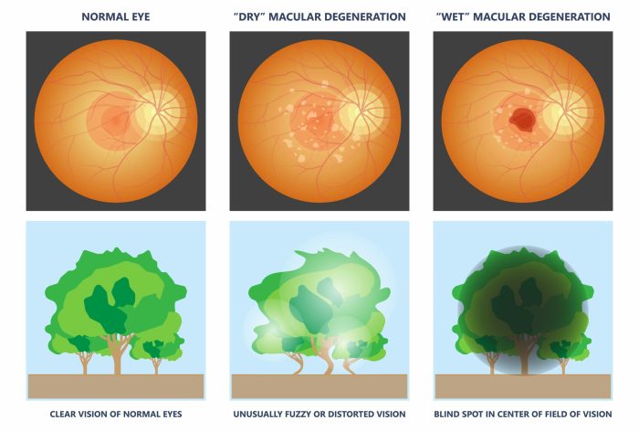
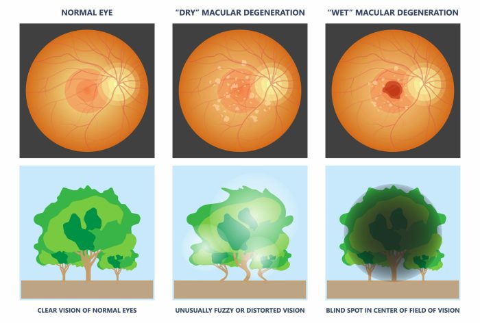
    Dry macular degeneration, the more common type, involves a gradual thinning of the macula. Wet macular degeneration, while less prevalent, is more aggressive and involves abnormal blood vessel growth under the macula, leading to leakage and scarring. Both forms result in vision loss, though the mechanisms and progression differ.Several factors increase the risk of developing macular degeneration.

    Age is a significant risk factor, with the prevalence increasing dramatically after the age of 50. Genetics also plays a role; individuals with a family history of macular degeneration are at a higher risk. Smoking, a diet deficient in certain nutrients (like antioxidants), and a history of cardiovascular disease are also associated with an increased likelihood of developing the condition.

    Types of Macular Degeneration and Associated Symptoms

    Different types of macular degeneration present with distinct symptoms, and the rate of progression can vary. Understanding these differences is crucial for early diagnosis and appropriate management.

    Type of Macular Degeneration Common Symptoms Approximate Age of Onset
    Dry Macular Degeneration Blurred central vision, distortion of straight lines, gradual loss of central vision, difficulty reading fine print, and reduced color perception. Typically after 50, but can occur earlier in some cases.
    Wet Macular Degeneration Sudden vision loss, distorted or blurry central vision, straight lines appearing wavy, and a sudden decrease in color perception. Typically after 50, but can occur earlier in some cases, often after dry MD.

    Note: The age of onset is an approximation. Individual experiences can vary significantly. Early diagnosis and management can help maintain vision and slow progression in most cases.

    Age of Onset Variability

    What age does macular degeneration start

    Macular degeneration, a leading cause of vision loss in older adults, isn’t a uniform experience. The age at which it manifests can vary significantly, impacting individuals differently and requiring tailored management strategies. Understanding this variability is crucial for early detection and proactive interventions.The range of ages at which macular degeneration appears highlights the complexity of this condition. While the typical onset is in later adulthood, individuals can experience the disease much earlier in life.

    This age disparity is due to a combination of genetic predispositions, environmental factors, and lifestyle choices.

    Typical Age of Onset for Different Types

    Age-related macular degeneration (AMD) typically presents in individuals over the age of 50, but the exact age of onset can vary based on the specific type. Dry AMD, the more common form, usually appears later than wet AMD. Wet AMD, characterized by the abnormal growth of blood vessels, tends to manifest at a somewhat earlier age than dry AMD, though this isn’t always the case.

    Furthermore, other less common forms of macular degeneration may have different typical age ranges, necessitating individualized assessment.

    Examples of Early Onset

    While less common, macular degeneration can emerge in individuals younger than the typical age of 50. Some individuals with a strong family history of AMD might develop symptoms earlier. Certain genetic mutations can significantly increase the risk of early-onset macular degeneration, even in those under 40. Additionally, certain lifestyle factors, such as prolonged exposure to intense sunlight or specific medical conditions, might also play a role.

    An example of early onset would be an individual diagnosed with wet AMD in their 30s due to a family history of the condition.

    Factors Contributing to Age Variation

    Several factors can influence the age at which macular degeneration develops. These factors interact in complex ways, and the precise contribution of each can vary from person to person.

    Factor Description Impact on Onset
    Genetics Inherited predisposition to the condition, including specific gene mutations. Can significantly accelerate or delay the onset, particularly in cases with strong family history.
    Lifestyle Factors Dietary habits, smoking, exposure to sunlight, and overall health conditions. Poor lifestyle choices can increase the risk and potentially lead to earlier onset.
    Environmental Factors Exposure to environmental toxins, pollution, and certain dietary deficiencies. Exposure to harmful substances can negatively affect retinal health, potentially accelerating macular degeneration.
    Underlying Medical Conditions Conditions such as diabetes, high blood pressure, and high cholesterol. Certain pre-existing health problems can increase the risk of macular degeneration and potentially accelerate its onset.
    Age The most significant risk factor, but not a predictor for onset in all cases. Generally associated with increased risk as the population ages.

    Genetic Predisposition and Macular Degeneration

    Genetics plays a significant role in determining an individual’s susceptibility to macular degeneration. While age remains the most significant risk factor, inherited variations in genes can influence the likelihood of developing the condition and, importantly, the age at which it might manifest. Understanding these genetic influences is crucial for identifying individuals at higher risk and potentially developing preventative strategies.The complex interplay between genetic predisposition and environmental factors dictates the development of macular degeneration.

    Environmental factors, such as smoking, diet, and exposure to sunlight, can influence the progression of the disease in individuals with a genetic predisposition. Conversely, individuals without a genetic predisposition might still develop the condition due to significant environmental factors, although the age of onset might be later. This interplay highlights the importance of both genetic and environmental factors in understanding the disease.

    Specific Genes Associated with Risk

    Several genes have been linked to an increased risk of macular degeneration. Research continues to uncover new associations, adding to our understanding of the genetic basis of the disease. This intricate genetic landscape suggests that multiple genes contribute to the overall risk.

    Genetic Mutations and Age of Onset

    While a precise correlation between specific genetic mutations and exact age of onset is often elusive, some patterns emerge. The presence of certain genetic mutations may lead to earlier-onset disease in some individuals compared to those without such mutations. The influence of other genetic variations and environmental factors can significantly modify the expected age of onset.

    Macular degeneration, a common eye condition, can start showing up at various ages, but it’s often more prevalent in older adults. While exploring different ways to manage eye health is important, it’s also crucial to consider support options like mobility aids with fibromyalgia if you or someone you know has fibromyalgia. The age of onset for macular degeneration is often associated with factors like genetics and lifestyle, so it’s a complex issue.

    Ultimately, maintaining a healthy lifestyle and getting regular eye checkups are key to detecting and managing the condition.

    Genetic Mutation Approximate Age of Onset (Example) Description
    CFH gene mutations 50-60s (often earlier in those with family history) The CFH gene is strongly associated with age-related macular degeneration. Mutations in this gene lead to a greater risk of the condition. The exact age of onset varies significantly, depending on the specific mutation and other contributing factors.
    ARMS2 gene mutations 40-50s (often earlier in those with family history) Mutations in the ARMS2 gene are also implicated in a heightened risk of macular degeneration. The age of onset, like in CFH, is not fixed and can vary based on individual factors.
    TIMP3 gene mutations 60-70s (may be earlier in individuals with other risk factors) The TIMP3 gene is involved in regulating the extracellular matrix. Mutations in this gene can increase the risk of macular degeneration. The observed age of onset, like in the other genes, is a general estimate.

    Note: The ages presented in the table are approximations and can vary significantly. Individual experiences are influenced by numerous genetic and environmental factors.

    Interaction Between Genetics and Environment

    The relationship between genetic predisposition and environmental factors is complex. Individuals with a strong genetic predisposition might experience earlier onset and more rapid progression of the disease if exposed to environmental risk factors. Conversely, individuals without a strong genetic predisposition might still develop macular degeneration if environmental factors are severe enough. This highlights the combined impact of these factors on the disease’s progression.

    Macular degeneration, a common eye condition, can start to affect vision at various ages, though it’s more prevalent in older individuals. Factors like genetics and lifestyle play a role, but maintaining a healthy red blood cell rbc count here might contribute to overall eye health, potentially delaying the onset of age-related macular degeneration. Ultimately, the precise age of onset varies significantly from person to person.

    Environmental Influences

    Environmental factors play a significant role in the development and progression of age-related macular degeneration (AMD). While genetics are a strong predictor, external influences can either exacerbate or mitigate the risk of AMD, potentially affecting the age of onset. Understanding these influences is crucial for developing preventative strategies and personalized approaches to managing the disease.

    Environmental Exposures Potentially Linked to AMD

    Environmental exposures, such as sunlight, smoking, diet, and certain infections, have shown a correlation with AMD. Sunlight, with its ultraviolet (UV) radiation, is believed to damage the retina over time, potentially contributing to AMD development. Similarly, exposure to certain pollutants in the air or water may also play a role. Smoking is a well-established risk factor, its impact on oxidative stress and inflammation likely contributing to the disease’s progression.

    A diet deficient in certain nutrients, or high in saturated fats, may also influence AMD development.

    Potential Mechanisms of Environmental Impact

    Environmental factors likely influence AMD through various mechanisms. Oxidative stress, the imbalance between free radicals and antioxidant defenses, is a key player. Exposure to UV radiation, air pollutants, and even certain dietary choices can increase oxidative stress in the eye. This stress can damage retinal cells, potentially accelerating the degeneration process. Inflammation is another critical factor.

    Chronic low-grade inflammation in the eye, induced by various environmental exposures, may lead to the breakdown of retinal tissues. Smoking, for example, directly contributes to inflammation and increases oxidative stress, creating a double-edged risk factor for AMD.

    Impact of Different Environmental Factors

    The impact of different environmental factors on the age of onset of AMD can vary. While some factors, like smoking, have a consistently strong correlation with earlier onset, others, like diet, might have a more nuanced impact, depending on the specific dietary choices and overall health. Factors such as UV exposure can increase the risk of AMD, but the degree of impact depends on the duration and intensity of the exposure.

    The influence of environmental factors is not always direct or predictable. Interactions between genetic predispositions and environmental factors can significantly alter the age of onset and disease progression.

    Comparison of Environmental Factors’ Impact on Age of Onset

    Environmental Factor Approximate Impact on Age of Onset (Note: This is a rough estimate and can vary greatly based on individual factors.)
    Smoking Potentially accelerates onset by several years.
    High UV Exposure May contribute to earlier onset, particularly in individuals with genetic predisposition.
    Poor Diet (High in Saturated Fats, Low in Antioxidants) Potentially accelerates onset by several years, primarily by increasing oxidative stress.
    Air Pollution Potentially increases risk, but the effect on onset age is less clear compared to other factors.
    Certain Infections Possible association with earlier onset, but more research is needed.

    Note: The table above provides a general overview. Individual responses to environmental factors can vary significantly due to genetic predisposition, lifestyle, and other factors.

    Lifestyle and Age of Onset

    What age does macular degeneration start

    Our lifestyle choices play a significant role in the development and progression of macular degeneration. While genetics and environmental factors are influential, the decisions we make daily can impact when and how severely this condition manifests. Understanding these connections is crucial for proactive measures to safeguard eye health.Maintaining a healthy lifestyle can delay the onset of macular degeneration or slow its progression.

    This involves adopting a balanced diet, regular exercise, and avoiding harmful habits like smoking. The impact of these choices on the disease’s trajectory is substantial, and understanding how they interact with the genetic predisposition is key to personalized strategies for prevention.

    Diet’s Impact on Macular Degeneration

    A balanced diet rich in antioxidants and essential nutrients is crucial for maintaining overall health, including eye health. Vitamins like lutein and zeaxanthin, found in leafy greens, are particularly beneficial for protecting the macula. Studies suggest that diets high in fruits, vegetables, and omega-3 fatty acids may contribute to delaying the onset and slowing the progression of macular degeneration.

    Conversely, diets high in saturated fats and processed foods might have an adverse effect. The specific nutrients that impact macular degeneration are still under investigation.

    Exercise and Macular Degeneration

    Regular physical activity is beneficial for overall health and may also play a role in protecting against macular degeneration. Exercise improves blood circulation, delivering essential nutrients to the eyes and potentially reducing inflammation. The exact mechanisms through which exercise influences macular degeneration are not fully understood, but evidence suggests a positive correlation between physical activity and reduced risk.

    For instance, a study found a lower incidence of macular degeneration in individuals who consistently engaged in moderate-intensity exercise.

    Smoking and Macular Degeneration

    Smoking significantly increases the risk of developing macular degeneration. The harmful chemicals in tobacco smoke damage blood vessels, impairing the delivery of nutrients to the eyes and potentially leading to accelerated macular degeneration. Individuals who smoke are more likely to experience a faster progression of the disease, resulting in greater vision loss compared to nonsmokers. Quitting smoking, even at later stages, can still have positive effects on the progression of macular degeneration.

    Importance of Regular Eye Exams

    Regular eye exams are vital for early detection of macular degeneration. Early diagnosis allows for timely intervention and management strategies, potentially slowing the progression of the disease and preserving vision. Even if there are no apparent symptoms, regular check-ups can identify early signs of macular degeneration. Early detection is essential because treatment options are often more effective when implemented in the initial stages.

    Lifestyle Factors Affecting Macular Degeneration

    Factor Description Impact on Onset
    Balanced Diet (Fruits, Vegetables, Omega-3s) High intake of antioxidants and essential nutrients. Positive
    Regular Exercise Maintaining a consistent physical activity routine. Positive
    Smoking Consumption of tobacco products. Negative
    Regular Eye Exams Routine check-ups to monitor eye health. Positive

    Early Detection and Diagnosis

    Catching macular degeneration early is crucial for managing its progression and preserving vision. Early intervention can significantly slow the rate of vision loss, allowing individuals to maintain independence and quality of life. Understanding the early signs and the diagnostic procedures available is key to proactive eye health.Early detection of macular degeneration relies heavily on regular eye exams, coupled with knowledge of potential symptoms.

    The process involves identifying subtle changes in vision that might initially go unnoticed. Different age groups may exhibit varying symptoms, and recognizing these differences is essential for prompt diagnosis and treatment.

    Recognizing Early Signs

    Early signs of macular degeneration may be subtle and easily missed, especially in the initial stages. Individuals may notice a blurring or distortion of central vision, particularly in activities requiring sharp focus like reading or driving. Straight lines might appear wavy, and colors may appear faded or less vibrant. Difficulty distinguishing fine details, such as facial features, is another possible symptom.

    Peripheral vision usually remains unaffected initially, which can mask the progression of the disease in its early stages.

    Common Diagnostic Procedures, What age does macular degeneration start

    Various diagnostic tools and procedures are used to detect macular degeneration. A comprehensive eye exam is the cornerstone of the diagnostic process. This involves evaluating the health of the entire eye, including the retina, optic nerve, and other structures. Detailed visual acuity tests are conducted to assess the sharpness and clarity of vision at different distances. Additional tests may include:

    • Amsler Grid Test: This test helps detect distortion or blurring in central vision. The Amsler grid is a grid-like pattern used to measure the clarity and straightness of the lines. Any deviation from the normal grid pattern can be a sign of macular degeneration. This test is often used as a screening tool.
    • Optical Coherence Tomography (OCT): OCT uses light waves to create detailed images of the retina, revealing the thickness and structure of the macula. This test can identify any changes or thinning in the macular region, which are indicative of macular degeneration. It can also help distinguish between different types of macular degeneration.
    • Fluorescein Angiography: This test involves injecting a dye into the bloodstream and taking pictures of the retinal blood vessels as the dye circulates. It helps to visualize the blood flow within the retina, highlighting any abnormalities that might be present. This can help diagnose macular degeneration by revealing any issues with the blood vessels supplying the macula.

    Vision Tests at Different Ages

    The approach to vision testing for macular degeneration varies based on the individual’s age. Children and young adults may undergo standard vision tests, similar to those performed for other refractive errors. As individuals age, the frequency and type of tests may increase. Older adults, particularly those with risk factors, might undergo more specialized tests, such as OCT or fluorescein angiography, to detect early signs of the disease.

    Regular comprehensive eye exams are essential regardless of age to detect and address any vision changes promptly.

    Diagnostic Procedures for Different Age Groups

    The diagnostic approach varies depending on the age of the patient. Young individuals often undergo standard vision tests to assess visual acuity and refraction. As people age, the diagnostic process might include more in-depth examinations, such as OCT or fluorescein angiography, to screen for macular degeneration. This tailored approach allows healthcare professionals to adapt their diagnostic strategies to the specific needs of each age group.

    Furthermore, family history and risk factors play a crucial role in determining the appropriate diagnostic steps for individuals of different ages.

    Treatment Options

    Unfortunately, there’s no cure for macular degeneration, but several treatments can help slow its progression and manage symptoms. These approaches aim to preserve vision and improve the quality of life for individuals affected by this condition. The specific treatment plan will depend on the type of macular degeneration, the stage of the disease, and the individual’s overall health.Current treatments primarily focus on slowing the disease’s progression rather than reversing it.

    The effectiveness of these treatments can vary significantly, and ongoing research continues to explore new and potentially more effective therapies.

    Available Treatment Options

    Various treatments are available for managing macular degeneration, each with its own set of benefits and drawbacks. The most common treatments involve medications, lifestyle adjustments, and supportive care.

    • Anti-VEGF Medications: These medications, administered through injections into the eye, target vascular endothelial growth factor (VEGF), a protein that contributes to the growth of abnormal blood vessels in the eye. By reducing VEGF levels, these injections can help slow the growth of these problematic vessels, thereby potentially reducing the damage to the macula. For example, ranibizumab (Lucentis) and aflibercept (Eylea) are common anti-VEGF medications used in treating macular degeneration.

    • Lifestyle Modifications: While not a cure, adopting a healthy lifestyle can potentially mitigate some risk factors associated with macular degeneration. These lifestyle changes can include maintaining a balanced diet rich in antioxidants and vitamins, regular exercise, and abstaining from smoking. For instance, a diet rich in fruits and vegetables high in antioxidants like lutein and zeaxanthin has shown promise in potentially reducing the risk of the disease.

    • Complementary and Alternative Therapies: Some individuals explore complementary and alternative therapies, such as nutritional supplements or specific diets, to potentially mitigate the progression of the disease. However, it’s crucial to consult with a healthcare professional before using these therapies, as their effectiveness has not been rigorously proven and can potentially interact with other medications.

    Effectiveness of Treatments

    The effectiveness of different treatments for slowing the progression of macular degeneration can vary significantly. Anti-VEGF medications, when administered as prescribed, are often quite effective in slowing the growth of abnormal blood vessels and preserving vision in many cases. However, the response to treatment is not uniform and may vary based on individual factors. Furthermore, treatment effectiveness may decrease over time as the disease progresses.

    The response to anti-VEGF medications can be measured through regular ophthalmological examinations.

    Treatment Variations

    The choice of treatment for macular degeneration will vary based on the type and stage of the disease. Early-stage dry macular degeneration might respond differently to treatment than wet macular degeneration, and the effectiveness of interventions may also differ depending on the specific characteristics of the individual’s condition. For example, a patient with early-stage dry macular degeneration might be more likely to benefit from dietary modifications and lifestyle changes, while a patient with wet macular degeneration might require more aggressive treatments like anti-VEGF injections.

    Comparison of Treatment Options

    Treatment Type Effectiveness Potential Side Effects
    Anti-VEGF Medications (e.g., Lucentis, Eylea) Often effective in slowing disease progression, preserving vision, and reducing fluid buildup in the macula. Eye infections, increased eye pressure, and bleeding in the eye are possible side effects, although they are usually temporary and manageable.
    Lifestyle Modifications (e.g., diet, exercise, smoking cessation) While not a cure, these modifications can mitigate some risk factors and improve overall health, which might have a positive impact on the disease’s progression. No direct side effects associated with these lifestyle modifications, but consistent adherence is crucial.
    Complementary and Alternative Therapies Effectiveness is not consistently proven, and interactions with other medications are possible. Potential side effects are dependent on the specific therapy and should be discussed with a healthcare professional.

    Conclusion

    In conclusion, macular degeneration’s onset is a multifaceted issue influenced by a complex interplay of genetic predisposition, environmental factors, and lifestyle choices. While a precise age of onset isn’t always predictable, understanding the contributing factors is crucial for early detection and potential intervention. This exploration has highlighted the significance of genetic testing, environmental awareness, and the importance of proactive lifestyle choices to mitigate risk.

    The key takeaway? Maintaining a healthy lifestyle, regular eye exams, and open communication with your eye care professional are paramount in safeguarding your vision.

  • Macular Degeneration Coping Support and Living Well

    Macular Degeneration Coping Support and Living Well

    Macular degeneration coping support and living well is a comprehensive guide to navigating the challenges and embracing the possibilities of life with this condition. It delves into understanding the different types, symptoms, and progression of macular degeneration, equipping you with practical coping strategies, support systems, and lifestyle choices. This journey will help you learn how to maintain independence, build a supportive community, and ultimately live a fulfilling life despite the challenges.

    This guide will explore the diverse aspects of managing macular degeneration, from the initial diagnosis to adapting to a life with reduced vision. We’ll cover practical techniques for maintaining daily tasks, assistive technologies, and important support systems, including family, friends, and healthcare professionals. We’ll also discuss the crucial role of maintaining a healthy lifestyle, emotional well-being, and connecting with a supportive community.

    Understanding Macular Degeneration

    Macular degeneration is a progressive eye disease that affects the macula, the central part of the retina responsible for sharp, detailed vision. This crucial area of the eye is essential for tasks like reading, driving, and recognizing faces. Understanding the different types of macular degeneration, their symptoms, and how they progress is vital for early detection and effective management.

    This knowledge empowers individuals to make informed decisions about their eye health and overall well-being.Macular degeneration, a significant cause of vision loss, is characterized by the gradual deterioration of the macula. This leads to a decline in central vision, impacting daily activities that rely on sharp, detailed sight. The disease progresses at different rates, and its impact on vision varies depending on the type and stage of the condition.

    Types of Macular Degeneration

    Macular degeneration manifests in two primary forms: dry and wet. Understanding the distinctions between these forms is crucial for appropriate management and treatment.

    Dry Macular Degeneration

    Dry macular degeneration, the more common type, typically begins with the gradual thinning and deterioration of the light-sensitive cells in the macula. This process can be slow and often asymptomatic in the early stages. The loss of vision is usually gradual and subtle. In many cases, dry macular degeneration progresses slowly over many years, and the impact on vision can be mild, but in some cases, it can lead to significant vision loss.

    This form is often associated with age-related changes in the eye.

    Wet Macular Degeneration

    Wet macular degeneration is a more aggressive form of the disease. In this type, abnormal blood vessels grow under the macula, leaking fluid and blood. This leakage damages the light-sensitive cells and leads to rapid vision loss. The vision loss associated with wet macular degeneration is often more significant and occurs more quickly than in dry macular degeneration.

    The abnormal blood vessels are responsible for the rapid progression of vision loss in wet macular degeneration.

    Symptoms and Signs of Macular Degeneration

    The symptoms of macular degeneration can vary, depending on the type and stage of the disease. Common signs and symptoms include:

    • Blurred central vision: Objects directly in front of the eyes may appear blurry or distorted.
    • Straight lines appearing wavy or distorted: This is a common early symptom of macular degeneration.
    • Difficulty reading or recognizing faces: Tasks requiring sharp central vision may become challenging.
    • Dark or empty spots in the center of vision: These are sometimes described as “blind spots.”

    Progression of Macular Degeneration

    The progression of macular degeneration varies significantly. In dry macular degeneration, the vision loss is often slow and gradual, with some individuals experiencing little to no impact on their daily activities. In contrast, wet macular degeneration can lead to a rapid decline in vision, potentially causing significant impairment within a short period. It’s important to note that the progression of macular degeneration is not uniform, and the rate of vision loss can differ from person to person.

    Comparison of Wet and Dry Macular Degeneration

    Feature Dry Macular Degeneration Wet Macular Degeneration
    Cause Thinning and breakdown of the light-sensitive cells in the macula. Abnormal blood vessel growth under the macula, causing leakage of fluid and blood.
    Progression Slow and gradual. Rapid and significant.
    Symptoms Often subtle, including blurred central vision and straight lines appearing wavy. Can include rapid central vision loss, distorted vision, and dark spots.
    Treatment Often supportive care, including eye exams and vitamin supplements. May involve treatments like injections or laser therapy.

    Coping Strategies for Vision Loss

    Navigating daily life with macular degeneration requires adapting to changes in vision. This involves not just adjusting to reduced sight, but also embracing strategies for maintaining independence, connection, and overall well-being. Understanding effective coping mechanisms and assistive technologies can empower individuals to live fulfilling lives despite the challenges posed by vision loss.Effective coping strategies are crucial for managing daily tasks with reduced vision.

    They encompass a wide range of approaches, from simple adjustments to utilizing advanced assistive technologies. Finding the right balance between adaptation and preservation of independence is key to maintaining a positive outlook.

    Practical Coping Mechanisms for Daily Tasks

    Managing daily tasks requires thoughtful adjustments. Simple modifications can significantly enhance daily life. For example, using larger print, strategically placed lighting, and organizing items in a predictable manner can ease navigation and reduce frustration. Utilizing a magnifying glass or a digital magnifier can greatly assist in tasks like reading, writing, or inspecting small objects. Creating a well-organized space with accessible items, reducing clutter, and maintaining clear pathways are crucial for minimizing obstacles and enhancing safety.

    Assistive Technologies for Daily Living

    Assistive technologies play a vital role in facilitating daily tasks and maintaining independence. These technologies range from simple tools to sophisticated computer programs. For example, screen readers, such as JAWS or NVDA, convert text on a computer screen into audible speech, enabling individuals with vision loss to access digital information and communications. Voice-activated devices and software are beneficial for controlling household appliances, managing calendars, and performing tasks that require minimal visual input.

    Adaptive switches and controls can be customized to accommodate various needs, enabling individuals to operate electronics and appliances with ease.

    Maintaining Independence and Social Engagement

    Maintaining independence is a key aspect of coping with vision loss. Strategies include relying on a support network of family and friends, utilizing public transportation systems with accessible features, and arranging transportation with care. Using adaptive devices such as talking clocks and calendars can help maintain daily routines and enhance time management. Learning to adapt and rely on technology for daily tasks will enable independent living.

    For social engagement, joining support groups and organizations focused on vision loss can provide a valuable network of peers and professionals who understand the challenges and offer encouragement.

    Resources for Support Groups and Organizations

    Numerous support groups and organizations offer invaluable assistance to individuals dealing with vision loss. These resources provide a platform for sharing experiences, gaining practical advice, and connecting with others who understand the unique challenges of living with macular degeneration. The National Federation of the Blind (NFB) and the American Foundation for the Blind (AFB) are prominent examples. Local chapters of these organizations and similar groups provide in-person support, resources, and advocacy opportunities.

    Many hospitals and community centers also offer support groups for vision loss. Connecting with these resources allows for building a strong support network, enabling individuals to share experiences and receive guidance.

    Navigating macular degeneration can be tough, but finding ways to cope and live well is key. Managing the day-to-day discomfort, like when a toothache strikes, can be surprisingly helpful. For instance, exploring over-the-counter dental pain relief options like otc dental pain relief can really ease some of that pain. Ultimately, prioritizing self-care and support groups remains crucial for a positive outlook and maintaining a good quality of life.

    Organization Description
    National Federation of the Blind (NFB) A prominent national organization advocating for the rights and needs of blind and visually impaired individuals.
    American Foundation for the Blind (AFB) Provides extensive resources, services, and advocacy for individuals with vision loss.

    Support Systems and Resources

    Navigating macular degeneration requires a multifaceted approach, encompassing medical care, emotional support, and practical assistance. This section highlights the vital roles of various support systems in helping individuals cope with the challenges and maintain a high quality of life. Understanding the available resources can significantly ease the burden and empower those affected by this condition.

    Family and Friends

    Family and friends play a crucial role in providing emotional support and practical assistance. They can offer companionship, encouragement, and a listening ear during difficult times. Their understanding and willingness to help with daily tasks, like transportation or household chores, can significantly improve the quality of life for someone with macular degeneration. Family members can also act as advocates, helping navigate medical appointments and understand treatment options.

    Building a strong support network among loved ones is invaluable in managing the emotional and practical aspects of the condition.

    Healthcare Professionals

    Healthcare professionals, including ophthalmologists, optometrists, and other specialists, are essential for managing macular degeneration. They provide crucial medical care, monitor disease progression, and recommend appropriate treatments. Beyond the medical aspect, they can offer valuable information about resources, support groups, and coping strategies. Regular communication with healthcare professionals is vital to stay informed and make informed decisions. This allows for prompt interventions and helps individuals maintain their independence.

    Government Assistance Programs and Financial Resources

    Numerous government assistance programs and financial resources are available to support individuals with macular degeneration. These programs can provide financial aid for medical expenses, assistive devices, and modifications to the home. Examples include Medicare, Medicaid, and state-specific programs for low-income individuals. Detailed information about eligibility criteria and application procedures can be obtained from local social service agencies and government websites.

    Exploring these options can alleviate financial burdens associated with the condition.

    Support Groups and Online Communities

    Support groups and online communities offer invaluable opportunities for connection and shared experiences. These platforms provide a safe space for individuals to connect with others who understand the challenges of macular degeneration. They can share practical tips, coping strategies, and emotional support. Participating in these groups can reduce feelings of isolation and foster a sense of community.

    Online forums and in-person support groups provide a valuable network of people who are navigating similar journeys.

    Navigating macular degeneration can be tough, but finding support groups and resources for living well is key. While dealing with a sudden injury like a gunshot wound requires immediate action, like knowing how to treat a gunshot wound, how to treat a gunshot wound is a different kind of challenge entirely. Focusing on practical strategies for coping with macular degeneration, like adapting daily routines and embracing assistive technology, is vital for maintaining a positive outlook and quality of life.

    Therapists and Counselors

    Therapists and counselors play a vital role in managing the emotional well-being of individuals with macular degeneration. They can provide guidance and support for dealing with the emotional challenges of vision loss, including feelings of frustration, anxiety, and depression. Counseling can help individuals develop coping mechanisms, adjust to lifestyle changes, and maintain a positive outlook. Therapy can empower individuals to manage the emotional impact of macular degeneration and navigate the changes in their lives.

    Maintaining a Healthy Lifestyle

    Living well with macular degeneration involves more than just managing vision loss. A holistic approach that prioritizes overall health plays a crucial role in maintaining quality of life. This includes making informed choices about diet, engaging in regular physical activity, and actively managing stress. These factors directly influence your physical and emotional well-being, allowing you to better cope with the challenges of the condition.A healthy lifestyle is fundamental in managing macular degeneration.

    By adopting positive habits, you can support your body’s natural ability to function optimally, thereby potentially slowing the progression of the disease and improving your overall quality of life. It’s not about a quick fix, but rather a sustained commitment to well-being that will yield long-term benefits.

    Dietary Recommendations for Eye Health

    A balanced diet rich in essential nutrients is vital for maintaining good eye health. Many nutrients contribute to the overall health and function of the eyes.

    • Antioxidants: Antioxidants, such as vitamins C and E, lutein, and zeaxanthin, play a crucial role in protecting the eyes from damage caused by free radicals. These nutrients are found in colorful fruits and vegetables like berries, leafy greens, and carrots. A diet rich in these foods can help maintain healthy eye tissues and potentially slow the progression of macular degeneration.

      For example, incorporating spinach, kale, and blueberries into your diet provides valuable antioxidants.

    • Omega-3 Fatty Acids: Omega-3 fatty acids are essential for eye health, contributing to the structural integrity of the retina. These are commonly found in fatty fish like salmon, tuna, and mackerel. Consider incorporating fish oil supplements as a way to obtain these essential nutrients.
    • Zinc: Zinc is an important mineral for eye health, and it is involved in the transport of vitamin A to the retina. Foods high in zinc include oysters, beef, and pumpkin seeds. A sufficient intake of zinc is vital for optimal retinal function.
    • Vitamin A: Vitamin A is essential for maintaining healthy eye tissue. Foods like sweet potatoes, carrots, and leafy greens are excellent sources of vitamin A. A balanced diet that includes a variety of fruits, vegetables, and whole grains can provide sufficient amounts of vitamin A.

    Importance of Regular Exercise

    Regular physical activity is crucial for overall health and well-being, and it plays a significant role in managing macular degeneration.

    • Cardiovascular Health: Regular exercise improves cardiovascular health, which is beneficial for blood flow to all parts of the body, including the eyes. This can help maintain healthy retinal blood vessels, which are vital for vision. For instance, brisk walking, swimming, or cycling can be beneficial for eye health.
    • Weight Management: Maintaining a healthy weight is crucial for overall health and can potentially slow the progression of macular degeneration. Exercise plays a vital role in weight management, helping you achieve and maintain a healthy body weight. For example, consistent exercise routines help control weight and improve overall health.
    • Stress Reduction: Exercise is an effective stress management technique, reducing stress hormones and promoting relaxation. This contributes to improved mental and emotional well-being. Physical activity, like yoga or meditation, can reduce stress levels.

    Stress Management Techniques

    Stress can have a significant impact on overall health, including eye health. Effective stress management techniques are essential for maintaining emotional well-being and coping with the challenges of macular degeneration.

    • Mindfulness and Meditation: Mindfulness and meditation practices help to cultivate awareness of the present moment, reducing stress and promoting relaxation. Regular mindfulness exercises can be beneficial for emotional regulation.
    • Yoga and Tai Chi: These practices combine physical postures, breathing techniques, and meditation, promoting relaxation, flexibility, and stress reduction. Yoga and Tai Chi are excellent stress reduction techniques.
    • Social Support: Maintaining strong social connections and seeking support from family and friends can provide emotional comfort and reduce stress levels. Connecting with others can be a crucial coping mechanism for stress.

    Living Well with Macular Degeneration

    Adjusting to macular degeneration requires proactive strategies to maintain independence and quality of life. This involves understanding how the condition affects daily routines and implementing modifications to the home, transportation, and social environment. Embracing new approaches to activities and social connections can be empowering and allow individuals to continue thriving despite vision loss.

    Sample Daily Routine for Reduced Vision

    A tailored daily routine can significantly improve the experience of living with macular degeneration. This structured approach can help individuals feel more in control and organized.

    • Morning Routine: Begin with a well-lit environment. Use large-button clothing and clocks with large numbers. Prepare a visual schedule of tasks with clear instructions. Ensure sufficient time for morning activities, allowing for potential delays due to reduced visual acuity.
    • Meal Preparation: Prepare meals in advance, using clear labels on containers and organized storage systems. Utilize kitchen aids with large handles and well-lit areas for food preparation. Consider pre-portioned meals or simple recipes that are easy to prepare.
    • Afternoon Activities: Schedule activities with ample time between tasks, allowing for adjustments and breaks. Prioritize tasks that require minimal visual effort in the afternoon. Utilize audio books, podcasts, or other auditory resources during downtime.
    • Evening Routine: Dim the lights gradually to prepare for sleep. Use a night light and a well-defined path to the bathroom or bedroom. Keep frequently used items within easy reach.

    Modifying Home Environments for Independent Living

    Adapting the home environment is crucial for maintaining independence. Careful modifications can significantly improve daily living.

    • Lighting: Maximize natural light. Use task lighting with adjustable brightness, and consider using bright, contrasting colors for rooms. Install strategically placed lamps and light fixtures to enhance visibility.
    • Furniture and Flooring: Ensure clear pathways, avoiding clutter. Choose contrasting colors for rugs and floors to delineate spaces. Utilize furniture with large, easily accessible features.
    • Kitchen and Bathroom: Install grab bars, non-slip mats, and adjustable counters for improved accessibility. Use contrasting colors for kitchen cabinets and appliances to enhance visibility. Organize frequently used items within easy reach.
    • Technology: Explore assistive technology like screen readers, magnifiers, and voice-activated devices. Ensure technology is user-friendly and easy to operate.

    Accessible Transportation Options

    Transportation options should be accessible and reliable, allowing for easy navigation and independence.

    • Public Transportation: Utilize public transportation systems with clear signage and well-marked routes. Learn the specific routes and schedules in advance. Communicate with transportation officials about specific needs.
    • Ride-Sharing Services: Research ride-sharing services and evaluate their accessibility features. Communicate specific needs to the drivers. Ensure clear communication and instructions.
    • Accessible Vehicles: Consider accessible vehicles with ramps and modifications for easier entry and exit. Consult with specialists for tailored vehicle recommendations.

    Maintaining Social Connections and Engagement

    Maintaining social connections and engagement in activities is vital for overall well-being. Adapting to vision loss should not limit participation in activities.

    • Social Activities: Engage in social activities that are accessible and adaptable to vision loss. Explore activities like group discussions, book clubs, or online communities. Seek out organizations and clubs with supportive environments.
    • Support Groups: Join support groups to connect with others facing similar challenges. Support groups provide a valuable platform for sharing experiences and gaining practical advice.
    • Adaptive Activities: Engage in activities that can be modified to accommodate vision loss. For example, consider group activities with auditory components or visual aids. Research modified options for hobbies and recreational pursuits.

    Accessible Communication and Information

    Navigating life with macular degeneration often involves adapting communication and information access methods. This section details various strategies to ensure you remain connected and informed despite vision loss. Understanding the options available allows you to maintain independence and build strong support networks.Effective communication and access to information are vital for maintaining a fulfilling life with macular degeneration. This includes adapting to different communication methods and learning to effectively communicate visual challenges to others.

    Access to accessible formats for information and media is also crucial for continuing to engage with the world around you.

    Methods for Receiving Accessible Information

    Different methods for receiving accessible information cater to various needs and preferences. These methods include audio descriptions, large print materials, braille, and electronic formats like screen readers. Choosing the best method often depends on your individual needs and learning styles.

    • Audio descriptions: Provide auditory descriptions of visual elements in movies, television shows, and other media. This allows viewers to understand the content despite not seeing it. Many streaming services offer audio description options. This is particularly useful for enjoying entertainment and staying informed about current events.
    • Large print materials: Enlarge text and images to make them easier to read. Many organizations and libraries offer materials in large print. This approach helps those with decreased visual acuity read documents and books more comfortably.
    • Braille: A tactile writing system that uses raised dots. Many books, magazines, and documents are available in braille. Braille offers an alternative for those with significant vision loss.
    • Electronic formats: Accessible documents, websites, and software with screen readers and other assistive technology help individuals with vision loss access digital content. Screen readers are software programs that convert text into speech. This approach allows those with impaired vision to navigate digital environments effectively.

    Assistive Devices for Communication

    Assistive devices are tools that aid in communication and information access. Many devices are designed to increase independence and help maintain social connections. Consider using them in combination with other methods to create a personalized approach.

    • Screen readers: Software that converts text on a computer screen into audio, allowing users to navigate and interact with digital content without relying on visual input. Many screen readers can be customized to suit individual needs.
    • Magnifying glasses: Optical aids that enlarge text and images, making them easier to read. Various types of magnifying glasses are available, ranging from handheld devices to tabletop models. These can significantly improve reading and viewing experiences.
    • Closed-captioning: Displaying text on a screen that synchronizes with audio content, enabling viewers to follow along with the dialogue and other spoken words. This is extremely helpful for understanding conversations and following along with lectures or presentations.
    • Text-to-speech software: Software that converts text into speech. This tool is particularly helpful for reading articles, emails, or documents. Text-to-speech can be used in conjunction with screen readers to improve accessibility.

    Communicating Effectively with Others

    Clear and open communication is crucial when discussing visual impairment. Understanding the specific challenges you face will help others offer appropriate support. Emphasize the need for adjustments and reasonable accommodations.

    Navigating macular degeneration can be tough, but finding support and ways to live well is key. Understanding your options for financial support is also crucial, and that includes exploring gold plan health insurance, like gold plan health insurance what is it. Knowing what’s covered can significantly ease the burden, which is essential for overall well-being in managing this condition.

    • Open communication: Explain your specific needs and challenges clearly and openly. Sharing your experience will help others understand your perspective and provide effective support.
    • Active listening: Pay attention to the other person’s responses. Ask clarifying questions to ensure you understand their perspectives and concerns. This will help foster a more collaborative and supportive environment.
    • Seeking accommodations: Explain the need for accommodations in various situations, such as at work or in social settings. Explain how certain changes will make interactions more inclusive.
    • Using appropriate language: Avoid using phrases that imply judgment or pity. Focus on practical solutions and strategies for support. Using respectful and considerate language enhances communication.

    Accessing Accessible Information and Media

    Finding accessible information and media requires awareness and exploration. Many organizations and websites offer accessible content. It’s important to actively seek these resources.

    • Online resources: Many websites provide accessible information, including audio descriptions and transcripts. Look for websites with accessibility features and use assistive technologies.
    • Libraries and community centers: Local libraries and community centers often have accessible materials and resources. Utilize these resources to acquire books, magazines, and other media in accessible formats.
    • Government agencies: Government agencies offer a wealth of accessible information on a variety of topics. Use these resources to find relevant information in accessible formats.
    • Organizations specializing in vision loss: Many organizations specifically cater to individuals with vision loss. These organizations provide accessible information, support groups, and other resources.

    Professional Guidance and Treatment

    Navigating macular degeneration requires a multi-faceted approach, including understanding the disease, adopting coping strategies, and seeking appropriate professional guidance. Expert advice and tailored treatment plans are crucial for managing the condition and maintaining quality of life. This section delves into the roles of ophthalmologists and optometrists, available therapies, and the importance of regular eye check-ups.Effective management of macular degeneration hinges on accurate diagnosis, personalized treatment strategies, and ongoing monitoring.

    This involves a collaboration between patients and their healthcare providers to ensure optimal outcomes and address the evolving needs of the individual.

    Roles of Ophthalmologists and Optometrists

    Ophthalmologists are medical doctors specializing in eye care, including the diagnosis and treatment of macular degeneration. They are trained to perform comprehensive eye exams, diagnose various eye conditions, and prescribe medications or conduct surgical procedures. Optometrists, on the other hand, are primary eye care providers who conduct routine eye exams, diagnose refractive errors (like nearsightedness or farsightedness), and prescribe corrective lenses.

    They play a vital role in early detection and monitoring of macular degeneration, often referring patients to ophthalmologists when necessary.

    Types of Therapies and Treatments

    Various therapies and treatments are available for managing macular degeneration, although there is no cure. These therapies aim to slow the progression of the disease, manage symptoms, and improve quality of life. Treatments include:

    • Anti-VEGF injections: These injections target and inhibit the growth of blood vessels that contribute to macular damage, thereby slowing the progression of the disease. They are often administered by ophthalmologists. For example, a patient with neovascular age-related macular degeneration might receive regular anti-VEGF injections to control the growth of new blood vessels.
    • Laser therapy: In some cases, laser treatment can help to reduce abnormal blood vessel growth. This is often a supplementary or complementary therapy used in conjunction with other treatments, such as anti-VEGF injections.
    • Low vision aids: These tools, such as magnifying glasses, large-print materials, and specialized computer software, help individuals with vision loss perform daily tasks and maintain independence. They are often recommended by optometrists and low vision specialists.
    • Lifestyle modifications: Adopting a healthy lifestyle, including a balanced diet, regular exercise, and stress management techniques, can contribute to overall well-being and potentially slow the progression of the disease. This may include dietary adjustments rich in antioxidants and vitamins.

    Importance of Regular Eye Check-ups and Monitoring

    Regular eye check-ups are essential for early detection and management of macular degeneration. Early diagnosis allows for prompt intervention and potential slowing of the disease’s progression. Monitoring helps track the progression of the condition and adjust treatment strategies as needed. A patient’s baseline vision and specific characteristics are considered to provide accurate monitoring.

    Comparison of Eye Care Professionals

    Professional Focus Role in Macular Degeneration Management
    Ophthalmologist Medical eye care, including surgery Diagnoses, treats, and monitors the disease, often prescribing medication and performing procedures like anti-VEGF injections.
    Optometrist Primary eye care, vision correction Conducts routine eye exams, detects early signs, and provides low vision aids and support. Refers patients to ophthalmologists when necessary.
    Low Vision Specialist Specialized care for vision loss Prescribes and fits assistive devices and techniques to maximize remaining vision. Provides support for adaptive strategies.

    Visual Aids and Adaptive Techniques

    Navigating daily life with macular degeneration can be challenging, but various visual aids and adaptive techniques can significantly improve independence and quality of life. These tools and strategies empower individuals to maintain their activities and connections, fostering a sense of control and participation in their communities.

    Visual Aids for Enhanced Vision

    Understanding the specific needs of each individual with macular degeneration is crucial when selecting visual aids. The table below Artikels common visual aids and their applications, offering a starting point for exploring options.

    Visual Aid Description Applications
    Large-print materials Printed text with larger font sizes Reading books, newspapers, and other documents
    Magnifiers Optical devices that enlarge text and images Examining small print, reading maps, and using computer screens
    Telescopic devices Optical aids that increase the apparent size of distant objects Viewing objects from a distance, such as during outdoor activities or while watching television
    Closed-circuit television (CCTV) Systems that magnify images projected onto a screen Reading documents, viewing small objects, and interacting with computer screens
    Screen readers and screen magnifiers Computer software that reads aloud text and enlarges screen elements Accessing digital information, navigating websites, and using software applications

    Adjusting Lighting and Contrast

    Proper lighting significantly impacts visibility for individuals with macular degeneration. Optimal lighting conditions reduce eye strain and improve clarity. Consider the following strategies:

    • Use bright, but not harsh, lighting. Avoid overly dim or flickering lights, as these can exacerbate eye fatigue.
    • Position lighting sources strategically to eliminate glare and shadows. For example, direct sunlight can be very harsh, and overhead lighting can cast shadows that obscure the intended reading material. A table lamp, placed to the side of the reading material, can provide a focused and balanced light source.
    • Optimize the contrast between the text or object and the background. High contrast between the subject and its background is vital for clarity. Dark text on a light background, or vice-versa, is typically easier to read.

    Large-Print Materials and Assistive Technologies

    Large-print materials are a readily available and effective means to enhance readability. They can be utilized for various tasks such as reading books, newspapers, and magazines. Furthermore, a variety of assistive technologies exist to assist with navigating digital information.

    • Many organizations provide large-print versions of books, magazines, and other documents. Libraries, bookstores, and specific online resources frequently offer such services.
    • Assistive technologies, such as screen readers and screen magnifiers, can be installed on computers and mobile devices to provide voice-over text and enlarge display elements. This significantly increases accessibility to digital information for individuals with macular degeneration.
    • Specialized computer software and tools are also available. These tools can include features that increase the font size, change text color, and improve the overall readability of digital content.

    Specialized Computer Software and Tools

    Numerous software applications and tools have been designed to enhance accessibility and usability for individuals with macular degeneration. These tools are invaluable for maintaining productivity and connectivity.

    • Screen readers convert text into audible formats, enabling individuals to access digital content through sound. These tools are available for various operating systems and can be integrated with assistive technology devices.
    • Screen magnifiers enlarge the content displayed on a computer screen, allowing individuals to clearly view and interact with digital materials. These tools can adjust zoom levels, font sizes, and other visual parameters.
    • Text-to-speech software converts written text into spoken language. This feature can aid individuals in understanding documents and interacting with electronic devices more effectively.

    Emotional Well-being and Mental Health

    Living with macular degeneration can bring about a range of emotional challenges, impacting mental well-being significantly. The adjustment to changing vision, the potential for decreased independence, and the fear of the future can all contribute to feelings of anxiety, sadness, or isolation. Understanding these emotional responses is crucial for effective coping strategies.Macular degeneration isn’t just a visual impairment; it can also affect an individual’s emotional and mental state.

    Coping with the loss of vision, and the uncertainty about the future, often necessitates a proactive approach to emotional well-being. This involves acknowledging the emotional responses, developing healthy coping mechanisms, and seeking support when needed.

    Emotional Challenges Associated with Macular Degeneration, Macular degeneration coping support and living well

    The emotional impact of macular degeneration is multifaceted. Individuals may experience feelings of grief, anger, frustration, and even depression as they adjust to the changing reality of their vision. It’s essential to acknowledge these feelings as a normal part of the adaptation process. Loss of independence, social isolation, and the fear of future limitations can also significantly impact mental health.

    Understanding these emotional responses is the first step towards developing effective coping strategies.

    Strategies for Managing Anxiety and Depression

    Managing anxiety and depression related to vision loss requires a holistic approach. A crucial strategy is to acknowledge and validate your emotions. Journaling can be helpful in processing feelings and identifying patterns. Regular exercise, a balanced diet, and sufficient sleep are fundamental to maintaining emotional well-being. Mindfulness and relaxation techniques, such as deep breathing exercises and meditation, can also effectively reduce stress and anxiety.

    Importance of Self-Care and Stress Reduction Techniques

    Self-care is not a luxury, but a necessity for managing the emotional toll of macular degeneration. Regular self-care practices, like engaging in hobbies, spending time in nature, or pursuing creative outlets, can significantly reduce stress. Prioritizing adequate sleep, maintaining a healthy diet, and regular exercise can contribute significantly to emotional well-being. Learning and practicing relaxation techniques, such as progressive muscle relaxation or guided imagery, can help individuals manage stress and anxiety more effectively.

    Connecting with others who understand the challenges can provide invaluable emotional support.

    Resources for Mental Health Support and Counseling

    Numerous resources are available to provide support and counseling for individuals dealing with the emotional challenges of macular degeneration. Support groups specifically for people with vision loss can offer a safe space to share experiences and connect with others facing similar challenges. Local organizations, eye care professionals, and mental health professionals can provide valuable guidance and resources. Online communities and forums can also offer a sense of connection and support.

    These resources can provide invaluable assistance in navigating the emotional journey of macular degeneration.

    Community Involvement and Support: Macular Degeneration Coping Support And Living Well

    Macular degeneration coping support and living well

    Connecting with others who understand the unique challenges of macular degeneration can be incredibly valuable. A supportive community provides emotional comfort, practical advice, and a sense of belonging. Shared experiences and mutual understanding can foster resilience and empower individuals to navigate the complexities of vision loss.Building a network of support is crucial for maintaining well-being. This community can offer invaluable assistance in accessing resources, sharing coping strategies, and providing encouragement during difficult times.

    It’s important to remember that you are not alone in this journey.

    Importance of Community Support

    A supportive community plays a vital role in the overall well-being of individuals living with macular degeneration. This support system can ease the burden of adjusting to vision loss, offering practical assistance, and emotional encouragement. A sense of belonging and shared experiences can reduce feelings of isolation and foster a stronger sense of resilience.

    Community Programs and Resources

    Numerous community programs and resources cater to the needs of individuals with visual impairments. Local organizations often provide support groups, workshops, and educational sessions. These initiatives are designed to help individuals cope with the emotional and practical aspects of vision loss.

    • Support Groups: These groups offer a safe space for sharing experiences, offering mutual support, and gaining valuable insights from others facing similar challenges. They provide a platform for discussing practical solutions and emotional well-being strategies.
    • Educational Workshops: Workshops are frequently offered by organizations dedicated to visual impairments. These sessions typically cover topics such as adaptive techniques, assistive technologies, and maintaining a healthy lifestyle. Such programs equip participants with knowledge and skills to enhance their daily living.
    • Advocacy Groups: These groups advocate for the rights and needs of individuals with visual impairments. They actively work to create more inclusive environments and improve access to resources and services.

    Connecting with Local Organizations

    Finding local organizations dedicated to visual impairment is often the first step in building a supportive community. Utilizing online search engines, contacting local eye care professionals, or visiting community centers are all effective strategies.

    • Online Search: Online search engines can yield a wealth of information about local organizations and programs. s such as “visual impairment support groups,” “low vision rehabilitation,” and “Macular Degeneration support” can be highly effective in locating relevant resources.
    • Local Eye Care Professionals: Eye care professionals, such as ophthalmologists and optometrists, are often well-versed in local resources and support networks. Asking them about available support groups or organizations can lead to immediate connections.
    • Community Centers and Libraries: Community centers and libraries are often hubs for local organizations and programs. They can provide information on support groups, workshops, and other resources tailored to visual impairments.

    Building Supportive Relationships

    Building supportive relationships with others who share similar experiences can greatly enhance the journey of living with macular degeneration. Initiating conversations, joining support groups, and participating in community events are all effective ways to build a strong support network.

    • Initiating Conversations: Don’t hesitate to reach out to others who share similar experiences. Sharing your story and concerns can foster a sense of connection and mutual understanding. It’s often a simple act of reaching out that can lead to profound support.
    • Joining Support Groups: Support groups offer structured environments for interaction with others facing similar challenges. Sharing experiences, learning coping strategies, and receiving encouragement can create meaningful relationships.
    • Participating in Community Events: Local organizations often host events that provide opportunities for socializing and networking with others facing similar circumstances. These gatherings can be a catalyst for building supportive relationships.

    Last Point

    Macular degeneration coping support and living well

    In conclusion, living well with macular degeneration is achievable through a combination of understanding the condition, adopting effective coping strategies, and nurturing a strong support network. This guide has provided a roadmap to navigate the complexities of vision loss and has emphasized the importance of maintaining a positive outlook, embracing available resources, and actively participating in a supportive community. By integrating the practical advice and strategies presented here, individuals can confidently manage their condition and continue to thrive in life.

  • What Does an Eye Doctor Do?

    What Does an Eye Doctor Do?

    What does an eye doctor do? This exploration dives into the fascinating world of eye care, revealing the diverse roles of ophthalmologists, optometrists, and opticians. From intricate diagnostic procedures to specialized treatments, we’ll uncover the essential steps in maintaining and restoring optimal vision.

    Understanding the different types of eye doctors, their educational paths, and the diagnostic procedures they utilize is crucial for anyone seeking quality eye care. This comprehensive guide will explore the vast landscape of eye health, equipping you with knowledge about various treatments, preventive measures, and the crucial role of regular check-ups.

    Introduction to Eye Doctor Roles

    Eye care is crucial for maintaining overall health and well-being. A comprehensive understanding of the various eye doctor roles and their responsibilities is essential for anyone seeking quality vision care. These professionals play a vital part in diagnosing, treating, and preventing eye-related issues, ensuring healthy vision for individuals of all ages.

    Different Types of Eye Doctors

    Different eye care professionals specialize in specific areas of eye health. Understanding the distinct roles and responsibilities of each type is key to choosing the right care for your needs.

    Doctor Type Education and Training Responsibilities
    Ophthalmologist Medical Doctor (MD) degree followed by residency training in ophthalmology, typically lasting 3-5 years. Ophthalmologists are medical doctors who specialize in the diagnosis and treatment of all eye diseases and conditions. They can perform surgery on the eye.
    Optometrist Doctor of Optometry (OD) degree, requiring extensive training in vision care. This involves extensive coursework and hands-on training. Optometrists are licensed to diagnose vision problems, prescribe eyeglasses and contact lenses, and treat certain eye conditions. They are trained to detect common eye diseases.
    Optician Specialized training in dispensing eyeglasses, contact lenses, and other vision aids. This often involves apprenticeships or formal training programs. Opticians work under the supervision of ophthalmologists or optometrists. They measure patients’ prescriptions, select and fit eyeglasses and contact lenses, and provide patient education about proper use and care of vision aids.

    Educational Requirements

    The educational requirements for each eye care professional vary significantly, reflecting the diverse scope of their practice. Each path requires rigorous training and dedication to develop the necessary skills for their specific roles. Understanding the educational backgrounds ensures patients choose professionals equipped to handle their unique eye health needs.

    Responsibilities of Eye Doctors

    The responsibilities of eye doctors are diverse and reflect the varying levels of medical training and specialization. Ophthalmologists, optometrists, and opticians each contribute to the comprehensive eye care ecosystem, working collaboratively to meet the diverse needs of patients. Their individual expertise and combined efforts ensure that patients receive the best possible care.

    Diagnostic Procedures

    Eye doctors, or ophthalmologists, employ a variety of diagnostic procedures to evaluate the health of the eyes and identify potential problems. These procedures are crucial for accurate diagnoses, allowing for timely interventions and appropriate treatment plans. Accurate diagnoses are essential for the well-being of the patient’s vision.Diagnostic procedures in ophthalmology range from simple assessments of visual acuity to more complex examinations of the inner eye structures.

    The specific procedures used depend on the patient’s symptoms, medical history, and the suspected issues. This ensures a comprehensive evaluation and targeted approach.

    Common Diagnostic Procedures

    A thorough examination often begins with basic tests to assess visual acuity and overall eye health. These preliminary steps help the ophthalmologist pinpoint areas that require further investigation. Following the initial assessment, more specific diagnostic procedures are implemented as needed.

    Visual Acuity Testing

    This fundamental test measures a person’s ability to see detail at various distances. It’s a critical initial step in identifying potential vision problems. Common tools include Snellen charts, which present letters of decreasing size. The patient reads the smallest line of letters they can clearly discern, providing a quantitative measure of their visual acuity.

    Slit Lamp Examination

    This procedure allows a magnified view of the front structures of the eye, including the cornea, iris, and lens. The slit lamp uses a beam of light to illuminate the eye’s anterior segment, providing a detailed view of any abnormalities. The equipment consists of a binocular microscope, a light source, and a slit lamp. This detailed examination aids in the detection of cataracts, corneal issues, and other anterior segment pathologies.

    Tonometry

    Tonometry measures the pressure inside the eye, known as intraocular pressure (IOP). Elevated IOP is a significant risk factor for glaucoma, a potentially blinding eye disease. Various tonometry methods exist, including applanation tonometry (using a small device to flatten a portion of the cornea) and non-contact tonometry (using air puff to measure pressure). These procedures are vital for early glaucoma detection and management.

    Fundus Examination

    The fundus is the interior part of the eye, including the retina, optic nerve, and blood vessels. This examination is crucial for diagnosing retinal diseases, such as macular degeneration and diabetic retinopathy. An ophthalmoscope, equipped with a light source and magnifying lens, allows visualization of the fundus. This allows for early detection of these diseases, which can impact vision significantly.

    Table of Diagnostic Procedures

    Procedure Purpose Relevant Equipment
    Visual Acuity Testing Measures sharpness of vision Snellen chart, eye chart
    Slit Lamp Examination Examines the front of the eye Slit lamp biomicroscope, light source
    Tonometry Measures intraocular pressure Applanation tonometer, non-contact tonometer
    Fundus Examination Examines the interior of the eye Ophthalmoscope

    Treatment Methods

    Eye care extends far beyond diagnosis. Effective treatment is crucial for managing and improving various eye conditions. This section delves into the diverse range of methods ophthalmologists utilize, from corrective lenses to surgical interventions. Understanding these options allows patients to make informed decisions about their eye health.

    Corrective Lenses

    Corrective lenses, a cornerstone of vision correction, come in various forms, each designed for specific refractive errors. The primary types include eyeglasses, contact lenses, and specialized lenses like orthokeratology lenses. Choosing the right type depends on individual factors, including lifestyle, comfort levels, and the severity of the condition.

    • Eyeglasses: These are the most common type of corrective lens. They use lenses of varying shapes and materials to refract light and focus it onto the retina, improving vision. Their versatility and affordability make them a popular choice for many.
    • Contact Lenses: These lenses are worn directly on the surface of the eye. They offer a wider field of vision compared to eyeglasses and are often preferred for activities requiring greater mobility. Different types exist for various needs, such as daily disposable, monthly, and extended-wear lenses. Their proper care is essential to prevent eye infections.
    • Orthokeratology Lenses: These are special contact lenses worn overnight. While wearing them, they reshape the cornea, thereby improving vision during the day. This approach is particularly useful for myopia correction.

    Surgical Procedures

    Surgical interventions provide another avenue for correcting vision problems. Laser refractive surgery, for example, uses precise laser beams to reshape the cornea, permanently altering its curvature to improve vision. Other surgical options address more complex conditions like cataracts or glaucoma. These procedures are performed under sterile conditions by experienced surgeons and typically involve a recovery period.

    • Laser Refractive Surgery: This procedure, such as LASIK (Laser-Assisted In Situ Keratomileusis), uses lasers to correct refractive errors. The procedure reshapes the cornea to improve focus. Results vary, but in many cases, it can provide long-term vision correction.
    • Cataract Surgery: This procedure replaces the clouded lens of the eye with an artificial intraocular lens (IOL). It’s a common procedure for restoring vision impaired by cataracts, which often occur with age.
    • Glaucoma Surgery: This addresses increased pressure within the eye. Surgical techniques aim to improve drainage of fluid from the eye, lowering pressure and preventing further damage.

    Comparison of Treatment Methods

    Treatment Method Effectiveness Suitability Potential Risks
    Eyeglasses Good for mild to moderate vision problems Versatile, affordable, and readily available May not be suitable for all activities, can cause discomfort in some
    Contact Lenses Good for moderate to severe vision problems and activities requiring a wider field of vision Suitable for active lifestyles, provides a clearer view Requires proper hygiene and care, potential for discomfort or infection
    Orthokeratology Lenses Effective for myopia correction Provides a solution for myopia without glasses or contact lenses during the day Requires consistent use and can cause discomfort for some users
    Laser Refractive Surgery High effectiveness, potentially permanent correction Suitable for individuals with stable eye conditions Risks include dry eye, infection, and complications in certain cases
    Cataract Surgery Restores vision significantly in most cases Suitable for individuals with cataracts impairing vision Recovery time and potential for complications exist, but generally safe
    Glaucoma Surgery Helps control intraocular pressure Suitable for individuals with glaucoma to prevent further damage Risks include complications and requires long-term management

    Eye Health and Prevention

    What does an eye doctor do

    Protecting your vision is crucial for a healthy and active life. Regular eye exams are not just about correcting vision; they are vital for detecting potential problems early, often before noticeable symptoms appear. By understanding common eye conditions and implementing preventive measures, you can significantly reduce your risk of developing serious eye issues. Early intervention often leads to better outcomes and improved quality of life.

    The Importance of Regular Eye Exams

    Regular eye exams are essential for maintaining good eye health. These checkups allow your eye doctor to assess the overall health of your eyes, identify potential problems, and provide timely interventions. Comprehensive eye exams often include tests to measure visual acuity, assess eye pressure, examine the optic nerve, and evaluate the structures within the eye. This thorough evaluation is crucial in detecting early signs of eye diseases, such as glaucoma, cataracts, and macular degeneration.

    Proactive care often leads to better management and outcomes.

    Common Eye Conditions and Symptoms

    Several common eye conditions can affect people of all ages. Understanding the symptoms can help you recognize potential problems and seek prompt medical attention. Cataracts, for example, often cause blurry vision, while glaucoma may present with no noticeable symptoms initially. Dry eye syndrome can lead to discomfort, burning, and a gritty feeling in the eyes. Age-related macular degeneration (AMD) is another concern, often causing central vision loss.

    Eye doctors, or ophthalmologists, are super important for our vision health. They examine our eyes, diagnose problems, and prescribe treatments, like glasses or surgery. Knowing the best time to take melatonin, for example, might help you get a better night’s sleep, which in turn can positively impact your eye health. Ultimately, eye doctors work hard to keep our peepers in tip-top shape.

    This is especially important for healthy vision. To learn more about the optimal time to take melatonin, check out this helpful guide: best time to take melatonin. They also provide crucial advice on maintaining overall eye health.

    Maintaining Good Eye Health

    Maintaining good eye health is a proactive approach that involves several key factors. A balanced diet rich in antioxidants and vitamins, like lutein and zeaxanthin, can contribute to eye health. Adequate hydration is also important, as dry eyes can lead to discomfort and potentially worsen existing conditions. Protecting your eyes from harmful UV radiation through the use of sunglasses is vital.

    Adequate sleep, stress management, and avoiding smoking also play a role in maintaining overall eye health. These habits are crucial in preventing and managing many eye conditions.

    Preventative Measures

    Implementing preventative measures can significantly reduce the risk of developing eye problems.

    • Regular Eye Exams: These exams are crucial for early detection and prompt treatment of potential eye issues. An ophthalmologist can identify potential problems before they significantly impact vision.
    • Healthy Diet: A diet rich in fruits, vegetables, and foods containing antioxidants is beneficial for overall eye health. Examples include leafy greens and colorful fruits like berries. These foods contribute essential nutrients for eye health.
    • UV Protection: Regular use of sunglasses that block harmful UV rays can help protect the eyes from sun damage. Sunglasses are essential for outdoor activities, particularly in bright sunlight.
    • Avoiding Smoking: Smoking is detrimental to overall health, including eye health. Smoking significantly increases the risk of several eye diseases.
    • Proper Hydration: Staying well-hydrated can help prevent dry eyes, a common eye issue that can cause discomfort and potential damage. Maintaining adequate hydration is essential.
    • Healthy Lifestyle: Maintaining a healthy lifestyle, including regular exercise and stress management, can promote good overall health, which is beneficial for eye health as well. Regular exercise is known to contribute to overall well-being and supports eye health.

    Specializations and Sub-Specialties

    Ophthalmology, the branch of medicine dedicated to the eyes, encompasses a wide range of specialized areas. Eye doctors don’t just treat blurry vision; they tackle a complex array of conditions affecting the entire visual system. Specialization allows practitioners to delve deeply into specific aspects of eye care, providing targeted and effective treatment for a diverse spectrum of eye problems.Sub-specialists within ophthalmology often possess advanced training and expertise in particular areas, enabling them to address intricate cases and offer comprehensive care to patients with complex needs.

    This dedication to a specific area of ophthalmology leads to a higher level of proficiency and knowledge in that field, allowing them to deliver the most precise and effective care.

    Oculoplastics

    Oculoplastics is a sub-specialty focusing on the structures surrounding the eye, including the eyelids, tear ducts, and orbital bones. This specialization requires a nuanced understanding of the intricate anatomy of the face and its relationship to the eye.

    Oculoplastics surgeons treat conditions like eyelid abnormalities, orbital tumors, and facial nerve palsies, often requiring delicate surgical procedures.

    These specialists are adept at performing reconstructive surgeries to restore normal function and appearance, often utilizing sophisticated microsurgical techniques.

    Glaucoma

    Glaucoma specialists diagnose and manage glaucoma, a group of eye conditions that damage the optic nerve. Early detection and treatment are crucial in preventing vision loss.

    Glaucoma often progresses silently, making regular eye exams essential for early diagnosis.

    Eye doctors, or ophthalmologists, are crucial for maintaining healthy vision. They examine eyes, diagnose issues like cataracts and glaucoma, and perform surgeries. Interestingly, recent research suggests that incorporating a healthy diet, like one potentially minimizing inflammation via avoiding certain seed oils, might positively impact overall health, including diabetes risk. A recent study, the seed oils lower inflammation and diabetes risk study , is exploring the potential link.

    Ultimately, while eye doctors focus on vision, a holistic approach to health, potentially incorporating dietary changes, is also a significant factor.

    Specialized equipment, like Goldmann applanation tonometers and optical coherence tomography (OCT), is used to measure intraocular pressure and assess the health of the optic nerve. These specialists are well-versed in various treatment approaches, including medication, laser therapy, and surgical interventions.

    Pediatric Ophthalmology

    Pediatric ophthalmologists specialize in diagnosing and treating eye conditions in infants, children, and adolescents. Early intervention is paramount for preserving vision and preventing long-term complications.

    Many eye problems in children can be successfully treated if detected and addressed early.

    These specialists possess the expertise to manage a broad spectrum of conditions, from strabismus (misaligned eyes) to amblyopia (lazy eye). They often collaborate with other specialists, such as pediatricians and neurologists, to ensure comprehensive care. Specialized equipment like retinoscopes and dilated fundus cameras are frequently employed in pediatric eye exams.

    Retina, What does an eye doctor do

    Retina specialists, also known as vitreoretinal surgeons, are experts in conditions affecting the retina, the light-sensitive tissue lining the back of the eye. These conditions often involve the vitreous humor (the gel-like substance filling the eye).

    The retina is a complex structure crucial for clear vision, and conditions affecting it can lead to significant vision loss.

    They are highly skilled in performing complex surgical procedures, such as vitrectomy (removal of the vitreous humor), laser treatments, and repair of retinal tears or detachments. Specialized equipment like advanced microscopes and laser systems is used in these delicate procedures.

    Eye Health Statistics

    What does an eye doctor do

    Our eyes are incredible organs, but they’re also vulnerable to a variety of conditions. Understanding the prevalence and trends of eye diseases is crucial for preventative measures and proactive healthcare. This section dives into key statistics, highlighting the importance of regular eye exams and healthy habits.

    Prevalence of Common Eye Conditions

    The prevalence of eye conditions varies significantly based on factors like age, ethnicity, and lifestyle. Understanding these variations is key to targeted preventative strategies.

    Eye Condition Prevalence (Estimated Percentage) Key Risk Factors
    Myopia (Nearsightedness) 25-50% (depending on location and age group) Genetics, prolonged near-work activities, environmental factors
    Hyperopia (Farsightedness) 20-30% (depending on location and age group) Genetics, shorter eye length, less common in childhood
    Cataracts >50% in individuals over 65 Age, UV exposure, smoking, certain medications
    Glaucoma 2-3% of the population over 40 Family history, high blood pressure, diabetes
    Age-related Macular Degeneration (AMD) >30% in individuals over 75 Age, genetics, smoking, obesity

    Demographic Variations in Eye Health

    Eye health disparities exist across different demographics. This table illustrates some of the variations in prevalence.

    Demographic Group Potential Risk Factors Prevalence Example
    Individuals of Asian Descent Higher prevalence of myopia, potentially linked to lifestyle factors Studies show higher rates of myopia in children and young adults
    Individuals with a Family History of Eye Diseases Increased risk of inheriting genetic conditions Family history of glaucoma or macular degeneration increases individual risk
    Individuals with Diabetes Increased risk of diabetic retinopathy, a serious complication Individuals with uncontrolled diabetes are at greater risk of eye damage
    Low-Income Individuals Limited access to preventative care, healthy nutrition, and eye exams Studies suggest disparities in access to care and treatment may exacerbate certain eye health conditions

    Global Trends in Eye Health

    The global landscape of eye health is constantly evolving.

    • Increased prevalence of myopia, particularly in developing nations with growing urbanization and changes in lifestyle.
    • Rising rates of diabetic retinopathy, mirroring the global rise in diabetes cases.
    • Greater awareness and improved access to eye care in some regions, leading to earlier diagnosis and treatment.
    • Ongoing research into innovative treatments and preventative measures for various eye conditions.

    Eye Conditions and Symptoms

    Our eyes are incredibly complex organs, and unfortunately, they are susceptible to a variety of conditions. Understanding the common eye conditions, their symptoms, and causes is crucial for proactive eye health. This knowledge empowers individuals to seek timely medical attention and potentially prevent further complications.

    Common Eye Conditions

    Various eye conditions can affect vision and overall eye health. Some are age-related, while others can be caused by various factors. Recognizing the symptoms and understanding the potential causes can be the first step towards effective treatment.

    Condition Symptoms Causes Risk Factors
    Glaucoma Progressive loss of peripheral vision, eye pain, halos around lights, headaches, nausea, and vomiting (in acute cases). Increased intraocular pressure (IOP) damages the optic nerve. This can be due to problems with fluid drainage or increased production of aqueous humor. Family history, aging, African descent, diabetes, high blood pressure, previous eye injuries, certain medications.
    Cataracts Cloudy or blurry vision, sensitivity to light, double vision, faded colors, difficulty seeing at night, and frequent changes in eyeglass prescriptions. Clouding of the lens of the eye. This can be due to aging, UV exposure, eye injuries, diabetes, smoking, prolonged use of certain medications, or certain systemic diseases. Aging, smoking, prolonged exposure to sunlight, diabetes, certain medications, previous eye injuries.
    Macular Degeneration Blurred central vision, distortion of straight lines, loss of fine details, difficulty reading, and reduced ability to recognize faces. There are two main types: dry and wet. Dry macular degeneration typically involves gradual vision loss, while wet macular degeneration progresses more rapidly. Damage to the macula, the central part of the retina responsible for sharp central vision. Dry macular degeneration is often associated with aging and gradual breakdown of cells in the macula. Wet macular degeneration occurs when abnormal blood vessels grow under the macula, leaking fluid and blood. Age, smoking, family history, obesity, high blood pressure, cardiovascular disease, and diet low in fruits and vegetables.
    Diabetic Retinopathy Blurred vision, floaters, spots, or dark areas in the vision, and in advanced cases, complete vision loss. High blood sugar levels damage the blood vessels in the retina. Uncontrolled diabetes, high blood pressure, high cholesterol, family history of diabetes, and a long duration of diabetes.

    Symptoms and Causes of Eye Conditions

    Recognizing the symptoms associated with different eye conditions is vital for timely intervention. The symptoms of glaucoma, for instance, might be subtle initially, making early detection challenging. Furthermore, the causes of eye conditions can range from age-related factors to underlying systemic diseases.

    Risk Factors for Eye Conditions

    Understanding the risk factors associated with each condition can help individuals take preventive measures. For example, maintaining healthy blood sugar levels can significantly reduce the risk of diabetic retinopathy. Similarly, regular eye exams are essential for early detection and management of age-related macular degeneration.

    Eye doctors, also known as ophthalmologists, handle a wide range of vision-related issues, from diagnosing and treating eye diseases to performing surgeries. Knowing how long supplements like fish oil stay in your system can be important for discussing your overall health with your eye doctor, as certain supplements might interact with medications or treatments. For example, understanding how long fish oil stays in your body can help you better understand potential impacts on your health and vision.

    Ultimately, an eye doctor’s role is to ensure optimal eye health and vision for their patients, and this includes discussing various factors influencing overall well-being, such as dietary supplements. how long does fish oil stay in your system. This information can help you have more informed conversations about your health and treatment plan.

    Tools and Equipment

    Eye examinations and treatments rely on a diverse array of tools and equipment, ranging from simple handheld instruments to sophisticated, high-tech machines. These tools allow ophthalmologists and optometrists to accurately diagnose eye conditions, monitor progress, and perform various treatment procedures. Understanding these tools is crucial for comprehending the depth and precision of modern eye care.

    Essential Handheld Instruments

    These instruments, often used in conjunction with other equipment, provide critical initial assessments and detailed observations. They are portable and versatile, allowing for quick diagnoses and adjustments during examinations.

    • Slit Lamp Biomicroscope: This instrument allows for magnified and detailed visualization of the front structures of the eye, including the cornea, iris, lens, and conjunctiva. It shines a narrow beam of light across the eye, revealing minute details and irregularities.
    • Ophthalmoscope: Used to examine the inside of the eye, particularly the retina, optic nerve, and choroid. It projects light into the eye, enabling visualization of the structures, allowing for detection of conditions such as diabetic retinopathy or glaucoma.
    • Tonometer: Measures intraocular pressure (IOP), a key factor in diagnosing glaucoma. Different types exist, including applanation tonometers that measure the pressure by flattening a small area of the cornea, and non-contact tonometers that use air puffs to estimate the pressure.
    • Autorefractor: This instrument measures the refractive error of the eye, determining the prescription for corrective lenses. It automatically determines the appropriate lens power for glasses or contact lenses.

    High-Tech and Specialized Equipment

    Advanced technology has revolutionized eye care, providing more detailed assessments and treatment options. These technologies allow for earlier diagnosis and more personalized treatment plans.

    • Optical Coherence Tomography (OCT) Scanners: These advanced imaging systems create detailed cross-sectional images of the retina and other eye structures. They provide highly detailed images, allowing for the detection of early signs of macular degeneration, glaucoma, and other retinal diseases.
    • Fundus Cameras: Capture high-resolution images of the retina, providing detailed documentation of the condition. These images are invaluable for tracking the progression of eye diseases and comparing findings over time.
    • Laser Ophthalmic Equipment: Used for various treatments, including laser surgery for correcting refractive errors (LASIK), treating diabetic retinopathy, and treating retinal tears. Different types of lasers, such as argon, diode, and excimer lasers, each have specific applications.
    • Intraocular Lens Implantation Systems: Advanced equipment used for precise placement of intraocular lenses (IOLs) during cataract surgery. These systems enhance the precision and accuracy of the procedure, improving the outcomes for patients.

    Categorization of Equipment

    The following table categorizes eye examination and treatment equipment based on function and type.

    Category Equipment Type Function
    Visual Acuity Testing Snellen chart, Jaeger card Assess clarity of vision at various distances and magnifications.
    Refraction Autorefractor, Phoropter Determine the refractive error of the eye to prescribe corrective lenses.
    Intraocular Pressure Measurement Tonometer (applanation, non-contact) Measure the pressure within the eye to detect glaucoma.
    Ophthalmic Imaging Slit Lamp Biomicroscope, Ophthalmoscope, OCT, Fundus Camera Visualize internal and external eye structures with high detail.
    Surgical Procedures Laser Ophthalmic Equipment, Intraocular Lens Implantation Systems Perform various surgical treatments, including laser corrections and lens implantations.

    Eye Care for Different Age Groups

    Our eyes, windows to the soul, require specialized care throughout our lives. From the delicate development of a child’s vision to the potential changes in a senior’s eye health, understanding the unique needs of each age group is crucial for maintaining optimal eye function and preventing potential problems. This section will explore the distinct eye care requirements for children, adults, and seniors, highlighting specific eye conditions and the importance of regular check-ups.

    Eye Care Needs of Children

    Children’s eyes are still developing, making them susceptible to various eye conditions. Early detection and intervention are vital for preventing vision problems from impacting a child’s overall development. Proper eye care during childhood can significantly influence their future visual health.

    • Early Detection of Vision Problems: Regular eye exams are crucial for identifying potential vision problems like nearsightedness (myopia), farsightedness (hyperopia), astigmatism, and amblyopia (lazy eye) in their early stages. Early diagnosis allows for timely intervention, preventing long-term vision impairment.
    • Importance of Routine Eye Exams: Children should have their first comprehensive eye exam around the age of 3-5 years old, followed by periodic check-ups throughout their school years. This helps monitor vision development and identify any changes.
    • Managing Children’s Eye Conditions: Treatments for children’s eye conditions may include eyeglasses, contact lenses, or vision therapy. The specific treatment will depend on the diagnosed condition and its severity.

    Eye Care Needs of Adults

    Adults face a different set of eye care concerns, often related to lifestyle choices and the cumulative effects of aging. Maintaining a healthy lifestyle and scheduling regular eye exams are crucial for preserving vision throughout adulthood.

    • Monitoring Age-Related Changes: Adults need to be aware of potential age-related eye conditions, including presbyopia (difficulty focusing on near objects), cataracts, and glaucoma. Regular check-ups allow for early detection and management of these conditions.
    • Importance of Lifestyle Choices: Diet, exercise, and avoiding prolonged screen time can significantly impact adult eye health. Maintaining a healthy lifestyle can reduce the risk of developing certain eye conditions.
    • Addressing Workplace Eye Strain: For adults who spend long hours working at computers or other devices, proper ergonomics and regular breaks are important to prevent eye strain and fatigue.

    Eye Care Needs of Seniors

    Seniors are more susceptible to age-related eye conditions, requiring tailored care and vigilance. Regular check-ups and prompt treatment are essential for maintaining good vision and overall eye health in later life.

    • Increased Risk of Age-Related Conditions: Conditions like cataracts, glaucoma, macular degeneration, and diabetic retinopathy are more prevalent in seniors. Regular eye exams can help detect these conditions early, enabling timely intervention.
    • Management of Existing Conditions: Seniors with pre-existing conditions like diabetes or high blood pressure should receive specialized care to monitor the potential impact on their eye health.
    • Importance of Comprehensive Eye Exams: Comprehensive eye exams are crucial for seniors, allowing for early detection of eye diseases and appropriate management strategies.

    Comparison of Eye Care Across Age Groups

    Age Group Key Eye Care Needs Common Eye Conditions Treatment Options
    Children Early detection of vision problems, routine eye exams, managing amblyopia Myopia, hyperopia, astigmatism, amblyopia Eyeglasses, contact lenses, vision therapy
    Adults Monitoring age-related changes, healthy lifestyle, addressing workplace eye strain Presbyopia, cataracts, glaucoma, dry eye Eyeglasses, contact lenses, surgery, medications
    Seniors Increased risk of age-related conditions, management of pre-existing conditions, comprehensive eye exams Cataracts, glaucoma, macular degeneration, diabetic retinopathy Eyeglasses, contact lenses, surgery, medications, laser treatments

    Closure: What Does An Eye Doctor Do

    In conclusion, understanding what an eye doctor does encompasses a wide range of responsibilities, from diagnosis and treatment to preventative care and specialized procedures. This in-depth look at the world of eye care highlights the vital role these professionals play in ensuring good eye health across all age groups. Armed with this knowledge, you can make informed decisions about your eye health and the importance of regular check-ups.