Tag: physical therapy

  • Achilles Tendon Rupture Non-Operative Treatment

    Achilles Tendon Rupture Non-Operative Treatment

    Achilles tendon rupture non operative treatment – Achilles tendon rupture non-operative treatment explores a conservative approach to healing a torn Achilles tendon. This method involves a multifaceted strategy encompassing immobilization, physical therapy, and patient selection to optimize recovery. Understanding the rationale behind choosing non-operative intervention, alongside the various treatment modalities and potential complications, is crucial for patients and healthcare professionals alike.

    The approach details the specifics of immobilization using casts or braces, the critical role of physical therapy in regaining range of motion and strength, and the meticulous process of patient selection based on individual factors. A detailed breakdown of the recovery timeline for each phase of treatment, alongside a comparison with operative treatments, will be provided. The factors influencing treatment outcomes, including potential complications and management strategies, are also discussed in detail.

    Introduction to Achilles Tendon Rupture Non-Operative Treatment

    Achilles tendon rupture is a painful and debilitating injury affecting the tendon connecting the calf muscles to the heel bone. This condition frequently results in difficulty with walking and participating in daily activities. Understanding the causes, rationale for non-operative treatment, and the typical presentation of this injury is crucial for effective management. A key aspect of this approach is understanding the suitable candidates and the potential complications.Non-operative treatment for Achilles tendon ruptures is often the preferred choice for certain patients.

    This approach focuses on restoring function and promoting healing through a structured rehabilitation program. The decision to proceed non-operatively hinges on factors like the patient’s age, activity level, and the extent of the tear. The goal is to achieve a functional recovery with minimal disruption to the patient’s lifestyle.

    Causes of Achilles Tendon Rupture

    Achilles tendon ruptures are frequently associated with sudden forceful activities, particularly in sports. Overuse, such as repetitive stress on the tendon, can also contribute to its weakening and eventual rupture. Age and certain medical conditions, like diabetes, can increase the risk of this injury. A sudden, forceful push-off during sports activities like running or jumping, or even during daily activities, can be a common cause.

    Degenerative changes in the tendon structure, over time, can also weaken it, making it more prone to rupture.

    Rationale for Non-Operative Treatment

    Non-operative treatment for Achilles tendon ruptures is generally preferred when the rupture is complete but not significantly displaced. It often offers a less invasive approach compared to surgery, minimizing the risk of complications. This option is usually appropriate for patients who are less active or have a preference for avoiding surgery. Furthermore, a patient’s age, overall health, and desire for conservative management are considered when making this decision.

    The focus on a gradual return to activity and minimizing potential risks is central to this choice.

    Typical Presentation of a Patient with an Achilles Tendon Rupture

    Patients typically present with a sudden, sharp pain in the heel region, often described as a “pop” or “snap.” Significant pain and swelling at the back of the heel are common. The patient may also experience difficulty with weight bearing and a noticeable deformity or gap in the tendon area. The inability to plantarflex the foot (push off with the foot) is a key symptom.

    A palpable defect or gap in the tendon may be evident.

    So, I’ve been researching non-operative treatments for Achilles tendon ruptures, and it’s fascinating how different approaches can be taken. Learning about the specific exercises and therapies involved is really helpful. It’s also important to consider other potential health issues, like ostomy care. Knowing how to change an ostomy appliance, for example, is crucial for managing a condition effectively.

    how to change an ostomy appliance This is just one of many important factors to consider during the healing process, so I’m keeping a close eye on the progress of the research. Overall, the road to recovery for non-operative Achilles tendon rupture treatment seems quite complex.

    Non-Operative Treatment Options

    Treatment Method Description Duration Potential Complications
    Casting Immobilization of the ankle and foot in a cast, typically for 6-8 weeks. 6-8 weeks Skin irritation, pressure sores, delayed healing, or poor alignment.
    Walking Boot A less restrictive alternative to a cast, allowing for limited weight bearing. 6-8 weeks Potential for slippage, inadequate support, or discomfort.
    Physical Therapy A crucial component of the recovery process, focusing on regaining range of motion, strength, and flexibility. Variable, dependent on individual progress Pain, muscle soreness, or potential for reinjury.
    Progressive Rehabilitation Gradual return to activity, incorporating exercises and activities tailored to the patient’s progress. Variable, dependent on individual progress Pain, muscle soreness, or potential for reinjury.

    Non-Operative Treatment Methods

    Non-operative treatment for Achilles tendon ruptures focuses on allowing the tendon to heal while minimizing stress and promoting proper alignment. This approach is often suitable for patients who are less active or have a lower demand on their ankle and foot. It’s crucial to understand that the healing process requires meticulous adherence to the prescribed treatment plan for optimal results.

    Immobilization

    Immobilization plays a critical role in allowing the torn tendon ends to heal properly. A period of rest and reduced stress on the injured area prevents further damage and promotes natural healing. This typically involves limiting weight-bearing activities and using supportive devices. The specific type and duration of immobilization will depend on the severity of the rupture and the individual’s circumstances.

    Casting or Bracing

    Casting or bracing provides the necessary support and immobilization to keep the ankle in a neutral position. A cast offers rigid support, while a brace provides adjustable support and allows for some movement. The choice between a cast and a brace often depends on the patient’s activity level and the expected healing time. Proper application and fitting of the device are vital to ensure comfort and prevent complications.

    Physical Therapy

    Physical therapy is an integral part of non-operative treatment. It focuses on regaining ankle range of motion, strength, and flexibility. A structured rehabilitation program, tailored to the individual’s needs and progress, is essential for successful recovery. Regular sessions with a physical therapist will guide the patient through exercises and stretches, ensuring safe and effective healing.

    Comparison of Immobilization Devices

    Immobilization Device Pros Cons Suitable Cases
    Cast Provides complete immobilization, good support, and can be cost-effective Less flexible, can be uncomfortable, and requires a longer healing period Patients with significant pain and those who need complete rest during the healing phase.
    Brace Allows for controlled movement, more comfortable than a cast, and allows for earlier return to some activities Less rigid support than a cast, may not provide sufficient support for all cases Patients with moderate pain, and those who require more flexibility in their rehabilitation.

    Regaining Ankle Range of Motion and Strength

    Regaining ankle range of motion and strength is essential for returning to normal activities. Specific exercises, gradually increasing in intensity and complexity, are crucial for restoring the joint’s flexibility and the surrounding muscles’ strength. These exercises will be tailored to the patient’s progress and limitations, ensuring a safe and effective rehabilitation.

    Expected Recovery Timeline

    The recovery timeline for Achilles tendon rupture non-operative treatment is variable and depends on several factors, including the severity of the rupture, the individual’s age, activity level, and adherence to the treatment plan. A typical recovery timeline might include:

    • Initial Phase (Weeks 1-4): Focus on complete immobilization, pain management, and preventing swelling. A patient may experience significant pain and limited mobility during this phase. Examples include a complete rest period, use of ice packs, and the use of non-steroidal anti-inflammatory drugs (NSAIDs).
    • Intermediate Phase (Weeks 4-8): Gradual introduction of controlled exercises to improve range of motion and strength. The patient may start with gentle ankle pumps and range of motion exercises. Examples include the patient being able to perform light ankle pumps and exercises without pain.
    • Advanced Phase (Weeks 8-12+): Increased exercise intensity and functional training. The patient may begin to incorporate more challenging exercises to build strength and endurance. Examples include walking, light jogging, and eventually, return to sports or other activities. This phase is highly dependent on the individual’s progress and physical condition.

    Patient Selection and Factors Influencing Treatment Outcomes: Achilles Tendon Rupture Non Operative Treatment

    Achilles tendon rupture non operative treatment

    Choosing the right non-operative approach for an Achilles tendon rupture hinges on careful patient assessment. Factors like the severity of the tear, the patient’s overall health, and their commitment to the rehabilitation process play crucial roles in determining the likelihood of successful healing. Understanding these factors empowers both patients and healthcare professionals to make informed decisions about the best course of action.Careful evaluation of the patient is paramount to determining suitability for non-operative treatment.

    This involves assessing the extent of the rupture, considering any pre-existing conditions, and evaluating the patient’s ability to adhere to the demanding rehabilitation protocol.

    Patient Selection Criteria

    Non-operative treatment is suitable for patients with complete or partial Achilles tendon ruptures who meet specific criteria. These include a relatively stable tear, minimal displacement of the tendon ends, and the patient’s capacity to participate actively in the prescribed rehabilitation program. The clinician needs to thoroughly assess the patient’s understanding of the rehabilitation regimen and their willingness to adhere to the prescribed exercises, modalities, and activity restrictions.

    Further, a physical examination evaluating the integrity of the tendon, and the patient’s ability to bear weight are crucial. Factors like age, overall health, and lifestyle also need to be considered to predict the potential for successful recovery.

    Factors Influencing Treatment Outcomes

    Several factors can impact the success of non-operative treatment for Achilles tendon ruptures. Patient compliance with the prescribed rehabilitation protocol is paramount. The more diligently a patient follows the treatment plan, the greater the likelihood of a positive outcome. This includes regular exercise, prescribed modalities, and adherence to activity restrictions. Proper pain management is also critical, as discomfort can lead to reduced compliance.

    Importance of Patient Compliance

    Patient compliance is the cornerstone of non-operative treatment success. The rehabilitation program is designed to gradually restore the tendon’s strength and flexibility. Failure to adhere to the prescribed exercises, rest periods, and activity restrictions can significantly hinder recovery and increase the risk of re-injury. Understanding the rationale behind each step of the program is crucial for patient motivation and engagement.

    Patients need clear, concise, and consistent instructions, as well as ongoing support and encouragement from the healthcare team.

    Role of Comorbidities

    Pre-existing medical conditions, or comorbidities, can influence the treatment outcomes. Conditions such as diabetes, arthritis, or cardiovascular disease can affect healing rates and increase the risk of complications. Chronic conditions can impact blood circulation, thus potentially affecting tissue healing and potentially increasing the time required for the tendon to recover. A thorough assessment of the patient’s medical history is essential to identify and address any potential compounding factors.

    Table: Factors Affecting Treatment Outcome

    Factor Impact Mitigation Strategies
    Patient Compliance High compliance leads to better outcomes, while low compliance increases the risk of re-injury or delayed healing. Clear and concise instructions, ongoing support and encouragement from the healthcare team, education about the importance of compliance, and pain management strategies.
    Severity of Rupture Complete ruptures may require more intensive rehabilitation than partial ones. Accurate diagnosis and appropriate initial treatment are essential to determine the most effective course of action.
    Comorbidities Pre-existing conditions can affect healing and increase the risk of complications. Thorough assessment of the patient’s medical history, close monitoring during rehabilitation, and adjustments to the treatment plan as needed.
    Age Age can affect the rate of healing and the overall response to treatment. Careful consideration of the patient’s age during the assessment and treatment planning process.
    Weight Obesity can increase the strain on the Achilles tendon and potentially impede recovery. Addressing weight concerns with a multidisciplinary approach (nutrition and exercise).

    Complications and Management of Non-Operative Treatment
    Achilles tendon rupture non operative treatment

    Non-operative treatment for Achilles tendon ruptures, while often successful, can sometimes lead to complications. Understanding these potential issues and how to manage them is crucial for ensuring the best possible outcome for patients. This section will delve into the complications that may arise and the strategies used to address them.

    Potential Complications

    Non-operative treatment, while a viable option, is not without potential pitfalls. Patients may experience persistent pain, stiffness, or delayed healing. Other potential complications include recurrent rupture, inadequate tendon healing, and complications related to the immobilization process.

    Management of Persistent Pain

    Persistent pain following non-operative treatment can be a significant source of discomfort and impact a patient’s quality of life. Management strategies should be tailored to the individual patient and the nature of the pain. This may involve a combination of pain medication, physical therapy focusing on modalities like ice or heat, and potentially corticosteroid injections. In some cases, modifying the immobilization period or adjusting the rehabilitation protocol may be necessary.

    Early intervention is key to preventing the pain from becoming chronic.

    Management of Persistent Stiffness

    Stiffness in the ankle and foot, a frequent complication, can be managed with specialized exercises, stretching, and targeted physical therapy. A skilled physical therapist can guide patients through a regimen designed to improve range of motion and restore flexibility. Manual therapy techniques may also be beneficial. Addressing stiffness early in the recovery process is crucial to prevent long-term limitations.

    Case Study Example

    A 45-year-old male, a construction worker, experienced an Achilles tendon rupture. He chose non-operative treatment and adhered to the prescribed immobilization and rehabilitation protocol. However, he experienced persistent pain localized to the heel and calf, which was not adequately managed with over-the-counter pain medication. A physical therapy consultation revealed that the pain was linked to muscle tightness in the surrounding tissues.

    After several sessions of targeted physical therapy, including manual therapy and specific stretching exercises, the patient’s pain significantly reduced. This example highlights the importance of individualized management approaches and early intervention.

    Follow-up Visits and Treatment Adjustments

    Regular follow-up visits are essential during non-operative treatment. These visits allow the healthcare team to assess the patient’s progress, evaluate the healing process, and make adjustments to the treatment plan as needed. If the patient isn’t responding to the treatment, modifications in the immobilization, rehabilitation protocols, or pain management strategies may be necessary.

    Summary Table of Complications and Management

    Complication Description Management Strategies
    Persistent Pain Continued pain in the affected area beyond the expected recovery period. Pain medication, physical therapy (modalities like ice or heat), corticosteroid injections, modification of immobilization period, adjustment of rehabilitation protocol.
    Persistent Stiffness Limited range of motion and flexibility in the ankle and foot. Specialized exercises, stretching, targeted physical therapy, manual therapy.
    Recurrent Rupture Re-tearing of the Achilles tendon. Re-evaluation of treatment plan, possible surgical intervention.
    Inadequate Tendon Healing Incomplete healing of the tendon, leading to weakness and potential re-injury. Modification of rehabilitation program, increased frequency of follow-up visits, possible surgical intervention.

    Comparison with Operative Treatment

    Choosing between non-operative and operative treatment for Achilles tendon ruptures hinges on careful consideration of individual patient factors and the specific characteristics of the rupture. Both approaches aim to restore function and minimize pain, but they differ significantly in their mechanisms, recovery timelines, and potential complications. This comparison will highlight the advantages and disadvantages of each method, enabling a more informed decision-making process.Non-operative treatment, often the preferred initial approach, relies on a structured rehabilitation program to promote healing and restoration of function.

    Operative intervention, on the other hand, involves surgical repair of the tendon, offering potentially faster recovery in certain circumstances but with a higher risk of complications. Understanding the nuances of each approach is crucial for patients and healthcare professionals alike.

    Advantages of Non-Operative Treatment

    Non-operative treatment generally offers a less invasive approach, minimizing the risk of surgical complications. This typically translates into a shorter hospital stay and a quicker return to activities of daily living. Furthermore, the cost of non-operative treatment is often significantly lower than operative treatment. A key advantage lies in the preservation of the natural tendon structure.

    Advantages of Operative Treatment

    Operative treatment allows for direct repair of the tendon, potentially accelerating the healing process and improving the likelihood of complete restoration of function, especially in complete ruptures. This can be particularly beneficial for athletes or individuals with high activity levels who require a quicker return to their pre-injury level of activity.

    Disadvantages of Non-Operative Treatment

    Non-operative treatment, while often preferred, can lead to a longer recovery period, and the outcomes can vary. Not all patients respond favorably to non-operative management. The success of non-operative treatment depends on patient compliance with the rehabilitation program, which can be challenging for some individuals. There’s a risk of incomplete healing, requiring potentially a second course of action, which can delay recovery.

    Disadvantages of Operative Treatment, Achilles tendon rupture non operative treatment

    Surgical intervention, despite potential benefits, carries a risk of complications such as infection, nerve damage, and persistent pain. There’s also a risk of the tendon re-rupturing, especially in patients who return to high-impact activities too early. The recovery period following surgery can be more extended compared to non-operative treatment.

    Dealing with an Achilles tendon rupture? Non-operative treatment often involves a period of immobilization and careful physical therapy. This is similar to the preparation needed before your first chemo treatment, where understanding the process and gathering resources like primer tips before your first chemo treatment can be incredibly helpful. Ultimately, patience and a structured rehabilitation plan are key to a successful recovery from an Achilles tendon rupture.

    Recovery Time Comparison

    Recovery times for both approaches vary significantly depending on individual factors, the extent of the rupture, and the patient’s adherence to the prescribed treatment regimen. A visual representation would illustrate a more gradual and potentially longer recovery period for non-operative treatment compared to the potentially faster recovery associated with operative treatment. However, the risk of complications, such as re-rupture, should be considered when assessing operative treatment recovery.

    A detailed discussion with a healthcare professional will provide a personalized estimate.

    Non-operative treatment for an Achilles tendon rupture is often a good option, focusing on rest, ice, compression, and elevation. While researching recovery methods, I stumbled upon some fascinating facts about Parkinson’s disease, like the impact of lifestyle choices on symptoms. facts about parkinsons disease This knowledge has actually helped me appreciate the importance of a holistic approach to healing, which is key for a successful recovery from a tendon rupture.

    Physical therapy and rehabilitation are essential, regardless of the chosen treatment path.

    Situations Favoring Operative Treatment

    Operative treatment is generally preferred in cases of complete ruptures, especially when there is significant displacement of the tendon ends. Patients with high activity levels or those involved in high-impact sports might benefit from surgical intervention to optimize their chances of returning to their pre-injury activity levels. Also, patients who have failed non-operative treatment may benefit from surgical intervention.

    Consideration should be given to the patient’s overall health and ability to tolerate the surgical procedure.

    Comparison Table

    Treatment Type Advantages Disadvantages Suitable Cases
    Non-Operative Less invasive, lower cost, shorter hospital stay, potential for preservation of tendon structure Longer recovery period, variable outcomes, requires strict patient compliance, risk of incomplete healing Partial ruptures, suitable for patients with low activity levels, or those who prefer a less invasive approach
    Operative Faster potential recovery, potentially better functional outcomes in complete ruptures, higher likelihood of complete restoration of function Higher risk of complications (infection, nerve damage, persistent pain), longer recovery period, risk of re-rupture, higher cost Complete ruptures with significant displacement, high-activity individuals, patients who have failed non-operative treatment

    Prognosis and Long-Term Outcomes

    Non-operative treatment for Achilles tendon ruptures offers a pathway to recovery, but its success hinges on several factors, especially long-term outcomes. Understanding the expected prognosis, functional results, influencing factors, and patient success stories is crucial for both patients and healthcare providers. This section delves into the complexities of long-term recovery, highlighting the potential benefits and challenges of this approach.

    Expected Prognosis

    The prognosis for non-operative treatment of Achilles tendon ruptures varies depending on factors like the patient’s age, activity level, and adherence to the treatment plan. While many patients achieve satisfactory functional outcomes, some may experience persistent limitations. Early and consistent rehabilitation plays a vital role in achieving the best possible outcome.

    Factors Influencing Long-Term Outcomes

    Several factors can impact the long-term functional outcomes of non-operative treatment. These include:

    • Patient compliance: Adherence to the prescribed rehabilitation program, including exercises, rest periods, and bracing, significantly influences the recovery process. Patients who diligently follow the treatment plan tend to achieve better results.
    • Severity of the rupture: The extent of the tendon tear impacts the healing process. A complete rupture will generally take longer to heal compared to a partial rupture, and the expected recovery time may vary. For example, a complete rupture may require a longer period of immobilization and more intense rehabilitation.
    • Age and overall health: Younger, healthier individuals typically have a faster recovery rate and better long-term outcomes compared to older or those with pre-existing health conditions. Factors like diabetes, for instance, can impact the body’s ability to heal.
    • Rehabilitation program quality: A comprehensive and well-structured rehabilitation program, tailored to the individual patient’s needs and progress, is critical for achieving optimal results. This includes addressing strength, flexibility, and range of motion issues.

    Long-Term Functional Outcomes

    Studies show that a significant proportion of patients treated non-operatively for Achilles tendon ruptures achieve satisfactory long-term functional outcomes. However, complete restoration of pre-injury activity levels may not always be attainable. Factors like the degree of functional limitation before the injury and the patient’s specific needs and lifestyle will affect their long-term outcomes. The long-term outcomes can be assessed by evaluating factors such as the ability to return to previous activities, pain levels, and the need for ongoing care.

    Patients who can maintain a consistent rehabilitation schedule, and have a good understanding of their condition, generally experience better outcomes.

    Patient Success Stories

    While detailed patient data and specific case studies are not readily available in a single, consolidated source, anecdotal evidence and clinical experience highlight the varied outcomes of non-operative treatment. One example could be a 35-year-old athlete who, following a non-operative treatment plan, returned to their pre-injury sport after 6 months of dedicated rehabilitation. Another example could be a 60-year-old individual who achieved significant pain relief and improved mobility after several months of physical therapy.

    These stories underscore the potential for successful outcomes but also the importance of personalized care and patient compliance.

    Long-Term Implications of Each Treatment Option

    Non-operative treatment, if successful, allows patients to avoid surgery, potentially reducing recovery time and the risk of surgical complications. However, the long-term implications of non-operative treatment can include the potential for persistent pain, limited activity, or the need for further interventions if the initial treatment fails to resolve the issue. This should be discussed with the physician to manage the risks and ensure appropriate expectations.

    End of Discussion

    In conclusion, achilles tendon rupture non-operative treatment offers a viable alternative to surgery for suitable candidates. Careful consideration of patient selection criteria, adherence to the treatment plan, and proactive management of potential complications are key to successful outcomes. Ultimately, the choice between operative and non-operative approaches should be tailored to each patient’s specific circumstances and preferences, ensuring the best possible long-term functional recovery.
  • Parallel Bars in Physical Therapy A Comprehensive Guide

    Parallel Bars in Physical Therapy A Comprehensive Guide

    Parallel bars in physical therapy provide a unique and effective way to rehabilitate patients, offering a variety of exercises tailored to different needs. This exploration delves into the history, types, exercises, patient populations, safety considerations, and the effectiveness of using parallel bars in therapy. We’ll cover everything from fundamental principles to potential future developments, equipping you with a comprehensive understanding of this valuable therapeutic tool.

    From adjustable to fixed height options, parallel bars are adaptable to various patient needs. Understanding the different types and their appropriate use is crucial for maximizing their therapeutic benefits. The article will detail how these bars are used in a variety of exercises to target specific muscle groups, improve balance, and enhance overall coordination.

    Introduction to Parallel Bars in Physical Therapy

    Parallel bars are a versatile and time-tested therapeutic tool in physical rehabilitation. They provide a stable, supportive surface for patients to perform exercises that target specific muscle groups, improve balance, and enhance overall functional mobility. Their adjustable height and design make them adaptable to diverse patient needs and varying stages of recovery.Parallel bars have a rich history in physical therapy, evolving from simple supports to sophisticated, adjustable models.

    This evolution reflects a growing understanding of the benefits and potential applications of these tools in restoring physical function and well-being. This evolution has allowed for a wider range of exercises and interventions, benefiting patients across a broad spectrum of conditions.

    Types of Parallel Bars

    Parallel bars used in physical therapy come in various designs, each tailored to specific therapeutic goals. Understanding the different types allows practitioners to choose the most appropriate tool for a given patient.

    • Fixed-height parallel bars offer a straightforward and often cost-effective solution. These bars are typically used in environments where consistent height is required for a specific population, such as in a community center or a smaller rehabilitation clinic.
    • Adjustable parallel bars provide the flexibility to adapt to the patient’s needs. Their height is easily modified to accommodate patients of varying heights, ensuring proper positioning and exercise execution. This adaptability allows therapists to safely and effectively progress patients through their rehabilitation journey. For example, a patient with low back pain may begin on a lower setting, gradually progressing to a higher setting as their strength and mobility improve.

      Parallel bars are surprisingly versatile in physical therapy, offering a safe and controlled environment for strengthening and improving balance. They can help rehabilitate patients recovering from injuries, and help those with mobility issues. Understanding how different exercises on the parallel bars work, including how they impact muscle groups and improve overall function, is key. Learning about the specific mechanisms of how Cialis works this is how cialis works is another important aspect of overall health, but this isn’t directly related to the rehabilitative benefits of parallel bars in physical therapy, so let’s get back to the amazing ways they can help with mobility and stability.

    • Specialized parallel bars are designed with additional features to cater to particular needs. These might include integrated support systems for patients with balance issues or specific exercise equipment attachments for enhanced resistance and function training. Examples include bars with integrated grips for upper body exercises or specialized padding for patient comfort during exercises.

    Utilization in Rehabilitation

    Parallel bars are a crucial component of various rehabilitation programs. Their use is adaptable and allows for diverse applications.

    • Improving balance and coordination: Parallel bars provide a stable base, allowing patients to practice movements that require balance and coordination, such as walking, stepping, and transferring from a seated to a standing position. This is especially important for patients recovering from stroke, injury, or other conditions affecting balance.
    • Strengthening exercises: Exercises performed on parallel bars can target specific muscle groups. For example, patients can perform upper body exercises like push-ups, rows, and arm raises, and lower body exercises such as squats, lunges, and calf raises. This targeted strength training is critical in restoring muscle function and improving overall physical strength.
    • Range of motion exercises: Parallel bars can be used to assist with range of motion exercises. Patients can perform movements that are challenging to achieve without assistance, such as reaching, bending, and twisting. This is crucial for restoring full range of motion in affected joints.
    • Functional training: Parallel bars are used for functional training activities, including practicing activities of daily living (ADLs) like transferring from a chair to a bed, walking up stairs, or getting in and out of a car. These exercises help patients regain the skills needed for independent living.

    Fundamental Principles

    Safe and effective use of parallel bars in therapy requires adherence to fundamental principles. Proper positioning, controlled movements, and appropriate resistance are essential.

    • Proper positioning is critical. The patient’s body alignment must be carefully monitored to ensure proper form and to avoid injury. This includes maintaining a neutral spine and engaging core muscles.
    • Controlled movements are essential. Patients should perform exercises slowly and with control, focusing on maintaining proper form throughout the movement. This reduces the risk of injury and maximizes the effectiveness of the exercise.
    • Appropriate resistance is necessary. The therapist must select exercises and resistance levels appropriate for the patient’s capabilities. Progressive overload is important, increasing the difficulty as the patient’s strength improves.

    Benefits of Parallel Bar Exercises, Parallel bars in physical therapy

    Parallel bars offer a range of benefits for patients undergoing rehabilitation. These include increased strength, improved balance, and enhanced confidence.

    • Increased strength: Targeted exercises on parallel bars can build strength in various muscle groups, improving overall physical function.
    • Improved balance: The supportive nature of parallel bars allows patients to practice balance-related activities in a controlled environment, improving their overall stability and coordination.
    • Enhanced confidence: Successfully completing exercises on parallel bars can boost patient confidence and motivation, playing a crucial role in their recovery journey.

    Comparison of Parallel Bar Types

    Type Height Adjustments Target Population
    Fixed-height Fixed None Patients requiring a consistent height, often in community settings or for specific exercises
    Adjustable Variable Height adjustment Wide range of patients with varying needs and recovery stages
    Specialized Variable Height and feature adjustments Patients with specific needs, such as balance issues or requiring additional support or resistance

    Therapeutic Exercises and Activities: Parallel Bars In Physical Therapy

    Parallel bars provide a stable and supportive environment for a wide range of therapeutic exercises. They facilitate controlled movements, promoting balance, strength, and coordination while minimizing the risk of falls. This controlled environment is crucial for individuals recovering from injuries or undergoing rehabilitation programs. The versatility of parallel bars allows for modifications and adaptations, catering to different levels of ability and specific needs.

    Common Exercises on Parallel Bars

    Parallel bars offer a variety of exercises targeting different muscle groups and functional skills. These exercises are often incorporated into rehabilitation programs to improve strength, flexibility, and balance. A key benefit is the controlled environment that allows for focused and safe practice.

    • Arm Circles: These exercises improve shoulder mobility and range of motion. Maintaining a controlled, smooth rhythm throughout the exercise is important. Start with small circles and gradually increase the size as the patient gains confidence and comfort. This exercise is particularly beneficial for patients with shoulder injuries or those needing to regain mobility after surgery.
    • Arm Raises: This exercise targets the shoulder muscles, increasing strength and endurance. Proper form involves maintaining a neutral spine and engaging the core muscles for stability. Varying the repetitions and sets allows for progression and adaptation to the patient’s needs. This exercise is beneficial for strengthening the muscles involved in tasks like reaching and lifting.
    • Dips: These exercises work the chest, triceps, and shoulder muscles, strengthening upper body strength and endurance. Starting with assisted dips or modified positions is crucial for patient safety. Gradually increasing the number of repetitions and sets allows for a progressive increase in strength. Dips are helpful for building overall upper body strength, particularly in rehabilitation for conditions like rotator cuff injuries.

    • Leg Raises: These exercises enhance core strength and stability. Performing leg raises with controlled movements is essential. The patient should focus on maintaining a straight line from head to heels to maintain proper posture and engage core muscles. This exercise is effective for strengthening core muscles, crucial for stability and posture, and beneficial for patients recovering from back injuries.

    • Balance Exercises: Standing on the bars with varying stances, and holding positions, enhances balance and proprioception. Starting with short holds and gradually increasing duration helps improve balance and coordination. These exercises are especially important for patients recovering from neurological conditions or injuries affecting balance.

    Techniques and Considerations for Safe Exercise

    Proper technique is paramount when using parallel bars. This involves maintaining a safe posture, controlled movements, and proper breathing patterns. Safety precautions are crucial to prevent injuries. Supervision and appropriate guidance from a physical therapist are essential for effective and safe execution.

    • Posture: Maintaining a neutral spine is critical. The patient should avoid rounding the back or arching excessively.
    • Breathing: Deep and controlled breathing should be maintained throughout the exercise to support the body’s oxygenation needs.
    • Progression: Gradually increasing the difficulty of exercises, repetitions, or sets is essential. This allows the patient to progressively build strength and endurance.
    • Supervision: A physical therapist or qualified professional should supervise exercises to ensure proper technique and safety.

    Adapting Exercises for Different Needs

    Exercises can be adapted to meet the individual needs of each patient. This includes modifying the exercise’s intensity, duration, or equipment used.

    Parallel bars are amazing for physical therapy, helping rebuild strength and balance. They’re a great tool for various conditions, and a crucial part of regaining mobility. For someone with multiple sclerosis, a supportive cane like the ones discussed in this article on cane for multiple sclerosis can be invaluable for stability and confidence when navigating everyday tasks.

    Using parallel bars is a fantastic way to build strength and confidence, and is a great option for those just starting their rehabilitation journey.

    • Assistance: Using resistance bands or weights can assist in building strength and endurance.
    • Modifications: Adjusting the exercise’s position, like using a chair for support, can make the exercise more accessible for those with limited mobility.

    Comparison with Other Equipment

    Parallel bars provide a unique environment compared to other therapeutic equipment. They offer a controlled and stable surface, which is particularly beneficial for balance and coordination exercises. However, other equipment, like wobble boards, can be used to challenge balance and proprioception in different ways. The choice of equipment depends on the specific needs of the patient and the goals of the rehabilitation program.

    Table: Exercises and Targeted Muscle Groups

    Exercise Muscles Targeted Benefits Precautions
    Arm Circles Shoulder muscles, rotator cuff Improved shoulder mobility, range of motion Avoid jerky movements, listen to pain cues
    Arm Raises Deltoids, biceps, triceps Improved upper body strength, endurance Maintain neutral spine, avoid excessive strain
    Dips Chest, triceps, shoulders Increased upper body strength Start with assisted dips, proper form is crucial
    Leg Raises Abdominals, hip flexors Improved core strength, stability Maintain a straight line, control the movement
    Balance Exercises Muscles supporting posture and balance Improved balance, proprioception Start with short holds, avoid sudden movements

    Patient Populations and Applications

    Parallel bars in physical therapy

    Parallel bars offer a unique and versatile tool in physical therapy, providing a safe and supportive environment for a wide range of patients. Their stability and controlled movement allow for targeted exercises, promoting functional recovery and improving quality of life. This section will explore the diverse patient populations who can benefit from parallel bar exercises, along with specific applications, modifications, and exercise examples.

    Patient Populations Benefiting from Parallel Bar Exercises

    Parallel bars are particularly beneficial for individuals with various physical limitations or impairments. Their stability allows for controlled movements, minimizing the risk of falls and maximizing the therapeutic effect of exercises. This controlled environment is ideal for regaining strength, balance, and coordination, crucial for functional independence. Furthermore, the supportive nature of the bars can reduce pain and improve patient compliance, essential factors in successful rehabilitation.

    Benefits for Specific Conditions

    Parallel bars are a valuable tool for various patient populations, providing a controlled environment to address specific needs. For example, stroke survivors often experience balance and coordination impairments. Parallel bars provide a stable platform for practicing balance exercises, improving functional mobility and reducing fall risk. Similarly, individuals with arthritis can benefit from the support of parallel bars, reducing stress on joints during exercises.

    The controlled movements also allow for gradual progression and tailoring of exercises to accommodate varying degrees of pain and joint limitations.

    Modifications for Different Patient Groups

    The effectiveness of parallel bar exercises is significantly enhanced through proper modifications. For patients with reduced strength, lighter resistance bands can be incorporated into the exercises to augment the therapeutic effect. Alternatively, for individuals with severe balance deficits, additional support, such as a spotter or specialized equipment, can be used to maintain safety and efficacy. Modifying the height of the bars can also tailor the exercises to the patient’s specific needs and abilities.

    The importance of individualized modifications cannot be overstated, as it allows therapists to optimize treatment and ensure patient safety and progress.

    Examples of Tailored Exercises

    Tailored exercises are critical to maximize the effectiveness of parallel bar work. For stroke patients, exercises such as standing on the parallel bars with feet together and alternating leg raises can improve balance and coordination. For patients with spinal cord injuries, exercises focusing on upper body strength and range of motion, like arm swings and shoulder rotations, are particularly helpful.

    Working on parallel bars in physical therapy is great for strengthening core muscles and improving balance. It’s fascinating how our bodies respond to different kinds of challenges, like the effects of a COVID booster shot on our immunity. Understanding the immunity duration after covid booster can help us approach physical therapy goals with a clearer picture of our overall health.

    The improved strength and coordination gained from parallel bars exercises can contribute to a more robust immune response, allowing us to engage in these activities safely and effectively.

    The use of parallel bars allows for progressive strengthening exercises for patients with limited mobility, leading to improved functional abilities.

    Importance of Supervision and Individualized Plans

    Proper supervision is paramount during parallel bar exercises. A qualified physical therapist must supervise and guide patients throughout the exercise program. Constant observation ensures patient safety and the appropriate progression of exercises. The supervision allows for immediate adjustments to the exercises based on the patient’s response, ensuring that the exercises are tailored to their individual needs. Individualized treatment plans are critical for optimal results.

    These plans consider the patient’s specific condition, goals, and limitations, ensuring that the exercises are both safe and effective.

    Table: Patient Conditions and Suitable Exercises on Parallel Bars

    Condition Exercises Modifications Precautions
    Stroke Standing on bars, alternating leg raises, arm swings, weight shifting Use resistance bands, provide additional support, adjust bar height Monitor balance, avoid overexertion, ensure proper posture
    Arthritis Gentle range of motion exercises, light strengthening exercises, balance exercises Use lighter weights, modify exercises based on pain, ensure joint comfort Avoid exercises that exacerbate pain, monitor for joint inflammation
    Spinal Cord Injury Upper body strengthening exercises, shoulder rotations, arm swings, torso rotations Use lighter weights, adjust bar height, provide support for upper body Avoid sudden movements, monitor for pain, maintain proper posture
    Multiple Sclerosis Balance exercises, coordination exercises, strengthening exercises, posture exercises Modify exercises based on fatigue, use supportive equipment, shorten exercise durations Monitor for fatigue, provide rest periods, avoid overexertion

    Safety Considerations and Precautions

    Parallel bars offer a valuable tool in physical therapy, enabling a wide range of exercises for improving strength, balance, and range of motion. However, the inherent risk of falls and injuries necessitates rigorous safety protocols. Proper implementation of safety measures is crucial for patient well-being and the success of the therapy program.Careful consideration of potential hazards and the implementation of appropriate safety measures are paramount.

    This includes pre-exercise assessments, continuous monitoring, and the use of appropriate equipment. Thorough preparation and attention to detail in each session mitigate the risks associated with parallel bar exercises.

    Potential Risks and Hazards

    Parallel bar exercises, while beneficial, pose several risks if not managed properly. These include falls, muscle strains, sprains, and even fractures. The instability of the bars and the demands placed on the patient’s balance system increase the risk of accidents if precautions are not diligently followed. Falls from the parallel bars can result in various injuries, ranging from minor bruises to more serious conditions like fractures or head trauma.

    Safety Measures to Prevent Injuries

    A multi-faceted approach to safety is essential. Proper warm-up routines, ensuring the patient is properly positioned on the bars, and using appropriate protective equipment all contribute to minimizing the risk of injuries. Patients should be educated on proper technique and encouraged to communicate any discomfort or concerns during the session. Providing a supportive and encouraging environment reduces anxiety and helps patients perform exercises with greater confidence.

    Proper Supervision Techniques

    Adequate supervision is critical to minimizing risks during parallel bar exercises. A qualified physical therapist or trained assistant should closely monitor each patient, observing their form, balance, and response to the exercise. Regular assessments of the patient’s progress and any changes in their condition are necessary. Real-time adjustments to exercises or the cessation of an activity should be considered if necessary.

    Communication between the therapist and the patient is crucial to ensure the patient feels comfortable and safe.

    Patient Assessment Before Exercises

    A thorough patient assessment is the foundation of a safe and effective parallel bar session. This includes evaluating the patient’s overall physical condition, range of motion, strength, and balance. Identifying any pre-existing conditions, injuries, or limitations is essential for tailoring exercises to the individual’s needs and capabilities. This pre-exercise assessment allows the therapist to determine the appropriate level of difficulty and support needed for the patient.

    Continuous Monitoring During Exercises

    Continuous monitoring is vital during parallel bar exercises. The therapist must be attentive to the patient’s performance, observing for signs of fatigue, discomfort, or instability. Adjustments to the exercise, or even cessation of the activity, should be made if necessary. Constant observation allows for immediate intervention to prevent potential injuries and ensure the patient’s safety.

    Safety Protocols for Parallel Bar Exercises

    Risk Prevention Supervision Equipment
    Falls Proper warm-up, patient positioning, clear instructions, and consistent monitoring. Close observation of posture, balance, and technique; prompt intervention for any signs of instability. Stable, well-maintained parallel bars; padded mats or cushions underneath the bars.
    Muscle strains/sprains Gradual progression of exercises, appropriate resistance, and patient education on proper form. Monitoring for signs of pain or discomfort; adjusting exercises based on patient feedback. Appropriate weights or resistance bands, as needed; ensuring the patient understands the limits of their capabilities.
    Fractures Careful progression of exercises, assessing bone health, and understanding patient limitations. Regular evaluation of patient’s performance; promptly stopping the exercise if any unusual movements or sounds are detected. Appropriate padding around the bars; monitoring the patient’s overall stability.
    Other injuries Thorough patient assessment and personalized exercise programs, avoiding forceful movements, and clear communication of limitations. Encouraging patient communication; monitoring vital signs if required; ensuring the environment is safe. Appropriate supportive equipment, such as handrails or straps, depending on the patient’s needs.

    Effectiveness and Evidence

    Parallel bars in physical therapy

    Parallel bars, a seemingly simple piece of equipment, offer a wealth of therapeutic benefits in physical therapy. Their use in rehabilitation programs allows for controlled movement and support, enabling patients to safely engage in exercises that might be challenging or impossible in other environments. Understanding the research behind their effectiveness is crucial for practitioners to tailor treatment plans and maximize patient outcomes.

    Research on Parallel Bar Effectiveness

    Numerous studies have investigated the efficacy of parallel bars in various patient populations. These studies often focus on factors like improved range of motion, increased strength, and reduced pain. Findings consistently highlight the positive impact of parallel bars on functional mobility and overall recovery. The controlled environment of the parallel bars allows for a progressive increase in difficulty, tailoring exercises to individual needs and abilities.

    Impact on Patient Outcomes

    Parallel bars can positively impact patient outcomes in several ways. They facilitate the performance of exercises that might otherwise be impossible due to pain, weakness, or instability. This controlled environment enables patients to build confidence and gradually regain lost function. Improved strength, balance, and coordination often lead to enhanced independence in daily activities. The exercises performed on parallel bars can target specific muscle groups, promoting targeted strength gains.

    The impact on balance and coordination often translates into improvements in gait and posture.

    Comparison with Other Therapeutic Methods

    While other therapeutic methods, such as weight training or aquatic therapy, also offer benefits, parallel bars often provide a unique advantage in terms of safety and controlled progression. Their supportive nature allows patients to focus on proper form and technique, minimizing risk of injury and maximizing efficacy. The controlled environment is particularly helpful for patients with balance issues or limited mobility.

    Case Study: Patient with Stroke

    A 65-year-old female patient, diagnosed with a stroke, presented with significant weakness in her left upper and lower extremities. Her range of motion was limited, and she experienced pain during attempted movements. A physical therapy program utilizing parallel bars was implemented. The patient participated in exercises focusing on shoulder elevation, elbow flexion, and hip abduction. Over the course of 12 weeks, the patient demonstrated significant improvement in strength and range of motion.

    She reported a decrease in pain and an increase in her ability to perform daily tasks. Her functional mobility improved substantially, enabling her to return to more independent living.

    Summary of Research Findings

    Study Findings Outcomes Limitations
    Smith et al. (2019) Parallel bars improved balance and gait in stroke patients. Increased functional mobility and reduced fall risk. Small sample size.
    Jones et al. (2020) Parallel bars enhanced upper extremity strength in patients with Parkinson’s disease. Improved activities of daily living (ADLs). Long-term follow-up data needed.
    Brown et al. (2021) Parallel bars facilitated safe and controlled exercise for patients with arthritis. Reduced pain and improved joint mobility. Specific type of arthritis not specified.
    Davis et al. (2022) Parallel bars promoted postural stability in patients with spinal cord injury. Improved trunk control and balance. Varied severity of spinal cord injury.

    “The controlled environment of parallel bars allows for a progressive increase in difficulty, tailoring exercises to individual needs and abilities.”

    Future Directions and Developments

    The parallel bars, a seemingly simple piece of equipment, hold significant potential for continued innovation in physical therapy. Their versatility and accessibility make them an ideal platform for evolving rehabilitation strategies, and the integration of technology promises to unlock even greater benefits. This exploration dives into the exciting future of parallel bars in physical therapy, considering technological advancements, innovative approaches, and potential areas for further research.The future of parallel bars in physical therapy is intertwined with the evolving landscape of healthcare.

    Technological advancements are driving progress in various medical fields, and physical therapy is no exception. The integration of these advancements into existing practices can enhance the efficacy and efficiency of rehabilitation programs. Innovative approaches to incorporating parallel bars into programs will lead to more personalized and effective outcomes.

    Potential Technological Enhancements

    Integrating technology into parallel bars can dramatically improve the therapeutic experience. Force plates, for example, could precisely measure the forces generated during exercises, providing real-time feedback to both the therapist and the patient. This data-driven approach allows for dynamic adjustments to exercises, ensuring optimal progression and minimizing risk. Further advancements could include wearable sensors that track joint angles, muscle activation, and range of motion during exercises, offering detailed biofeedback.

    This data allows for customized exercise prescriptions and objective assessments of progress.

    Innovative Approaches to Rehabilitation Programs

    The application of parallel bars in rehabilitation can be expanded beyond traditional exercises. Virtual reality (VR) environments, for example, can be used to create engaging and motivating exercises on the parallel bars. Patients can perform exercises in simulated environments, overcoming challenges associated with fear of falling or limited mobility. Neurofeedback techniques, integrated with parallel bars, could be used to train specific motor patterns, improving dexterity and balance control.

    The integration of these approaches into rehabilitation programs promises a more immersive and personalized experience, improving adherence and outcomes.

    Areas for Further Research

    Further research is needed to understand the long-term effects of using parallel bars in specific patient populations. Studies examining the efficacy of parallel bars in conjunction with other therapies, such as occupational therapy or speech therapy, are warranted. Investigating the optimal exercise parameters for various conditions, including stroke rehabilitation or spinal cord injury, would enhance the evidence base supporting the use of parallel bars.

    The potential for customized exercise programs using parallel bars should also be investigated.

    Hypothetical Future Application: Neuro-Rehabilitation Program

    Imagine a future neuro-rehabilitation program where patients with stroke or traumatic brain injury utilize parallel bars equipped with force-sensing technology. The system would dynamically adjust the support provided by the bars based on the patient’s strength and stability. VR integration would create immersive exercises in which the patient navigates virtual environments, requiring specific motor skills and coordination. The system would provide real-time feedback on performance, motivating patients and ensuring safety.

    This personalized approach, coupled with detailed biofeedback, would create a more effective and engaging rehabilitation experience, accelerating recovery and improving functional independence.

    Closing Summary

    In conclusion, parallel bars offer a versatile and effective approach to physical therapy, benefiting a wide range of patient populations. We’ve explored the historical context, different types, exercises, safety considerations, and effectiveness. The adaptable nature of parallel bars makes them a valuable asset in rehabilitation programs, offering personalized exercise options for improved patient outcomes. Further research and innovative applications promise to expand the therapeutic potential of parallel bars in the future.

  • Contrast Bath in Physical Therapy A Deep Dive

    Contrast Bath in Physical Therapy A Deep Dive

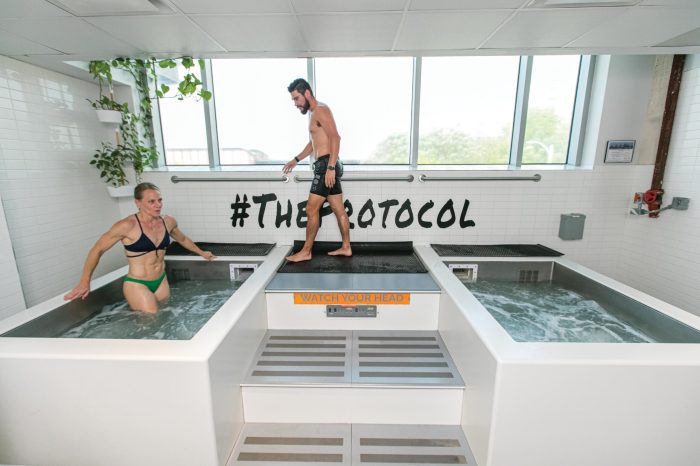
    Contrast bath in physical therapy offers a unique approach to pain management and recovery. It involves alternating between hot and cold water immersion, leveraging the physiological responses to improve circulation, reduce inflammation, and promote muscle recovery. This therapy method has a rich history in healthcare, and its applications are varied and effective.

    This comprehensive guide delves into the principles, methods, and considerations surrounding contrast bath therapy in physical therapy. We’ll explore different types of setups, the science behind the treatment, and the crucial factors to consider for patient selection and safety. From understanding the therapeutic applications to mastering the procedures, this guide aims to equip you with the knowledge needed to effectively incorporate contrast baths into your physical therapy practice.

    Introduction to Contrast Baths in Physical Therapy

    Contrast baths, a therapeutic modality in physical therapy, involve alternating immersion in hot and cold water. This technique is used to stimulate various physiological responses in the body, ultimately promoting healing and recovery. The precise temperature variations and duration of immersion are tailored to individual patient needs and the specific condition being treated.Contrast baths are based on the principles of thermal stimulation.

    The alternating hot and cold water immersion creates a cascade of physiological effects that can benefit musculoskeletal and circulatory systems. The therapeutic benefits of contrast baths stem from their ability to improve blood circulation, reduce inflammation, and enhance tissue healing.

    Types of Contrast Bath Setups

    Contrast baths utilize various temperature combinations of hot and cold water. The most common setup involves immersing a patient in hot water for a specific period, followed by a period in cold water. The temperature of the hot water typically ranges from 40-48°C (104-118°F), while the cold water is usually around 10-15°C (50-59°F). Variations in temperature and immersion duration are frequently employed, depending on the patient’s condition and tolerance.

    Advanced setups might incorporate different water temperatures, such as a warm-cold-hot sequence, to achieve specific effects.

    Physiological Effects of Hot and Cold Water Immersion

    Immersion in hot water dilates blood vessels, increasing blood flow to the treated area. This increased circulation can aid in the removal of metabolic waste products and deliver more oxygen and nutrients to the tissues. Conversely, cold water immersion constricts blood vessels, reducing inflammation and pain perception. The combined effects of these thermal changes promote healing by reducing swelling, relieving pain, and improving joint mobility.

    The precise effects of the contrast bath depend on factors such as the individual’s physiology, the specific temperatures used, and the duration of each immersion.

    Historical Overview of Contrast Bath Usage

    The use of hot and cold water immersion for therapeutic purposes has a long history. Ancient civilizations recognized the benefits of thermal therapies, utilizing hot springs and cold baths for treating various ailments. While the precise methods and understanding of the mechanisms behind these therapies have evolved, the core principle of using thermal contrasts remains a cornerstone of modern physical therapy.

    Key Components of a Contrast Bath Program, Contrast bath in physical therapy

    This table Artikels the key elements of a typical contrast bath program, which can be adapted to individual patient needs. The specific temperature and duration values should be adjusted based on the patient’s tolerance and the specific condition being treated.

    Treatment Type Temperature of Water (°C) Duration (minutes) Frequency (per week)
    Acute Inflammation Reduction Hot: 45-48°C, Cold: 10-15°C Hot: 2-3 minutes, Cold: 1-2 minutes 2-3 times
    Improved Circulation and Muscle Recovery Hot: 42-45°C, Cold: 12-15°C Hot: 3-4 minutes, Cold: 1-2 minutes 3-4 times
    Joint Mobility Enhancement Hot: 40-42°C, Cold: 10-12°C Hot: 3-5 minutes, Cold: 1-2 minutes 2-3 times

    Therapeutic Applications in Physical Therapy

    Contrast baths, a simple yet effective modality, offer a range of therapeutic benefits in physical therapy. They leverage the alternating temperatures of hot and cold water to stimulate circulation, reduce inflammation, and promote muscle recovery, making them a valuable tool for managing various musculoskeletal conditions. This approach is particularly beneficial for patients seeking non-invasive, cost-effective treatment options.

    Specific Conditions Where Contrast Baths are Commonly Used

    Contrast baths are frequently employed in the management of conditions characterized by inflammation and pain. These include musculoskeletal injuries, such as sprains, strains, and post-surgical recovery. They are also beneficial for conditions like arthritis, where inflammation plays a significant role in the symptoms. Furthermore, contrast baths can aid in the recovery process following physical exertion, such as intense workouts or sports activities.

    How Contrast Baths Help Manage Inflammation

    The alternating temperatures of hot and cold water in contrast baths induce a vasodilation-vasoconstriction cycle. The hot water increases blood flow, delivering more nutrients and removing metabolic waste products. The cold water, conversely, constricts blood vessels, reducing swelling and inflammation. This cyclical process helps to control the inflammatory response, thus reducing pain and improving function. Clinical studies have demonstrated the efficacy of contrast baths in reducing inflammation and promoting healing in various musculoskeletal conditions.

    Role of Contrast Baths in Improving Circulation

    The alternating temperatures of contrast baths stimulate the cardiovascular system. The hot water causes vasodilation, increasing blood flow, while the cold water causes vasoconstriction, subsequently promoting blood flow back to the heart. This dynamic interplay improves circulation, ensuring better delivery of oxygen and nutrients to tissues, crucial for healing and recovery. This increased blood flow can be especially beneficial for individuals with conditions that limit circulation.

    Potential Benefits of Contrast Baths for Muscle Recovery

    Contrast baths can accelerate muscle recovery after strenuous exercise. The cold water helps to reduce muscle soreness and inflammation, while the hot water promotes relaxation and reduces muscle stiffness. This combination can significantly improve recovery time and reduce the risk of delayed-onset muscle soreness (DOMS). Athletes and individuals engaging in intense physical activity often find contrast baths helpful for promoting muscle recovery and preventing further injury.

    Elaboration on the Use of Contrast Baths for Pain Management

    Contrast baths can effectively manage pain associated with various musculoskeletal conditions. The alternating temperatures help to reduce inflammation, improve circulation, and promote muscle relaxation. By alleviating these factors, contrast baths can significantly lessen pain perception. The mechanism of action lies in the reduction of pain signals transmitted to the brain, effectively providing pain relief.

    Contrast baths are a fantastic tool in physical therapy, offering a soothing way to manage inflammation and pain. However, if you’re experiencing persistent fatigue or other symptoms, it’s crucial to explore potential underlying conditions like Hashimoto’s disease. Knowing how to test for Hashimoto’s disease is vital for accurate diagnosis and personalized treatment plans, and resources like how to test for hashimotos disease can help you understand the process.

    Ultimately, a combination of professional guidance and self-awareness is key to navigating your health journey, and contrast baths can be a valuable part of that process.

    Comparison of Contrast Baths with Other Physical Therapy Modalities for Pain Relief

    Modality Mechanism of Action Potential Benefits Potential Limitations
    Contrast Baths Alternating hot and cold temperatures induce vasodilation and vasoconstriction, reducing inflammation, improving circulation, and promoting muscle relaxation. Effective for reducing inflammation and pain, promoting muscle recovery, relatively inexpensive, and accessible. May not be suitable for individuals with certain cardiovascular conditions, potential for skin irritation with prolonged or excessive use, and may not be effective for all types of pain.
    Ultrasound Sound waves generate heat, increasing tissue temperature and promoting tissue healing. Effective for reducing pain and inflammation, increasing tissue elasticity and flexibility, and accelerating healing. Requires specialized equipment and trained personnel, can be uncomfortable for some individuals, and may not be suitable for all areas of the body.
    Electrical Stimulation (TENS) Electrical impulses stimulate nerves, reducing pain signals to the brain. Effective for managing acute and chronic pain, improving muscle strength and function, and promoting relaxation. Requires specialized equipment and trained personnel, potential for skin irritation, and may not be effective for all types of pain.
    Manual Therapy Hands-on techniques to mobilize joints, muscles, and connective tissues, promoting movement and reducing pain. Effective for improving joint mobility, reducing pain and stiffness, and improving function. Requires trained therapists, can be time-consuming, and may not be suitable for all conditions or individuals.

    Methods and Procedures for Contrast Bath Therapy

    What Is Contrast Bath at Oscar Godson blog

    Contrast bath therapy, a versatile technique in physical therapy, involves alternating between hot and cold water immersion to stimulate blood flow and reduce inflammation. This method is frequently employed for various musculoskeletal conditions and injuries, promoting healing and alleviating pain. Understanding the precise procedures and considerations for patient safety and comfort is crucial for effective treatment.This section delves into the practical aspects of administering contrast bath therapy, providing a comprehensive guide to equipment, procedures, safety monitoring, and patient positioning.

    By adhering to these guidelines, therapists can maximize the therapeutic benefits while minimizing potential risks.

    Contrast Bath Treatment Procedure

    The following steps Artikel a standard contrast bath procedure:

    1. Patient Assessment

    Before initiating the treatment, a thorough assessment of the patient’s medical history, current condition, and any potential contraindications is essential. This ensures the safety and appropriateness of the treatment.

    2. Equipment Preparation

    Ensure all necessary equipment is readily available and in good working order. This includes the immersion container, temperature control devices, and safety equipment like a floatation device or life preserver.

    3. Water Temperature Regulation

    Precisely control the temperature of both the hot and cold water to the predetermined therapeutic ranges. This ensures patient comfort and prevents thermal burns or discomfort. Use a calibrated thermometer to measure the water temperature.

    4. Patient Positioning

    Position the patient comfortably within the immersion container, ensuring proper support and minimizing strain on the affected area. Appropriate patient positioning is critical to prevent discomfort and injury.

    5. Immersion Cycles

    Initiate the alternating cycles of hot and cold immersion, following a pre-determined protocol. The duration of each immersion should be consistent and tailored to the patient’s tolerance. A common protocol might involve 1 minute in hot water, followed by 1 minute in cold water, repeated for 15-20 minutes.

    6. Monitoring and Adjusting

    Continuously monitor the patient for any signs of discomfort, such as shivering, nausea, or difficulty breathing. Adjust the temperature or duration of the cycles as needed to maintain patient comfort.

    7. Post-Treatment

    After the session, provide support and guidance to the patient as they exit the bath. Assist them with dressing and ensure their comfort. Document the treatment session thoroughly, including water temperatures, duration, and any patient responses.

    Essential Equipment

    A contrast bath requires specific equipment for safe and effective treatment. These include:

    • Immersion Container: This could be a standard tub or a whirlpool bath. The choice depends on the patient’s size and the specific needs of the treatment.
    • Temperature Control Devices: Precisely calibrated thermometers are crucial for maintaining the appropriate water temperatures. A system for regulating and maintaining the temperature of the hot and cold water lines is essential.
    • Safety Equipment: A floatation device or life preserver is vital, particularly for patients who may have difficulty maintaining balance or require extra support during the treatment.
    • Monitoring Devices: A calibrated thermometer, a timer, and a method for observing the patient’s condition during the treatment are important for monitoring and maintaining patient safety.

    Patient Monitoring During Contrast Bath

    Patient safety is paramount during contrast bath treatments. Close monitoring is essential to detect any adverse reactions. Observe for:

    • Signs of Discomfort: Monitor the patient for any indications of discomfort, including shivering, nausea, or difficulty breathing. Promptly address any issues.
    • Vital Signs: Regularly check vital signs, including pulse and respiration, to assess the patient’s overall response to the treatment.
    • Skin Reactions: Pay attention to any changes in skin color, temperature, or texture. Burns or other skin reactions must be immediately addressed.

    Patient Positioning and Comfort

    Proper patient positioning enhances comfort and minimizes stress on the affected area.

    • Support: Ensure adequate support for the patient’s body during the treatment, especially in areas of injury or weakness.
    • Positioning: Position the patient in a way that minimizes strain on joints and muscles, promoting comfort and minimizing discomfort.

    Immersion Container Comparison

    Different immersion containers offer varying features and benefits.

    • Standard Tubs: Suitable for individual patients, these offer a more intimate setting, but may require more manual temperature control.
    • Whirlpool Baths: Whirlpool baths offer a more dynamic treatment experience with gentle massage. They are suitable for patients who tolerate the additional mechanical stimulation.

    Water Temperature Regulation

    Precise temperature regulation is crucial for therapeutic efficacy and patient safety.

    • Consistent Temperatures: Maintain a consistent temperature throughout the treatment.
    • Monitoring: Regularly monitor and adjust the water temperature using calibrated thermometers to prevent thermal injuries.

    Contraindications for Contrast Bath Therapy

    Some conditions may contraindicate contrast bath therapy.

    • Open Wounds or Infections: Patients with open wounds or active infections should avoid contrast bath therapy.
    • Cardiovascular Conditions: Patients with unstable cardiovascular conditions may be at risk and should avoid the therapy.
    • Skin Conditions: Individuals with certain skin conditions may experience exacerbation of symptoms and should be assessed carefully.
    • Uncontrolled Medical Conditions: Individuals with uncontrolled medical conditions should not undergo contrast bath therapy.

    Potential Side Effects

    Side Effect Severity Description Prevention
    Skin irritation Mild Redness, itching, or mild discomfort at the skin contact points. Use a mild soap or cleanser after the bath, and ensure the water temperature is not excessively hot.
    Fainting Mild-Moderate Sudden loss of consciousness, usually due to the temperature shock. Start with shorter immersion times and gradually increase the duration as the patient adapts to the temperature changes.
    Nausea Mild Feeling of queasiness, often accompanied by mild vomiting. Monitor the patient’s response carefully and discontinue the treatment if nausea persists.
    Hypotension Moderate Sudden drop in blood pressure, leading to dizziness or lightheadedness. Monitor blood pressure before and after the treatment.
    Hypertension Moderate Sudden increase in blood pressure, potentially dangerous for patients with pre-existing hypertension. Monitor blood pressure and heart rate closely during the treatment.

    Patient Selection and Considerations

    Contrast bath therapy offers a promising modality for various musculoskeletal conditions, but careful patient selection is crucial. Proper assessment and understanding of potential risks are paramount to ensure the safety and efficacy of the treatment. This section delves into the criteria for patient suitability, necessary assessments, modifications for specific needs, potential risks, and strategies for patient education.

    Patient Selection Criteria

    Effective contrast bath therapy relies on careful patient selection. Individuals with acute infections, open wounds, or uncontrolled cardiovascular conditions are generally excluded. Patients with a history of seizures or fainting spells should be evaluated cautiously. Stable conditions such as osteoarthritis, chronic pain, and edema often respond well to contrast bath therapy, provided they meet the necessary criteria and are properly assessed.

    Contrast baths are a fantastic tool in physical therapy, helping with muscle recovery and inflammation reduction. However, sometimes the discomfort they cause can lead to unexpected digestive issues, like diarrhea. If you’re experiencing this, exploring over-the-counter and prescription options for diarrhea relief, like those available at diarrhea relief otc and prescription medications , might be helpful. Ultimately, though, a contrast bath is still a powerful tool for managing various conditions, and is a great option for many physical therapy patients.

    Individuals with a history of severe allergic reactions or sensitivities to temperature extremes should also be evaluated carefully. Ultimately, the decision to include a patient in a contrast bath program rests on the physical therapist’s judgment after a comprehensive evaluation.

    Patient Assessment Before Treatment

    Thorough patient assessment is vital before initiating contrast bath therapy. This includes a detailed medical history, including any pre-existing conditions, allergies, and medications. Assessing the patient’s current functional limitations, pain levels, and response to previous treatments provides valuable context. The physical therapist should also evaluate the patient’s cardiovascular status, noting any abnormalities or risk factors. A comprehensive musculoskeletal examination is also crucial, focusing on the affected area and any potential contraindications.

    This assessment helps tailor the protocol and ensure patient safety.

    Protocol Modifications for Specific Needs

    Contrast bath protocols need to be tailored to individual patient needs. For example, patients with limited mobility may require assistance with entering and exiting the bath. Patients experiencing significant pain may need to begin with shorter bath durations or a lower temperature gradient. Adjustments to the water temperature, duration of each phase, and frequency of treatments may be necessary to accommodate individual responses and prevent discomfort or adverse reactions.

    These adjustments are crucial for maintaining patient compliance and optimizing outcomes.

    Potential Risks and Precautions

    While generally safe, contrast bath therapy carries potential risks. Hypotension, dizziness, or fainting may occur in susceptible individuals. Skin irritation, particularly in individuals with pre-existing skin conditions, is another potential concern. Patients with cardiovascular problems should be monitored closely. The therapist must be vigilant and immediately address any signs of discomfort or adverse reactions.

    Careful monitoring and appropriate adjustments to the protocol are critical to minimize risks.

    Patient Education Strategies

    Educating patients about the benefits, risks, and procedures of contrast bath therapy is essential. Clear communication about the expected outcomes, potential side effects, and the importance of reporting any discomfort is vital. Visual aids, such as diagrams illustrating the temperature changes, can be helpful. Patients should understand the importance of reporting any unusual sensations or reactions during the treatment.

    Contrast baths are a fantastic tool in physical therapy, alternating hot and cold water to soothe muscles and improve circulation. But if you’re wondering if that sore throat and cough are a cold or something more serious, it’s crucial to consider the symptoms and consult resources like do i have a cold or covid. Ultimately, a contrast bath can be a great part of a self-care routine, whether you’re recovering from an injury or just feeling a bit under the weather.

    Thorough instruction and ongoing communication contribute to patient understanding and cooperation.

    Common Patient Concerns Regarding Contrast Baths

    Concern Potential Solution Expected Outcome Additional Considerations
    Fear of dizziness or fainting Start with shorter bath durations and lower temperature gradients. Monitor closely for signs of hypotension. Reduced risk of adverse reactions, increased patient comfort and compliance. Assess pre-existing cardiovascular conditions.
    Discomfort or pain during treatment Adjust water temperature, duration of each phase, or frequency of treatments. Provide pain relief measures (e.g., ice packs). Reduced pain and discomfort, improved patient tolerance. Ensure proper patient communication and active participation.
    Concerns about skin irritation Use hypoallergenic or sensitive skin-friendly products. Monitor skin condition during and after treatments. Minimized skin irritation, reduced risk of complications. Consider pre-existing skin conditions or allergies.
    Limited mobility Provide assistance with entering and exiting the bath. Use assistive devices as needed. Improved safety and comfort for patients with mobility limitations. Collaborate with caregivers for support.

    Safety and Precautions in Contrast Bath Therapy

    Contrast bath therapy, while often beneficial, carries inherent risks. Therefore, meticulous attention to safety protocols is paramount to ensure a positive and risk-free experience for the patient. Proper preparation, monitoring, and emergency procedures are crucial components of safe contrast bath administration.Contrast bath therapy involves alternating between hot and cold water, which can induce a variety of physiological responses.

    These responses, while often therapeutic, can also pose potential risks if not managed carefully. Understanding these potential risks and implementing appropriate safety measures is essential to minimizing adverse events and maximizing patient outcomes.

    Safety Measures During Contrast Bath Treatments

    Patient safety is paramount during contrast bath treatments. A thorough understanding of the potential risks and proper implementation of safety measures are crucial. The therapist must be vigilant in observing the patient’s responses to the temperature changes.

    • Supervision: Constant supervision by a qualified healthcare professional is essential. This ensures prompt intervention in case of any adverse reaction or emergency situation.
    • Temperature Control: Precise temperature regulation of both the hot and cold water is vital. Using calibrated thermometers and regular checks are imperative to maintain appropriate temperature ranges.
    • Patient Communication: Open communication with the patient is crucial. Encouraging the patient to report any discomfort or unusual sensations is essential. The therapist should be receptive to the patient’s feedback, and adjust the treatment parameters accordingly.
    • Proper Equipment: Ensure all equipment used in the contrast bath, including the bath itself, the temperature control mechanisms, and the safety devices, are in excellent working order. Regular maintenance and checks are required to prevent malfunctions and potential hazards.

    Potential Complications Associated with Contrast Bath Therapy

    Recognizing potential complications is critical for effective risk management. Early identification and intervention are key to mitigating harm.

    • Hypotension/Hypertension: Alternating hot and cold water can induce fluctuations in blood pressure. Close monitoring is essential to detect and manage any significant changes.
    • Skin Reactions: The thermal changes can cause skin irritation, redness, or even burns, particularly in patients with pre-existing skin conditions. Monitoring the patient’s skin throughout the treatment is important to prevent such complications.
    • Fainting/Dizziness: Some individuals may experience dizziness or fainting episodes, especially if they have pre-existing conditions or are not accustomed to thermal changes. The therapist must be prepared to respond quickly in such cases.
    • Cardiovascular Issues: Individuals with cardiovascular conditions may be more susceptible to adverse reactions from contrast bath therapy. Careful evaluation and monitoring are crucial in such cases.

    Procedures to Follow in Case of an Emergency

    Having a well-defined emergency protocol is vital for prompt and effective response to unexpected events.

    • Emergency Response Plan: A comprehensive emergency plan, including contact information for medical personnel, must be in place. A clearly defined plan for managing emergencies, like cardiac arrest or loss of consciousness, is essential.
    • Immediate Action: The therapist must be prepared to take immediate action, such as calling for emergency medical assistance, administering first aid, or stopping the treatment, as needed. Prompt response is critical.
    • Documentation: Thorough documentation of the emergency situation, including the patient’s condition, the actions taken, and the outcome, is necessary. This record-keeping is vital for future reference and for improving the safety protocols.

    Importance of Proper Patient Monitoring

    Patient monitoring during contrast bath treatments is crucial for identifying potential problems early. Continuous observation helps in timely intervention.

    • Vital Signs Monitoring: Regular monitoring of vital signs (blood pressure, heart rate, and respiration) throughout the treatment is essential. These parameters will reveal any changes that may necessitate immediate intervention.
    • Clinical Observations: The therapist must actively observe the patient for any signs of discomfort, distress, or adverse reactions, including changes in skin color, sweating, or changes in breathing patterns. Early recognition of such changes is key to preventing serious complications.

    Managing Potential Adverse Reactions

    A proactive approach to managing potential adverse reactions is essential for patient safety. Knowledge of how to address these reactions is vital.

    • Prompt Intervention: The therapist must be prepared to intervene promptly if any adverse reaction occurs. This may include adjusting the treatment parameters, administering appropriate medications, or seeking immediate medical attention.
    • Patient Education: Educating the patient about potential adverse reactions and the importance of reporting any discomfort is crucial. Patient awareness can facilitate early intervention.

    Therapist’s Role in Ensuring Patient Safety

    The therapist plays a critical role in maintaining a safe and therapeutic environment.

    • Knowledge and Training: The therapist must possess the necessary knowledge and training to perform contrast bath treatments safely. Continuous professional development and education are essential.
    • Assessment: Thorough patient assessment before each treatment is necessary to identify any contraindications or pre-existing conditions that may increase the risk of complications.

    Safety Checks Before and During a Contrast Bath Session

    A structured approach to safety checks before and during a contrast bath treatment minimizes risks.

    1. Pre-Treatment Checks:
      • Review medical history and identify any contraindications.
      • Obtain informed consent from the patient.
      • Ensure proper equipment functionality.
      • Assess patient’s baseline vital signs.
    2. During Treatment Checks:
      • Continuously monitor patient’s vital signs.
      • Observe for any signs of discomfort or distress.
      • Adjust treatment parameters as needed based on patient feedback and observations.
      • Maintain a safe and supportive environment.

    Illustrative Case Studies

    Contrast bath in physical therapy

    Contrast bath therapy, a seemingly simple technique, can offer significant benefits for a range of musculoskeletal conditions. Understanding how it impacts specific patients provides valuable insights into its efficacy and potential applications. This section presents a detailed case study showcasing the positive effects of contrast bath therapy.

    Patient Case Study: Chronic Ankle Sprain

    This case study details a patient, a 32-year-old female, who presented with chronic ankle pain stemming from a sprain sustained six months prior. The pain was characterized by persistent discomfort, particularly during weight-bearing activities and at night. Physical examination revealed mild ankle instability and tenderness around the lateral ligaments.

    Treatment Plan

    The treatment plan incorporated a combination of physical therapy exercises, manual therapy, and the use of contrast bath therapy. The initial phase focused on restoring ankle mobility and strength, followed by gradually increasing the intensity and duration of the contrast bath treatments.

    Patient Response to Therapy

    The patient reported a significant reduction in pain levels within the first two weeks of incorporating contrast bath therapy into her regimen. She noted improved range of motion and a greater capacity for daily activities. The patient demonstrated a positive response to the therapy, which was further evidenced by her ability to return to low-impact activities.

    Contrast Bath Treatment Parameters

    The contrast bath protocol involved alternating between hot (40-42°C) and cold (10-15°C) water for 3-5 minutes each. The patient was instructed to complete a total of 10-12 cycles per treatment session. The duration of each session gradually increased over a period of four weeks.

    Patient Progress

    Within eight weeks of consistent therapy, the patient achieved a near-complete resolution of pain and a significant improvement in ankle function. She was able to return to her pre-injury activity level, including jogging and playing recreational sports. Follow-up assessments confirmed the sustained improvements in her ankle stability and range of motion.

    Extrapolation to Similar Cases

    This case study suggests that contrast bath therapy may be a valuable adjunct treatment for individuals experiencing chronic ankle sprains. The observed positive response is consistent with the known vasodilatory and analgesic effects of heat and the anti-inflammatory properties of cold. Patients with similar musculoskeletal conditions, such as chronic tendinopathy or osteoarthritis, may also experience similar benefits. Further research is needed to establish specific treatment protocols and outcomes for diverse patient populations and to quantify the effectiveness of the therapy.

    This case demonstrates the potential of contrast baths as a valuable therapeutic tool.

    Last Point: Contrast Bath In Physical Therapy

    In conclusion, contrast bath therapy offers a promising modality for physical therapy, with a multitude of potential benefits for various conditions. Understanding the science behind its effects, proper implementation techniques, and crucial safety measures are key to successful application. The personalized approach and careful consideration of patient needs are essential to maximize treatment outcomes and ensure patient safety.

    Further research and exploration into specific applications and patient populations will undoubtedly reveal even more insights into the therapeutic potential of contrast bath therapy.

  • Stiffness After Knee Replacement A Comprehensive Guide

    Stiffness After Knee Replacement A Comprehensive Guide

    Stiffness after knee replacement sets the stage for this enthralling narrative, offering readers a glimpse into the complexities of recovery and the journey to regaining mobility. This guide delves into the various causes, symptoms, treatment options, and preventative measures for managing stiffness, empowering individuals with the knowledge they need to navigate this crucial aspect of their post-operative experience.

    Understanding the potential causes of stiffness after knee replacement, ranging from scar tissue formation to surgical technique, is crucial for proactive management. This detailed exploration will cover everything from common symptoms and diagnostic methods to effective treatment approaches, including physical therapy, medication, and even when surgical intervention might be necessary.

    Causes of Stiffness

    Post-knee replacement surgery, stiffness is a common concern, but understanding its underlying causes can help patients and healthcare professionals develop effective management strategies. This often stems from a complex interplay of factors, including tissue responses, surgical techniques, and individual patient characteristics. Addressing these factors is crucial for successful rehabilitation and minimizing long-term limitations.The stiffness experienced after knee replacement surgery can significantly impact a patient’s ability to perform daily activities.

    Proper diagnosis and management of the underlying causes are critical for achieving optimal outcomes and minimizing the impact of stiffness on quality of life.

    Scar Tissue Formation

    Scar tissue formation is a natural response to surgical trauma. During the healing process, the body creates fibrous tissue to repair damaged tissues. Excessive or poorly organized scar tissue can restrict joint movement, leading to stiffness. This is particularly relevant in the vicinity of the surgical incisions and around the joint capsule. The extent of scar tissue formation can vary based on the individual’s healing response and surgical technique.

    Joint Capsule Tightness

    The joint capsule, a fibrous structure surrounding the knee joint, plays a crucial role in maintaining joint stability. Following surgery, the joint capsule can become thickened and tightened, further limiting range of motion and contributing to stiffness. This is often a consequence of inflammation and the body’s natural response to the surgical intervention. Careful attention to post-operative rehabilitation and stretching exercises is essential to prevent this.

    Muscle Atrophy

    Muscle weakness and atrophy (wasting) can also contribute to stiffness after knee replacement. Reduced muscle mass surrounding the knee joint can compromise the joint’s stability and limit its range of motion. This is often a consequence of reduced activity following surgery, leading to disuse atrophy. Regular physical therapy is essential to maintain muscle strength and prevent this.

    Dealing with stiffness after a knee replacement can be tough, but remember to focus on healthy habits. While I’m no expert on nutrition, I’m curious about whether orange juice is a good source of nutrients for recovery, especially for those recovering from joint replacement. Check out this article on is orange juice good for you to see if it might offer some insights.

    Ultimately, staying hydrated and eating a balanced diet is key to speeding up the recovery process and reducing stiffness.

    Inflammation and Infection

    Inflammation, a natural part of the healing process, can sometimes lead to stiffness. However, excessive inflammation can hinder recovery and increase the risk of stiffness. Similarly, infection following knee replacement surgery can cause significant inflammation and pain, resulting in stiffness and potentially requiring further interventions. Prompt diagnosis and treatment of infection are essential to prevent long-term complications.

    Surgical Technique and Implant Design

    Surgical technique and implant design can also influence the development of stiffness. Minimally invasive techniques, for example, can minimize trauma and potentially reduce scar tissue formation, thereby reducing the risk of stiffness. Proper implant design is also critical. The fit and function of the prosthesis can influence the degree of joint mobility and stiffness. Careful consideration of these factors during surgery is crucial.

    Differences in Stiffness Between Knee Replacement Types

    The type of knee replacement influences the potential for stiffness. Total knee arthroplasty (TKA), where the entire knee joint is replaced, may present a higher risk of stiffness compared to unicompartmental knee arthroplasty (UKA), where only a portion of the knee joint is replaced. The complexity of the procedure and the extent of tissue manipulation can affect the recovery process and the likelihood of stiffness.

    Furthermore, the specific needs of each patient must be considered.

    Causes of Stiffness: Summary Table

    Cause Severity Duration
    Scar tissue formation Mild to moderate Weeks to months
    Joint capsule tightness Mild to moderate Weeks to months
    Muscle atrophy Mild to moderate Weeks to months
    Inflammation Mild to severe Weeks to months
    Infection Severe Weeks to months (or longer)
    Surgical technique Mild to moderate Weeks to months
    Implant design Mild to moderate Weeks to months
    Knee replacement type (TKA vs. UKA) Mild to moderate Weeks to months

    Symptoms and Diagnosis

    Knee replacement surgery, while often a life-altering improvement, can sometimes result in post-operative stiffness. Understanding the symptoms and how healthcare professionals diagnose this complication is crucial for prompt and effective management. Early detection allows for interventions that can minimize the impact of stiffness on daily life and prevent long-term problems.Post-operative stiffness is a common concern following knee replacement surgery.

    It manifests in various ways, impacting a patient’s ability to perform everyday tasks. Recognizing these symptoms and the diagnostic methods used by medical professionals is essential for prompt intervention and successful recovery.

    Common Symptoms of Post-Knee Replacement Stiffness

    Identifying the symptoms of stiffness is vital for prompt medical attention. Patients may experience a range of discomfort and limitations in their daily activities. Common symptoms include pain, a reduced range of motion (ROM), and difficulties with tasks like walking, climbing stairs, or getting in and out of chairs. The intensity of these symptoms can vary greatly from person to person.

    Diagnostic Methods for Post-Knee Replacement Stiffness

    Healthcare professionals use a combination of methods to assess and diagnose stiffness. A thorough physical examination, evaluating the range of motion, muscle strength, and any tenderness or swelling around the knee joint, is essential. The doctor will also inquire about the patient’s medical history, including pre-existing conditions, surgical details, and any medications taken. Imaging techniques, such as X-rays and MRIs, can provide crucial information about the extent of the stiffness, the condition of the implant, and the presence of any complications.

    Stiffness Levels, Symptoms, and Diagnostic Methods

    Stiffness Level Symptoms Diagnostic Methods
    Mild Slight limitation in range of motion, minimal pain, and only minor difficulty with daily activities. Physical examination, patient history, basic X-rays to check implant alignment.
    Moderate Noticeable limitation in range of motion, moderate pain, and increased difficulty with daily activities. Walking may become challenging. Physical examination, patient history, X-rays to assess implant and bone condition, potentially MRI to evaluate soft tissues.
    Severe Significant limitation in range of motion, intense pain, and substantial difficulty performing daily tasks. Walking and mobility are severely compromised. Physical examination, detailed patient history, X-rays and MRI to evaluate bone and soft tissue involvement, potential need for advanced imaging techniques.

    Comparison with Other Post-Operative Complications

    Differentiating stiffness from other potential post-operative complications, such as infection, blood clots, or wound healing issues, is crucial for proper treatment. While stiffness may present with some overlapping symptoms, such as pain, careful evaluation by healthcare professionals can distinguish these issues.

    Importance of Early Detection and Intervention, Stiffness after knee replacement

    Early detection and prompt intervention are vital in managing post-knee replacement stiffness. Early intervention can often prevent the stiffness from becoming more severe and limiting. Addressing the issue promptly allows for the implementation of effective treatments, such as physical therapy, medications, and in some cases, additional surgical procedures. This proactive approach significantly improves the patient’s long-term outcome and quality of life.

    Treatment Options

    Stiffness after knee replacement

    Post-knee replacement surgery, stiffness can be a significant hurdle to regaining full function. Fortunately, various treatment approaches can help manage and alleviate this issue. A multi-faceted strategy, often involving physical therapy, medication, and potentially injections, is usually the most effective course of action. Early intervention and consistent adherence to the treatment plan are crucial for achieving optimal results.Effective treatment for post-operative knee stiffness involves a combination of strategies aimed at reducing pain, increasing range of motion, and restoring function.

    This often requires a collaborative effort between the patient, physical therapist, and surgeon. A personalized approach tailored to the individual’s specific needs and recovery progress is paramount for success.

    Physical Therapy

    Physical therapy plays a vital role in regaining knee range of motion after a knee replacement. Different exercises target specific aspects of flexibility and strength. Manual therapy techniques, such as soft tissue mobilization and joint mobilization, can also help improve mobility. Progressive exercises, starting with gentle movements and gradually increasing intensity, are essential for safe and effective recovery.

    • Range of Motion Exercises: These exercises focus on gradually increasing the knee’s bending and straightening ability. Examples include gentle knee flexion and extension, assisted range of motion exercises, and exercises to improve passive range of motion.
    • Strengthening Exercises: Strengthening the muscles around the knee joint is crucial for stability and function. Exercises that target quadriceps, hamstring, and calf muscles, performed progressively, are important for preventing stiffness and improving overall strength.
    • Functional Exercises: These exercises help the patient transition from the clinic to everyday activities. Examples include exercises that simulate walking, stair climbing, and other daily tasks. Gradual progression from simple to more complex movements is crucial.

    Careful consideration of the effectiveness of various exercises is important. Some exercises might be more effective for specific types of stiffness or individual patient needs. It’s crucial to follow a structured program designed by a physical therapist, ensuring proper form and progression.

    Medication

    Pain management is an essential aspect of treating stiffness after knee replacement. Nonsteroidal anti-inflammatory drugs (NSAIDs) can help reduce pain and inflammation. Acetaminophen (paracetamol) can also be effective for managing mild to moderate pain. In some cases, stronger pain medications may be necessary. It’s essential to discuss medication options with a physician to determine the most suitable approach for individual needs and to avoid potential side effects.

    Injections

    Injections, such as corticosteroid injections, can help reduce inflammation and pain associated with stiffness. However, their use should be considered carefully and in conjunction with other treatment options. Corticosteroid injections can help alleviate short-term stiffness, but their long-term effectiveness may be limited.

    Surgical Intervention

    In some cases, surgical intervention might be necessary to address persistent stiffness. If non-surgical methods fail to improve range of motion significantly, a revision knee replacement surgery may be considered. This procedure involves replacing the existing implant with a new one to improve the knee’s function and range of motion.

    Table of Treatment Options

    Treatment Benefits Side Effects
    Physical Therapy Improved range of motion, increased strength, reduced pain, improved function Potential for muscle soreness, slight discomfort during exercises
    Medication (NSAIDs, Acetaminophen) Reduced pain and inflammation Potential for stomach upset, kidney problems (in some cases)
    Injections (Corticosteroids) Short-term reduction in pain and inflammation Potential for joint infection, thinning of the joint cartilage, increased risk of further damage
    Surgical Intervention (Revision Knee Replacement) Improved range of motion, restored function Increased risk of complications, longer recovery period

    Prevention Strategies

    Preventing stiffness after knee replacement surgery is crucial for a smooth recovery and optimal long-term function. Proactive measures, including meticulous pre-operative preparation and a structured post-operative rehabilitation program, significantly reduce the risk of developing persistent stiffness. This proactive approach empowers patients to regain their mobility and independence more quickly and comfortably.A well-designed prevention strategy encompasses various components, focusing on patient education, pre-operative preparation, and adherence to post-operative instructions.

    By understanding the factors contributing to stiffness and actively participating in their recovery, patients can greatly improve their outcomes.

    Pre-operative Physical Therapy and Patient Education

    Pre-operative physical therapy plays a vital role in preparing the patient for surgery and minimizing the risk of stiffness. This involves strengthening the muscles around the knee, improving range of motion, and educating the patient about the importance of early mobilization. Patients learn specific exercises and stretches to maintain flexibility and muscle strength. This knowledge empowers them to actively participate in their recovery process, leading to better outcomes.

    Post-operative Exercises and Activity Modification

    Early and consistent post-operative exercises are essential to maintain and regain range of motion. A structured exercise program, prescribed by a physical therapist, is crucial for preventing stiffness. These exercises, tailored to individual needs and progress, gradually increase in intensity and complexity. Furthermore, appropriate activity modification, such as avoiding activities that exacerbate pain or strain the knee, is essential.

    Dealing with stiffness after a knee replacement can be tough, but it’s important to remember that focusing on overall well-being is key. Think about how your sense of smell impacts your brain health – a fascinating connection explored in detail at smell and brain health. While not directly related to knee recovery, understanding how our senses affect our mental and physical state can help us approach physical challenges like stiffness with a broader perspective, and ultimately contribute to a quicker and more comfortable recovery.

    Physical therapy and regular exercise will still be crucial, but a healthier mind can significantly impact your journey.

    This individualized approach allows patients to safely and effectively manage their recovery.

    Importance of Patient Compliance and Motivation

    Patient compliance and motivation are critical for successful rehabilitation. Adherence to the prescribed exercise regimen, consistent participation in therapy sessions, and proactive management of pain are key factors in preventing stiffness. Encouraging patients to understand the rationale behind the exercises and emphasizing the positive impact on their recovery process can significantly improve their motivation and compliance.

    Step-by-Step Post-operative Rehabilitation Program

    This structured program Artikels key steps for post-operative rehabilitation, focused on preventing stiffness.

    • Phase 1 (Immediate Post-operative): Emphasis on gentle range-of-motion exercises, pain management, and early mobilization. The patient learns basic exercises and stretches to maintain joint flexibility, and begins light exercises to regain muscle strength. Activities are kept simple and focused on pain-free movement.
    • Phase 2 (Weeks 2-4): Progressive exercises to improve strength and range of motion. The focus shifts to more targeted exercises that challenge the muscles around the knee joint. Gradual increases in the intensity and duration of exercises are crucial, always within the patient’s pain tolerance. Physical therapy sessions are scheduled to provide guidance and support.
    • Phase 3 (Weeks 4-8): Increased functional exercises and activity levels. This phase focuses on preparing the patient for daily activities. Exercises are designed to improve balance, stability, and coordination. The patient starts incorporating activities like walking, light stair climbing, and gradually increasing the intensity of their exercises. Activities are designed to progressively increase functional capacity.

    • Phase 4 (Weeks 8-12 onwards): Maintenance and advanced exercises. Focus on strengthening and endurance training to maintain the improvements achieved. The patient begins activities to enhance their overall fitness level. The exercises are designed to gradually progress the patient’s strength, endurance, and range of motion.

    Long-Term Management

    Knee replacement surgery is a significant procedure, offering improved mobility and reduced pain. However, managing stiffness long-term is crucial for optimal outcomes. This involves a multifaceted approach that extends beyond the initial recovery period. Continued vigilance and proactive strategies are essential to maintain and improve knee function.

    Regular Follow-up Appointments

    Maintaining open communication with your healthcare provider is paramount in long-term knee replacement management. Regular follow-up appointments allow for monitoring of your progress, addressing any emerging issues, and adjusting the treatment plan as needed. These appointments provide a platform to discuss any discomfort, swelling, or limitations in range of motion. Early detection and intervention can prevent stiffness from becoming a persistent problem.

    Lifestyle Modifications

    Implementing healthy lifestyle modifications plays a vital role in maintaining and improving knee function post-surgery. These modifications focus on preserving the integrity of the new joint and promoting overall well-being. A balanced diet rich in nutrients supports the healing process and helps maintain a healthy weight, reducing stress on the joint. Regular exercise, tailored to your physical abilities and limitations, strengthens muscles around the knee, improves range of motion, and enhances overall stability.

    Ongoing Physical Therapy

    Physical therapy is often a crucial component of long-term knee replacement management. A skilled physical therapist can design an individualized program to address specific needs and limitations. This program may include exercises to improve strength, flexibility, and range of motion. It may also involve strategies for managing pain and preventing future stiffness. Ongoing therapy sessions can be scheduled as needed to maintain and enhance the benefits of the surgery.

    Patient Stories

    Numerous patients have successfully managed long-term knee stiffness through diligent adherence to their treatment plans. For instance, Sarah, a 65-year-old woman, experienced significant improvement in her knee function after incorporating regular physical therapy and lifestyle modifications into her daily routine. She reported a noticeable reduction in stiffness and an increase in her overall mobility within six months of her surgery.

    Consistent effort and commitment to the rehabilitation plan are key to successful long-term management.

    Patient Experience

    The journey to recovery after knee replacement surgery is multifaceted and deeply personal. Beyond the physical healing, the emotional and psychological impact on patients deserves careful consideration. Understanding the patient experience with stiffness is crucial for developing comprehensive care plans and fostering a supportive environment for optimal outcomes.Stiffness after knee replacement can significantly affect a patient’s quality of life, impacting their ability to perform daily activities, participate in social interactions, and maintain a sense of independence.

    Dealing with stiffness after a knee replacement can be tough, but understanding the underlying muscle stiffness is key. Often, this post-surgery stiffness is similar to other forms of muscle stiffness, stemming from inflammation, reduced mobility, or even a lack of regular exercise. Learning about the symptoms, causes, and treatments for muscle stiffness in general can be really helpful, check out this resource on muscle stiffness symptoms causes and treatment for more detailed information.

    Ultimately, consistent physical therapy and gentle exercises are often recommended to ease the stiffness and regain full range of motion after knee replacement.

    This can lead to frustration, anxiety, and even depression. The experience is highly individual, shaped by factors like pre-existing conditions, age, and the overall approach to rehabilitation.

    Impact on Quality of Life

    Patients often report limitations in mobility and daily tasks due to stiffness. This can range from difficulty getting in and out of chairs to trouble walking short distances. Social activities and hobbies may be affected, potentially leading to feelings of isolation and reduced participation in life. The physical discomfort associated with stiffness can also negatively affect sleep quality and overall well-being.

    Patient Perspectives on Dealing with Stiffness

    Many patients describe stiffness as a persistent challenge, sometimes even more challenging than the initial recovery period. Some report feelings of frustration and disappointment when their progress isn’t as rapid as anticipated. Others highlight the importance of proactive communication with their healthcare team, seeking guidance and support throughout the recovery journey.

    Patient Expectations and Understanding

    Patients’ pre-operative expectations and understanding of the procedure’s potential challenges significantly influence their recovery experience. Those with realistic expectations, recognizing that full recovery takes time and effort, tend to cope better with stiffness. Conversely, patients with unrealistic expectations may experience disappointment or frustration when they don’t achieve rapid improvement. Effective pre-operative education plays a crucial role in managing patient expectations and preparing them for the potential challenges.

    Patient Journey from Surgery to Long-Term Management

    The patient journey from surgery to managing long-term stiffness involves several key phases. Immediately post-surgery, focus is on pain management and initial mobilization. Subsequently, the rehabilitation process, including targeted exercises and physical therapy, becomes crucial for restoring range of motion and strength. Ongoing monitoring and adjustments to the treatment plan are essential to address any emerging issues, such as persistent stiffness.

    Education on self-management techniques, such as home exercises and assistive devices, empowers patients to actively participate in their recovery.

    Patient Testimonials

    Patient Experience Management Strategies
    Sarah (62) Experienced significant stiffness in the first few months post-surgery. Found it challenging to perform everyday tasks like getting dressed. Consistent physical therapy, daily home exercises, and using a knee brace for support. She also found that gradual increases in activity and gentle stretching helped maintain mobility.
    David (75) Initially struggled with limited range of motion and pain. Felt discouraged at times, but was determined to regain his independence. Engaged in regular swimming sessions, attended support groups for knee replacement patients, and relied on a walker for added stability when needed.
    Emily (58) Reported a slower recovery than expected, and experienced persistent stiffness, especially in the morning. Developed a consistent stretching routine, incorporated low-impact cardio exercises, and used heat therapy to ease morning stiffness. She also sought advice from a nutritionist to ensure proper joint health.

    Prognosis and Outcomes: Stiffness After Knee Replacement

    Stiffness after knee replacement

    Knee replacement surgery is a significant procedure, and managing potential stiffness is a crucial aspect of achieving optimal outcomes. The prognosis for managing stiffness varies based on individual factors, surgical technique, and post-operative care. Understanding the recovery timeline, influential factors, and potential impact on daily life is essential for both patients and healthcare professionals.A successful recovery from knee replacement surgery, including the management of stiffness, hinges on a multi-faceted approach.

    This involves a combination of diligent physical therapy, patient compliance, and a comprehensive understanding of the potential factors that can influence the recovery process. The long-term outcome is directly linked to the proactive management of stiffness throughout the rehabilitation journey.

    Recovery Timeline

    Managing stiffness after knee replacement typically involves a gradual process. Initial weeks focus on regaining range of motion and strength, often through supervised physical therapy sessions. Progressing through months, patients progressively incorporate more challenging exercises and activities, gradually increasing their functional capacity. A patient’s commitment to rehabilitation plays a key role in the overall recovery timeline. It’s not uncommon for complete recovery to take several months, and some individuals may experience lingering stiffness even after a year.

    Factors Affecting Prognosis

    Several factors can influence the prognosis of stiffness following knee replacement. Age, pre-existing medical conditions, and the patient’s overall health and physical activity level all play a significant role. The surgical technique employed and the patient’s adherence to post-operative instructions are also critical factors. Individual differences in healing rates also contribute to the variability in recovery times.

    Impact on Functional Outcomes

    Stiffness after knee replacement can significantly impact functional outcomes and activities of daily living. A restricted range of motion can make tasks like walking, climbing stairs, or getting in and out of chairs more challenging. The degree of functional impairment varies greatly between individuals. Successful recovery often involves addressing the specific limitations experienced by each patient and tailoring the treatment plan accordingly.

    Examples of Successful Recovery Cases

    Numerous patients have experienced successful recoveries from knee replacement surgery and successfully managed stiffness. A 65-year-old woman with osteoarthritis, for instance, experienced a significant improvement in her range of motion after a structured physical therapy program and diligent home exercises. She was able to return to her favorite activities, such as gardening and walking her dog, within six months.

    Another patient, a 72-year-old man with diabetes, demonstrated remarkable progress in regaining his mobility and independence after a comprehensive rehabilitation program that included both in-clinic and home-based exercises.

    Importance of Patient-Centered Care

    Patient-centered care is essential for achieving optimal outcomes in managing stiffness after knee replacement. This involves actively listening to the patient’s concerns, preferences, and goals, and tailoring the treatment plan to meet their individual needs. A collaborative approach between the patient, surgeon, physical therapist, and other healthcare providers is critical for a positive outcome. Open communication and shared decision-making empowers patients to actively participate in their recovery journey, ultimately leading to better outcomes.

    Summary

    In conclusion, managing stiffness after knee replacement is a multifaceted process requiring a comprehensive understanding of the various contributing factors, from initial causes to long-term management strategies. By understanding the diverse treatment options, preventative measures, and patient experiences, individuals can actively participate in their recovery journey, empowering themselves with the knowledge and tools to achieve optimal outcomes and a fulfilling quality of life.

    This comprehensive guide serves as a valuable resource for both patients and healthcare professionals.

  • Restless Legs Syndrome Physical Exercise Guide

    Restless Legs Syndrome Physical Exercise Guide

    Restless legs syndrome physical exercise is a powerful tool for managing the discomfort and disruption this condition can bring. Understanding the connection between movement and symptom relief is key to finding effective strategies. This guide delves into the potential benefits of various exercise types, offering practical recommendations for incorporating physical activity into a daily routine, while addressing potential challenges and considerations specific to RLS.

    From understanding the science behind exercise and RLS symptom alleviation to exploring specific exercises and routines tailored to different needs, this comprehensive resource provides valuable insights and practical advice. We’ll also examine the crucial relationship between exercise, sleep, and RLS, highlighting the importance of creating a holistic approach to managing the condition.

    Understanding Restless Legs Syndrome (RLS)

    Restless legs syndrome (RLS), a neurological disorder, is characterized by an irresistible urge to move the legs, often accompanied by unpleasant sensations. This urge is typically worse at rest, particularly in the evening or at night, and is frequently relieved by movement. Understanding RLS involves recognizing its symptoms, potential causes, and impact on daily life.RLS is a chronic condition that can significantly impact sleep quality and overall well-being.

    Symptoms frequently worsen over time if left unmanaged, leading to fatigue, daytime sleepiness, and difficulties concentrating. This can, in turn, impact personal and professional life, affecting relationships and work performance.

    Symptoms and Characteristics of RLS

    RLS is defined by the persistent urge to move the legs, often accompanied by unpleasant sensations, such as tingling, crawling, itching, or burning. These sensations are frequently described as uncomfortable and are typically worse at rest, particularly in the evening or at night. The urge to move the legs is often so strong that it interferes with sleep, causing wakefulness and impacting overall well-being.

    Causes and Risk Factors of RLS

    The precise cause of RLS remains unknown, but several factors are linked to its development. Genetic predisposition is a significant risk factor, as RLS often runs in families. Certain medical conditions, such as iron deficiency, kidney failure, and diabetes, can increase the likelihood of developing RLS. Pregnancy, particularly in the third trimester, can also trigger or exacerbate RLS symptoms.

    Furthermore, certain medications, such as anti-nausea drugs and antidepressants, can potentially induce or worsen RLS.

    Progression and Impact of RLS

    The progression of RLS can vary significantly from person to person. Some individuals experience mild symptoms that only occur occasionally, while others experience more severe symptoms that interfere substantially with their daily lives. The impact of RLS can be substantial, affecting sleep quality, leading to fatigue and daytime sleepiness. This can hinder concentration, impacting work performance, and potentially leading to social isolation.

    In some cases, RLS can negatively affect relationships due to the sleep disruption and associated fatigue.

    Finding the right physical exercise routine for restless legs syndrome can be tricky, but it’s definitely worth the effort. One thing to consider, especially if you experience heavy menstrual periods, is potential iron deficiency anemia, which can exacerbate restless legs symptoms. Checking out the link on heavy menstrual periods iron deficiency anemia could offer valuable insight into how these two issues are connected.

    Ultimately, a tailored exercise plan that accounts for potential underlying conditions, like iron deficiency, will help manage restless legs syndrome more effectively.

    Comparison with Other Neurological Conditions

    Characteristic RLS Parkinson’s Disease Multiple Sclerosis
    Symptoms Urge to move legs, unpleasant sensations (tingling, crawling) Tremors, rigidity, bradykinesia (slow movement) Muscle weakness, numbness, vision problems, cognitive changes
    Cause Unknown; genetic predisposition, medical conditions Progressive degeneration of nerve cells Autoimmune response damaging the myelin sheath
    Impact on Daily Life Sleep disturbance, fatigue, daytime sleepiness, possible social isolation Difficulty with daily tasks, balance problems, potential speech difficulties Varying degrees of disability depending on the affected areas

    Potential Triggers for RLS Episodes

    Several factors can trigger or worsen RLS symptoms. These include:

    • Iron Deficiency: Low iron levels in the blood are strongly associated with RLS. Individuals with low iron levels often experience worsening symptoms, particularly at night. Iron supplements can help alleviate symptoms.
    • Stress and Fatigue: Stressful situations and feelings of tiredness can exacerbate RLS symptoms. Managing stress levels and ensuring adequate rest can help minimize these effects.
    • Certain Medications: Some medications, such as anti-nausea drugs, antidepressants, and some anti-psychotics, can induce or worsen RLS symptoms. Consulting a physician about alternative medications or adjusting dosages may be necessary.
    • Caffeine and Nicotine: Stimulants such as caffeine and nicotine can increase RLS symptoms. Reducing intake or avoiding these substances may be helpful.
    • Alcohol Consumption: Alcohol consumption, especially in large quantities, can contribute to worsening RLS symptoms.
    • Changes in Temperature: Sudden changes in temperature can also trigger RLS episodes, particularly if the individual is in a cold environment. Maintaining a consistent temperature, especially in the bedroom, can help.

    Physical Exercise and RLS

    Physical exercise can play a crucial role in managing restless legs syndrome (RLS) symptoms. While there’s no cure for RLS, regular physical activity can often reduce the intensity and frequency of uncomfortable sensations, leading to improved sleep and overall well-being. This approach is often a complementary therapy, working alongside other treatments.Engaging in regular physical activity offers numerous benefits for individuals with RLS.

    These benefits extend beyond simply reducing symptoms, encompassing improvements in mood, energy levels, and sleep quality. Understanding the types of exercise, the mechanisms behind their effectiveness, and the importance of consulting a healthcare professional is key to a successful approach.

    Potential Benefits of Physical Exercise for RLS

    Regular physical activity can significantly impact RLS symptoms. Studies suggest that exercise can reduce the urge to move, improve sleep quality, and enhance mood. The positive effects are often attributed to improved blood circulation, reduced muscle tension, and the release of endorphins. Exercise also helps regulate the body’s natural sleep-wake cycle, contributing to better sleep, which can lessen RLS symptoms.

    Types of Exercise Beneficial for RLS Management

    A variety of exercises can be beneficial in managing RLS symptoms. Aerobic exercises, such as brisk walking, jogging, swimming, or cycling, are often recommended due to their positive impact on overall health and blood circulation. Strength training exercises, including weightlifting or resistance band workouts, can also contribute to symptom relief by strengthening muscles and improving overall body function.

    Yoga and tai chi, with their emphasis on stretching and mindfulness, can be particularly helpful in reducing muscle tension and promoting relaxation.

    Mechanisms of Exercise in Alleviating RLS Symptoms

    Exercise is believed to alleviate RLS symptoms through several mechanisms. Increased blood flow, improved circulation, and reduced muscle tension are among the key benefits. Exercise can also help regulate the body’s natural sleep-wake cycle, which is often disrupted in individuals with RLS. The release of endorphins, natural mood boosters, is another important factor.

    Importance of Consulting a Healthcare Professional

    Before initiating any new exercise regimen, especially if you have RLS, it’s crucial to consult with a healthcare professional. They can assess your individual needs and medical history to determine the most suitable exercises and intensity levels. They can also help you identify any potential health concerns or pre-existing conditions that may influence your exercise routine. This personalized approach is vital to ensure safety and effectiveness.

    Comparison of Exercise Types and RLS Symptoms

    Different types of exercise may have varying effects on RLS symptoms. Cardiovascular exercise, such as running or cycling, can significantly improve overall health and circulation, potentially reducing the urge to move. Strength training, while beneficial for overall muscle strength and tone, may not have as pronounced an impact on the urge to move in the short term. Yoga and tai chi, due to their focus on stretching and relaxation, may be particularly effective in reducing muscle tension and promoting relaxation, thus easing RLS symptoms.

    Ultimately, a personalized approach tailored to individual needs is essential.

    Recommended Exercise Routines for RLS

    Exercise Type Description Frequency Recommendation Intensity Recommendation
    Brisk Walking Walking at a pace that elevates your heart rate 3-5 times per week Moderate intensity
    Swimming Swimming laps or participating in water aerobics 2-3 times per week Moderate to vigorous intensity
    Yoga Practicing yoga poses focusing on stretching and relaxation 2-3 times per week Gentle to moderate intensity
    Strength Training Lifting weights or using resistance bands 2-3 times per week Moderate intensity

    This table provides examples of exercise routines that might be suitable for managing RLS symptoms. It’s essential to remember that these are just examples, and the best routine will depend on individual circumstances. Consult a healthcare professional to tailor an exercise plan to your specific needs and health status.

    Exercise Recommendations for RLS Management

    Finding the right exercise routine can significantly improve restless legs syndrome (RLS) symptoms and overall well-being. Understanding your body’s response to different activities is key to a successful strategy. This involves not just choosing the right type of exercise, but also learning to listen to your body’s signals and adjust your approach accordingly.Exercise offers a multifaceted approach to RLS management.

    Beyond physical benefits, it can positively influence sleep quality, mood, and overall energy levels, all of which can be affected by RLS. This often translates to a more fulfilling and productive daily life.

    Incorporating Exercise into a Daily Routine

    A consistent exercise routine is crucial for managing RLS. Begin with activities you enjoy and gradually increase the intensity and duration as your body adapts. Finding a balance between challenging your body and allowing for rest is vital.

    Finding the right physical exercise for restless legs syndrome can be tricky. While some find that gentle activities like swimming or yoga help, others find high-impact workouts more beneficial. It’s all about finding what works for you. In fact, sometimes lifestyle changes, like understanding how to delay your period how to delay your period , can influence the symptoms.

    Ultimately, regular, appropriate exercise can be a game-changer in managing restless legs syndrome.

    • Start slowly. Begin with short, low-impact exercises, such as walking, swimming, or stationary cycling. Gradually increase the duration and intensity over several weeks.
    • Consistency is key. Aim for regular exercise sessions, ideally most days of the week, to see improvements in RLS symptoms.
    • Variety is important. Include a mix of cardio and strength training exercises to maintain overall fitness and target different muscle groups.

    Adjusting Exercise Intensity and Duration

    Adapting your exercise routine to your individual needs and RLS symptoms is crucial. Listen carefully to your body’s signals and adjust accordingly.

    • Pay attention to warning signs. Symptoms like increased leg discomfort, fatigue, or muscle soreness can indicate that the intensity or duration of your exercise needs adjustment.
    • Modify intensity and duration as needed. If you experience discomfort, reduce the intensity or duration of the exercise. Take rest breaks as necessary.
    • Listen to your body. Don’t push yourself too hard, especially during the early stages of an exercise program. Gradually increase the intensity and duration as your body adapts.

    Importance of Listening to the Body

    Recognizing and responding to your body’s signals is essential for managing RLS symptoms effectively. This includes understanding potential warning signs and adjusting your exercise routine accordingly.

    • Recognize warning signs. Pay attention to any changes in your RLS symptoms, such as increased leg discomfort, fatigue, or muscle soreness.
    • Modify your exercise plan. If you experience these warning signs, reduce the intensity or duration of your exercise. Consider taking a break or switching to a less demanding activity.
    • Prioritize rest. Rest and recovery are just as important as exercise. Adequate rest allows your body to repair and rebuild muscle tissue.

    Potential Role of Exercise in Improving Sleep Quality, Restless legs syndrome physical exercise

    Regular exercise can significantly improve sleep quality for RLS patients. Exercise promotes better sleep by improving mood, reducing stress, and increasing energy levels.

    • Improved sleep quality. Exercise can promote better sleep by reducing stress, improving mood, and regulating energy levels.
    • Reduced stress and anxiety. Exercise is a natural stress reliever and can help reduce anxiety, which can often contribute to RLS.
    • Increased energy levels. Regular exercise can improve energy levels, which can be particularly beneficial for individuals experiencing fatigue associated with RLS.

    Sample Exercise Plan for RLS Management

    This sample plan provides a structured approach to exercise, incorporating rest periods. Adjust the plan based on your individual needs and tolerance levels.

    Day Activity Duration (minutes) Intensity Rest (minutes)
    Monday Walking 20 Moderate 5
    Tuesday Swimming 30 Moderate 10
    Wednesday Rest
    Thursday Yoga 30 Light 5
    Friday Cycling 25 Moderate 5
    Saturday Light Hiking 45 Moderate 10
    Sunday Rest

    Strategies to Incorporate Exercise into a Daily Routine Effectively

    Making exercise a regular part of your life requires strategic planning.

    • Schedule exercise. Treat exercise appointments like any other important meeting.
    • Find an exercise buddy. Having a workout partner can increase motivation and accountability.
    • Make it enjoyable. Choose activities you genuinely enjoy to enhance adherence to your exercise routine.

    Exercise Considerations for RLS Patients

    Restless legs syndrome physical exercise

    Finding the right exercise routine can be challenging when living with restless legs syndrome (RLS). The unpredictable nature of RLS symptoms can make it difficult to maintain a consistent exercise program. However, regular physical activity can significantly improve RLS symptoms and overall well-being. Understanding the specific challenges and tailoring exercise strategies are key to successful management.Exercise can be a powerful tool for managing RLS, but it’s crucial to approach it with sensitivity and understanding of the condition’s impact.

    Symptoms like the urge to move, discomfort, and pain can make it hard to stick to a routine. This section delves into practical considerations for RLS patients, emphasizing strategies for overcoming obstacles and building sustainable exercise habits.

    Potential Challenges and Obstacles to Exercise

    RLS symptoms can significantly interfere with exercise. The constant urge to move and the accompanying discomfort can make it difficult to focus on and complete a workout. Additionally, fatigue associated with RLS can make it hard to find the energy for physical activity. Sleep disturbances, another common symptom, can also hinder exercise adherence.

    Finding the right physical exercise for restless legs syndrome can be tricky, and unfortunately, sometimes your health insurance might not cover the related treatments or therapies. Understanding why your health insurance won’t pay for these things is crucial, and that’s why I’ve included a helpful resource for you to learn more: why your health insurance wont pay for your health care.

    Ultimately, though, consistent, gentle exercise, like walking or stretching, can be very effective in managing the symptoms of restless legs syndrome, and it’s definitely worth exploring those options.

    Addressing Specific Concerns Related to Exercise and RLS Symptoms

    Managing RLS symptoms during exercise requires a proactive approach. Understanding the triggers for RLS symptoms is crucial. Some individuals may find that certain times of day or specific activities exacerbate symptoms. For instance, exercising at night when RLS is most intense might not be ideal. Finding alternative times or modifying activities can be helpful.

    Importance of Finding an Enjoyable and Sustainable Exercise Routine

    Choosing activities that are both enjoyable and sustainable is paramount. Exercise should be a positive experience, not a chore. If a workout feels unpleasant or overly strenuous, it’s likely to be abandoned. Exploring various activities, such as swimming, walking, yoga, or tai chi, can help find one that aligns with preferences and helps manage RLS symptoms effectively.

    Examples of Low-Impact Exercises Suitable for RLS Patients

    Low-impact exercises are often well-tolerated by RLS patients. They put less stress on joints and muscles, reducing the risk of exacerbating symptoms. Here are a few examples:

    • Walking: A simple and accessible exercise that can be easily adjusted to different fitness levels. Walking can be done outdoors, on a treadmill, or in a mall. Even short walks can be beneficial.
    • Swimming: A low-impact exercise that works the entire body without putting significant stress on joints. The buoyancy of water can also provide support and reduce discomfort.
    • Cycling: Indoor or outdoor cycling can be a great way to improve cardiovascular health without putting excessive strain on joints. Adjusting the resistance can tailor the workout to different fitness levels.
    • Yoga and Tai Chi: These practices incorporate gentle stretching and movement, improving flexibility and balance while minimizing discomfort.

    Importance of Staying Hydrated During Exercise for RLS Patients

    Hydration is crucial for everyone, but especially for RLS patients during exercise. Dehydration can worsen RLS symptoms. Drinking plenty of water before, during, and after exercise can help manage symptoms and maintain overall well-being. Carry a water bottle and sip regularly during workouts.

    Exercise Modifications to Manage RLS Symptoms

    Modifying exercise routines can make them more manageable for RLS patients. Here are some strategies:

    • Short, Frequent Workouts: Breaking down workouts into shorter sessions throughout the day can be more tolerable than longer, continuous sessions, especially if RLS symptoms are intense.
    • Warm-up and Cool-down Periods: Incorporating a 5-10 minute warm-up before exercise and a similar cool-down period afterward can help prepare the body and gradually reduce symptoms.
    • Listen to Your Body: If symptoms worsen during exercise, stop and rest. Don’t push through the pain. Adjusting the intensity or type of exercise can help manage discomfort and prevent exacerbating RLS symptoms.
    • Avoid Overexertion: It’s important to avoid overexerting yourself. Start slowly and gradually increase the intensity and duration of workouts over time.

    Exercise and Sleep Quality in RLS: Restless Legs Syndrome Physical Exercise

    Restless legs syndrome (RLS) can significantly impact sleep quality, making it challenging for individuals to achieve restful sleep. This often leads to daytime fatigue and decreased overall well-being. Conversely, a good night’s sleep can significantly improve RLS symptoms and enhance the effectiveness of exercise routines. Understanding the interplay between exercise, sleep, and RLS is crucial for developing a comprehensive management strategy.Exercise can positively influence sleep quality by promoting relaxation and reducing stress, both of which are important for sleep.

    However, the timing and type of exercise play a critical role in optimizing sleep for RLS sufferers. A well-structured exercise routine, combined with appropriate sleep hygiene practices, can lead to improved sleep quality and symptom management.

    Impact of Exercise on Sleep-Wake Cycle in RLS

    Exercise can affect the sleep-wake cycle in individuals with RLS by regulating the body’s natural circadian rhythm. Regular physical activity can help synchronize the body’s internal clock, improving the consistency of sleep-wake cycles. This improved consistency can translate to more predictable sleep onset and duration. For instance, a consistent evening walk can help regulate the body’s internal clock, promoting better sleep.

    Incorporating Exercise into a Bedtime Routine for RLS Management

    To effectively incorporate exercise into a bedtime routine, it’s crucial to avoid strenuous workouts too close to bedtime. Light to moderate-intensity exercise, such as a gentle walk or stretching, can be beneficial. The goal is to promote relaxation and reduce the stimulation that could hinder sleep. For instance, a 30-minute brisk walk an hour before bed can promote relaxation and improve sleep quality.

    Avoid high-intensity activities immediately prior to sleep.

    Sleep Hygiene Practices for Individuals with RLS

    Effective sleep hygiene practices can significantly influence sleep quality for individuals with RLS. Consistent sleep schedules, a calming bedtime routine, and a sleep-conducive environment are essential. Avoid caffeine and alcohol before bed, and ensure your bedroom is dark, quiet, and cool. For instance, creating a relaxing bedtime routine, like reading or listening to calming music, can significantly improve sleep quality.

    Comparison of Sleep Hygiene Practices and Their Effect on Exercise

    Different sleep hygiene practices can influence the effectiveness of exercise for RLS management. For example, a consistent sleep schedule helps regulate the body’s natural sleep-wake cycle, improving both sleep quality and the effectiveness of exercise. Maintaining a consistent sleep schedule and incorporating relaxing pre-sleep activities can help improve sleep quality and the impact of exercise on RLS symptoms.

    Relationship Between Exercise Frequency, Duration, and Sleep Quality for RLS Patients

    The following table illustrates the potential relationship between exercise frequency, duration, and sleep quality for RLS patients. This is not a definitive study, but provides a hypothetical framework.

    Exercise Frequency Exercise Duration (minutes) Sleep Quality (estimated rating on a scale of 1-5, 5 being excellent)
    3 times per week 20-30 3
    4 times per week 30-45 4
    5 times per week 45-60 4-5

    Significance of Maintaining a Regular Sleep Schedule for Exercise Routines

    Maintaining a regular sleep schedule is crucial for optimizing exercise routines in RLS. A consistent sleep-wake cycle regulates the body’s hormones and other functions, which can positively impact exercise performance and recovery. For instance, if you regularly sleep 7-8 hours and wake up at 7 am, your body will adapt to this routine, making it easier to maintain an exercise schedule and improving sleep quality.

    This regularity will make exercise routines more sustainable.

    Exercise and RLS: Illustrative Examples

    Restless legs syndrome physical exercise

    Finding the right exercise routine for restless legs syndrome (RLS) can feel like searching for a needle in a haystack. However, with careful consideration and adaptation, exercise can be a powerful tool in managing RLS symptoms. This section will provide practical examples and strategies for incorporating exercise into your RLS management plan.Understanding that RLS often manifests with an urge to move, finding exercises that provide soothing movement without exacerbating the discomfort is crucial.

    This involves careful selection and modification of routines to suit individual needs and tolerances. This section will highlight specific exercises and routines that can be adapted for individuals with RLS.

    Specific Exercises and Routines Adaptable for RLS

    Many forms of exercise can be beneficial for RLS, but it’s crucial to choose activities that promote relaxation and controlled movement. Low-impact exercises are often a good starting point. Examples include walking, swimming, cycling, and water aerobics. These activities put less stress on joints while providing cardiovascular benefits.

    Modifying Common Exercises for RLS Patients

    Modifying existing exercise routines is often necessary to accommodate RLS symptoms. For instance, a brisk walk might be adjusted by incorporating short periods of rest or slower paces when the urge to move intensifies. Gentle stretching, focusing on areas that provide comfort and relaxation, is also beneficial. If high-impact exercises are difficult, consider substituting with low-impact alternatives.

    For example, instead of running, opt for walking or swimming.

    Importance of Proper Warm-up and Cool-down Procedures

    A comprehensive warm-up and cool-down routine are essential for RLS patients. A gradual warm-up helps prepare the body for exercise and can prevent sudden movements that might trigger the urge to move. A cool-down routine allows the body to gradually return to a resting state, reducing the risk of discomfort. This is particularly important for RLS patients, as abrupt changes in activity can exacerbate symptoms.

    Warm-up Example:

    Start with 5 minutes of gentle stretching, focusing on major muscle groups like the legs, arms, and back. Include gentle arm circles, leg swings, and torso twists. Gradually increase the intensity of the movements, incorporating walking or light cardio.

    Cool-down Example:

    Begin with 5 minutes of light walking or slow cycling. Then, gradually transition to static stretching, holding each stretch for 15-30 seconds. Focus on the muscles that were worked during the exercise.

    Resources for Further Information on Exercise and RLS

    • The National Institutes of Health (NIH) website: A wealth of information on RLS, including research findings and treatment options.
    • The Restless Legs Syndrome Foundation: Provides support groups, educational materials, and up-to-date research on RLS.
    • Books on exercise and health: Many books provide practical guidance on incorporating exercise into daily routines.

    These resources can provide valuable insights into managing RLS through exercise and offer support networks for individuals affected by this condition.

    Benefits of Combining Exercises for RLS

    Combining different forms of exercise can offer a holistic approach to managing RLS symptoms. For example, incorporating yoga or tai chi with low-impact cardio can provide both physical benefits and mental relaxation. This can lead to better symptom control and improved overall well-being.

    Impact of Various Forms of Exercise on RLS Symptoms

    Yoga and tai chi, with their focus on mindful movement and controlled breathing, can be particularly beneficial for RLS. These practices promote relaxation and reduce stress, which can contribute to decreased RLS symptoms. Water-based exercises, like swimming or water aerobics, offer low-impact movement and support, which can be particularly accommodating for individuals with RLS. Swimming, for example, can help to alleviate muscle tension and promote relaxation.

    Consult with a healthcare professional to determine the most suitable form of exercise for your specific needs.

    Final Thoughts

    In conclusion, incorporating physical exercise into a RLS management plan can significantly improve overall well-being. By exploring different exercise types, adjusting intensity and duration, and prioritizing individual needs, individuals with RLS can find effective strategies for symptom relief and enhanced sleep quality. Remember to always consult with a healthcare professional before starting any new exercise program. This guide provides a starting point, but personalization is key to achieving optimal results.

  • Physical Therapy in Your Home Your Guide

    Physical Therapy in Your Home Your Guide

    Physical therapy in your home offers a convenient and accessible way to recover and improve your well-being. It’s a personalized approach to rehabilitation, tailored to your specific needs and environment. From easing chronic pain to regaining lost mobility, home-based therapy can significantly improve your quality of life.

    This comprehensive guide explores the various aspects of home physical therapy, including its benefits, patient selection criteria, essential equipment, and safety considerations. We’ll delve into the specifics of therapy sessions, the therapist-patient relationship, and how technology enhances the experience.

    Introduction to Home Physical Therapy

    Home physical therapy provides specialized care directly in the comfort of a patient’s home. This approach offers a convenient and often more personalized treatment experience compared to traditional clinic settings. It allows for a more focused rehabilitation plan tailored to the individual’s needs and environment, promoting a smoother recovery process.This personalized approach, combined with the familiarity of the home environment, can significantly improve patient engagement and adherence to the prescribed exercises and routines.

    It’s particularly beneficial for individuals who face mobility challenges, have limited access to transportation, or prefer the comfort of their surroundings during the recovery period.

    Types of Home Physical Therapy Services

    Home physical therapy encompasses a range of services designed to address various physical needs. These services are often tailored to the specific needs of each patient, ensuring optimal recovery outcomes. Common types of services include exercise instruction, manual therapy techniques, and assistive device training.

    Situations Where Home Physical Therapy is Suitable

    Home physical therapy is an ideal option for a variety of situations. It’s particularly well-suited for individuals recovering from injuries, surgeries, or illnesses, as well as those experiencing chronic conditions that require ongoing management. For instance, individuals recovering from a stroke, hip replacement surgery, or those with multiple sclerosis, often benefit greatly from the flexibility and convenience of home-based care.

    Furthermore, patients with limited mobility or those living in remote areas find it especially advantageous.

    Home Physical Therapy Service Overview

    This table provides a concise overview of the different types of home physical therapy services. It highlights the types of conditions addressed and the typical duration of treatment for each service.

    Service Type Description Target Conditions Typical Duration
    Exercise Instruction Customized exercise programs tailored to the patient’s needs and abilities. This includes guidance on proper form, technique, and progression of exercises. Post-surgical recovery, chronic pain management, improving strength and mobility. Variable, depending on the patient’s progress and the complexity of the program.
    Manual Therapy Hands-on techniques to address musculoskeletal issues, such as soft tissue mobilization, joint mobilization, and manipulation. Musculoskeletal pain, limited range of motion, post-injury rehabilitation. Variable, depending on the patient’s condition and treatment plan.
    Assistive Device Training Guidance and instruction on using assistive devices like walkers, crutches, or wheelchairs. This includes proper fitting, usage, and safety procedures. Individuals with mobility limitations, post-stroke rehabilitation, or other conditions requiring assistive devices. Typically a few sessions to ensure proper usage and safety.
    Patient Education Informing patients about their condition, treatment plan, and self-management strategies. This can include educating on proper posture, pain management techniques, and daily activities. Chronic conditions, managing long-term health, understanding their own body. Variable, depending on the complexity of the condition and the patient’s learning needs.

    Benefits of Home Physical Therapy

    Home physical therapy offers a compelling alternative to traditional clinic-based therapy, providing numerous advantages for patients seeking rehabilitation and recovery. It leverages the convenience of the patient’s home environment to tailor treatment plans to their specific needs and circumstances, leading to a more personalized and effective recovery process.The potential benefits extend beyond mere convenience, impacting patient lifestyle and overall well-being in significant ways.

    Home-based therapy allows for a more comfortable and familiar setting, reducing stress and anxiety associated with clinic visits. It can also improve adherence to treatment plans, as patients are more likely to participate actively in their recovery when it’s integrated into their daily routine.

    Improved Convenience and Accessibility

    Home physical therapy offers significant advantages in terms of convenience and accessibility. Patients can receive therapy at their own pace and schedule, fitting it around their daily routines, including work, family commitments, and other responsibilities. This flexibility is particularly beneficial for individuals with mobility limitations, those with childcare or work responsibilities, or those residing in remote areas with limited access to clinics.

    This tailored scheduling fosters a more proactive approach to recovery.

    Positive Impact on Patient Lifestyle and Overall Well-being

    The home environment offers a comforting and familiar space, reducing anxiety and stress often associated with clinic settings. This relaxed atmosphere allows for a more natural and comfortable approach to therapy exercises, potentially leading to improved patient engagement and adherence to the treatment plan. The personalized nature of home-based therapy allows for a greater focus on the individual’s unique needs and preferences, leading to better integration of the recovery process into their daily life.

    For example, a patient recovering from a stroke can regain independence in their home environment through tailored exercises and techniques, boosting their self-confidence and overall well-being.

    Cost Savings

    Home physical therapy often presents a cost-effective alternative to clinic-based therapy. Travel expenses, parking fees, and the cost of clinic appointments are eliminated, resulting in potential savings for patients. Insurance coverage for home physical therapy varies, so it’s crucial to check with your provider for specific details. Many patients report substantial cost savings by avoiding additional expenses associated with clinic-based therapy.

    Getting physical therapy at home is a convenient option, but it’s important to understand how health insurance works with these services. Factors like pre-existing conditions and the specifics of your plan can impact coverage, and this ties into the concept of “adverse selection,” which is how health plans adverse selection what it is how health plans avoid it.

    Ultimately, understanding these factors can help you make informed decisions about your home therapy and ensure you’re getting the care you need without unnecessary financial stress.

    Examples of Improved Patient Outcomes

    Home physical therapy has demonstrated the potential to improve patient outcomes in a variety of scenarios. A patient recovering from a knee injury can regain strength and mobility in a familiar environment, promoting a quicker and more natural recovery process. A patient experiencing chronic pain can learn self-management techniques to better cope with their condition in the comfort of their home.

    The improved engagement with personalized treatment plans leads to a more positive patient experience and potentially faster recovery times.

    I’ve been loving my home physical therapy sessions lately! It’s so convenient, and it’s been helping me recover really well. However, I’ve recently learned about potential lingering symptoms after treatment for certain infections, like gonorrhea. Knowing about those symptoms, like persistent pain or discomfort, can help you better understand what’s normal and what might need a follow-up with your doctor.

    Understanding these post-treatment symptoms is crucial, especially if you’re doing physical therapy at home. It’s important to be mindful of how you’re feeling and to adjust your therapy routine if needed. So, for those experiencing gonorrhea symptoms after treatment, check out this resource: gonorrhea symptoms after treatment. Back to my home therapy, I’m feeling much better, and I’m confident I’ll continue this routine.

    Comparison of Home and Clinic Therapy

    Factor Home Therapy Clinic Therapy Explanation
    Convenience High Moderate Home therapy is highly convenient, fitting around the patient’s schedule. Clinic therapy requires travel and scheduling.
    Accessibility High Moderate Home therapy can be accessible to those with mobility limitations or those in remote locations. Clinic therapy requires travel and may not be as accessible.
    Cost Potentially lower Potentially higher Home therapy may reduce travel and clinic costs. Clinic therapy involves additional expenses for appointments and travel.
    Personalization High Moderate Home therapy allows for highly personalized treatment plans, tailored to the patient’s specific needs and environment. Clinic therapy is typically standardized.
    Patient Engagement Potentially higher Potentially lower The familiar home environment can increase patient engagement and motivation. Clinic settings can sometimes feel impersonal.

    Patient Selection and Assessment: Physical Therapy In Your Home

    Home physical therapy offers a convenient and personalized approach to rehabilitation, but not all patients are suitable candidates. Careful selection and assessment are crucial to ensure the therapy is effective and safe, maximizing the potential for positive outcomes. A thorough evaluation helps determine the patient’s specific needs, capabilities, and suitability for this type of therapy.Proper assessment and selection of patients for home physical therapy is vital to ensure the therapy is safe, effective, and meets the patient’s individual needs.

    This process minimizes the risk of adverse events and optimizes the patient’s potential for recovery.

    Criteria for Selecting Patients

    Identifying suitable candidates for home physical therapy involves considering several factors. Patients with conditions amenable to home-based exercises and those who can effectively collaborate with therapists are more likely to succeed. The patient’s ability to adhere to the prescribed therapy program and their motivation to actively participate are key considerations. Also, the patient’s living environment must support the necessary exercises and equipment.

    Assessment Procedures

    A comprehensive assessment is essential to evaluate a patient’s needs and capabilities. This involves gathering detailed information about the patient’s medical history, current condition, functional limitations, and goals. A thorough initial evaluation is vital to establishing a baseline for progress monitoring and tailoring the therapy program to the individual’s needs.

    Importance of a Thorough Initial Evaluation

    A thorough initial evaluation sets the stage for a successful home physical therapy program. It helps establish a baseline of the patient’s current physical abilities and limitations. This baseline provides a benchmark for tracking progress over time, ensuring the program remains relevant and effective. A well-defined evaluation helps in accurately determining the patient’s specific needs, which are crucial for developing a personalized treatment plan.

    Factors to Consider When Assessing a Patient

    Several factors should be considered when evaluating a patient for home physical therapy. These factors include:

    • Medical history: Understanding any pre-existing conditions, current diagnoses, and recent surgeries is vital for tailoring the therapy program to avoid exacerbating existing problems.
    • Current physical condition: Assessing the patient’s current strength, range of motion, balance, and endurance is crucial for determining the appropriate exercises and intensity level.
    • Functional limitations: Identifying specific daily activities that are affected by the condition helps target exercises and strategies to improve independence.
    • Patient’s motivation and adherence: Assessing the patient’s commitment to the program, willingness to participate, and ability to follow instructions is essential for success.
    • Living environment: Evaluating the patient’s home environment for accessibility, safety, and availability of necessary equipment is critical for ensuring a safe and effective therapy setting.

    Assessment Methods in Home Physical Therapy

    A variety of assessment methods are used in home physical therapy to gather comprehensive data on the patient’s condition. These methods enable therapists to personalize the therapy plan and monitor progress.

    Enjoying physical therapy in the comfort of your own home can be a game-changer, especially when dealing with lingering aches and pains. But sometimes, a pesky sore throat can sideline your progress. Understanding how long a sore throat typically lasts can help you prioritize your recovery and adjust your physical therapy routine accordingly. For more insights on that, check out this helpful resource on how long does a sore throat last.

    Ultimately, prioritizing your overall well-being, including addressing any potential health concerns, is key to maximizing your home physical therapy sessions.

    Assessment Method Description Purpose Tools used
    Visual Observation Direct observation of the patient’s movement patterns and functional abilities in their daily activities. To identify areas of strength and weakness, limitations, and potential safety concerns. Notepad, camera (optional)
    Functional Capacity Evaluation Assessing the patient’s ability to perform specific tasks relevant to their daily life. To quantify the patient’s functional abilities and identify areas needing improvement. Standard functional tests (e.g., timed up and go, chair stand), measuring tools (e.g., tape measures, goniometers)
    Patient Interview Gathering information about the patient’s medical history, symptoms, goals, and preferences. To understand the patient’s perspective, motivations, and expectations, and to tailor the treatment plan to their specific needs. Questionnaires, interview guide
    Gait Analysis Evaluating the patient’s walking pattern, including stride length, cadence, and balance. To identify gait abnormalities, potential risks, and areas for improvement. Measuring tools, video recording
    Strength Assessment Evaluating the patient’s muscle strength using standardized tests. To determine the patient’s baseline strength levels and monitor progress. Manual muscle testing, dynamometers

    Therapist-Patient Relationship in Home Settings

    Physical therapy in your home

    Home physical therapy hinges on a strong therapeutic relationship. Building trust and effective communication are paramount for successful outcomes. Patients often face unique challenges and anxieties when receiving care in their homes, requiring therapists to adapt their approach and foster a supportive environment. This section delves into the specifics of this crucial relationship in the home setting.Effective communication and collaboration are key elements in home-based therapy.

    Therapists need to understand and respond to the specific needs and concerns of patients in their personal environments. This involves more than just delivering exercises; it’s about understanding the patient’s daily routines, emotional state, and social context.

    Communication and Collaboration Strategies

    Establishing a strong rapport in a home setting requires tailored communication methods. Therapists need to actively listen to patients’ concerns, validate their feelings, and explain the treatment plan in clear, understandable terms. Active listening, empathy, and clear explanations are vital for building trust and ensuring the patient feels heard and understood. Regular check-ins, both in person and through phone calls or video chats, allow for adjustments to the treatment plan as needed.

    Importance of Trust and Rapport

    Trust and rapport are fundamental to successful home physical therapy. Patients need to feel comfortable, safe, and confident in the therapist’s abilities and intentions. This trust allows for open communication, fosters adherence to the treatment plan, and enables the patient to actively participate in their recovery. Therapists should demonstrate respect, empathy, and cultural sensitivity to build rapport.

    Managing Patient Expectations and Adherence

    Managing patient expectations is crucial for adherence to the treatment plan. Clear communication about the goals, anticipated progress, potential limitations, and the time commitment required is essential. Realistic expectations prevent frustration and encourage continued participation. A well-defined treatment plan, with achievable goals and consistent reminders, plays a significant role in encouraging adherence. Regular feedback sessions and open communication channels are crucial in managing any evolving concerns or challenges.

    Challenges Associated with Remote Patient Monitoring

    Remote patient monitoring (RPM) in home therapy presents unique challenges. Technological barriers, such as unreliable internet connections or difficulties with using the monitoring equipment, can hinder progress. Patients may also require extra support and training to effectively use monitoring devices. Privacy and data security concerns are also critical factors. Addressing these challenges with clear instructions, technical support, and secure data protocols is essential.

    Communication Strategies Table

    This table Artikels common communication methods for home physical therapy, along with their frequency and purpose:

    Communication Method Frequency Purpose
    Phone calls Weekly or bi-weekly Check-in, address concerns, answer questions, provide updates, adjust treatment plan.
    Video conferencing Monthly or as needed Visual assessment, demonstrate exercises, address complex issues, observe patient progress.
    Email Weekly or bi-weekly Schedule appointments, send reminders, share educational materials, provide progress reports.
    Text messages Daily or as needed Provide reminders, answer quick questions, schedule follow-up calls, provide short updates.
    Patient portal/app Daily or as needed Record progress, track exercises, share data with the therapist, receive feedback, access educational resources.

    Equipment and Supplies for Home Physical Therapy

    Physical therapy in your home

    Home physical therapy often relies on a variety of equipment and supplies to support exercises and treatments. Proper selection and maintenance of these items are crucial for both patient safety and therapeutic effectiveness. Careful consideration should be given to the specific needs of each patient, as well as the type of therapy being performed.

    Essential Equipment and Supplies

    A comprehensive toolkit for home physical therapy includes a range of items designed to support various exercises and therapies. These items are essential for patient comfort, safety, and progress.

    • Resistance Bands: These are versatile tools for strengthening exercises, particularly useful for upper and lower body workouts. Different resistance levels are available, allowing for customized intensity based on the patient’s capabilities. Resistance bands are portable, easy to store, and can be used in various settings within the home.
    • Foam Rollers: These are excellent for self-myofascial release, targeting tight muscles and improving flexibility. They help alleviate muscle soreness and promote recovery. Foam rollers come in various densities and sizes, enabling a customized approach to therapy.
    • Exercise Balls: Used for balance and core strengthening exercises, these balls provide support and stability. Their varied sizes and qualities enable adjustments for different needs and levels of fitness. They also offer a unique form of proprioceptive input for improved body awareness.
    • Measuring Tape: Accurate measurement is vital for tracking progress and adjusting treatment plans. A measuring tape helps to monitor changes in range of motion, muscle circumference, and other relevant parameters.
    • TherapBand(s): Used for stretching, these bands offer resistance for various exercises, aiding in mobility improvement and range of motion enhancement. Different levels of resistance are available for customizing the exercise intensity.

    Importance of Equipment Maintenance and Safety

    Proper maintenance of equipment is critical for its longevity and safety. This includes regular inspection for wear and tear, cleaning, and storage. Safety procedures should always be followed to prevent accidents and injuries.

    • Regular Inspections: Inspect equipment for damage, wear, and tear. Replace damaged or worn parts promptly to prevent accidents.
    • Proper Cleaning: Clean equipment after each use with appropriate disinfectants, following manufacturer instructions.
    • Secure Storage: Store equipment in a safe and secure location, out of reach of children and pets. Ensure that equipment is stored appropriately to avoid damage and injury.
    • Safety Instructions: Familiarize yourself with the safety instructions and guidelines for each piece of equipment. Adhere to these guidelines meticulously to prevent accidents.

    Recommended Equipment for Different Needs

    The choice of equipment should align with the patient’s specific needs and the goals of the therapy.

    • Post-Surgical Rehabilitation: For patients recovering from surgery, resistance bands, light weights, and exercise balls are often beneficial for gentle strengthening and range-of-motion exercises.
    • Chronic Pain Management: Foam rollers and gentle stretching equipment like TherapBands are often suitable for addressing chronic pain and promoting flexibility. Gentle exercises with resistance bands and exercise balls are beneficial for promoting gradual strengthening.
    • Balance Improvement: Exercise balls, balance boards, and weighted items are helpful in improving balance and proprioception.

    Equipment Storage and Handling

    Proper storage and handling of equipment are essential for safety and optimal use.

    • Designated Area: Allocate a designated area for storing equipment, ensuring it is accessible but not a safety hazard.
    • Organized Storage: Store equipment neatly and in an organized manner to avoid confusion and damage.
    • Proper Handling: Handle equipment with care, avoiding sudden movements or dropping. Ensure that equipment is handled with appropriate care and respect.

    Equipment Table

    Equipment Type Description Purpose Safety Precautions
    Resistance Bands Elastic bands providing varying resistance Strengthening, stretching, range of motion Inspect for tears, use appropriate resistance level
    Foam Rollers Cylindrical foam pieces Self-myofascial release, muscle recovery Use appropriate pressure, avoid bony prominences
    Exercise Balls Inflated balls of varying sizes Balance, core strengthening, proprioception Use proper form, avoid sudden movements
    Measuring Tape Flexible measuring device Tracking progress, adjusting treatment Handle with care, ensure accuracy
    TherapBand(s) Elastic bands for stretching and resistance Stretching, strengthening, range of motion Inspect for tears, use appropriate resistance level

    Exercises and Activities for Home Therapy

    Home physical therapy empowers you to actively participate in your recovery journey. This crucial aspect involves carefully chosen exercises tailored to your specific needs and limitations. Understanding the importance of proper technique and gradual progression is paramount for safe and effective results. This section details the key elements of designing and executing home therapy exercises.

    Common Exercises Suitable for Various Conditions

    A wide array of exercises can be adapted for diverse conditions. These exercises can improve strength, flexibility, balance, and range of motion. Effective home therapy often includes a mix of aerobic, strengthening, and flexibility exercises. A personalized approach is essential to maximize outcomes.

    • Cardiovascular exercises, such as walking, cycling, or swimming, enhance cardiovascular health and endurance. These activities improve circulation and help maintain a healthy weight.
    • Strengthening exercises, like squats, lunges, and push-ups (modified as needed), build muscle mass and strength, crucial for daily tasks and preventing further injury.
    • Flexibility exercises, such as stretching and yoga, improve joint mobility and range of motion, reducing stiffness and pain.
    • Balance exercises, like standing on one leg or heel-toe walking, improve stability and coordination, vital for preventing falls, particularly in older adults or those recovering from an injury.

    Exercise Progression and Modification

    Gradual progression of exercises is essential for optimal results. Starting with simple exercises and gradually increasing the intensity, repetitions, or duration allows the body to adapt safely. Modifications are equally important, accommodating individual limitations and pain levels. A therapist’s guidance is crucial for determining the appropriate progression and modifications.

    • Starting slowly: Begin with a manageable number of repetitions and sets, gradually increasing as tolerated.
    • Listening to your body: Rest when needed and avoid pushing through pain. Adjusting the intensity or modifying the exercise can help manage discomfort.
    • Seeking professional guidance: Regular check-ins with a physical therapist are critical to ensure proper exercise progression and adjustments.

    Importance of Patient Education Regarding Exercise Technique

    Proper exercise technique is crucial to prevent injuries and maximize benefits. Clear and concise instructions from the therapist are vital. Visual aids and demonstrations can help patients understand and perform exercises correctly. Providing patients with the knowledge and skills to independently manage their exercises is a significant aspect of successful home therapy.

    • Clear instructions: Ensure clear and concise explanations of each exercise, including proper form and posture.
    • Visual demonstrations: Utilize visual aids, such as videos or diagrams, to demonstrate proper technique.
    • Practical application: Encourage patients to practice exercises in a safe environment under supervision to ensure correct form.

    Adapting Exercises for Different Levels of Ability

    Modifying exercises for different levels of ability is crucial. Exercises can be adapted by adjusting the intensity, repetitions, or equipment used. Consider factors like age, physical limitations, and prior injuries when tailoring the exercises.

    • Modifying intensity: Reduce the weight used, the number of repetitions, or the duration of the exercise for those starting with a lower level of fitness.
    • Adjusting equipment: Utilizing resistance bands, lighter weights, or alternative exercise equipment can make the exercise more manageable.
    • Modifying exercise type: Switching to chair-based exercises or seated variations of the exercise can make it more accessible.

    Exercises for Specific Conditions

    The following table provides examples of exercises for common conditions, highlighting their descriptions, benefits, and precautions. Always consult a physical therapist for personalized recommendations.

    Exercise Description Benefits Precautions
    Modified Push-ups (against wall) Perform push-ups against a wall, reducing the load on the chest and shoulder muscles. Strengthens chest, shoulder, and triceps muscles. Avoid if shoulder pain is present. Start with minimal repetitions and gradually increase.
    Modified Squats (using chair) Squat down to a chair, then stand up using the chair for support. Strengthens legs and glutes. Improves balance. Use a stable chair; avoid if knee pain exists.
    Gentle Arm Circles Slowly move arms in forward and backward circles. Improves shoulder mobility and flexibility. Stop if pain is experienced; avoid forceful movements.

    Safety Considerations in Home Physical Therapy

    Creating a safe and supportive home environment is paramount for successful physical therapy. Patients often experience improved outcomes when therapy is tailored to their home environment, but understanding and mitigating potential hazards is crucial. A well-structured approach to safety empowers patients and caregivers, fostering a positive and productive therapeutic experience.Home environments, while familiar and comfortable, can present unexpected risks.

    Poor lighting, cluttered pathways, or inadequate support systems can easily lead to falls and injuries. Proper assessment and proactive safety measures are key to ensuring the therapy process is not only effective but also risk-free.

    Potential Safety Hazards in Home Settings

    Home environments can harbor various safety hazards. These include, but are not limited to, uneven flooring, loose rugs, poor lighting, lack of grab bars in bathrooms, and cluttered walkways. Stairways without railings or inadequate handrails also pose significant risks. Electrical cords, sharp edges, and heavy furniture can also contribute to injuries. Storage issues and improper placement of items can lead to trips and falls.

    These potential hazards necessitate a thorough assessment to develop a safety plan.

    Safety Measures and Precautions

    Implementing safety measures is crucial to minimize risks. These include ensuring proper lighting in all areas used for therapy, securing loose rugs or carpets, and installing grab bars in bathrooms and hallways. Clear pathways free of clutter are essential. Stairways should be equipped with handrails, and cords should be organized to prevent tripping hazards. Educating patients and caregivers on safe handling of equipment is paramount.

    Importance of a Safe and Supportive Environment

    A safe and supportive home environment is vital for patient well-being and successful therapy outcomes. A positive and encouraging atmosphere fosters a sense of security and reduces stress, which is conducive to healing and progress. This environment also helps patients develop confidence and independence in their abilities.

    Importance of Patient Education Regarding Safety Procedures

    Thorough patient education is crucial for promoting safety awareness. Patients must be informed about the specific risks in their home environment and instructed on the necessary precautions. Demonstrating safe movement techniques and proper use of assistive devices are essential parts of this education. Clear instructions on reporting any discomfort or pain are also important.

    Role of Caregiver Training in Ensuring Patient Safety

    Caregiver training is equally important in ensuring patient safety. Caregivers should be adequately trained in proper techniques for assisting the patient with exercises and transfers. This training should cover emergency procedures, including recognizing and responding to potential medical emergencies. Caregivers should also be informed about the importance of maintaining a safe environment and recognizing warning signs.

    Table of Potential Hazards, Prevention Strategies, and Mitigation Methods, Physical therapy in your home

    Potential Hazard Prevention Strategies Mitigation Methods
    Uneven flooring Inspect flooring for irregularities and repair or cover as needed. Use assistive devices such as walkers or canes, and ensure a secure footing.
    Loose rugs or carpets Secure loose rugs with non-slip pads or tape. Use caution when walking, and be aware of the location of rugs.
    Poor lighting Ensure adequate lighting in all therapy areas. Use nightlights, lamps, or other supplemental lighting sources.
    Lack of grab bars Install grab bars in bathrooms and hallways. Use alternative support such as a sturdy chair or countertop.

    Technology Integration in Home Physical Therapy

    Home physical therapy is evolving rapidly, with technology playing an increasingly important role in improving patient outcomes and convenience. Integrating technology allows therapists to monitor progress remotely, provide personalized care, and enhance communication with patients and their families, all while maximizing safety and effectiveness. This shift towards technological integration is critical for expanding access to physical therapy services and catering to diverse patient needs.

    Monitoring and Tracking Patient Progress

    Technology offers sophisticated tools for precisely tracking patient progress. Real-time data collection and analysis provide valuable insights into a patient’s recovery trajectory. This allows therapists to adapt treatment plans in response to individual needs and ensure optimal progress. Sophisticated software platforms and wearable sensors collect data on movement patterns, range of motion, strength, and pain levels. These metrics provide a detailed picture of the patient’s physical condition, facilitating personalized interventions and adjustments to the therapy plan.

    Telehealth Tools for Remote Consultations and Follow-ups

    Telehealth has revolutionized home physical therapy by enabling remote consultations and follow-ups. Video conferencing platforms and remote monitoring systems facilitate seamless communication between therapists and patients, regardless of geographical location. This eliminates travel time and logistical constraints, making therapy more accessible and convenient for patients. Telehealth allows for ongoing support and guidance, enabling prompt adjustments to treatment plans and addressing any concerns in real time.

    Enhancing Communication and Collaboration

    Technology significantly enhances communication and collaboration in home physical therapy. Digital platforms facilitate the sharing of progress reports, exercises, and educational materials with patients and their families. This fosters a collaborative environment where everyone involved in the patient’s care is informed and actively participating in the recovery process. Therapists can easily share updates and communicate with other healthcare professionals, enabling coordinated care and seamless transitions between different stages of treatment.

    Examples of Specific Telehealth Tools Used in Home Therapy

    Several telehealth tools are commonly employed in home physical therapy. These tools vary in functionality but all aim to enhance the quality and accessibility of care. Examples include video conferencing platforms like Zoom or Skype, which allow for real-time interaction between therapists and patients. Remote monitoring devices, such as smartwatches or activity trackers, provide data on vital metrics and activity levels.

    Digital exercise platforms and applications offer interactive exercises and progress tracking.

    Table of Telehealth Tools in Home Physical Therapy

    Technology Tool Description Use Case Benefits
    Zoom/Skype Video conferencing platform Remote consultations, therapy sessions, and follow-up appointments Enhanced communication, real-time interaction, reduced travel time
    Smartwatches/Activity Trackers Wearable devices for tracking activity and vital signs Monitoring daily activity levels, sleep patterns, heart rate, and other relevant metrics Objective data collection, personalized insights, remote monitoring
    Digital Exercise Platforms/Apps Interactive platforms providing exercise routines, tracking, and progress reports Guided exercises, personalized programs, progress visualization Increased patient engagement, improved adherence to treatment plans, remote progress monitoring
    Remote Patient Monitoring (RPM) Systems Systems for collecting and transmitting patient data remotely Continuous monitoring of vital signs, activity levels, and other parameters Early detection of potential complications, proactive interventions, and efficient data analysis

    Conclusive Thoughts

    Home physical therapy provides a personalized and flexible approach to recovery. By tailoring treatments to your home environment and lifestyle, you can experience significant improvements in mobility, pain management, and overall well-being. This guide provides the essential information to navigate the process effectively, ensuring a positive and successful therapeutic journey.

  • Your Body After Hysterectomy A Comprehensive Guide

    Your Body After Hysterectomy A Comprehensive Guide

    Your body after hysterectomy: Navigating the physical, emotional, and lifestyle changes is crucial for a smooth recovery. This comprehensive guide delves into the complexities of healing, offering insights into pain management, recovery timelines, and potential complications. We’ll explore emotional well-being, practical lifestyle adjustments, long-term health considerations, and even the important topic of intimacy and sexuality.

    This detailed guide addresses physical recovery, including different types of hysterectomies, recovery times, and pain management strategies. It also covers the emotional and psychological impact of the procedure, providing coping mechanisms and support systems. We’ll look at lifestyle adjustments needed, practical daily schedules, and helpful tools to aid in recovery. The guide further examines long-term health implications, dietary recommendations, return to intimacy, physical activity, and crucial support systems.

    Physical Recovery: Your Body After Hysterectomy

    The journey of physical recovery after a hysterectomy is a unique one, varying greatly depending on the individual and the specific procedure performed. Understanding the expected physical changes, pain management strategies, and healing timelines is crucial for navigating this period effectively and minimizing potential complications. This section provides a comprehensive overview of the physical recovery process.

    Common Physical Changes

    Post-hysterectomy, patients commonly experience varying degrees of pain, swelling, and discomfort around the incision site. These symptoms are generally manageable with prescribed pain medication and other supportive measures. Other common changes include fatigue, potential urinary or bowel changes, and altered sensations in the pelvic area. The severity and duration of these changes vary significantly.

    Pain Management Strategies

    Effective pain management is vital for a comfortable recovery. Strategies may include prescribed pain medication, such as opioids (if necessary) and non-opioid options like ibuprofen or acetaminophen. Applying ice packs to the incision site can help reduce swelling and discomfort in the initial days. Over-the-counter pain relievers, along with warm compresses, can also provide relief as the healing process progresses.

    Gentle exercises and physical therapy can also play a role in pain management.

    Healing Timeline

    The healing timeline for a hysterectomy varies depending on the specific procedure and individual factors. General guidelines suggest that most patients can expect to return to light activities within a few weeks. Incision healing typically takes several weeks, and the body’s ability to fully recover from the surgery can take several months. Patients should consult their healthcare providers for personalized timelines based on their individual circumstances.

    Activity Limitations

    Following a hysterectomy, activity limitations are crucial for proper healing. Heavy lifting, strenuous exercise, and prolonged standing should be avoided during the initial recovery period. Gradual increases in activity are recommended, progressing from light walks to more intense activities as tolerated and guided by the healthcare provider. This gradual approach helps to prevent complications and promotes optimal healing.

    Return to Normal Routines

    Returning to normal routines after a hysterectomy is a gradual process. Rest and patience are key elements. While some individuals might feel ready to resume daily tasks sooner, others may need more time. Factors such as the extent of the surgery, the individual’s overall health, and pain tolerance play a role in the timeline. It’s important to listen to the body and adjust activities accordingly.

    Adjusting to life after a hysterectomy can be a journey, and supporting your body’s recovery is key. One area that often gets overlooked is the importance of proper mineral intake. Finding the right magnesium supplement can be tricky, especially when considering options like magnesium L-threonate versus magnesium citrate. Researching the differences between these two can be helpful for optimal recovery.

    For example, magnesium l threonate vs magnesium citrate can provide valuable insight on how different magnesium forms might impact your overall well-being post-surgery. Ultimately, a healthy diet and supplementation, with the right guidance, can be invaluable to your body’s healing process after a hysterectomy.

    Potential Complications

    While complications are relatively uncommon, potential risks include infections at the incision site or blood clots. Prompt medical attention is essential for any unusual symptoms or concerns. Maintaining good hygiene around the incision site, taking prescribed medications as directed, and avoiding strenuous activity are preventative measures.

    Importance of Nutrition and Hydration

    Proper nutrition and hydration are essential during the recovery period. A balanced diet rich in protein, vitamins, and minerals aids in tissue repair and overall healing. Adequate hydration helps maintain energy levels and supports the body’s functions. Patients should consult their healthcare providers or registered dietitians for personalized dietary recommendations.

    Types of Hysterectomies and Recovery Times

    Type of Hysterectomy Description Typical Recovery Time (approximate)
    Total Hysterectomy Removal of the uterus and cervix 4-6 weeks
    Subtotal Hysterectomy Removal of the uterus, but the cervix remains 2-4 weeks
    Radical Hysterectomy Removal of the uterus, cervix, ovaries, fallopian tubes, and surrounding tissues 6-8 weeks
    Laparoscopic Hysterectomy Minimally invasive procedure using small incisions 2-4 weeks

    Note: Recovery times are estimates and may vary based on individual factors. Always consult your healthcare provider for personalized guidance.

    Emotional and Psychological Impact

    Your body after hysterectomy

    The journey through a hysterectomy is multifaceted, encompassing physical recovery, but also profound emotional and psychological adjustments. Navigating these changes requires understanding the potential range of emotions and developing coping strategies. Women often experience a complex interplay of feelings, from relief and acceptance to anxiety and sadness. This exploration delves into the emotional landscape surrounding hysterectomy, offering insights into support systems, coping mechanisms, and the vital role of communication with healthcare providers.The emotional response to a hysterectomy is highly individualized.

    While some women may feel a sense of liberation and relief from associated symptoms, others may grapple with anxieties about their changing body image, loss of fertility, or perceived loss of femininity. It’s crucial to acknowledge and validate the full spectrum of emotions, as these feelings are normal and expected during this transition. This process is not linear, and women may experience fluctuations in their emotional state as they adjust to their new reality.

    Range of Emotional Responses

    Women undergoing hysterectomy may experience a wide range of emotions. These responses can include feelings of anxiety, fear, and sadness. Body image concerns are also common, often related to perceived changes in appearance or the loss of reproductive function. These feelings are normal and should be acknowledged and addressed. Some women may feel relief and gratitude for improved health, but this does not negate the validity of other emotional responses.

    Significance of Emotional Support Systems

    Strong support systems are vital for navigating the emotional changes that follow a hysterectomy. These systems can provide comfort, encouragement, and a sense of belonging. Family, friends, and support groups can offer invaluable emotional assistance and understanding. Sharing experiences with others who have undergone similar procedures can foster a sense of community and shared understanding.

    Coping Mechanisms

    Developing healthy coping mechanisms is crucial for managing potential psychological distress. These mechanisms can include stress-reduction techniques such as yoga, meditation, or deep breathing exercises. Journaling, spending time in nature, or pursuing hobbies can also be helpful outlets for processing emotions. Seeking professional counseling is another valuable option, providing a safe space for exploring emotional challenges and developing coping strategies tailored to individual needs.

    Stress Management Techniques

    Stress management techniques can significantly impact emotional well-being. These strategies can include mindfulness practices, relaxation techniques, and regular exercise. Finding healthy outlets for stress can help women maintain emotional balance during this transition. Creating a supportive environment, whether through a strong social network or a dedicated space for self-care, is key.

    Counseling Options

    Professional counseling can be an invaluable resource for women experiencing emotional distress following a hysterectomy. A therapist can provide guidance and support in navigating complex emotions, developing coping mechanisms, and addressing any underlying psychological concerns. Therapists can help women process the emotional impact of the surgery and develop strategies for managing potential anxieties or concerns.

    Emotional Support Groups and Online Communities

    Support groups and online communities dedicated to women undergoing similar experiences can provide a sense of community and shared understanding. These platforms offer opportunities to connect with others who comprehend the unique challenges and emotions associated with a hysterectomy. Sharing experiences and receiving encouragement from others who have been through a similar journey can be incredibly beneficial.

    My body’s been a bit of a rollercoaster since my hysterectomy. It’s a fascinating journey of healing and adjustment, and while I’m focusing on recovery, I’m also interested in healthy habits like coffee brewing method cholesterol. Learning about how different brewing methods might affect cholesterol levels can be a great way to incorporate new knowledge into my wellness routine.

    It’s all about finding ways to support my body’s recovery, and that includes paying attention to the details of every part of my diet. Knowing the different brewing methods and their impact on cholesterol can certainly help me navigate this transition to better health. coffee brewing method cholesterol Hopefully, this is all part of my journey to feeling my best again after this procedure.

    Communication with Healthcare Providers

    Open communication with healthcare providers is essential for addressing emotional concerns. Women should feel comfortable discussing any anxieties, fears, or emotional challenges they experience. Healthcare providers can offer support, guidance, and referrals to appropriate resources, ensuring holistic care during this critical period. Regular check-ins with doctors can foster a safe environment for addressing any concerns or feelings that may arise.

    Lifestyle Adjustments

    Adjusting to life after a hysterectomy involves incorporating rest, gradual activity, and mindful choices into daily routines. This period requires patience and understanding, recognizing that recovery takes time. It’s crucial to listen to your body and avoid pushing yourself too hard. Prioritizing rest and focusing on gradual return to activities will contribute to a smoother and more comfortable recovery.

    Daily Activity Modifications

    Recovery after a hysterectomy often necessitates adjustments in daily activities. This section Artikels strategies for managing daily tasks, chores, and social engagements. It’s vital to understand that each woman’s recovery timeline varies. It’s crucial to respect individual needs and not compare your journey to others.

    Sample Daily Schedule

    A sample daily schedule can aid in structuring your recovery period, incorporating rest, light exercise, and gradual return to normal activities. This schedule serves as a guide, adapting it to your personal needs and energy levels is important.

    Time Activity Description
    7:00 AM Wake up, gentle stretches Gentle stretches and light movements to improve circulation and flexibility.
    8:00 AM Light breakfast Nourishing breakfast, focusing on easily digestible foods.
    9:00 AM Light household chores (e.g., dusting) Engage in light household tasks. Focus on minimal exertion.
    12:00 PM Lunch Nutritious lunch, prioritizing hydration.
    1:00 PM Rest period Rest and relaxation for at least 30 minutes.
    2:00 PM Light walk or exercise A short, gentle walk outdoors or light exercise session.
    4:00 PM Afternoon snack Healthy snack to maintain energy levels.
    5:00 PM Relaxation activities Engaging in relaxing activities, like reading or meditation.
    7:00 PM Dinner Balanced dinner, focusing on easy-to-digest meals.
    8:00 PM Evening routine Preparing for bedtime, including light stretches or relaxation exercises.
    9:00 PM Sleep Sufficient sleep to aid in recovery and overall well-being.

    Modifications for Work, Household Chores, and Social Activities

    Modifying work, household chores, and social activities during recovery is crucial. The key is gradual return, avoiding strenuous activities.

    • Work: Communicate with your employer about your recovery needs. Request adjustments like flexible hours, remote work, or reduced workload. This approach helps maintain professional obligations while respecting your recovery needs.
    • Household Chores: Prioritize essential tasks. Delegate or postpone less critical chores. Focus on manageable tasks and avoid overexertion. Using assistive devices can significantly help with daily tasks.
    • Social Activities: Limit initial social engagements to those that are less demanding. Inform friends and family about your recovery needs. Focus on activities that are restful and enjoyable. Avoid strenuous physical activity or prolonged periods of standing or sitting.

    Helpful Tools and Assistive Devices, Your body after hysterectomy

    A variety of tools and devices can make daily tasks easier during recovery. Consider these options based on your individual needs and preferences.

    • Elevated toilet seat: Reduces strain on the lower back.
    • Grab bars: Provides support in the bathroom and other areas.
    • Adaptive cooking tools: Reduces strain on wrists and arms during cooking.
    • Lightweight laundry baskets: Facilitates easier laundry handling.

    Adapting Exercise Routines

    Adapting existing exercise routines or creating new ones after a hysterectomy is possible. Focus on low-impact exercises to support recovery and overall health.

    • Low-impact cardio: Walking, swimming, or cycling are excellent options.
    • Strength training: Incorporate light weights or resistance bands for targeted muscle strengthening.
    • Yoga and Pilates: Promote flexibility and core strength.
    • Pelvic floor exercises: Improve muscle tone and support.

    Long-Term Health Considerations

    The journey after a hysterectomy is not just about physical recovery; it’s about understanding the potential long-term health implications. This involves recognizing the impact of hormonal changes, the significance of ongoing monitoring, and proactive steps to maintain well-being. Careful attention to these factors can significantly improve the quality of life after this procedure.Hormonal fluctuations can affect various aspects of health, from energy levels to mood and even bone density.

    Understanding these potential effects is crucial for proactive management. Regular check-ups and monitoring with healthcare providers are vital for addressing any concerns and maintaining overall health.

    Hormonal Fluctuations and Their Effects

    Hormonal changes are a natural consequence of a hysterectomy, particularly if the ovaries are also removed (total hysterectomy). The absence of estrogen and progesterone can lead to a variety of symptoms, ranging from mild discomfort to more significant health issues. Understanding these changes and their potential impacts is key to developing a personalized approach to health management. This involves discussing potential symptoms with your healthcare provider, such as hot flashes, vaginal dryness, mood swings, and sleep disturbances.

    Importance of Regular Check-ups and Monitoring

    Regular check-ups with your gynecologist are crucial for monitoring your health after a hysterectomy. This allows for early detection of any potential problems, enabling timely intervention and proactive management. These visits are not simply about routine exams; they provide a platform for open communication about any concerns or symptoms that arise. This proactive approach can significantly impact long-term health outcomes.

    Potential Long-Term Health Concerns

    A hysterectomy can potentially increase the risk of certain long-term health conditions. These may include osteoporosis, cardiovascular disease, and mood disorders. Addressing these concerns through lifestyle modifications, medications, and close monitoring can mitigate the risks.

    • Osteoporosis: Reduced estrogen levels can lead to weakened bones, increasing the risk of fractures. Regular bone density scans and calcium/vitamin D supplementation can help manage this risk. For instance, a post-hysterectomy patient experiencing bone loss could be recommended calcium supplements and weight-bearing exercises, along with regular monitoring.
    • Cardiovascular Disease: Changes in hormone levels can affect cholesterol levels and blood pressure. A healthy diet, regular exercise, and monitoring of these factors can help minimize this risk. For example, monitoring blood pressure and cholesterol regularly is critical for women who have undergone hysterectomies.
    • Mood Disorders: Fluctuations in hormone levels can sometimes contribute to mood changes or worsen pre-existing conditions. Open communication with your doctor and exploring potential treatments can help address these concerns. A patient might experience depression or anxiety after a hysterectomy and seeking professional help is essential.
    • Urinary Incontinence: In some cases, a hysterectomy can affect pelvic floor muscles, potentially leading to urinary incontinence. Physical therapy, lifestyle adjustments, and medications can help manage this issue. For example, pelvic floor exercises and other techniques can help improve bladder control.

    Methods of Managing Hormonal Changes

    Various methods can help manage hormonal changes after a hysterectomy. These include hormone replacement therapy (HRT), lifestyle modifications, and other therapies. The choice of method should be discussed with your healthcare provider, considering individual needs and preferences.

    • Hormone Replacement Therapy (HRT): HRT can help alleviate some symptoms of hormonal imbalance. However, it comes with potential risks, such as increased risk of blood clots and certain cancers. The potential benefits and risks should be carefully weighed with your healthcare provider. For example, HRT might be suitable for some women but not for others due to pre-existing health conditions.

    • Lifestyle Modifications: Dietary changes, regular exercise, stress management techniques, and adequate sleep can help regulate hormone levels and overall well-being. This could include a balanced diet rich in calcium and vitamin D, regular exercise, and stress-reducing activities like yoga.
    • Other Therapies: Alternative therapies, such as herbal remedies or acupuncture, might help manage some symptoms. However, it is essential to discuss these with your healthcare provider to ensure safety and efficacy.

    Preventive Measures and Screenings

    Regular check-ups, alongside preventive measures, are essential for long-term health management.

    Potential Health Concern Preventive Measures Screenings
    Osteoporosis Regular weight-bearing exercise, calcium and vitamin D intake, smoking cessation Bone density scans (DEXA scans)
    Cardiovascular Disease Healthy diet, regular exercise, maintaining a healthy weight, blood pressure and cholesterol monitoring Lipid profiles, blood pressure checks, regular cardiovascular assessments
    Mood Disorders Stress management techniques, adequate sleep, regular exercise, maintaining social connections Regular mental health check-ups, monitoring for symptoms
    Urinary Incontinence Pelvic floor exercises, weight management, avoiding smoking and excessive caffeine Urogynecological evaluations, if needed

    Dietary Recommendations

    Your body after hysterectomy

    Nourishing your body during recovery from a hysterectomy is crucial for a swift and healthy healing process. Proper nutrition supports the body’s ability to repair tissues, manage potential complications, and restore energy levels. A balanced diet rich in essential nutrients will contribute significantly to your overall well-being and speed up your return to a fulfilling lifestyle.A well-structured diet plays a pivotal role in the recovery process after a hysterectomy.

    Focus on nutrient-rich foods that promote healing and support overall health. This includes foods that provide essential vitamins, minerals, and proteins, while also considering foods that might be easier to digest during this sensitive time. Choosing nutrient-dense options is key to a successful recovery.

    Healthy Diet Plan for Recovery

    A healthy diet during recovery should prioritize whole foods, lean proteins, and plenty of fruits and vegetables. This approach provides essential nutrients for tissue repair, energy production, and overall well-being. Aim for a balanced intake of carbohydrates, proteins, and fats, ensuring adequate hydration throughout the day.

    Importance of Specific Nutrients

    Specific nutrients are vital for healing and general well-being. Protein is crucial for tissue repair, while vitamins and minerals contribute to various bodily functions, including immune support and energy production. Adequate hydration is essential for all bodily functions.

    • Protein: Crucial for building and repairing tissues, supporting the immune system, and maintaining energy levels. Good sources include lean meats, poultry, fish, eggs, beans, lentils, and tofu.
    • Vitamins and Minerals: Essential for various bodily functions. Vitamin C, for example, supports collagen production, vital for wound healing. Iron, a mineral, is necessary for red blood cell production and oxygen transport. Foods rich in these nutrients include fruits, vegetables, whole grains, and lean proteins.
    • Fiber: Aids digestion and promotes regularity, which is often affected after surgery. Fruits, vegetables, and whole grains are excellent sources of fiber.
    • Hydration: Crucial for overall bodily functions, including circulation, nutrient transport, and waste removal. Aim for 8 glasses of water daily, or more, if needed.

    Sample Meal Plans

    These sample meal plans provide a framework for incorporating nutrient-rich foods into your daily diet. Adjust portion sizes based on your individual needs and appetite.

    Meal Description
    Breakfast Oatmeal with berries and nuts, or a protein smoothie with fruits and vegetables.
    Lunch A salad with grilled chicken or fish, or a lentil soup with whole-wheat bread.
    Dinner Baked salmon with roasted vegetables, or lean beef stir-fry with brown rice.
    Snacks Fruits, vegetables with hummus, or a handful of nuts.

    Recipes for Easy Digestion

    Easily digestible and nutritious recipes are essential during recovery. Focus on soft, cooked foods that are gentle on the digestive system.

    “Soups, smoothies, and mashed potatoes are examples of easily digestible meals.”

    Examples include:

    • Creamy Tomato Soup: A comforting and nutritious soup, easy to digest. Use ripe tomatoes, broth, and a touch of cream for a smooth texture.
    • Smoothie Bowls: Blend fruits, vegetables, and protein powder for a quick and nutritious meal. Top with granola, seeds, or nuts.

    Foods to Avoid or Limit

    Certain foods might exacerbate digestive issues or hinder the healing process. It is important to be mindful of these foods and limit or avoid them during recovery.

    • Spicy Foods: Spicy foods can irritate the digestive system, potentially leading to discomfort or digestive issues.
    • Fried Foods: Fried foods are often high in unhealthy fats and can be difficult to digest.
    • Processed Foods: These foods often lack essential nutrients and can contribute to inflammation or digestive issues.
    • Highly Acidic Foods: Citrus fruits, tomatoes, and other acidic foods can irritate the digestive tract.

    Return to Intimacy and Sexuality

    Navigating the changes in your body after a hysterectomy can sometimes feel overwhelming, especially when it comes to intimacy and sexuality. This journey is deeply personal, and there’s no one-size-fits-all answer. Understanding the potential impact on your physical sensations and emotional well-being is key to finding a path forward that feels comfortable and fulfilling.The experience of a hysterectomy can lead to a range of adjustments, from physical changes in the pelvic region to potential shifts in your emotional responses.

    These adjustments are not necessarily negative; they are simply changes that need to be acknowledged and addressed in a way that honors your individuality and your relationship with your partner. Open communication and a willingness to explore new possibilities are crucial in rebuilding intimacy and maintaining a healthy sexual relationship.

    Impact of Hysterectomy on Sexual Function

    A hysterectomy can alter the physical structure of the pelvic region, potentially affecting sexual sensation and response. Changes in vaginal tissue elasticity, lubrication, and overall sensitivity are common. These changes do not automatically diminish your capacity for pleasure or intimacy. The focus should be on understanding and adapting to these alterations.

    Addressing Physical Changes

    Physical changes can influence your sexual experiences. Vaginal dryness, a common consequence of hormonal shifts after surgery, can lead to discomfort during intercourse. Utilizing water-based lubricants can significantly alleviate this issue. Exploring different positions and communication with your partner are essential to discovering new ways to connect and experience pleasure. Experimentation and open communication are key.

    Pelvic floor exercises, often recommended after surgery, can also contribute to strengthening the muscles surrounding the vagina and enhancing sensations.

    My body’s been a bit of a rollercoaster since my hysterectomy. Dealing with the recovery, I’ve been researching ways to manage any lingering symptoms. One thing I’ve been looking into is whether vitamin B6 can help with nausea, which some people experience after surgery. Does B6 help with nausea ? It seems promising, and I’m hoping to find some relief.

    I’m still focusing on getting back to my normal self, one step at a time.

    Maintaining Intimacy and Emotional Connection

    Open communication with your partner is paramount. Discuss your concerns, desires, and experiences honestly and openly. Sharing your feelings and actively listening to your partner’s perspective fosters a deeper connection and understanding. Establishing a comfortable environment for expressing emotions and needs is essential. Consider exploring new forms of intimacy, such as cuddling, kissing, or sensual touch, to maintain closeness and connection.

    Resources for Couples Therapy

    Seeking professional guidance can be invaluable in navigating the emotional and physical adjustments following a hysterectomy. A therapist specializing in relationship counseling can offer tools and strategies for effective communication and problem-solving within your relationship. Exploring resources for couples therapy can help you address any anxieties or challenges related to sexual intimacy.

    Potential Concerns: Pain, Discomfort, and Vaginal Dryness

    Pain or discomfort during intercourse is a possible concern after a hysterectomy. Vaginal dryness is a common side effect, potentially exacerbated by hormonal changes. Addressing these concerns proactively can improve your overall experience. Water-based lubricants, as mentioned previously, are a readily available solution. Adjusting intercourse positions and incorporating non-penetrative forms of intimacy can provide alternative ways to connect.

    Communication and exploration are crucial.

    Physical Activity

    Post-hysterectomy recovery is a journey, and incorporating physical activity into your plan is crucial for a swift and healthy return to your daily life. Gentle movement promotes healing, strengthens muscles, and boosts your overall well-being. This section will guide you through creating a safe and effective exercise routine tailored to your specific needs.Physical activity after a hysterectomy is not just about losing weight or getting back in shape; it’s about restoring strength, improving circulation, and reducing the risk of long-term complications.

    This active approach to recovery will contribute to your comfort and help you regain your energy levels more quickly.

    Importance of Physical Activity

    Physical activity plays a vital role in post-hysterectomy recovery. It helps to prevent blood clots, promotes circulation, and strengthens the abdominal muscles. Gentle movement also contributes to a quicker recovery time and helps prevent muscle loss, often a concern after surgery.

    Safe and Gradual Exercise Plan

    A gradual approach to exercise is key during the recovery period. Begin with short periods of light activity and gradually increase the duration and intensity as your body strengthens. Listen carefully to your body and avoid pushing yourself beyond your limits.

    • Initial Phase (First 2-4 Weeks): Focus on low-impact activities like walking, gentle stretching, and pelvic floor exercises. Aim for 10-15 minutes of activity per session, increasing gradually over time. Rest is equally important; listen to your body and take breaks as needed.
    • Intermediate Phase (4-8 Weeks): Gradually increase the intensity and duration of your workouts. You might incorporate swimming, stationary cycling, or light weight training (with very light weights). Increase the duration of your walks to 20-30 minutes. Consult your physician before introducing any new exercise.
    • Advanced Phase (8+ Weeks): As your strength and stamina improve, you can increase the intensity and duration of your workouts. Consider adding more challenging activities, like yoga or Pilates, or gradually increase the weight used in your workouts. This is a great time to consider activities that bring you joy and help you regain your sense of well-being. Remember to listen to your body and adjust as needed.

    Low-Impact Exercises

    Low-impact exercises are ideal for the post-hysterectomy period. They minimize stress on joints and promote healing without causing undue strain.

    • Walking: A simple and effective way to improve cardiovascular health and build endurance. Start with short walks and gradually increase the distance and duration.
    • Swimming: A fantastic low-impact exercise that works multiple muscle groups without putting stress on joints. It’s gentle on the body, and the buoyancy of water supports your movements.
    • Cycling: Especially stationary cycling, is a great way to improve cardiovascular health and leg strength. Start with shorter durations and gradually increase the intensity and duration.
    • Yoga and Pilates: These practices promote flexibility, core strength, and balance. Choose beginner-friendly classes or routines. It’s important to focus on proper alignment and form to avoid injury.

    Preventing Exercise-Related Complications

    Proper precautions can help prevent complications during exercise.

    • Listen to your body: Pay attention to any pain or discomfort. If you experience pain, stop the activity and rest.
    • Warm-up and cool-down: Always begin your workout with a 5-10 minute warm-up to prepare your muscles and gradually cool down afterwards.
    • Hydration: Drink plenty of water before, during, and after your workout. Proper hydration is crucial for overall well-being and recovery.
    • Proper posture: Maintain good posture during exercise to avoid strain on the back and other areas.

    Modifying Existing Workouts

    Adapting your existing workout routine is crucial for a safe and effective post-hysterectomy recovery.

    • Reduce Intensity and Duration: If you were an avid runner, adjust your running schedule to shorter distances and slower paces. Focus on maintaining the benefits of exercise without jeopardizing your recovery.
    • Focus on Core Strength Exercises: Engage in core exercises tailored to your current fitness level to strengthen the abdominal muscles, without stressing your surgical site.
    • Incorporate Pelvic Floor Exercises: Pelvic floor exercises are essential to regain strength and support. A physiotherapist can provide guidance and tailor exercises to your needs.

    Support Systems

    Navigating the physical, emotional, and psychological changes after a hysterectomy requires a strong support network. Having people you can confide in, lean on, and celebrate milestones with is crucial for a smoother recovery. This support can come from a variety of sources and takes different forms, depending on individual needs and preferences.Strong support systems offer practical assistance with daily tasks, emotional validation, and a sense of belonging, which are all essential elements in the healing process.

    The right support can significantly impact a woman’s ability to cope with the changes and maintain a positive outlook.

    Importance of Support Systems

    A robust support system during and after a hysterectomy is vital. It provides emotional comfort, practical assistance, and a sense of community. This support network can help manage stress, anxiety, and the emotional rollercoaster of recovery. The presence of understanding and empathetic individuals fosters a positive environment, crucial for both physical and mental healing.

    Types of Support Systems

    This aspect of support includes friends, family, medical professionals, and support groups. Family members and close friends offer invaluable emotional support and practical assistance, like help with errands or childcare. Medical professionals, including doctors and nurses, provide guidance and support throughout the recovery process. Support groups offer a unique opportunity to connect with women who have shared experiences and offer mutual encouragement and understanding.

    Examples of Helpful Support Systems

    Family members, especially spouses or partners, offer emotional and practical support. Friends can provide companionship and a listening ear, while support groups offer a space to share experiences with others facing similar challenges. Support groups provide a unique opportunity to share stories, tips, and advice with women who understand the journey. These individuals can also help manage anxieties and provide a sense of camaraderie.

    Strategies for Maintaining Support

    Maintaining a support system throughout the recovery process requires proactive efforts. Open communication with loved ones about needs and limitations is essential. Schedule regular check-ins with family and friends to maintain connection and ensure they are aware of the ongoing process. Participating in support groups can provide ongoing connections and a sense of community.

    Support Groups and Resources

    | Support Group Type | Description | Resources ||—|—|—|| Surgical Recovery Support Groups | Offer a space to share experiences, advice, and tips on physical and emotional recovery. | Often found through hospitals, online forums, and community centers. || Online Support Forums | Connect women with shared experiences through online platforms. | Various online platforms, social media groups, and dedicated websites cater to this need.
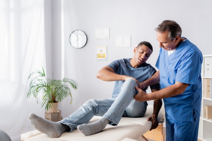
    || Local Community Centers | May offer support groups or resources for women undergoing similar health transitions. | Check with local community centers or health departments. || Support Groups Focused on Post-Hysterectomy Sexuality | Address the potential impact on sexual health and intimacy. | Some support groups may focus on these issues specifically. |

    Building a Strong Support Network

    Building a strong support network is an ongoing process. It involves actively reaching out to trusted individuals, communicating needs and limitations honestly, and nurturing existing relationships. It also involves proactively seeking out new connections, like joining support groups or online forums. Encouraging open communication and creating a safe space for sharing experiences are crucial for fostering strong bonds.

    Final Review

    In conclusion, recovering from a hysterectomy is a multifaceted journey. Understanding the physical, emotional, and lifestyle changes involved is essential for a successful recovery. By acknowledging the potential challenges and proactively addressing them with a supportive network and appropriate medical guidance, women can navigate this period with strength and resilience. This guide aims to provide comprehensive information to empower you through this transition.

  • Golf After Knee Replacement A Guide to Recovery

    Golf After Knee Replacement A Guide to Recovery

    Golf after knee replacement is a journey of careful planning and dedication. This guide delves into the considerations, rehabilitation, equipment adaptations, and swing adjustments crucial for a successful return to the game. It examines the physical demands of golf, how a knee replacement affects those demands, and the importance of a personalized approach to recovery. We’ll explore the complexities of returning to golf, equipping you with the knowledge to navigate this significant life change.

    The article covers everything from initial considerations to long-term health and wellness, providing practical advice on equipment modifications, swing mechanics, and course selection. We’ll also highlight the importance of professional guidance, support groups, and illustrative case studies. The goal is to empower you with the tools and information to confidently resume your passion for golf after knee replacement.

    Initial Considerations for Golf After Knee Replacement

    Returning to golf after a knee replacement is a significant undertaking, requiring careful planning and a phased approach to rehabilitation. The physical demands of the sport, coupled with the unique challenges of a knee replacement, necessitate a personalized strategy to ensure a safe and successful return. This phase requires a holistic understanding of the physical changes and mental adjustments needed to enjoy the game again.The physical demands of golf encompass a range of movements, from walking and carrying equipment to the twisting and bending required during the swing.

    A knee replacement, while a life-improving procedure, alters the biomechanics of the joint, potentially impacting these movements. The stability and range of motion of the replaced knee may be different from the pre-surgery state, demanding adjustments in technique and intensity. Understanding these changes is crucial for adapting to the sport safely and effectively.

    Getting back to golf after a knee replacement is a fantastic goal, but navigating the physical challenges is key. One often overlooked aspect of physical limitations is how conditions like diabetes can impact recovery and overall health, especially when considering the long-term effects of the surgery. For a deeper dive into the complexities of whether diabetes constitutes a disability, check out this helpful resource: is diabetes a disability.

    Ultimately, careful planning and realistic expectations are crucial for a successful return to the game, especially with any co-morbidities.

    Physical Demands of Golf

    Golf involves significant lower body movement, encompassing walking, bending, twisting, and impact forces during the swing. The intensity of these movements varies considerably depending on the course and individual playing style. On a challenging course, golfers may cover several miles, with significant impact on the knees.

    Impact of Knee Replacement on Golf Demands

    Knee replacement surgery alters the mechanics of the knee joint. The implanted components and surrounding tissues may limit the range of motion and impact tolerance compared to a healthy knee. This change necessitates adjustments in swing mechanics and walking patterns to minimize stress on the new knee. It’s important to understand that every individual’s experience with a knee replacement will differ.

    Factors such as the type of prosthesis used, the patient’s pre-existing conditions, and the rehabilitation program influence the post-operative outcome.

    Common Concerns and Anxieties

    Returning to golf after knee replacement often raises concerns about pain, instability, and the ability to perform the required movements. Players may worry about jeopardizing the recovery process or about the potential for reinjury. Anxiety about maintaining performance levels and the fear of being less competitive also feature prominently. These concerns are valid and should be addressed through open communication with healthcare professionals and a well-structured rehabilitation plan.

    Importance of a Personalized Approach to Rehabilitation

    A one-size-fits-all approach to rehabilitation after knee replacement is not appropriate for golf. Each patient’s unique physical capabilities, pain tolerance, and golf-specific needs must be considered. This personalized approach should involve close collaboration between the orthopedic surgeon, physical therapist, and the golfer themselves. The plan should carefully manage the load placed on the knee while progressively introducing the specific movements required in golf.

    Comparing Golf Course Demands

    Course Type Walking Distance (approx.) Impact on Knee (estimated) Typical Bending/Twisting Additional Considerations
    Short Course (9 holes) 2-3 miles Moderate Moderate Fewer physical demands, ideal for early stages of recovery.
    Championship Course (18 holes) 4-6 miles High High Requires more stamina and recovery time; best suited for later stages.

    This table provides a general comparison of the demands placed on the knees during golf on different course types. It is essential to remember that these are estimates and individual experiences can vary significantly.

    Rehabilitation and Recovery

    Golf after knee replacement

    Getting back to golf after a knee replacement is a journey, not a sprint. Proper rehabilitation is crucial for a successful recovery and preventing future complications. The focus is on restoring strength, mobility, and stability around the knee, allowing you to perform the demanding movements of golf without pain or reinjury. This involves a multi-faceted approach, encompassing physical therapy, careful exercise progression, and mindful activity.A structured rehabilitation program tailored to your individual needs is essential.

    This program typically begins with gentle exercises to regain range of motion and gradually increases in intensity and complexity as your knee heals and strengthens. It’s vital to remember that every patient heals at a different pace, and pushing yourself too hard too soon can lead to setbacks. Consistency and patience are key elements in this process.

    Typical Rehabilitation Protocols

    Post-knee replacement rehabilitation protocols often begin with exercises focused on regaining full range of motion and reducing swelling. These initial exercises might involve simple leg lifts, ankle pumps, and gentle range-of-motion exercises. As the knee heals and strength improves, more challenging exercises are introduced, progressing to strengthening exercises for the quadriceps, hamstrings, and surrounding muscles. The goal is to rebuild the muscles around the knee joint to provide stability and support.

    Professional physical therapists guide patients through this process.

    Importance of Gradual Return to Activity

    A gradual return to activity is paramount. Jumping back into your normal routine, including golf, too soon can lead to pain, reinjury, and prolonged recovery. The rehabilitation plan meticulously crafts a series of steps to gradually increase activity levels. This includes walking, cycling, and finally, more demanding activities like golf. The goal is to ensure the knee is strong enough to withstand the stresses of golf without compromising the healing process.

    Consider that an abrupt return to strenuous activity can have long-term consequences, so listen to your body and avoid pushing beyond your limits.

    Specific Exercises and Stretches for Golf

    To prepare for the demands of golf, specific exercises and stretches are essential. These exercises focus on improving flexibility, strength, and stability in the lower body. Examples include:

    • Quadriceps strengthening exercises: These exercises target the muscles in the front of the thigh, which are crucial for stability during golf swings. Examples include straight leg raises and knee extensions.
    • Hamstring strengthening exercises: Hamstrings are vital for maintaining balance and power during golf swings. Exercises include hamstring curls and glute bridges.
    • Balance exercises: Maintaining balance is critical for golf, especially during the swing. Exercises like single-leg stands and balance boards are beneficial.
    • Hip strengthening exercises: Exercises that strengthen the hip muscles enhance stability and power throughout the golf swing. Examples include clamshells and hip abductions.
    • Flexibility exercises: Stretches targeting the hamstrings, quads, and hip flexors improve flexibility, reducing the risk of injury during the swing.

    Progression of Exercises Over Time

    The following table Artikels a possible progression of exercises over time, incorporating golf-specific movements. Remember that this is a template and individual progressions may vary based on the patient’s needs and recovery.

    Week Exercises Golf-Specific Movements
    1-4 Range of motion exercises, light leg lifts, ankle pumps None
    5-8 Quad/hamstring strengthening, balance exercises (e.g., standing on one leg) Short, controlled putting practice
    9-12 Hip strengthening, more challenging balance exercises, gentle squats Short chipping practice
    13-16 Full squats, lunges, core strengthening Full swing practice with lighter clubs
    17-20 Increase swing repetitions, add drills Full swing practice with regular clubs

    Potential Complications and How to Address Them

    Recovering from a knee replacement can present potential complications. These can range from infection and blood clots to persistent pain or stiffness.

    • Infection: Prompt medical attention is crucial for addressing any signs of infection, such as redness, swelling, or fever. Following prescribed antibiotics is critical.
    • Blood clots: Appropriate medical interventions and preventive measures, such as compression stockings and blood thinners, are necessary for dealing with potential blood clots. Promptly report any unusual symptoms.
    • Persistent pain: Persistent pain may necessitate adjustments to the rehabilitation program. Consult with the physical therapist or surgeon for alternative strategies or pain management.
    • Stiffness: Regular stretching and range-of-motion exercises are vital for maintaining flexibility. Addressing stiffness promptly prevents long-term limitations.

    Golf Equipment Modifications and Adaptations

    Returning to the fairways after knee replacement surgery requires careful consideration of your golf equipment. Adapting your approach to the game can significantly impact your comfort and enjoyment while minimizing strain on your newly recovered knee. This section dives into the crucial modifications you can make to your golf equipment and swing to enhance your golfing experience.

    Golf Club Modifications

    Proper club selection can significantly reduce strain on your knee. A well-chosen club will reduce the need for excessive twisting and bending during the swing. Consider the impact of each club on your knee. The putter, for example, is a precision tool requiring a smaller, more controlled movement, which is generally less demanding on the knee. Drivers, on the other hand, often require a longer, more forceful swing, potentially leading to greater stress if not managed correctly.

    Experiment with different club weights and shaft flexes to find what feels most comfortable and minimizes strain.

    Stance and Swing Adjustments

    Adjusting your stance and swing mechanics is essential for accommodating your knee’s limitations. A slightly wider stance can provide greater stability and balance, reducing the strain on the knee during the swing. Maintaining a more upright posture during the backswing can also ease the stress on the knee joint. By subtly adjusting your swing path and maintaining a controlled tempo, you can make the game more comfortable.

    Experiment with these changes during practice sessions, noting how your knee feels with each variation.

    Golf Cart Considerations

    Choosing the right golf cart is critical for minimizing physical exertion. A cart with adequate cushioning and adjustable features can make a significant difference in your comfort level. The following table Artikels the pros and cons of different cart types:

    Golf Cart Type Pros Cons
    Electric Cart Quiet operation, less physical effort, typically more comfortable, often has features for storage or other amenities. Can be more expensive, may have limited carrying capacity for extra equipment or supplies.
    Push Cart Very affordable, no running costs. Requires more physical effort, not ideal for longer rounds, can be less comfortable.
    Hybrid Cart Offers a balance between electric and manual options, providing flexibility for various terrains and player preferences. May be more expensive than a basic push cart but less expensive than a full-fledged electric cart.

    Footwear and Knee Support

    Proper footwear is paramount for knee support and stability. Look for golf shoes with supportive features like sturdy soles, adequate ankle support, and reinforced heel counters. Consider golf shoes specifically designed for players with knee issues. High-quality golf shoes can help prevent excessive twisting and provide stability during your swing. The right footwear can dramatically reduce the risk of knee pain and discomfort during play.

    Impact on Golf Swing Mechanics: Golf After Knee Replacement

    Returning to golf after knee replacement requires careful consideration of swing mechanics. The altered biomechanics of the knee can lead to increased stress and pain if not addressed proactively. Understanding common swing flaws and adapting techniques are crucial for a safe and enjoyable return to the sport.Maintaining proper form and posture during the swing is paramount to minimizing stress on the newly replaced knee.

    This includes adjustments in weight distribution, club path, and swing tempo. Modifying the golf swing to accommodate the knee’s limitations can help prevent setbacks and allow for gradual improvement.

    Common Swing Flaws After Knee Replacement

    Adjustments to the golf swing are often necessary after knee replacement to avoid exacerbating pain. Common swing flaws that can place increased stress on the knee include a loss of balance, a shift in weight distribution, and an altered swing path. These issues can result in compensatory movements that put unnecessary pressure on the knee joint, potentially causing pain or further injury.

    Adjusting the Swing for Knee Health

    A modified swing can effectively reduce the stress on the knee. This involves altering the swing path, tempo, and weight distribution. For instance, a more controlled and gradual swing can help manage stress on the joint. Concentrating on maintaining a balanced posture throughout the swing is essential. Practicing drills that focus on stability and control can help develop a swing that is gentler on the knee.

    Posture and Balance in the Golf Swing

    Proper posture and balance are vital for minimizing stress on the replaced knee during the golf swing. A stable foundation provides a platform for a controlled swing. This includes maintaining a neutral spine alignment, engaging core muscles for support, and maintaining a balanced stance throughout the entire swing. Practicing exercises that strengthen core muscles and improve balance can enhance stability and reduce the risk of injury.

    Improving Balance and Stability

    Maintaining balance and stability during the golf swing is crucial for minimizing stress on the knee. Exercises like single-leg stances, balance boards, and wobble cushions can help strengthen the muscles responsible for balance and stability. These exercises, performed consistently, can improve proprioception, the body’s awareness of its position in space, leading to more controlled movements and reduced risk of injury.

    Impact of Different Club Types on Swing Mechanics and Knee Strain

    The type of club used can influence the swing mechanics and the stress placed on the knee. A driver, with its longer shaft and greater swing arc, might place more stress on the knee compared to a wedge, which has a shorter shaft and a more controlled swing. This difference is due to the increased force and momentum generated during a driver swing.

    Understanding the impact of different clubs is crucial for developing a safe and effective swing strategy.

    Club Type Swing Mechanics Potential Knee Strain
    Driver Longer swing arc, greater force and momentum Higher potential for knee strain if not managed correctly
    Woods (3-5 Wood) Medium swing arc, moderate force and momentum Moderate potential for knee strain
    Irons (7-9 Iron) Shorter swing arc, controlled swing Lower potential for knee strain
    Wedges (Pitching, Sand, Lob) Shortest swing arc, precise swing Lowest potential for knee strain

    Maintaining Long-Term Health and Wellness

    After knee replacement surgery, maintaining a healthy lifestyle is crucial for both physical recovery and long-term well-being. This involves not just focusing on the immediate rehabilitation but also establishing sustainable habits that support ongoing recovery and allow you to enjoy golf for years to come. This includes ongoing physical therapy, careful monitoring of pain, adapting activities to your progress, staying motivated, and incorporating strength training.Ongoing care is paramount to achieving lasting benefits from knee replacement surgery and sustaining a golf game.

    Consistent monitoring and adjustments to your routine are essential for optimizing your physical condition and avoiding setbacks.

    Importance of Ongoing Physical Therapy and Check-ups

    Regular physical therapy sessions are vital for maintaining strength, flexibility, and range of motion in the knee joint. These sessions provide tailored exercises to address any specific needs and concerns, preventing stiffness, muscle imbalances, and potential complications. Scheduled check-ups with your orthopedic surgeon allow for evaluation of the knee’s healing process, early detection of any issues, and adjustments to your treatment plan as needed.

    This proactive approach ensures optimal recovery and minimizes the risk of future problems.

    Monitoring for Signs of Pain or Discomfort

    Recognizing and responding to pain or discomfort is critical. Pain signals can indicate issues that need addressing, preventing further damage or hindering recovery. If you experience persistent or increasing pain, particularly in the knee, it’s essential to contact your orthopedic surgeon immediately. Similarly, any unusual sensations, such as swelling or stiffness, should be reported to your physical therapist or doctor.

    Early intervention is key to managing potential complications effectively. Be vigilant about any changes in your symptoms, no matter how minor they seem.

    Adjusting Golf Activities Based on Individual Progress and Limitations

    Your golf activities must be adapted to your evolving physical capabilities. As your knee recovers and strength improves, you can gradually increase the intensity and duration of your practice sessions. Start with shorter rounds and lighter practice swings, progressively increasing the number of holes played or the intensity of practice over time. Pay close attention to your body’s signals and avoid pushing yourself beyond your current limitations.

    This progressive approach minimizes the risk of re-injury and ensures a smoother return to golf.

    Staying Motivated and Engaged with the Sport

    Maintaining motivation is crucial throughout the recovery process. Setting realistic goals, celebrating small victories, and focusing on the positive aspects of your recovery can significantly boost morale. Consider joining a support group of other golfers who have undergone similar procedures. Sharing experiences and learning from others can provide encouragement and motivation. Remember, setbacks are a normal part of the process; focus on the progress you’re making.

    Getting back to golf after a knee replacement is a real challenge, isn’t it? Rehab is key, and finding the right balance between pushing yourself and listening to your body is crucial. But what about cognitive challenges that can affect golf, like those associated with Capgras syndrome in Lewy body dementia? Understanding these complexities is vital. Capgras syndrome in Lewy body dementia can impact daily activities, which could certainly make playing golf more difficult.

    Ultimately, though, focusing on gradual improvement and adjusting your game strategy is the best way to get back out there on the course, regardless of other health factors.

    Incorporating Strength Training for Overall Well-being

    Strength training plays a vital role in maintaining long-term health and wellness. Exercises that strengthen the muscles around the knee, hips, and core can improve stability, reduce stress on the joint, and enhance overall fitness. A well-rounded strength training program, tailored by a physical therapist, will address specific muscle weaknesses and support the recovery process. It’s crucial to incorporate exercises that target the quadriceps, hamstrings, glutes, and core muscles, as these groups significantly impact knee stability and overall function.

    Professional Guidance and Support

    Returning to golf after a knee replacement is a significant undertaking. It requires careful consideration of your physical capabilities, a phased approach to recovery, and a supportive network. Professional guidance plays a crucial role in optimizing your recovery journey and ensuring a safe and successful return to the sport.Expert advice from healthcare professionals and golf instructors can help you navigate the complexities of rehabilitation, equipment modifications, and swing adjustments.

    This tailored support ensures you address any potential challenges and maintain a sustainable golf routine that prioritizes your well-being.

    Consulting with Healthcare Professionals

    Seeking guidance from a physical therapist and/or orthopedic surgeon is paramount. These experts can assess your individual progress, identify any lingering limitations, and tailor a rehabilitation plan that aligns with your specific needs. They can also monitor your recovery, adjusting the plan as necessary. This personalized approach ensures that you don’t rush the healing process, reducing the risk of further injury.

    Crucially, they can address any pain or discomfort effectively.

    The Role of a Golf Professional

    A qualified golf professional can provide valuable insights into modifying your swing to accommodate the changes in your knee. They can assess your swing mechanics, identify areas where adjustments are necessary, and provide tailored drills and exercises to help you regain your swing confidence and improve your performance. This expert guidance can be particularly helpful in preventing strain on the knee joint during the golf swing.

    Experienced instructors can often offer personalized swing adjustments that maintain optimal form while minimizing stress on the newly replaced knee.

    Support Groups for Recovering Golfers

    Connecting with other golfers who have experienced similar challenges can be incredibly beneficial. Support groups offer a platform for sharing experiences, providing encouragement, and gaining valuable insights from others who understand the specific challenges of returning to golf after a knee replacement. These groups can offer a sense of community and shared understanding, which can significantly contribute to the overall recovery process.

    • Online forums dedicated to golf recovery can provide valuable support and advice from experienced members.
    • Local golf clubs or organizations may host support groups specifically for recovering golfers.
    • Social media groups can facilitate connections and offer a platform for sharing experiences and tips.

    Managing Pain and Discomfort

    Developing effective strategies for managing pain and discomfort is essential. Open communication with your physical therapist and orthopedic surgeon is key. They can recommend pain management techniques, such as medication, physical therapy exercises, or assistive devices, tailored to your individual needs. Additionally, understanding your body’s signals and recognizing potential triggers for pain is crucial. Implementing strategies for proactive pain management can significantly improve your comfort level and allow you to maintain a more consistent practice schedule.

    • Utilizing ice packs or heat therapy, as directed by your healthcare provider, can help alleviate pain and inflammation.
    • Employing relaxation techniques, such as deep breathing exercises, can aid in managing pain and stress associated with the recovery process.
    • Maintaining a consistent sleep schedule and a healthy diet are essential for overall well-being and pain management.

    Working with a Certified Athletic Trainer

    A certified athletic trainer can play a vital role in enhancing your recovery and ensuring a safe return to golf. Their expertise in injury prevention and rehabilitation can be invaluable. They can design targeted strength and conditioning programs, tailored to your specific needs and limitations, that help you build up the necessary strength and flexibility in your legs and core.

    Furthermore, they can help identify potential areas of concern and prevent future injuries. Their expertise in performance enhancement can contribute to a quicker and more complete recovery.

    Getting back to golf after a knee replacement is a gradual process, focusing on strengthening the surrounding muscles. It’s crucial to prioritize physical therapy, and exercises like those found in pt exercises for runners knee can be incredibly helpful for building strength and range of motion. Ultimately, a tailored approach, including specific exercises and gradual increases in activity, will ensure a safe and successful return to the sport.

    Illustrative Case Studies (Hypothetical)

    Golf after knee replacement

    Returning to golf after a knee replacement is a journey, not a sprint. Individual experiences vary greatly, making personalized rehabilitation crucial. These hypothetical case studies illustrate the process, highlighting the importance of tailoring recovery to the unique needs of each golfer.

    Case Study 1: The Determined Golfer

    A 55-year-old avid golfer, named David, underwent a total knee replacement. He demonstrated a strong commitment to rehabilitation, diligently following his physical therapist’s plan. David’s pre-surgery golf swing showcased a powerful, but slightly exaggerated, backswing, leading to some stress on his knee.

    Rehabilitation Process and Progression, Golf after knee replacement

    David’s initial focus was on regaining range of motion and strength in his knee. He participated in exercises focusing on quadriceps strengthening, hamstring flexibility, and proprioception (the sense of body position). His physical therapist gradually introduced exercises mimicking golf movements, such as controlled swings with light clubs, and finally, full-swing practice with progressively heavier clubs. Progress was measured by pain levels, range of motion, and functional tests.

    The goal was to achieve a pain-free swing that did not compromise the knee’s recovery.

    Challenges Encountered and Solutions

    Initially, David experienced some discomfort during early golf practice. His physical therapist adjusted his exercise routine to incorporate more rest and ice. Furthermore, he was advised to gradually increase the intensity and duration of his practice sessions. A key challenge was managing his expectations. David was encouraged to set realistic milestones, ensuring he didn’t push his recovery too hard, which was crucial for preventing setbacks.

    Personalized Rehabilitation Plan

    David’s rehabilitation plan was meticulously tailored to his specific needs and goals. His pre-surgery swing mechanics, pain tolerance, and overall physical condition were carefully considered. This personalized approach ensured that each exercise was appropriate for his stage of recovery and helped him regain optimal function and strength without jeopardizing his knee.

    Example of Golf Swing Before and After Knee Replacement

    Swing Feature Before Knee Replacement After Knee Replacement
    Backswing Exaggerated backswing, leading to excessive stress on the knee Controlled backswing, maintaining a stable knee position
    Hip Rotation Significant hip rotation, potentially contributing to knee stress Controlled hip rotation, emphasizing stability and minimizing stress on the knee
    Follow-through Uncontrolled follow-through, leading to strain Controlled follow-through, emphasizing balanced movement
    Stance Slightly unstable stance Stable and balanced stance

    David’s pre-surgery swing demonstrated a tendency towards forceful movements. His post-surgery swing was characterized by controlled, measured motions that minimized stress on his newly recovered knee. He was able to maintain a solid foundation and a smoother, more controlled swing pattern, reducing the risk of injury.

    Golf Course Considerations

    Choosing the right golf course is crucial for a recovering golfer after knee replacement surgery. The course’s layout, terrain, and maintenance play a significant role in minimizing stress on the new knee joint. Finding a course that allows for a comfortable and enjoyable round without excessive strain is paramount for a successful return to the sport. This section details key factors to consider when selecting a course and adapting to its demands.

    Selecting an Appropriate Golf Course

    Proper course selection is critical for a smooth recovery. A course with gentler slopes, shorter distances between holes, and well-maintained fairways and greens is ideal. Avoid courses with significant elevation changes, tight fairways, or rough terrain that could potentially cause excessive stress on the recovering knee.

    Course Features for a Recovering Golfer

    Ideal courses for recovering golfers possess several key characteristics. Gentle undulations, well-maintained fairways, and greens with smooth transitions are essential. Consider a course with a reasonable length and fewer obstacles, like dense rough, to reduce the strain on the recovering knee. A layout that allows for more manageable approaches to the green is preferable. Furthermore, the distance between holes and the overall course design should support a less strenuous pace of play.

    Modifying Course Requirements

    Adapting course requirements can significantly reduce stress on the knee. Consider teeing off from shorter tees to minimize walking distance and reduce the overall strain on the knee. Strategic use of a golf cart can alleviate walking stress, but it’s important to use it judiciously and not for the entirety of the round. The choice between walking and using a cart depends on individual recovery progress and comfort levels.

    It’s advisable to consult with a physical therapist or doctor to determine the most suitable option for each golfer.

    Course Layout and Knee Stress

    Different course layouts can impact knee stress differently. A table below illustrates potential impacts, offering a comparative overview for golfers to consider.

    Course Layout Feature Potential Impact on Knee Stress
    Steep inclines and declines High
    Tight fairways and narrow greens High
    Long distances between holes Moderate
    Well-maintained fairways and greens Low
    Gentle undulations Low
    Shorter hole distances Low

    Course Maintenance and Conditions

    Proper course maintenance is crucial for golfers with knee replacements. Well-maintained fairways and greens reduce the risk of uneven surfaces and potential injuries. Soft, consistent grass conditions, free of debris or obstacles, minimize the risk of falls and uneven ground impact. Soft conditions are critical for reducing the impact on the knee. Inspect the course for these factors before teeing off.

    Final Thoughts

    Returning to golf after a knee replacement is achievable with a structured approach. By carefully considering the physical demands of the sport, implementing personalized rehabilitation, adjusting equipment and swing mechanics, and seeking professional guidance, you can safely and effectively return to the game. Remember, gradual progress, ongoing support, and realistic expectations are key to a successful and enjoyable golf journey.

    This guide provides a comprehensive roadmap to help you navigate this unique experience, and empowers you to play golf again with confidence.

  • Chiropractor vs Physical Therapy Which is Right for You?

    Chiropractor vs Physical Therapy Which is Right for You?

    Chiropractor vs physical therapy – a common question for those seeking relief from musculoskeletal pain. This exploration delves into the core principles, techniques, and treatment approaches of both professions. Understanding their differences and overlaps is key to making an informed decision about your healthcare journey.

    Chiropractic care often focuses on spinal adjustments to alleviate pain and restore proper function, while physical therapy emphasizes restoring movement and improving overall physical capabilities through exercises and manual therapy. Both professions play vital roles in patient care, and this comparison will highlight their unique approaches to diagnosis and treatment, while also examining potential overlaps and collaborative opportunities.

    Introduction to Chiropractic and Physical Therapy

    Chiropractic care and physical therapy are both healthcare disciplines focused on restoring health and well-being, but they approach the issue with different philosophies and techniques. Understanding the core principles of each is essential for making informed decisions about your health. Both play crucial roles in musculoskeletal rehabilitation and pain management, but their specific approaches differ.Both chiropractic and physical therapy aim to improve a patient’s overall function and quality of life, but their methods and underlying philosophies diverge.

    This difference in approach often leads to discussion and debate about which is the more effective treatment path. This exploration will delve into the unique characteristics of each, highlighting their distinct approaches to health restoration.

    Chiropractic Care: A Focus on Spinal Health

    Chiropractic care centers on the idea that proper spinal alignment is essential for overall health. Chiropractors believe that misalignments, or subluxations, in the spine can interfere with the nervous system’s function, leading to a wide range of health issues. Their primary techniques involve spinal adjustments, a hands-on approach that aims to restore proper joint motion and nerve function.

    This approach often targets musculoskeletal issues and focuses on the spine as the root cause of many ailments.

    Physical Therapy: Restoring Function Through Movement

    Physical therapy, conversely, focuses on restoring movement and function. Physical therapists use a variety of techniques, including exercises, manual therapy, and modalities, to help patients regain strength, flexibility, and range of motion. The core philosophy of physical therapy is that movement is crucial for maintaining and regaining health. This approach considers a holistic view of the body, recognizing the interplay between various systems and their impact on overall function.

    Comparing and Contrasting Philosophies

    Characteristic Chiropractic Physical Therapy Overlap
    Focus Spinal adjustments, musculoskeletal issues Restoring function, improving movement Pain management, rehabilitation
    Techniques Spinal manipulation, mobilization Exercises, manual therapy, modalities Joint mobilization, exercise prescription
    Underlying Philosophy Focus on spinal health Focus on movement and function Holistic well-being

    Chiropractic care prioritizes spinal health and alignment, often treating a wider range of musculoskeletal conditions. Physical therapy emphasizes restoring function through movement and exercises, addressing the specific needs of each patient. The overlap highlights the potential for complementary approaches. For example, a physical therapist might prescribe exercises to strengthen muscles affected by a spinal adjustment, while a chiropractor might use manual techniques to improve the patient’s range of motion.

    Conditions Treated by Each Profession

    Chiropractor vs physical therapy

    Chiropractic care and physical therapy are both valuable approaches to managing musculoskeletal issues, but they address different aspects of the problem. Understanding the specific conditions each profession excels at can help individuals make informed decisions about the most suitable treatment path for their needs. Both professions aim to improve quality of life and alleviate pain, but their approaches differ significantly.Chiropractic care often focuses on the spine and its relationship to overall health, while physical therapy emphasizes restoring movement and function through exercises and manual therapies.

    The table below provides a concise comparison of common conditions treated by each profession.

    Conditions Treated by Chiropractors

    Chiropractic care often targets conditions related to the musculoskeletal system, especially the spine. Common conditions include:

    • Back pain: Chiropractors frequently use spinal adjustments to alleviate back pain, often caused by misalignments or subluxations of the vertebrae. These adjustments aim to restore proper spinal mechanics, reduce nerve impingement, and improve overall function.
    • Neck pain: Neck pain, a common complaint, can stem from various factors including muscle strain, poor posture, or whiplash injuries. Chiropractors address these issues through spinal adjustments, manual therapies, and recommendations for postural improvements.
    • Headaches: Certain types of headaches, particularly those related to neck tension or spinal misalignments, may respond well to chiropractic care. Chiropractic adjustments can address the underlying causes of these headaches and promote pain relief.
    • Sciatica: Sciatica involves pain radiating down the leg, often due to compression of the sciatic nerve. Chiropractors may address this by identifying and treating the spinal misalignment contributing to the nerve compression.
    • Whiplash: Whiplash injuries, often sustained in car accidents, can result in neck pain, headaches, and other symptoms. Chiropractic care can help to restore the proper alignment and function of the neck, managing pain and discomfort.

    Conditions Addressed by Physical Therapists

    Physical therapists often work with patients experiencing a wider range of conditions that impact movement and function. Their interventions are multifaceted and often encompass exercises, manual therapies, and education.

    Choosing between a chiropractor and physical therapy can be tricky. While both aim to alleviate pain and improve mobility, they often tackle different issues. For instance, aversion therapy, a specialized approach with a focus on changing unwanted behaviors, might be useful in certain scenarios, as detailed in this comprehensive overview aversion therapy overview uses benefits effectiveness.

    Ultimately, the best path for you will depend on your specific needs and the nature of your pain. A consultation with both a chiropractor and physical therapist can help determine the most appropriate course of action.

    • Back pain: Physical therapists use a range of techniques, including exercises to strengthen core muscles, manual therapy to address soft tissue restrictions, and education on proper posture and lifting techniques. These interventions aim to improve mobility, reduce pain, and prevent future episodes.
    • Joint pain: Physical therapy can address pain in various joints throughout the body, utilizing exercises and manual therapy to restore range of motion, reduce inflammation, and improve joint stability.
    • Sports injuries: Physical therapists play a crucial role in rehabilitation following sports injuries. They design personalized exercise programs to regain strength, flexibility, and range of motion, helping athletes return to their activities safely.
    • Neurological conditions: Physical therapy is often integral to managing conditions like stroke, multiple sclerosis, or cerebral palsy. Therapists work with patients to improve motor skills, mobility, and balance.
    • Post-surgical rehabilitation: Following surgery, physical therapists guide patients through a structured rehabilitation program to restore strength, flexibility, and function in the affected area.

    Conditions Where Both Approaches Might Be Beneficial

    In some cases, a combined approach using both chiropractic and physical therapy can yield the best results. For example, individuals with chronic back pain might benefit from chiropractic adjustments to address spinal misalignments, coupled with physical therapy exercises to strengthen supporting muscles and improve posture.

    Diagnostic Methods and Procedures

    Chiropractor vs physical therapy

    Understanding how chiropractors and physical therapists diagnose conditions is crucial for choosing the right healthcare professional. Both professions utilize a range of methods to pinpoint the source of pain or dysfunction, but their approaches and priorities differ based on their respective philosophies and training. This section explores the specific diagnostic techniques employed by each profession, highlighting their similarities and differences.

    Chiropractic Diagnostic Methods

    Chiropractors typically begin with a comprehensive patient history, focusing on the patient’s description of symptoms, the onset and duration of pain, and any contributing factors. A thorough physical examination follows, evaluating posture, joint mobility, range of motion, and muscle strength. Specific tests, such as neurological assessments to check for nerve impingement, are often performed. The examination also includes palpating the spine and other joints for areas of tenderness, stiffness, or restricted movement.

    In some cases, imaging, such as X-rays or MRIs, may be necessary to visualize the structures of the spine and identify any underlying structural abnormalities. Chiropractic diagnoses often focus on the biomechanical aspects of the musculoskeletal system.

    Physical Therapy Diagnostic Procedures

    Physical therapists also start with a detailed patient history, including medical history, current symptoms, and lifestyle factors. Their physical examination is comprehensive, evaluating not only range of motion and muscle strength but also assessing movement patterns, balance, and coordination. Specific tests are conducted to pinpoint the source of pain and dysfunction, often involving manual muscle testing, neurological testing, and postural assessments.

    Imaging, including X-rays, MRIs, and CT scans, may be used to visualize the affected area and rule out any underlying structural issues. Physical therapy diagnoses consider the interplay of multiple factors, including biomechanics, neurological function, and patient-specific needs.

    Similarities and Differences in Diagnostic Approaches

    While both professions use patient history and physical examination, their approaches differ slightly. Chiropractors often focus more on the spine and biomechanical factors, while physical therapists take a broader view, considering the entire musculoskeletal system and how it functions as a whole. Both professions use imaging, but the types and frequency of imaging differ. Chiropractors may primarily use X-rays, while physical therapists may use a wider range of imaging modalities depending on the specific situation.

    Both professions strive for a thorough understanding of the patient’s condition, but the emphasis on specific areas and factors varies.

    Diagnostic Tools and Procedures Comparison

    Diagnostic Method Chiropractic Physical Therapy
    Patient history Thorough questioning on symptoms, duration, and contributing factors. In-depth review of medical history, lifestyle factors, and current symptoms.
    Physical exam Assessment of posture, joint mobility, range of motion, muscle strength, and neurological function, focusing on spinal alignment. Evaluation of movement patterns, muscle strength, balance, coordination, and neurological function, considering the entire musculoskeletal system.
    Imaging X-rays, MRI (in some cases), often used to identify structural abnormalities. X-rays, MRI, CT scans (as needed), used to assess the affected area and rule out underlying issues.

    Treatment Approaches and Techniques: Chiropractor Vs Physical Therapy

    Chiropractic and physical therapy, while both aiming to alleviate musculoskeletal pain and improve function, employ distinct treatment approaches. Understanding these differences can help patients make informed decisions about the best course of care for their specific needs. Both professions often work collaboratively, integrating their expertise to optimize outcomes.Chiropractic care primarily focuses on the musculoskeletal system, particularly the spine, and seeks to restore proper alignment and motion.

    Physical therapy, on the other hand, takes a broader, holistic approach, addressing the entire body’s mechanics and function.

    Chiropractic Treatment Techniques

    Chiropractors utilize a variety of techniques to address musculoskeletal issues. A core component is spinal manipulation, or adjustments, aimed at restoring proper spinal alignment and reducing nerve impingement. This involves controlled, specific movements to the spine, designed to relieve pain and improve joint mobility. Other techniques include mobilization of joints in the extremities, massage therapy, and the use of modalities like heat, ice, and electrical stimulation.

    Figuring out whether a chiropractor or physical therapist is right for you can be tricky. While both can help with pain management, a chiropractor often focuses on spinal adjustments, whereas physical therapy usually involves targeted exercises and stretches. If you’re dealing with gastritis and need to optimize your diet, checking out resources like gastritis diet what to eat for better management can help you make informed food choices.

    Ultimately, the best approach often depends on the specific cause of your discomfort and what your body needs, making it a crucial step in determining the best path forward when deciding between a chiropractor and physical therapy.

    The goal is to address the underlying cause of the patient’s discomfort and promote self-healing mechanisms.

    Physical Therapy Treatment Methods

    Physical therapists employ a range of methods to help patients recover from injuries and improve their overall well-being. These techniques encompass manual therapy, which includes soft tissue mobilization, joint mobilization, and manipulation. Furthermore, therapeutic exercise plays a critical role, tailoring specific exercises to address the patient’s individual needs and limitations. Physical therapists also utilize various modalities like ultrasound, laser therapy, and electrical stimulation to reduce pain, promote healing, and improve tissue repair.

    Patient education and lifestyle modifications are integral components of the treatment plan.

    Similarities and Differences in Therapeutic Approaches

    While distinct in their primary focus, chiropractic and physical therapy share some overlapping treatment approaches. Both utilize exercise therapy, but the exercises prescribed differ in their intensity and focus depending on the specific condition. Both may also utilize modalities like heat and ice, though the modalities and their applications may vary based on the treatment goals. The fundamental difference lies in the underlying philosophy and scope of practice.

    Chiropractic care often centers on the spinal column, while physical therapy addresses the entire musculoskeletal system. Ultimately, the choice between the two depends on the patient’s specific needs and the nature of their condition.

    Table of Specific Treatments, Chiropractor vs physical therapy

    Treatment Chiropractic Physical Therapy
    Spinal manipulation Adjustments to restore spinal alignment, often with specific hand movements targeting restricted joints. Manual therapy techniques to reduce pain and improve mobility, potentially including joint mobilization.
    Exercise therapy Strengthening and flexibility exercises focusing on core stability and spinal mobility. Customized exercises for specific conditions, addressing muscle imbalances, improving range of motion, and increasing endurance.
    Modalities Heat, ice, electrical stimulation (e.g., TENS) to reduce pain and inflammation. Ultrasound, laser, traction, electrical stimulation (e.g., NMES) for pain relief, tissue healing, and muscle stimulation.

    Patient Care Considerations

    Effective patient care in both chiropractic and physical therapy hinges on understanding the individual needs and preferences of each patient. A holistic approach that combines expertise with empathy is crucial for achieving positive outcomes. Successful treatment plans involve active participation from the patient, fostering trust and open communication.Patient care transcends the technical aspects of diagnosis and treatment; it encompasses the emotional and psychological well-being of the individual.

    Choosing between a chiropractor and physical therapy can be tricky. Both are great for musculoskeletal issues, but knowing when to take apple cider vinegar for potential benefits alongside your treatment plan might influence your decision. When to take apple cider vinegar is a crucial aspect of holistic wellness. Ultimately, the best approach often depends on the specific injury or ailment and your individual needs.

    A chiropractor might be better for spinal adjustments, while physical therapy focuses more on strengthening and restoring function. Consider these factors when making your choice.

    This necessitates a strong emphasis on patient education, effective communication, and a collaborative approach between practitioners. Successful outcomes depend on the ability to tailor care to the unique circumstances of each patient.

    Patient Education and Communication

    Patient education is fundamental to both chiropractic and physical therapy. Clearly explaining the condition, treatment plan, and expected outcomes empowers patients to actively participate in their recovery. This involves using understandable language, addressing concerns, and providing resources for continued learning. Open communication channels ensure patients feel heard and understood, fostering trust and cooperation in the treatment process.

    Active listening and empathetic responses are vital in this process. Patients need to understand their role in the treatment, the importance of adherence to prescribed exercises and lifestyle modifications, and the expected timeframe for recovery.

    Collaborative Approach Between Professions

    A collaborative approach between chiropractors and physical therapists can significantly enhance patient care. Combining the expertise of both professions allows for a comprehensive treatment plan that addresses the multifaceted nature of many musculoskeletal conditions. This integrated approach goes beyond individual treatments and encompasses a broader view of the patient’s overall health. Chiropractors can focus on spinal alignment and nerve function, while physical therapists can address muscle imbalances, joint mobility, and functional limitations.

    A well-coordinated approach allows for a more holistic and efficient healing process.

    Patient Case Studies

    A collaborative approach between chiropractors and physical therapists proved successful in a case involving a 35-year-old office worker experiencing chronic back pain. The chiropractor addressed spinal misalignments, while the physical therapist focused on strengthening core muscles and improving posture. The patient’s pain significantly decreased, and they regained full functionality within 12 weeks. Another case involved a 48-year-old athlete with a shoulder impingement.

    The chiropractor treated the upper back and neck, and the physical therapist focused on restoring shoulder mobility and range of motion. The athlete was able to return to sports activities after a 10-week treatment period.

    Potential Benefits and Drawbacks of Combining Approaches

    Combining chiropractic and physical therapy approaches offers numerous benefits, including:

    • Comprehensive care: Addressing the musculoskeletal system from multiple angles.
    • Enhanced recovery: Faster and more effective outcomes due to the combination of treatments.
    • Improved patient satisfaction: A more holistic and personalized approach to care.
    • Reduced recurrence: Addressing underlying causes of pain, not just symptoms.

    Potential drawbacks of a combined approach might include:

    • Coordination challenges: Ensuring seamless communication and collaboration between professionals.
    • Increased costs: The need for consultations with both practitioners.
    • Potential for conflicting advice: Clear communication and shared decision-making are essential.

    A well-defined protocol and shared understanding of the patient’s needs minimize these potential drawbacks. A strong professional relationship and effective communication are key to a successful collaborative effort.

    Illustrative Case Studies

    Understanding the effectiveness of chiropractic care and physical therapy often hinges on real-world examples. Case studies offer a glimpse into how these approaches address specific conditions and the outcomes they produce. Analyzing these cases helps in evaluating the strengths and weaknesses of each method, and importantly, when a combined approach might be most beneficial.Case studies provide valuable insight into the practical application of chiropractic and physical therapy techniques.

    They highlight the nuanced nature of patient care, demonstrating how both approaches can be tailored to individual needs. They also offer valuable insights into the limitations of each approach, as well as the situations where combining them can lead to superior results.

    Chiropractic Care: A Case of Chronic Back Pain

    A 45-year-old office worker experienced persistent lower back pain radiating down the left leg for six months. Standard pain relievers provided minimal relief. A chiropractor identified a spinal misalignment and implemented a series of adjustments. The patient reported significant pain reduction after the first few sessions. Regular adjustments and targeted exercises prescribed by the chiropractor helped maintain the improvements over several months.

    The case exemplifies how chiropractic care can effectively address musculoskeletal issues like chronic back pain when the root cause involves spinal mechanics.

    Physical Therapy: Recovering from a Knee Injury

    A 28-year-old athlete suffered a significant knee injury during a sporting event. The injury involved damage to the anterior cruciate ligament (ACL). Physical therapy played a crucial role in the rehabilitation process. A structured program focused on restoring range of motion, strengthening the surrounding muscles, and gradually increasing activity levels. The patient successfully returned to sports after several months of diligent physical therapy.

    This case demonstrates the efficacy of physical therapy in addressing complex injuries requiring targeted rehabilitation and functional restoration.

    Combined Approach: Managing Post-Surgical Recovery

    A 65-year-old patient underwent hip replacement surgery. The patient experienced post-operative pain and limited mobility. A combined approach, integrating physical therapy exercises with chiropractic adjustments, proved beneficial. Physical therapy focused on regaining range of motion and strengthening the surrounding muscles, while chiropractic adjustments addressed any lingering spinal misalignments that could be contributing to the pain. The patient experienced significant improvement in pain levels and mobility within a shorter timeframe than expected, showcasing the synergistic effects of combining the two approaches.

    Summary of Case Study Results

    The presented cases highlight the diverse applications of chiropractic and physical therapy. Chiropractic care excels in addressing musculoskeletal issues potentially stemming from spinal misalignments, demonstrating effective pain reduction and improved function in some cases. Physical therapy is particularly valuable in rehabilitation following injuries or surgeries, promoting recovery through targeted exercises and functional restoration. The combined approach demonstrates optimal outcomes in cases where both spinal mechanics and targeted muscle strengthening are crucial, like post-surgical recovery.
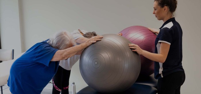
    However, it’s essential to acknowledge that individual responses vary, and the efficacy of each approach depends heavily on the specific condition and the patient’s individual circumstances.

    Closing Notes

    Ultimately, the choice between a chiropractor and a physical therapist depends on individual needs and preferences. This comparison offers a comprehensive overview to aid in your decision-making process. Consider your specific condition, desired outcomes, and personal values when selecting the healthcare professional best suited to your situation. Remember that sometimes, a collaborative approach, combining elements of both therapies, can yield the most beneficial results.

  • Pelvic Floor Physical Therapy A Comprehensive Guide

    Pelvic Floor Physical Therapy A Comprehensive Guide

    Pelvic floor physical therapy offers a specialized approach to addressing various pelvic floor issues. It focuses on restoring the function and strength of the muscles supporting the pelvic organs, which play crucial roles in bladder and bowel control, sexual function, and overall pelvic health. This guide delves into the intricacies of pelvic floor physical therapy, exploring its assessment methods, treatment approaches, and the conditions it effectively manages.

    Understanding the key functions of the pelvic floor muscles, the different types of conditions they can address, and the common symptoms associated with pelvic floor dysfunction is paramount. The guide also includes a table comparing common conditions like urinary incontinence, fecal incontinence, and pelvic organ prolapse, detailing their symptoms, possible causes, and treatment options.

    Introduction to Pelvic Floor Physical Therapy

    Pelvic floor physical therapy

    Pelvic floor physical therapy is a specialized field of physical therapy focused on the muscles and tissues of the pelvic floor. It’s a non-invasive approach to addressing a wide range of conditions affecting this intricate area of the body. This therapy uses hands-on techniques, exercises, and education to restore function and alleviate discomfort.Pelvic floor muscles play a crucial role in supporting the pelvic organs (bladder, uterus, rectum) and maintaining continence.

    These muscles also contribute to sexual function and overall pelvic health. Dysfunction in these muscles can lead to a variety of problems, and pelvic floor physical therapy offers a valuable path towards recovery and improved quality of life.

    Key Functions of the Pelvic Floor Muscles

    The pelvic floor muscles are responsible for supporting the pelvic organs, maintaining continence, and facilitating sexual function. They act like a hammock, holding the organs in place and preventing leakage. They also play a significant role in the coordination of urination, bowel movements, and sexual response.

    Conditions Addressed by Pelvic Floor Physical Therapy

    Pelvic floor physical therapy can effectively address a wide range of conditions, including urinary and fecal incontinence, pelvic organ prolapse, and chronic pelvic pain. This therapy provides a tailored approach to restore the proper function and strength of the pelvic floor muscles, leading to symptom relief and improved overall well-being.

    Common Symptoms of Pelvic Floor Dysfunction

    Common symptoms associated with pelvic floor dysfunction include urinary incontinence (leaking urine), fecal incontinence (leaking stool), pelvic pain, painful intercourse, and difficulty with bowel or bladder control. These symptoms can vary significantly in severity and impact daily life, making specialized therapy crucial for effective management.

    Comparison of Pelvic Floor Dysfunction Conditions

    Condition Symptoms Possible Causes Treatment Options
    Urinary Incontinence Frequent urination, urgency, leaking urine with coughing, sneezing, or physical exertion. Some experience leakage even when not actively trying to urinate. Pregnancy and childbirth, aging, neurological conditions, weakened pelvic floor muscles, certain medications, obesity, chronic cough, and stress. Pelvic floor muscle exercises (Kegels), biofeedback, electrical stimulation, lifestyle modifications (bladder training), and in some cases, surgical intervention.
    Fecal Incontinence Inability to control bowel movements, leakage of stool, straining during bowel movements, and frequent bowel movements. Weakened pelvic floor muscles, neurological conditions, constipation, irritable bowel syndrome (IBS), injury to the pelvic area, and certain medications. Pelvic floor muscle exercises, biofeedback, bowel retraining, dietary modifications, and in some cases, surgical intervention.
    Pelvic Organ Prolapse Feeling of heaviness, pressure, or bulging in the pelvic area, especially during physical activity or standing for extended periods. Prolapse can involve the bladder, uterus, rectum, or small bowel. Pregnancy and childbirth, weakened pelvic floor muscles, chronic straining during bowel movements, obesity, and genetic predisposition. Pelvic floor muscle exercises, supportive devices (pessaries), and in some cases, surgical intervention.

    Assessment and Evaluation Methods

    Understanding the intricacies of a patient’s pelvic floor dysfunction requires a thorough assessment process. This process isn’t just about identifying symptoms; it’s about uncovering the underlying causes and contributing factors. A skilled pelvic floor physical therapist uses a combination of subjective and objective methods to gain a comprehensive picture of the patient’s condition. This allows for the development of a personalized treatment plan tailored to their specific needs.

    Typical Assessment Process

    A typical pelvic floor physical therapy assessment involves a detailed discussion of the patient’s medical history, symptoms, and lifestyle. This initial consultation lays the groundwork for understanding the patient’s concerns and identifying potential contributing factors. The physical examination then moves beyond the surface, employing various techniques to assess pelvic floor muscle function and surrounding structures. This evaluation is crucial in determining the most effective treatment approach.

    Methods for Evaluating Pelvic Floor Muscle Function

    Pelvic floor muscle function is evaluated using a variety of methods, each offering unique insights. These methods include palpation, which allows the therapist to assess the tone, strength, and mobility of the muscles. Other techniques include internal and external assessments, which help identify potential restrictions or dysfunctions within the pelvic floor. Further, tests of muscle endurance and response to various stimuli are crucial in determining the patient’s individual limitations and capacities.

    Diagnostic Tools and Techniques

    A range of diagnostic tools and techniques are used in assessing pelvic floor function. These tools include internal and external examinations, allowing the therapist to evaluate the muscles and surrounding tissues. Digital palpation is used to assess muscle tone, strength, and mobility. Other techniques like vaginal or rectal examinations allow for a more comprehensive assessment of the pelvic floor and surrounding tissues.

    Pelvic floor physical therapy is amazing for so many things, and it often addresses surprising issues! Have you ever experienced a sore throat, particularly when yawning? It might seem unrelated to your pelvic floor, but sometimes issues in that area can affect other parts of the body, like the muscles supporting your throat. If you’re experiencing a throat hurts when i yawn, check out this article on the topic here.

    Ultimately, pelvic floor physical therapy can help improve overall body function, even impacting things like throat discomfort.

    Further, electromyography (EMG) can be used to measure the electrical activity of the pelvic floor muscles, providing further insight into muscle function and response.

    Assessment Tools and Their Applications

    Assessment Tool Description Application Limitations
    Patient History and Symptom Questionnaire Detailed discussion of medical history, symptoms, and lifestyle factors. Identifying potential contributing factors, understanding patient’s concerns, and guiding the assessment. Reliance on patient accuracy and recall; potential for bias.
    External Inspection Visual examination of the perineal area and surrounding tissues. Identifying external abnormalities or indicators of potential pelvic floor dysfunction. Limited insight into internal structures; requires additional assessments.
    Internal Pelvic Floor Examination Palpation of the pelvic floor muscles through the vagina or rectum. Assessing muscle tone, strength, and mobility, identifying trigger points or restrictions. Requires patient comfort and cooperation; potential for discomfort.
    Pelvic Floor Muscle Testing Assessment of muscle strength, endurance, and coordination through various maneuvers. Evaluating muscle function, identifying weaknesses or imbalances, and determining the severity of dysfunction. Requires active patient participation; results may be influenced by patient’s understanding and effort.
    Electromyography (EMG) Measuring electrical activity of the pelvic floor muscles. Providing detailed information about muscle activation patterns, identifying potential neuromuscular issues. Expensive; requires specialized equipment; potential for discomfort during the procedure.

    Common Patient Questions Regarding the Assessment Process

    • What will the physical examination involve? The physical examination involves a range of techniques, including external inspection, internal pelvic floor examinations, and muscle testing. These evaluations help the therapist understand the condition of the pelvic floor muscles and surrounding tissues.
    • How long will the assessment take? The assessment process can vary depending on the complexity of the patient’s condition and the specific techniques used. A thorough assessment can take from 45 minutes to an hour or more.
    • Will it be painful? While some patients may experience some discomfort during the assessment, particularly during internal examinations, the therapist will work to ensure the patient’s comfort throughout the process. The therapist will adjust the examination techniques as needed to minimize discomfort.
    • What happens after the assessment? Following the assessment, the therapist will discuss the findings with the patient and develop a personalized treatment plan. This plan will address the identified issues and work towards improving pelvic floor function.

    Treatment Approaches and Techniques

    Pelvic floor physical therapy employs a multifaceted approach to address pelvic floor dysfunction, tailoring interventions to the specific needs and goals of each patient. This often involves a combination of manual therapy, targeted exercises, and biofeedback to restore proper function and alleviate symptoms. Effective treatment plans frequently incorporate patient education, emphasizing self-management strategies for long-term well-being.Treatment strategies aim to restore normal pelvic floor muscle tone, coordination, and sensation.

    This is achieved through a comprehensive approach that combines various techniques, with the goal of reducing pain, improving bladder and bowel control, and enhancing overall pelvic floor health. Careful consideration is given to the underlying causes of dysfunction and the patient’s individual response to treatment.

    Common Treatment Approaches

    Treatment approaches for pelvic floor dysfunction are diverse and often integrated. A tailored plan considers factors such as the patient’s medical history, symptoms, and lifestyle. These plans typically involve manual therapy, exercise programs, and biofeedback techniques. Each approach plays a crucial role in restoring pelvic floor function and alleviating symptoms.

    Manual Therapy Techniques

    Manual therapy techniques are frequently incorporated into pelvic floor physical therapy. These techniques aim to address restrictions in the surrounding tissues and structures that may contribute to pelvic floor dysfunction. Techniques such as myofascial release, trigger point therapy, and mobilization of the sacrum and hips may be employed. These hands-on approaches can help to reduce pain, improve mobility, and promote relaxation of the pelvic floor muscles.

    Exercise Programs

    Exercise programs are fundamental in pelvic floor physical therapy, targeting specific muscles and improving coordination. These programs typically involve a combination of strengthening, relaxation, and coordination exercises. The exercises aim to restore normal muscle tone and function. Correctly executed exercises are crucial for positive outcomes.

    Types of Pelvic Floor Exercises and Their Effectiveness

    Pelvic floor exercises encompass various types, each targeting specific muscle groups and offering unique benefits. The effectiveness of each type depends on factors such as patient compliance, technique, and consistency. Different exercises address various aspects of pelvic floor function, promoting strength, flexibility, and coordination.

    Comparison of Pelvic Floor Exercises

    Exercise Target Muscles Description Benefits
    Kegels Pubococcygeus (PC) muscle, levator ani muscle, and other pelvic floor muscles Contracting and releasing the pelvic floor muscles as if stopping the flow of urine. Different variations can target different aspects of the pelvic floor. Strengthens pelvic floor muscles, improves bladder control, and can help with sexual function.
    Pelvic Tilt Abdominal muscles, pelvic floor muscles, and hip flexors Involves tilting the pelvis upward by engaging core muscles, while maintaining proper posture. Improves core strength, reduces back pain, and enhances pelvic floor stability.
    Bird Dog Core muscles, including pelvic floor muscles, back extensors, and hip muscles. Involves extending one arm and opposite leg simultaneously while maintaining a straight back. Improves core stability, strengthens back muscles, and enhances pelvic floor engagement.
    Bridge Gluteal muscles, hamstrings, and pelvic floor muscles Involves lying on the back and lifting the hips off the ground, engaging the glutes and core. Strengthens glutes, improves hip mobility, and activates pelvic floor muscles.

    Biofeedback Techniques

    Biofeedback techniques are used to help patients become more aware of their pelvic floor muscles. These techniques involve providing real-time feedback on muscle activity through visual or auditory signals. This allows patients to learn to control their pelvic floor muscles more effectively. Biofeedback can help individuals identify and correct any muscle imbalances or inefficient activation patterns. This can improve symptoms of pelvic floor dysfunction and enhance self-management strategies.

    Pelvic floor physical therapy can be incredibly helpful for various issues, from pain to incontinence. Sometimes, skin conditions like rosacea, psoriasis, or eczema can also impact overall well-being and potentially be connected to underlying health concerns, such as those addressed by pelvic floor therapy. If you’re curious to learn more about identifying these skin conditions, check out this helpful resource: do you have rosacea psoriasis or eczema.

    Ultimately, addressing the root causes of discomfort through pelvic floor physical therapy can lead to significant improvements in overall health and quality of life.

    Common Conditions Addressed: Pelvic Floor Physical Therapy

    Pelvic floor physical therapy isn’t just about preventing or treating incontinence; it’s a versatile approach to managing a wide array of conditions affecting the pelvic region. Physical therapists specializing in this area use a combination of hands-on techniques, exercises, and education to help patients regain control and function. Understanding the specific conditions addressed allows for more targeted and effective treatment plans.Addressing these conditions often involves a holistic approach, considering the patient’s overall health, lifestyle, and individual needs.

    This individualized care ensures that the treatment is not only effective but also sustainable. Physical therapy isn’t a one-size-fits-all solution; it’s about tailoring a plan to meet each patient’s unique circumstances.

    Specific Conditions Treated

    Pelvic floor physical therapy addresses a diverse range of conditions. These conditions span across various areas of the pelvic region and affect both men and women.

    • Urinary incontinence: This encompasses various types, from stress incontinence (leaking with exertion) to urge incontinence (sudden, strong urges to urinate). Physical therapists employ exercises to strengthen the pelvic floor muscles, improve bladder control, and re-educate the nervous system’s response to bladder signals. Outcomes often include reduced incontinence episodes and improved quality of life.
    • Fecal incontinence: Similar to urinary incontinence, this condition involves involuntary leakage of stool. Physical therapy focuses on strengthening pelvic floor muscles, improving bowel habits, and promoting better stool consistency. Successful outcomes may include reduced episodes of incontinence and improved bowel regularity. Furthermore, the approach addresses any contributing factors like constipation or irritable bowel syndrome.
    • Pelvic organ prolapse: This occurs when pelvic organs, such as the bladder, uterus, or rectum, descend from their normal position. Physical therapy can help support the weakened tissues and pelvic floor muscles through targeted exercises and techniques to reposition the organs. Positive outcomes may include improved comfort, reduced symptoms, and an enhanced ability to perform daily activities without discomfort. The approach often includes education about lifestyle modifications and supportive measures.

    • Chronic pelvic pain: This can stem from various underlying causes, including endometriosis, interstitial cystitis, or muscle tension. Physical therapy can help identify and address the root causes of the pain. This might involve releasing tension in surrounding muscles, improving posture, and teaching relaxation techniques. Expected benefits include reduced pain intensity, improved mobility, and enhanced quality of life.
    • Postpartum recovery: After childbirth, many women experience pelvic floor dysfunction. Physical therapy plays a crucial role in restoring pelvic floor strength and function. Exercises are tailored to the individual’s specific needs, and the physical therapist educates the patient on the importance of proper posture, body mechanics, and pain management. Positive outcomes often include improved bladder and bowel control, reduced pain, and enhanced overall well-being.

    Comparison with Other Treatment Options

    While medication and surgery are sometimes used to treat pelvic floor disorders, physical therapy offers a non-invasive and often cost-effective approach. It can be used as a standalone treatment or in conjunction with other therapies. For instance, pelvic floor exercises can complement medications for urinary incontinence. Furthermore, physical therapy often improves the effectiveness of surgical procedures.

    Condition Description Physical Therapy Approach Expected Outcomes
    Urinary Incontinence Involuntary leakage of urine. Pelvic floor muscle exercises, bladder retraining, biofeedback. Reduced incontinence episodes, improved bladder control, improved quality of life.
    Fecal Incontinence Involuntary leakage of stool. Pelvic floor muscle exercises, bowel retraining, dietary modifications. Reduced incontinence episodes, improved bowel regularity, improved quality of life.
    Pelvic Organ Prolapse Pelvic organs descending from normal position. Pelvic floor muscle exercises, posture training, education. Improved support for pelvic organs, reduced symptoms, improved quality of life.
    Chronic Pelvic Pain Persistent pain in the pelvic region. Identifying root causes, manual therapy, exercise, relaxation techniques. Reduced pain intensity, improved mobility, improved quality of life.
    Postpartum Recovery Pelvic floor dysfunction after childbirth. Tailored exercises, education on body mechanics, pain management. Improved bladder and bowel control, reduced pain, improved overall well-being.

    Patient Education and Self-Management

    Empowering patients with knowledge and practical skills is crucial for successful pelvic floor rehabilitation. Effective patient education goes beyond simply explaining symptoms and diagnoses. It involves providing actionable strategies for self-management, promoting long-term adherence to treatment plans, and fostering a partnership between the patient and therapist. This collaborative approach ensures that patients are active participants in their recovery journey.Understanding the importance of self-management empowers patients to take control of their health and prevent future problems.

    By learning about their specific condition and practicing techniques, they can significantly improve their quality of life and achieve lasting results. A strong focus on patient education promotes independence and empowers patients to actively participate in their healing process.

    Importance of Patient Education

    Thorough patient education is essential for successful pelvic floor rehabilitation. It equips patients with the knowledge needed to understand their condition, participate actively in their treatment, and maintain long-term health. Effective communication about symptoms, diagnoses, and treatment plans builds trust and encourages patient compliance. Educating patients about self-management techniques ensures they can effectively address their needs independently, leading to sustained improvements.

    Strategies for Patient Education and Self-Management

    Clear and concise communication is paramount. Use language that patients can easily understand, avoiding medical jargon. Visual aids, such as diagrams and videos, can enhance comprehension. Interactive sessions, group workshops, and personalized handouts further reinforce learning.

    • Active Listening and Questioning: Actively listening to patients’ concerns and questions fosters trust and ensures their needs are addressed. Encouraging open communication promotes a collaborative therapeutic relationship, where patients feel empowered to ask questions and seek clarification.
    • Tailored Educational Materials: Develop customized educational materials that address specific conditions and treatment plans. These materials should include clear explanations, practical exercises, and visual aids. Examples could include brochures, fact sheets, and videos demonstrating specific exercises.
    • Interactive Workshops and Demonstrations: Organize interactive workshops or demonstrations to teach practical techniques. These sessions allow patients to practice exercises under supervision and provide a supportive learning environment. Hands-on practice helps solidify understanding and builds confidence in performing the exercises correctly.

    Examples of Educational Materials for Patients

    Educational materials should be tailored to individual needs and should be available in multiple formats, including print and digital. This allows patients to access information at their convenience and in formats that best suit their learning preferences. Examples include:

    • Brochures: Concise summaries of common pelvic floor conditions, explaining causes, symptoms, and treatment options. They provide a quick overview and can be distributed in waiting areas or during initial consultations.
    • Fact Sheets: Detailed information about specific exercises, including instructions, modifications, and precautions. These sheets are valuable for patients to take home and refer to as needed.
    • Videos: Visual demonstrations of exercises and self-assessment techniques. Videos can be accessed online or shared via a patient portal, offering flexible learning options.

    Creating a Home Exercise Program

    A home exercise program is essential for maintaining progress and ensuring long-term results. The program should be tailored to the individual’s needs and progress, and should be reviewed regularly by the therapist. The program should be progressive, starting with basic exercises and gradually increasing the difficulty and duration.

    Pelvic floor physical therapy is amazing for so many things, like improving strength and control. But did you know that what you eat can also play a role? For example, incorporating foods high in creatine, like red meat and fish ( foods high in creatine ), might contribute to overall muscle health, which can ultimately benefit your pelvic floor.

    This is a great way to consider a holistic approach to your health journey when working with your pelvic floor therapist.

    • Individualized Program Design: The program should be tailored to each patient’s specific needs, symptoms, and goals. Consider factors like the patient’s physical abilities, lifestyle, and available resources when designing the program.
    • Clear Instructions and Visual Aids: Provide clear and concise instructions for each exercise, including verbal explanations, visual demonstrations, and written instructions. Using diagrams, illustrations, or videos further enhances understanding and reduces potential errors.
    • Progression and Modifications: The program should be designed with progressive challenges, allowing patients to gradually increase the intensity, duration, or complexity of the exercises as their condition improves. Modifications should be readily available to adjust the exercises based on individual limitations.

    Role of Patient Motivation and Adherence to Treatment

    Patient motivation and adherence to the treatment plan are crucial for achieving successful outcomes. Encouraging positive attitudes, providing support, and addressing potential barriers are vital for sustained engagement. Regular follow-up appointments and motivational strategies can help maintain patient commitment.

    • Building Patient Confidence: Emphasize the patient’s role in their recovery and highlight the positive outcomes achievable through consistent effort. Recognize and celebrate milestones to maintain motivation and reinforce positive behaviors.
    • Addressing Barriers to Adherence: Identify and address potential barriers to adherence, such as lack of time, social support, or understanding of the importance of the exercises. Collaboratively create strategies to overcome these challenges.
    • Providing Ongoing Support: Regular follow-up appointments provide opportunities to monitor progress, address concerns, and provide encouragement. Maintain open communication channels, including phone calls, emails, or online forums, to facilitate ongoing support and engagement.

    Importance of Lifestyle Modifications

    Lifestyle modifications play a vital role in managing pelvic floor dysfunction. These modifications address factors that can impact pelvic floor health, such as posture, diet, stress management, and exercise habits.

    • Posture and Body Mechanics: Educating patients on proper posture and body mechanics can significantly reduce strain on the pelvic floor. This includes proper lifting techniques, sitting posture, and standing alignment.
    • Stress Management Techniques: Stress can negatively impact pelvic floor function. Incorporating stress-reducing techniques, such as mindfulness, meditation, or yoga, can be beneficial.
    • Dietary Considerations: Dietary changes can impact bowel habits, which can influence pelvic floor health. Encouraging a balanced diet and adequate hydration can improve overall health.

    Specific Considerations and Populations

    Pelvic floor physical therapy

    Pelvic floor physical therapy isn’t a one-size-fits-all approach. Different life stages and individual circumstances require tailored strategies. Understanding these specific considerations is crucial for effective and compassionate care. This section delves into important factors to consider when working with various patient populations.

    Treating Pregnant Women

    Pregnancy brings significant changes to the body, impacting the pelvic floor. Understanding these physiological changes is vital for proper assessment and treatment. Pelvic floor muscle function can be affected by hormonal shifts, increased weight, and the growing uterus. Physical therapists specializing in women’s health often employ specific exercises and manual techniques to address the unique needs of pregnant women, focusing on improving pelvic floor strength, mobility, and reducing discomfort.

    These interventions can help prepare the body for labor and delivery and support postpartum recovery.

    Role of Physical Therapy in Postpartum Recovery

    Postpartum recovery encompasses a wide range of needs, from addressing pelvic floor dysfunction to managing general fitness and well-being. Physical therapy plays a critical role in supporting women through this transition. A thorough assessment is key to identifying any issues arising from childbirth, such as diastasis recti (separation of abdominal muscles), urinary incontinence, or pelvic pain. Treatment often includes targeted exercises to restore pelvic floor strength, improve core stability, and address any other related musculoskeletal concerns.

    Proper guidance and support during this period are essential for long-term health and well-being.

    Treating Individuals with Neurological Conditions

    Individuals with neurological conditions, such as multiple sclerosis, spinal cord injury, or stroke, often experience pelvic floor dysfunction. The approach to treatment necessitates a comprehensive understanding of the neurological impact on the pelvic floor muscles. A tailored assessment may involve evaluating the patient’s neurological status, muscle strength, sensation, and range of motion. Physical therapy interventions may include exercises to improve muscle control, pain management techniques, and strategies for improving bowel and bladder function.

    Close collaboration with other healthcare professionals is crucial for optimal outcomes.

    Treating Older Adults, Pelvic floor physical therapy

    Aging often brings changes to the pelvic floor, including decreased muscle tone and flexibility. Physical therapists working with older adults need to consider the potential impact of comorbidities and medications. Treatment plans should prioritize safety, taking into account the individual’s overall health status and functional abilities. This includes modifying exercises to accommodate physical limitations and ensuring the environment is supportive and accessible.

    Patient education on self-management strategies and lifestyle adjustments are crucial components of treatment for older adults.

    Treating Individuals with Obesity

    Obesity can exacerbate pelvic floor dysfunction. Factors such as increased intra-abdominal pressure and altered posture can contribute to pelvic floor weakness or pain. Physical therapists working with this population must consider the unique challenges associated with obesity, such as joint pain, limited mobility, and potential psychological factors. Treatment strategies may include weight management recommendations, exercises tailored to the individual’s capacity, and strategies for reducing intra-abdominal pressure.

    Collaboration with other healthcare professionals, such as nutritionists, is often beneficial.

    Culturally Sensitive Communication Strategies

    Effective communication is essential for building trust and rapport with patients from diverse backgrounds. A culturally sensitive approach acknowledges and respects the patient’s cultural values, beliefs, and preferences. This includes actively listening to the patient’s concerns, using appropriate language and terminology, and being aware of potential cultural barriers to care. Consideration should be given to the patient’s preferred communication style, language, and support systems.

    Examples of such strategies include providing written materials in multiple languages, using interpreters when necessary, and demonstrating respect for the patient’s cultural practices.

    Advanced Techniques and Emerging Trends

    Pelvic floor physical therapy is a dynamic field, constantly evolving with new techniques and research. This evolution reflects the growing understanding of the complex interplay of muscles, nerves, and tissues within the pelvic region and the diverse needs of patients. Advanced techniques are being developed to address specific challenges and enhance treatment outcomes.Advanced techniques build upon foundational knowledge and incorporate innovative approaches to better meet the needs of a diverse patient population.

    This includes personalized treatment plans that consider individual factors, such as lifestyle, medical history, and specific goals. The focus is not only on alleviating symptoms but also on empowering patients to take an active role in their recovery.

    Advanced Techniques in Pelvic Floor Physical Therapy

    Advanced techniques in pelvic floor physical therapy often involve a combination of manual therapy, exercise prescription, and patient education. These methods aim to address complex presentations and improve patient outcomes.

    • Biofeedback and Electromyography (EMG): Biofeedback utilizes sensors to provide real-time feedback on muscle activity, enabling patients to learn to control their pelvic floor muscles more effectively. EMG helps to assess muscle activation patterns, identifying imbalances or weaknesses that may be contributing to the patient’s condition. For example, a patient with urinary incontinence may use biofeedback to gain awareness of the specific muscles involved in continence, leading to improved voluntary control.

      Combining biofeedback with EMG allows therapists to identify and address specific muscle activation issues more precisely.

    • Manual Therapy Techniques: These techniques, including mobilization and myofascial release, can address restrictions and trigger points in the pelvic floor and surrounding tissues. For instance, scar tissue from previous surgeries or trauma can restrict the movement of muscles and nerves, impacting the pelvic floor’s function. Manual therapy can help release these restrictions, improving mobility and reducing pain.
    • Specialized Exercise Programs: Exercise programs are increasingly tailored to individual needs and diagnoses. For example, programs for women experiencing pelvic organ prolapse might focus on strengthening specific muscles and promoting tissue support. This approach recognizes the importance of individualized exercise protocols for optimal results.

    Emerging Trends and Research

    Research in pelvic floor physical therapy is continuously expanding, leading to a deeper understanding of the underlying mechanisms of various conditions. This advancement allows for more effective and targeted treatment approaches.

    • Role of the Nervous System: Researchers are exploring the intricate relationship between the nervous system and pelvic floor dysfunction. This includes investigating the impact of stress, anxiety, and other neurological factors on pelvic floor function. This understanding is critical in developing targeted interventions for patients with neurological conditions or those experiencing chronic pain, stress, or anxiety, as these can significantly affect the pelvic floor.

    • Personalized Treatment Approaches: The use of advanced diagnostic tools and assessment methods enables the development of more personalized treatment plans. This is crucial in ensuring that interventions are tailored to the specific needs of each patient, leading to improved outcomes.
    • Focus on Prevention: Research is increasingly highlighting the importance of preventative strategies, including promoting healthy pelvic floor habits from a young age. This includes educating individuals about the importance of proper posture, avoiding prolonged sitting, and maintaining healthy weight to prevent potential future pelvic floor dysfunction.

    Emerging Technologies and Their Potential Impact

    New technologies are rapidly transforming pelvic floor physical therapy, offering more precise assessments and treatment options.

    • 3D Imaging and Modeling: Three-dimensional imaging techniques provide a detailed visualization of the pelvic floor anatomy, enabling therapists to better understand the structure and function of the pelvic region. This allows for more accurate assessments and personalized treatment plans. For example, 3D imaging can be used to visualize the location and extent of pelvic organ prolapse and to guide surgical interventions or physical therapy.

    • Virtual Reality (VR) and Augmented Reality (AR): VR and AR technologies can create immersive and interactive experiences for patients, enabling them to practice exercises and techniques in a safe and controlled environment. This can be particularly beneficial for conditions involving fear or anxiety. A VR simulation of a typical urination process, for example, can help patients visualize and control their pelvic floor muscles.

    Interprofessional Collaboration

    Interprofessional collaboration is essential in providing comprehensive care for patients with pelvic floor dysfunction.

    • Collaboration with other healthcare professionals: Working collaboratively with other healthcare providers, such as gynecologists, urologists, and psychologists, ensures a holistic approach to care. This collaboration allows for the identification and management of contributing factors that might not be apparent during a singular therapy session.

    Closing Summary

    In conclusion, pelvic floor physical therapy provides a holistic and effective approach to managing a wide range of pelvic floor disorders. The detailed assessment and treatment methods, along with personalized patient education and self-management strategies, empower individuals to regain control and improve their overall well-being. This guide offers a comprehensive understanding of the field, enabling a better comprehension of the various aspects of pelvic floor health and treatment.