Category: Health Conditions

  • Risk Factors for Rheumatoid Arthritis A Deep Dive

    Risk Factors for Rheumatoid Arthritis A Deep Dive

    Risk factors for rheumatoid arthritis play a crucial role in understanding this complex autoimmune disease. This exploration delves into the multifaceted nature of RA risk, examining genetic predispositions, environmental influences, immunological factors, and lifestyle choices. We’ll also investigate how these factors intertwine to affect disease severity.

    Rheumatoid arthritis, a chronic inflammatory condition, affects the lining of the joints, causing pain, swelling, and stiffness. Understanding the risk factors is essential for prevention, early detection, and better management strategies. This comprehensive overview explores the various elements that contribute to the development and progression of this debilitating disease.

    Introduction to Rheumatoid Arthritis

    Risk factors for rheumatoid arthritis

    Rheumatoid arthritis (RA) is a chronic autoimmune disease that primarily affects the joints. It’s characterized by inflammation and pain, which can progressively damage the joint tissues, leading to long-term disability if left untreated. Understanding the nature of RA, from its initial symptoms to its potential progression, is crucial for effective management and improved quality of life.The key characteristics of RA include persistent inflammation, often symmetrical, targeting multiple joints.

    This chronic inflammatory process can cause a wide range of symptoms, impacting daily activities and overall well-being. Early recognition and prompt medical intervention are vital in slowing disease progression and mitigating long-term complications.

    Key Characteristics and Symptoms of RA

    RA manifests with a range of symptoms that can vary in severity and duration. Common symptoms include joint pain, swelling, stiffness, and tenderness. These symptoms frequently affect multiple joints, often symmetrically (meaning both sides of the body are affected). Early symptoms may be subtle and easily overlooked, but they can escalate to significant joint damage over time.

    Progression of Rheumatoid Arthritis

    The progression of RA is typically characterized by periods of remission and flare-ups. Early stages often feature mild symptoms affecting a few joints, with occasional periods of relief. As the disease progresses, more joints become involved, and symptoms intensify, leading to increasing pain and stiffness. Established RA can cause significant joint deformity, functional limitations, and potentially affect other organs.

    Common Misconceptions about RA

    Some common misconceptions about RA include believing it’s solely a disease of the elderly, or that it only affects women. While RA can affect people of all ages, and women are more commonly affected, it’s not limited to any single demographic. Also, RA is not simply a “wear and tear” condition of the joints, but rather a complex autoimmune response.

    The disease can affect many individuals and impact their lives.

    Symptoms Table, Risk factors for rheumatoid arthritis

    Symptom Description Severity Scale Typical Onset Age
    Joint Pain Persistent aching or throbbing sensation in the joints. Mild (occasional discomfort) to Severe (unbearable pain). Usually between 30 and 50 years, but can occur at any age.
    Joint Swelling Inflammation of the joint lining, causing visible or palpable enlargement. Mild (slight puffiness) to Severe (significant swelling). Usually between 30 and 50 years, but can occur at any age.
    Joint Stiffness Difficulty moving the affected joints, often worse in the morning. Mild (slight limitation) to Severe (immobility). Usually between 30 and 50 years, but can occur at any age.
    Fatigue Extreme tiredness and lack of energy. Mild (occasional tiredness) to Severe (inability to perform daily tasks). Usually between 30 and 50 years, but can occur at any age.

    Genetic Predisposition

    Risk factors for rheumatoid arthritis

    Understanding rheumatoid arthritis (RA) requires acknowledging the significant role of genetics. While environmental factors undoubtedly play a part, a predisposition towards the disease often stems from inherited variations in certain genes. This genetic component significantly influences an individual’s risk of developing RA.The intricate dance between genetic makeup and environmental triggers shapes the development of RA. Individuals inheriting specific gene variations may be more susceptible to the disease, but environmental factors, such as infections or lifestyle choices, can ultimately determine whether these genes manifest into clinical RA.

    This interplay highlights the complexity of the disease and underscores the importance of a multifaceted approach to understanding and managing it.

    Specific Genes Associated with Increased RA Risk

    Certain genes are strongly linked to an increased risk of developing rheumatoid arthritis. These genes are often involved in the body’s immune response, playing a crucial role in the autoimmune processes that characterize RA. A variety of genes, some more strongly associated than others, influence RA risk. Some examples include genes encoding proteins involved in the inflammatory response, such as those related to the major histocompatibility complex (MHC) region, and genes regulating immune cell function.

    Understanding risk factors for rheumatoid arthritis is key to preventative measures. While genetics play a role, environmental factors can also influence susceptibility. Interestingly, some studies suggest a potential connection between certain lung conditions, like those affecting the pleura, and rheumatoid arthritis. For a deeper dive into the pleura and the various conditions that can affect the lungs, check out this informative article on pleura lungs definition conditions.

    Ultimately, a holistic approach to understanding risk factors is crucial for managing this complex autoimmune disease.

    Mechanisms of Gene Contribution to RA Development

    The mechanisms by which these genes contribute to RA development are complex and not fully understood. However, several key pathways are implicated. For instance, genetic variations within the MHC region can influence the presentation of antigens to immune cells, leading to an aberrant immune response. Further, genetic variations in genes regulating immune cell function can lead to abnormal activation and proliferation of immune cells, resulting in chronic inflammation.

    These genetic predispositions, combined with environmental triggers, can lead to the autoimmune cascade characteristic of RA. The interplay between these genetic factors and environmental triggers is a crucial area of ongoing research.

    Comparison and Contrast of Genetic Risk Factors Across Different Populations

    The genetic risk factors for RA vary across different populations. Studies have shown that certain gene variants are more prevalent in specific ethnic groups, which can contribute to variations in RA prevalence across different populations. For example, specific MHC alleles may be more frequent in certain populations, potentially explaining variations in the disease’s incidence.

    Role of Family History in RA Susceptibility

    A strong family history of rheumatoid arthritis significantly increases an individual’s risk of developing the disease. This observation highlights the heritability of RA and the crucial role of genetic predisposition. If a close relative has RA, the risk of an individual developing the disease is significantly higher than in the general population.

    Prevalence of RA in Different Genetic Backgrounds

    Genetic Background Prevalence of RA (Approximate)
    European descent 0.5-1.0%
    African descent 0.2-0.5%
    Asian descent 0.1-0.5%

    The table above provides a general overview of RA prevalence across different populations. It is important to note that these figures are approximate and can vary based on several factors, including the specific study design and population sampled. The wide range of prevalence emphasizes the complexity of RA’s manifestation and the influence of genetic factors in these variations. Additional factors such as lifestyle and environmental exposures further complicate the precise prediction of risk.

    Environmental Factors

    Beyond genetics, environmental factors play a significant role in the development of rheumatoid arthritis (RA). While the precise mechanisms are still being investigated, exposure to certain substances and conditions can trigger or exacerbate the autoimmune response that characterizes RA. Understanding these environmental influences is crucial for identifying potential risk factors and developing preventive strategies.

    Impact of Environmental Exposures on RA Risk

    Environmental exposures can interact with genetic predispositions to RA, potentially tipping the balance towards disease development. This interaction suggests that even individuals with a genetic predisposition might not develop RA if not exposed to certain environmental triggers. Conversely, individuals without a strong genetic link could still develop RA if exposed to a sufficiently potent combination of environmental factors.

    This complex interplay highlights the importance of considering both genetic and environmental components in the overall risk assessment for RA.

    Specific Environmental Factors Increasing RA Likelihood

    Numerous environmental factors have been linked to an increased risk of RA. These include exposure to certain chemicals, infections, and even occupational hazards. While the precise mechanisms aren’t fully understood, the evidence strongly suggests a correlation.

    While researching risk factors for rheumatoid arthritis, I stumbled upon the interesting correlation between stress and inflammation. It got me thinking about how mental health plays a role in overall well-being, and specifically, how medications for anxiety and depression might impact the body’s inflammatory response. This connection highlights the intricate interplay between physical and mental health, and emphasizes the importance of addressing all potential factors in managing rheumatoid arthritis.

    Occupational Hazards Potentially Linked to RA

    Certain occupational environments can expose individuals to substances that may trigger or exacerbate RA. Examples include exposure to silica dust, certain heavy metals, and even some pesticides. The specific mechanisms linking these exposures to RA development are often complex and involve interactions with the immune system. These occupational hazards highlight the importance of workplace safety measures and monitoring for potential RA risk factors in specific industries.

    Table of Environmental Factors

    Factor Mechanism of Impact Evidence Level
    Silica Dust Inhalation of silica dust can lead to inflammation and immune system activation, potentially triggering or exacerbating RA in susceptible individuals. Moderate
    Heavy Metals (e.g., cadmium, lead) Exposure to heavy metals may disrupt immune system regulation, increasing the likelihood of an autoimmune response, potentially contributing to RA development. Moderate
    Pesticides Certain pesticides may contain chemicals that trigger an inflammatory response in the body, potentially influencing the development of RA. Low to Moderate (more research needed)
    Infections (e.g., Epstein-Barr virus) Infections, particularly persistent or chronic infections, can trigger or modulate the immune system, potentially influencing the development of RA. Moderate
    Smoking Smoking has been linked to a higher risk of developing RA and influencing disease progression. The exact mechanism is not fully understood, but it is thought to involve immune system dysregulation. High

    Comparison of Environmental Triggers

    The impact of different environmental triggers on RA risk varies. While some, like silica dust, might directly cause inflammation, others, such as infections, may act by altering the immune system’s response. Smoking, for example, seems to have a more generalized impact on immune function, increasing the risk of several autoimmune diseases, not just RA. The variability in mechanisms highlights the complex interplay between environmental factors, genetic predisposition, and the development of RA.

    Immunological Factors

    Rheumatoid arthritis (RA) is fundamentally an autoimmune disease, meaning the body’s immune system mistakenly attacks its own tissues. Understanding the intricacies of this immune response is crucial to comprehending the disease’s pathogenesis and developing effective treatments. This section delves into the immune cells, molecules, and pathways involved in RA, highlighting the dysregulation that drives the chronic inflammation characteristic of the disease.The immune system, normally tasked with defending the body against pathogens, malfunctions in RA, leading to the persistent attack on the synovial lining of joints.

    This chronic inflammation eventually damages cartilage and bone, resulting in the debilitating joint destruction that defines RA.

    The Role of the Immune System in RA Pathogenesis

    The immune system’s primary function is to distinguish self from non-self. In RA, this critical distinction breaks down, resulting in an inappropriate and sustained immune response against the body’s own tissues. This dysregulation begins with the activation of specific immune cells and the production of autoantibodies, ultimately leading to chronic inflammation.

    Immune Cells and Molecules Involved in the Autoimmune Response

    The complex interplay of immune cells and molecules is essential in RA’s development. Key players include:

    • T cells: A critical component of the adaptive immune system, T cells are activated by antigen-presenting cells (APCs) like macrophages and dendritic cells. Activated T cells, specifically Th1 and Th17 cells, release cytokines, proteins that orchestrate the inflammatory response. The abnormal activation and proliferation of these T cells contribute significantly to the pathogenesis of RA.
    • B cells: B cells are responsible for producing antibodies. In RA, B cells are activated and produce autoantibodies, particularly rheumatoid factor (RF) and anti-citrullinated protein antibodies (ACPA). These autoantibodies target specific proteins within the joints, exacerbating inflammation.
    • Macrophages and dendritic cells: These APCs play a crucial role in presenting antigens to T cells, initiating the immune response. Their activation and sustained presence in the synovium are hallmarks of RA’s inflammatory process. They contribute to the amplification and maintenance of the inflammatory cascade.
    • Cytokines: Cytokines are signaling molecules that regulate the immune response. In RA, there’s an overproduction of pro-inflammatory cytokines like TNF-α, IL-1, and IL-6. These cytokines amplify the inflammatory cascade and contribute to the damage of joint tissues.
    • Antigen-presenting cells (APCs): These cells, such as dendritic cells and macrophages, process and present antigens to T cells, initiating the immune response. In RA, APCs in the synovium are often activated and dysfunctional, leading to persistent immune activation.

    Dysregulation of Immune Responses Contributing to RA

    The immune response in RA is characterized by chronic and excessive inflammation, a deviation from the typical regulated immune response. This dysregulation stems from several factors, including:

    • Genetic predisposition: Certain genetic variations predispose individuals to RA, impacting the function and activation of immune cells.
    • Environmental triggers: Environmental factors, like infections or exposure to certain chemicals, may contribute to the development of RA by influencing the immune system.
    • Immune cell dysfunction: The ability of immune cells to regulate themselves is compromised, leading to a persistent and uncontrolled inflammatory response.
    • Autoantibody production: The production of autoantibodies, such as RF and ACPA, is excessive and targets healthy tissues, further driving the inflammatory process.

    Immunological Pathways Targeted by Current Therapies

    Current therapies for RA aim to modulate the dysregulated immune responses. These therapies target key inflammatory pathways:

    • TNF-α inhibitors: These medications block the activity of TNF-α, a key cytokine involved in inflammation.
    • IL-1 and IL-6 inhibitors: These drugs target other inflammatory cytokines crucial in RA pathogenesis.
    • B-cell depletion therapies: These strategies aim to reduce the number of B cells and consequently the production of autoantibodies.
    • T-cell modulation therapies: These treatments aim to regulate the activity of T cells, reducing their contribution to the inflammatory response.

    Illustrative Immunological Cascade Leading to RA

    (Note: This is a simplified representation of a complex process.)

    Step Event
    1 Environmental triggers or genetic predisposition activate the immune system.
    2 Antigen-presenting cells (APCs) present antigens to T cells.
    3 T cells, particularly Th1 and Th17 cells, become activated and proliferate.
    4 Activated T cells release cytokines, amplifying inflammation.
    5 Cytokines stimulate B cells to produce autoantibodies (e.g., RF, ACPA).
    6 Autoantibodies bind to target tissues (e.g., joint tissues).
    7 Inflammation becomes chronic, leading to joint damage.

    Lifestyle Factors

    Beyond genetics and environmental exposures, lifestyle choices play a significant role in the development and progression of rheumatoid arthritis (RA). Understanding how diet, exercise, and stress impact RA risk is crucial for proactive management. Adopting healthy habits can potentially mitigate the risk and lessen the severity of symptoms, contributing to a better quality of life for those affected.

    The Influence of Diet on RA Risk

    A balanced diet rich in fruits, vegetables, and whole grains is associated with a reduced risk of developing various chronic inflammatory conditions, including RA. Anti-inflammatory foods, such as fatty fish rich in omega-3 fatty acids, can help modulate the immune system. Conversely, diets high in processed foods, saturated fats, and refined sugars may contribute to inflammation, potentially increasing RA risk.

    The precise mechanisms linking diet to RA are still being investigated, but evidence suggests a strong correlation.

    Understanding risk factors for rheumatoid arthritis is crucial for prevention and early intervention. While genetics play a role, environmental factors also contribute. Interestingly, similar to rheumatoid arthritis, certain factors can increase the likelihood of developing macular degeneration, like age and family history. Learn more about the factors that contribute to macular degeneration at macular degeneration causes risk factors.

    Ultimately, pinpointing risk factors for rheumatoid arthritis is a complex puzzle, with ongoing research helping us piece together the picture.

    The Impact of Exercise on RA Risk

    Regular physical activity is crucial for overall health and well-being, and it can also positively influence RA risk. Exercise helps maintain a healthy weight, which is important because obesity is a recognized risk factor for several inflammatory conditions. Furthermore, exercise improves cardiovascular health and can potentially reduce systemic inflammation. Moderate-intensity exercise, such as brisk walking, swimming, or cycling, can be beneficial.

    It’s essential to consult with a healthcare professional before starting any new exercise program, especially if RA symptoms are present. Exercise should be tailored to individual needs and capabilities.

    The Role of Stress in RA Development

    Chronic stress can negatively impact the immune system, potentially increasing susceptibility to autoimmune diseases like RA. Stress hormones can exacerbate inflammation and hinder the body’s natural ability to regulate the immune response. Stress management techniques, such as meditation, yoga, and deep breathing exercises, may help reduce stress levels and potentially mitigate the risk of RA or help manage symptoms.

    Lifestyle Interventions for Managing RA Risk

    Implementing specific lifestyle interventions can contribute to reducing RA risk and managing its symptoms. These include adopting a balanced diet rich in fruits, vegetables, and whole grains; engaging in regular physical activity; and practicing stress management techniques. Maintaining a healthy weight through a combination of balanced nutrition and regular exercise is also vital.

    Recommended Lifestyle Changes for Reducing RA Risk

    Lifestyle Aspect Recommended Changes
    Diet Prioritize whole, unprocessed foods, including fruits, vegetables, and whole grains. Limit processed foods, saturated fats, and refined sugars. Include foods rich in omega-3 fatty acids, like fatty fish.
    Exercise Engage in regular moderate-intensity exercise, such as brisk walking, swimming, or cycling. Consult with a healthcare professional before starting a new exercise program.
    Stress Management Practice stress-reducing techniques like meditation, yoga, deep breathing exercises, or spending time in nature. Prioritize sufficient sleep.
    Weight Management Maintain a healthy weight through a balanced diet and regular exercise.

    Other Potential Risk Factors: Risk Factors For Rheumatoid Arthritis

    While genetic predisposition, environmental triggers, and immunological dysregulation play significant roles in rheumatoid arthritis (RA) development, other factors may also contribute. Infections, trauma, and hormonal fluctuations are potential risk factors that warrant further investigation. Understanding the possible mechanisms behind these associations and the current research landscape is crucial for developing a more comprehensive picture of RA etiology.The interplay between these potential factors and the underlying mechanisms involved in RA development remains complex.

    While definitive causal links are often elusive, emerging research provides valuable insights into the intricate pathways that might connect these factors to the onset and progression of the disease. This exploration delves into the role of infections, trauma, and hormonal influences, exploring potential mechanisms and highlighting areas where further research is needed.

    Infections

    Certain infections have been linked to an increased risk of developing RA. These infections may trigger an inflammatory response that could contribute to the autoimmune process associated with RA. The exact mechanisms involved in this connection remain under investigation, but research suggests that some infections may mimic autoimmune responses, potentially leading to the activation of the immune system in a way that targets healthy tissues.

    Trauma

    While seemingly unrelated, trauma has been implicated as a possible risk factor for RA. Certain types of trauma, particularly those involving joints, may lead to tissue damage and inflammation. This inflammation could potentially trigger or exacerbate the autoimmune process in genetically predisposed individuals. Research into the relationship between joint trauma and RA is ongoing, exploring whether specific types of trauma or locations of injury might be associated with a higher risk.

    Hormonal Influences

    Hormonal fluctuations, particularly in women, have been suggested as potential risk factors for RA. The role of hormones, such as estrogen and progesterone, in immune regulation is well-established. Changes in hormone levels during menstruation, pregnancy, or menopause might influence the immune system’s activity and potentially contribute to the development or exacerbation of RA symptoms. Research is actively exploring the specific hormonal pathways involved and how they might interact with other risk factors.

    Summary Table of Potential Risk Factors

    Potential Risk Factor Possible Mechanisms Supporting Evidence
    Infections Mimicking autoimmune responses, triggering inflammatory cascades, molecular mimicry Studies showing associations between specific infections (e.g., Epstein-Barr virus) and RA development
    Trauma Joint damage and inflammation, triggering or exacerbating autoimmune responses in genetically susceptible individuals Observational studies suggesting links between joint injuries and RA onset
    Hormonal Influences Alterations in immune regulation, impacting the balance between pro-inflammatory and anti-inflammatory processes, particularly in women Studies showing increased RA risk in women during specific hormonal transitions

    Risk Factors and Disease Severity

    Understanding rheumatoid arthritis (RA) severity is crucial for effective management. While various factors contribute to the development of RA, the interplay between these factors significantly impacts the disease’s progression and the degree of inflammation and joint damage. This section explores how different risk factors influence RA severity and the potential correlations between them.The severity of rheumatoid arthritis can range from mild, impacting daily life minimally, to severe, causing significant disability and affecting multiple organ systems.

    The factors influencing this range are complex and interconnected, requiring a holistic approach to understanding and managing the disease.

    Influence of Genetic Predisposition on Disease Severity

    Genetic factors play a substantial role in determining an individual’s susceptibility to RA. Certain genes, particularly those involved in the immune system, increase the risk of developing the disease. Individuals with a stronger genetic predisposition might experience a more aggressive form of RA, characterized by faster progression, greater joint destruction, and an earlier onset. For example, a person inheriting a higher number of RA-associated genes may experience more severe symptoms like inflammation and pain at a younger age compared to someone with a weaker genetic predisposition.

    Impact of Environmental Factors on Disease Progression

    Environmental triggers, like infections, smoking, and exposure to certain chemicals, can exacerbate the effects of a genetic predisposition. Smoking, for instance, is a well-established environmental risk factor strongly associated with more aggressive disease courses. The combination of a genetic predisposition to RA and environmental exposures, like smoking, can lead to a more rapid progression and increased joint damage.

    Individuals with a genetic predisposition might experience an earlier and more severe onset of RA symptoms when exposed to environmental triggers.

    Role of Lifestyle Factors in Modifying Risk

    Lifestyle choices can either exacerbate or mitigate the impact of genetic and environmental factors. A diet rich in processed foods and a lack of physical activity can contribute to systemic inflammation, potentially accelerating RA progression. Conversely, a balanced diet, regular exercise, and stress management techniques can potentially help to control inflammation and slow down the disease’s progression. For instance, a person with a genetic predisposition to RA and a sedentary lifestyle may experience a more severe and rapidly progressing form of the disease than someone with a similar genetic profile but who maintains an active lifestyle and a healthy diet.

    Combinations of Risk Factors and Aggressive Disease

    Certain combinations of risk factors can lead to a more aggressive form of RA. For example, a strong genetic predisposition coupled with heavy smoking and exposure to certain environmental pollutants can result in a rapidly progressive disease with significant joint destruction. The interplay between these factors creates a complex scenario, where one factor can amplify the effects of another.

    This is not to say that every individual with a combination of risk factors will experience severe disease, but it does highlight the importance of understanding the potential impact of these factors in combination.

    Table: Potential Impact of Risk Factor Combinations on Disease Severity

    Risk Factor Combination Potential Impact on Disease Severity
    Strong genetic predisposition + Heavy smoking + Environmental pollutants High risk of aggressive disease, rapid progression, and significant joint damage
    Moderate genetic predisposition + Sedentary lifestyle + Unhealthy diet Increased risk of moderate disease progression, with potential for exacerbating inflammation
    Strong genetic predisposition + Infections + Certain medications Increased risk of flare-ups and potential for more aggressive disease in susceptible individuals

    Closure

    In conclusion, risk factors for rheumatoid arthritis are a complex interplay of genetic, environmental, immunological, and lifestyle elements. While some factors are unchangeable, understanding these risks empowers individuals to take proactive steps towards reducing their susceptibility. Continued research into the intricate relationships between these factors is crucial for improving diagnostic tools, preventative measures, and ultimately, treatment outcomes for those affected by this chronic condition.
  • Diverticulitis Signs, Symptoms, and Complications

    Diverticulitis Signs, Symptoms, and Complications

    Diverticulitis signs symptoms and complications – Diverticulitis signs, symptoms, and complications are a significant concern for many. This comprehensive guide delves into the intricacies of this digestive condition, from its fundamental causes and anatomical location to the various symptoms, potential complications, and effective treatment strategies. We’ll explore the spectrum of symptoms, ranging from mild discomfort to severe complications, and provide a clear understanding of how to approach diagnosis and treatment.

    This detailed overview aims to equip you with the knowledge to better navigate this health concern.

    Understanding diverticulitis involves comprehending the structure of the colon and its role in digestion. This knowledge is crucial in recognizing the potential issues that can arise, from the initial signs of diverticulitis to the potential complications that can develop. The discussion will also compare and contrast diverticulosis with diverticulitis, highlighting the key differences and providing a clearer picture of each condition.

    Introduction to Diverticulitis

    Diverticulitis signs symptoms and complications

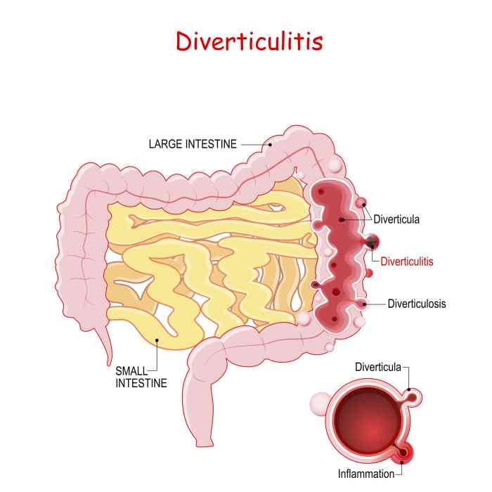
    Diverticulitis is a condition characterized by inflammation and infection of small pouches (diverticula) that can form in the lining of the colon. These pouches, typically located in the sigmoid colon, are essentially outpouchings of the colon’s wall. Understanding diverticulitis involves recognizing its prevalence, risk factors, the anatomy of the colon, and the differences between diverticulosis and diverticulitis.The prevalence of diverticulitis varies geographically and increases with age.

    Factors such as a low-fiber diet, smoking, and a sedentary lifestyle are associated with a higher risk of developing the condition. Understanding the structure of the digestive system, specifically the colon, is crucial to comprehend diverticulitis.

    Diverticulitis can be a real pain, with symptoms like abdominal pain, fever, and changes in bowel habits. Understanding the complications is crucial, but did you know that conditions like a missing pancreas can also affect your digestive system? Learning about how to manage diverticulitis signs, symptoms, and complications is important for your overall health, and it’s worth exploring how different digestive issues relate to each other.

    For example, can you live without a pancreas and other similar questions can highlight potential challenges in digestive health, which is essential when managing diverticulitis. So, understanding the bigger picture is key to managing these issues effectively.

    Anatomy of the Colon and its Role in Digestion

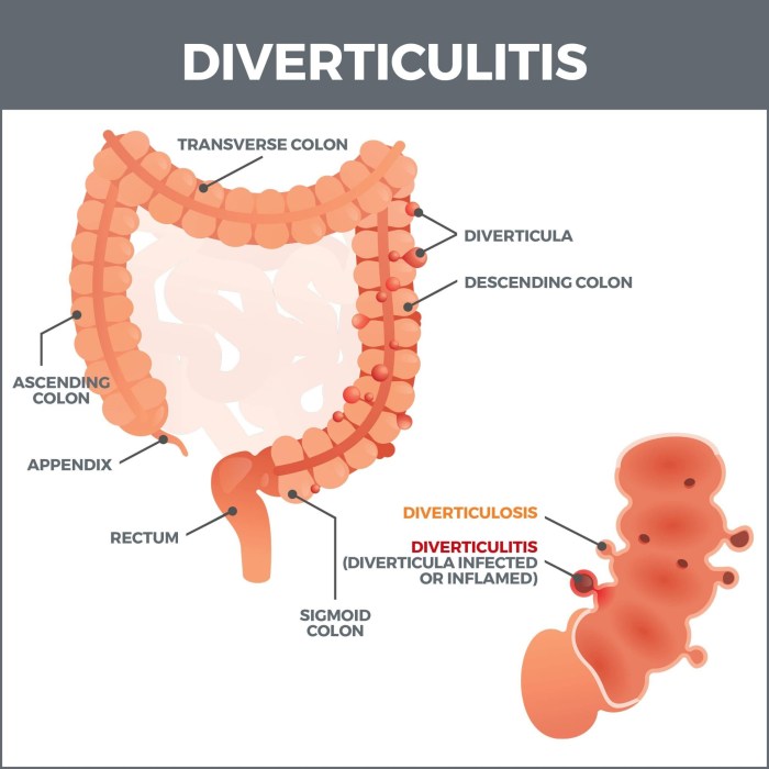
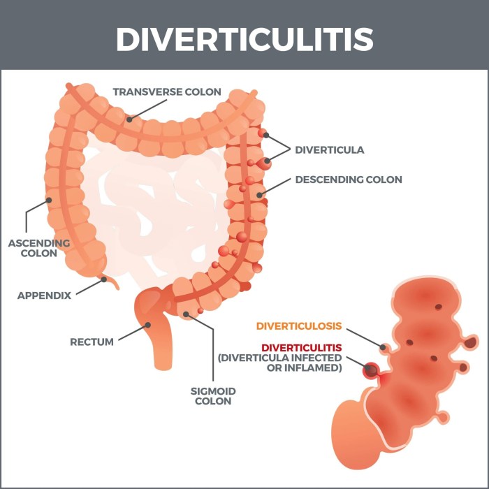
    The colon, a vital part of the large intestine, is responsible for absorbing water and electrolytes from the digested food, forming and storing feces. It’s a muscular tube approximately 5 feet long, extending from the small intestine to the rectum. The colon is further subdivided into sections: the ascending colon, transverse colon, descending colon, and sigmoid colon. The sigmoid colon, located at the lower left quadrant of the abdomen, is where diverticula are most frequently found.

    Infographic: Location of Diverticula

    Imagine a simplified drawing of the large intestine, resembling a curved tube. The sigmoid colon, the S-shaped portion at the lower end of the large intestine, is highlighted. Small, balloon-like protrusions, the diverticula, are depicted emerging from the inner wall of the sigmoid colon. The drawing clearly shows the location of these pouches within the colon’s lining.

    This graphic emphasizes the specific area of the colon where diverticula tend to form.

    Diverticulosis vs. Diverticulitis

    This table compares the key differences between diverticulosis and diverticulitis, highlighting the presence or absence of inflammation and infection.

    Characteristic Diverticulosis Diverticulitis
    Definition Presence of diverticula in the colon’s wall, without inflammation or infection. Inflammation and infection of one or more diverticula.
    Symptoms Typically asymptomatic; may cause mild discomfort or bloating. Often presents with abdominal pain, fever, nausea, and changes in bowel habits.
    Complications Rare complications include bleeding. Potential complications include abscesses, fistulas, bowel obstruction, and perforation.
    Treatment Often managed with dietary adjustments. Usually requires antibiotics and, in some cases, surgery.

    Signs and Symptoms of Diverticulitis

    Diverticulitis, an inflammation of the diverticula (small pouches that can form in the digestive tract), often presents with a range of symptoms. Understanding these signs is crucial for prompt diagnosis and treatment. Recognizing the patterns of acute and chronic diverticulitis, as well as the variations based on location, can significantly aid in proper medical care.Identifying the specific symptoms and their severity can help distinguish between simple digestive issues and a more serious condition.

    This understanding allows for quicker intervention, preventing potential complications. The symptoms can be influenced by various factors, including the location of the infection and the presence of any associated complications.

    Common Signs and Symptoms of Acute Diverticulitis

    Acute diverticulitis is characterized by a sudden onset of symptoms, often requiring immediate medical attention. Common symptoms include lower abdominal pain, typically located in the lower left quadrant, but it can also occur in other areas. This pain is often described as cramping, throbbing, or a constant ache.

    Comparison of Acute and Chronic Diverticulitis Symptoms

    Acute diverticulitis, as mentioned, presents with sudden, intense symptoms, whereas chronic diverticulitis symptoms tend to be less severe and more intermittent. The pain in chronic diverticulitis might be milder and occur less frequently. Chronic diverticulitis often includes a history of recurrent, milder symptoms.

    Symptoms Based on Infection Location

    The location of the infected diverticulum can influence the specific symptoms experienced. For instance, if the infection is in the sigmoid colon (the lower part of the large intestine), pain is often concentrated in the lower left abdomen. In contrast, diverticulitis in other parts of the large intestine might manifest with pain in a different quadrant of the abdomen.

    Symptoms Associated with Diverticulitis Complications

    Complications of diverticulitis, such as abscess formation or fistula development, can manifest with distinct symptoms. For example, a palpable mass in the abdomen might indicate an abscess. Fever, chills, and nausea are also common signs associated with complications.

    Table of Diverticulitis Symptoms

    Symptom Severity Potential Causes
    Lower abdominal pain (LLQ most common) Moderate to severe, often cramping or throbbing Inflammation of diverticula, infection, obstruction
    Fever Mild to high Infection, complications (e.g., abscess)
    Nausea and vomiting Mild to moderate Inflammation, infection, complications
    Chills Mild to moderate Infection, complications
    Change in bowel habits (e.g., diarrhea or constipation) Mild to moderate Inflammation, obstruction
    Loss of appetite Mild to moderate Inflammation, infection, pain
    Tenderness to the touch in the abdomen Moderate to severe Inflammation, infection, complications

    Complications of Diverticulitis

    Diverticulitis, while often manageable, can lead to serious complications if left untreated or if the condition becomes severe. Understanding these potential complications is crucial for individuals at risk and healthcare providers to ensure timely intervention and prevent further health problems. Early recognition and appropriate management are key to minimizing the impact of these complications.

    Potential Complications

    Diverticulitis complications range from mild to severe, requiring varying degrees of medical attention. Recognizing these potential complications is essential for effective management and minimizing the risk of long-term health consequences. The severity and likelihood of complications depend on several factors, including the extent of the infection, individual health status, and promptness of treatment.

    • Abscess Formation: A localized collection of pus can develop at the site of the diverticulitis. This can be a result of the infection spreading and becoming walled off. Abscesses can cause significant pain, fever, and tenderness in the abdomen. The likelihood of abscess formation is moderate to high, especially in individuals with poorly controlled inflammation or delayed treatment.

      Management typically involves drainage of the abscess, either surgically or through percutaneous drainage, guided by imaging studies. Prompt diagnosis and treatment are vital to prevent the spread of infection and complications like sepsis.

    • Fistula Formation: A fistula is an abnormal connection between two organs or between an organ and the skin. In diverticulitis, a fistula can develop between the colon and another organ, such as the bladder or vagina, or even the skin. Fistulas can cause significant drainage and discomfort, along with potential infection. The likelihood of fistula formation is moderate, often associated with recurrent or severe diverticulitis episodes.

      Management can range from conservative measures like antibiotics and dietary changes to surgical intervention, depending on the location and severity of the fistula. Preventing recurrence involves managing underlying diverticulitis effectively and addressing any contributing factors.

    • Sepsis: A life-threatening systemic inflammatory response to an infection. In diverticulitis, severe infection can lead to sepsis, characterized by high fever, rapid heart rate, low blood pressure, and altered mental status. The likelihood of sepsis is high, particularly with severe diverticulitis or delayed treatment. Sepsis requires immediate medical attention, including aggressive fluid resuscitation, antibiotics, and intensive care support.

      Prompt diagnosis and intervention are crucial to save lives.

    • Bowel Perforation: A rupture in the colon wall can occur due to severe inflammation or pressure. This can lead to the leakage of intestinal contents into the abdominal cavity, causing peritonitis, a serious inflammation of the lining of the abdomen. The likelihood of perforation is moderate to high in cases of untreated or severe diverticulitis. Management typically involves emergency surgery to repair the perforation and control infection.

      Preventing perforation is paramount, emphasizing timely intervention and aggressive management of the diverticulitis.

    • Obstruction: Inflammation can cause narrowing or blockage of the colon, hindering the passage of stool. This can lead to severe pain, nausea, vomiting, and abdominal distension. The likelihood of obstruction is moderate, often occurring with recurrent diverticulitis. Management can include intravenous fluids, bowel rest, and potentially surgical intervention to restore bowel function. Proactive management of diverticulitis can reduce the risk of obstruction.

    Risk Factors for Complications

    Several factors increase the risk of complications from diverticulitis. These include the severity of the initial infection, pre-existing medical conditions, and delayed or inadequate treatment. Factors like advanced age, a compromised immune system, and inadequate fluid intake also contribute to a higher risk.

    Management Strategies, Diverticulitis signs symptoms and complications

    The management of diverticulitis complications varies depending on the specific complication and its severity. This often involves a combination of medical interventions, including intravenous fluids, antibiotics, pain management, and sometimes surgical procedures. Early diagnosis and prompt treatment are essential to minimize the risk of severe complications.

    Prevention Strategies

    Preventing diverticulitis complications hinges on proactive measures to manage the underlying condition. A high-fiber diet, adequate hydration, and maintaining a healthy weight can help prevent diverticulitis flare-ups. Regular check-ups with a healthcare provider can also help detect and manage diverticulitis early, reducing the risk of complications.

    Severity and Treatment Approach

    Severity Level Complication Treatment Approach
    Mild Abscess (small), mild fistula Antibiotics, drainage (percutaneous or surgical), close monitoring
    Moderate Moderate fistula, bowel obstruction, recurrent diverticulitis Antibiotics, bowel rest, pain management, possible surgical intervention
    Severe Sepsis, bowel perforation, large abscess Emergency surgery, intensive care, aggressive antibiotic therapy, fluid resuscitation

    Diagnosis of Diverticulitis

    Diverticulitis signs symptoms and complications

    Pinpointing diverticulitis relies on a combination of patient history, physical examination, and diagnostic imaging. A thorough understanding of the potential symptoms and their presentation is crucial for accurate diagnosis and timely treatment. The goal is to distinguish diverticulitis from other conditions that can mimic its symptoms, ensuring the right course of action for the patient.

    Diagnostic Methods

    A variety of methods contribute to the diagnosis of diverticulitis. These methods aim to confirm the presence of inflammation in the diverticula, assess the extent of the inflammation, and rule out other possible causes for the patient’s symptoms. Careful consideration of all available information leads to an accurate diagnosis.

    Role of Imaging Techniques

    Imaging plays a vital role in confirming the diagnosis of diverticulitis. Advanced imaging techniques, such as CT scans, allow healthcare professionals to visualize the affected area and identify any complications. These images offer detailed information about the presence, extent, and location of inflammation, assisting in the development of an appropriate treatment plan.

    Summary of Diagnostic Tests and Procedures

    Several diagnostic tests and procedures are used in the diagnosis of diverticulitis. These include:

    • History and Physical Examination: A detailed medical history, including a description of the patient’s symptoms, and a physical examination, including assessment of vital signs and abdominal tenderness, provide valuable initial information. This information helps to identify potential risk factors and symptoms consistent with diverticulitis.
    • Blood Tests: Complete blood count (CBC) and inflammatory markers (e.g., C-reactive protein, erythrocyte sedimentation rate) can help assess the presence of infection and inflammation. Elevated levels of these markers may indicate an active inflammatory process.
    • CT Scan: A computed tomography (CT) scan is often the gold standard imaging modality for diagnosing diverticulitis. It provides detailed cross-sectional images of the abdomen, enabling visualization of the colon and surrounding structures. Contrast material is frequently used to enhance the visualization of the affected area, highlighting any inflammation or complications such as abscesses or fistulas.
    • Ultrasound: Ultrasound is a less commonly used imaging technique for diagnosing diverticulitis. While it is useful in some cases, it often does not provide the same level of detail as CT scans in assessing the extent of inflammation or identifying complications.

    Typical Clinical Presentation of Suspected Diverticulitis

    A patient with suspected diverticulitis often presents with a combination of symptoms. These symptoms may include:

    • Abdominal pain: Often localized to the lower left quadrant of the abdomen, the pain may be sharp, cramping, or a dull ache.
    • Fever: A rise in body temperature is a common sign of infection, which is often present in cases of diverticulitis.
    • Nausea and vomiting: These symptoms may accompany the abdominal pain and fever, indicating the body’s response to the inflammation.
    • Changes in bowel habits: Patients may experience altered bowel movements, including constipation, diarrhea, or a change in stool consistency.

    Comparison of Diagnostic Imaging Techniques

    The following table compares and contrasts different diagnostic imaging techniques for diverticulitis:

    Imaging Technique Advantages Disadvantages
    CT Scan High resolution, detailed images of the colon and surrounding structures; can identify complications like abscesses and fistulas; widely available. Exposure to ionizing radiation; contrast material may cause allergic reactions in some patients; more expensive than ultrasound.
    Ultrasound Non-invasive; does not involve ionizing radiation; relatively inexpensive. Lower resolution images; may not adequately visualize all structures; less sensitive for identifying complications compared to CT.

    Treatment Options for Diverticulitis

    Diverticulitis, an inflammation of the pouches in the colon, requires careful management. Effective treatment hinges on the severity of the infection and the individual patient’s overall health. Early intervention and appropriate treatment choices are crucial for preventing complications and promoting recovery.

    Diverticulitis can be a real pain, with symptoms like abdominal pain, fever, and changes in bowel habits. It’s important to understand the signs and potential complications, like infections or bowel blockages. While we’re on the topic of digestive issues, have you ever wondered if a drink like whiskey might help with a cold? The question of whether or not alcohol is beneficial for colds is a fascinating one, and if you’d like to know more about the supposed benefits of alcohol for colds, you can check out this interesting article on does whiskey help a cold.

    Ultimately, seeking professional medical advice is crucial for managing diverticulitis symptoms and preventing further complications.

    Medications for Managing Diverticulitis

    Medications play a vital role in treating diverticulitis, particularly in cases of mild to moderate inflammation. Antibiotics are often prescribed to combat the bacterial infection. These medications aim to reduce inflammation and prevent further complications. Specific antibiotics and their dosages are tailored to the individual patient’s needs. For example, a common approach involves a course of oral antibiotics, like metronidazole or ciprofloxacin, to clear the infection.

    Pain relievers, such as ibuprofen or acetaminophen, can help manage discomfort. Dietary modifications, including a clear liquid diet, may also be recommended to allow the bowel to rest and heal.

    Surgical Options for Severe Cases

    Surgical intervention becomes necessary in cases of severe diverticulitis, complications like abscesses or fistulas, or recurrent episodes. The choice of surgical procedure depends on the specific situation and the extent of the damage to the colon. Surgical options can range from minimally invasive procedures, like laparoscopic surgery, to open surgeries. Laparoscopic surgery typically involves smaller incisions, leading to less post-operative pain and a quicker recovery.

    However, open surgery may be necessary for more extensive procedures or complex cases.

    Recovery Process and Expected Outcomes

    The recovery process following treatment for diverticulitis varies based on the severity of the condition and the chosen treatment approach. Patients undergoing medical management typically experience a return to normal activities within a few days to a couple of weeks. Surgical intervention often necessitates a longer recovery period, with patients typically needing several weeks or even months to fully recover.

    In general, complete recovery is possible for most individuals with appropriate medical care.

    Comparison of Treatment Options

    The effectiveness and side effects of different treatment approaches for diverticulitis vary. Medications are generally effective for mild cases, minimizing the need for surgery. However, in severe cases, surgical intervention might be the only option to resolve the issue. The choice of treatment depends on the individual patient’s condition and the judgment of the medical professional. Surgical options offer a definitive solution for severe cases but carry a higher risk of complications, such as infection or bleeding.

    Medication-based treatment has fewer risks but may not always be sufficient. For example, if a patient experiences a recurring infection or an abscess, surgery might be required to address the underlying cause and prevent future complications.

    Summary Table of Treatment Options

    Treatment Option Effectiveness Potential Side Effects Suitable for
    Medications (Antibiotics, Pain Relievers) Effective for mild-moderate cases Possible allergic reactions, gastrointestinal upset Patients with uncomplicated diverticulitis
    Laparoscopic Surgery Effective for severe cases, complications Infection, bleeding, pain, longer recovery time Patients with abscesses, fistulas, or recurrent episodes
    Open Surgery Definitive solution for severe cases Increased risk of infection, bleeding, pain, longer recovery time Patients with extensive damage or complex cases

    Prevention and Management Strategies

    Diverticulitis, while often manageable, can be prevented or its impact lessened through proactive measures. Understanding the factors that contribute to its development and implementing strategies to mitigate these risks are key to improving overall health and well-being. This section delves into preventive measures, dietary recommendations, lifestyle adjustments, and the critical role of follow-up care in managing diverticulitis.A proactive approach is essential for preventing diverticulitis or reducing its severity.

    By understanding risk factors and implementing lifestyle changes, individuals can significantly decrease their likelihood of experiencing this condition. This includes not only dietary adjustments but also a focus on overall well-being.

    Preventive Measures to Reduce Diverticulitis Risk

    Maintaining a healthy weight, exercising regularly, and avoiding smoking are crucial steps in reducing the risk of diverticulitis. These lifestyle choices positively impact gut health and overall systemic well-being, making them integral components of a preventative strategy.

    Speaking of digestive issues, diverticulitis can manifest with pain, fever, and changes in bowel habits. While the recent tuberculosis outbreak in Kansas tuberculosis outbreak in kansas highlights the importance of overall health, it’s crucial to remember that diverticulitis, a common condition, requires prompt medical attention. Severe cases can lead to complications like abscesses or fistulas, so recognizing the signs is key.

    Dietary Recommendations for Managing Diverticulitis

    A high-fiber diet is vital for preventing and managing diverticulitis. It promotes healthy bowel movements and reduces strain on the digestive system. A diet rich in fiber can help prevent the formation of diverticula, which are the small pouches that can become inflamed in diverticulitis.

    • Increase Fiber Intake Gradually: Sudden increases in fiber intake can lead to gas and bloating. It’s best to gradually increase fiber-rich foods over time to allow the digestive system to adjust. This approach minimizes discomfort and allows the body to better utilize the increased fiber.
    • Prioritize Whole Grains: Opt for whole-grain breads, cereals, and pasta over refined options. Whole grains provide more fiber and nutrients, promoting digestive health.
    • Embrace Fruits and Vegetables: Include a variety of colorful fruits and vegetables in your diet. Fruits and vegetables are excellent sources of fiber and vitamins, essential for overall health.
    • Choose Legumes Wisely: Legumes, such as beans, lentils, and peas, are excellent sources of fiber. However, they can cause gas in some individuals. Consider introducing them gradually into your diet.
    • Limit Foods High in Refined Carbohydrates: Foods like white bread, white rice, and sugary drinks should be consumed in moderation. These foods lack the fiber needed for optimal digestive function.

    Lifestyle Modifications for Diverticulitis Prevention

    Maintaining a healthy weight through balanced nutrition and regular exercise can greatly reduce the risk of developing diverticulitis. Exercise promotes overall health, strengthens the body, and supports healthy digestion.

    • Regular Exercise: Aim for at least 30 minutes of moderate-intensity exercise most days of the week. Physical activity strengthens the body, improves digestion, and aids in maintaining a healthy weight.
    • Manage Stress: Stress can negatively impact digestive health. Employ stress-reduction techniques such as meditation, yoga, or deep breathing exercises.
    • Maintain a Healthy Weight: Obesity increases the risk of diverticulitis. Maintaining a healthy weight through a balanced diet and exercise is crucial for preventing this condition.

    Follow-up Care and Monitoring

    Regular checkups are essential for monitoring diverticulitis and preventing complications. This includes addressing any symptoms promptly and adhering to prescribed treatment plans.

    • Regular Checkups: Schedule regular checkups with your doctor, especially if you have a history of diverticulitis or risk factors. These appointments allow for early detection of any potential issues and enable proactive management.
    • Adherence to Treatment Plans: Strictly follow any prescribed medications or dietary recommendations to effectively manage diverticulitis and minimize the risk of complications.
    • Prompt Symptom Reporting: Report any new or worsening symptoms to your doctor immediately. Early intervention is crucial for preventing complications and ensuring optimal health.

    Dietary Recommendations for Individuals with Diverticulitis

    Following a specific diet can help manage symptoms and prevent future episodes.

    • Soft Foods: During an acute flare-up, a soft diet that’s easy to digest can ease discomfort. This often includes cooked fruits and vegetables, well-cooked grains, and dairy products.
    • Avoid Irritating Foods: Foods like popcorn, nuts, seeds, and certain vegetables (e.g., broccoli, cabbage) can be irritating and exacerbate diverticulitis symptoms. These should be avoided or consumed in moderation during flare-ups.
    • Adequate Hydration: Staying well-hydrated is crucial for digestive health. Drink plenty of water throughout the day to help prevent constipation and maintain overall bodily functions.
    • Limit High-Fiber Foods During Flare-Ups: While a high-fiber diet is generally beneficial, reducing fiber intake temporarily during an acute flare-up may be necessary to minimize discomfort and allow the digestive system to heal.

    Patient Education and Support

    Navigating a diagnosis of diverticulitis can be overwhelming. Understanding the condition, its treatment, and the importance of self-care is crucial for successful management. This section provides essential information to empower patients and facilitate a positive recovery journey.Effective management of diverticulitis relies heavily on patient understanding and active participation. By equipping patients with knowledge and support, healthcare providers can foster adherence to treatment plans and promote long-term well-being.

    Importance of Patient Education

    Patient education plays a vital role in managing diverticulitis effectively. A thorough understanding of the condition, including its causes, symptoms, and potential complications, empowers patients to actively participate in their care. This knowledge allows patients to recognize early warning signs, communicate effectively with their healthcare team, and make informed decisions about their treatment and lifestyle choices. Proper education also helps reduce anxiety and promotes a sense of control over the condition.

    Resources for Patients

    Accessing reliable information and support is essential for patients with diverticulitis. Numerous resources can provide valuable insights and guidance. Local support groups offer a platform for connecting with others who share similar experiences, providing encouragement, and exchanging practical tips. Many hospitals and healthcare organizations have patient education websites or materials available online. These resources often contain detailed information about the condition, treatment options, and lifestyle modifications.

    Patient Guide to Diverticulitis

    This guide aims to provide a comprehensive understanding of diverticulitis, encompassing various aspects of the condition and its management. Understanding the cause, symptoms, and complications empowers patients to actively participate in their care. Detailed information on treatment options, including dietary changes, medication, and surgical interventions, helps patients make informed decisions. Self-care strategies and pain management techniques are also included to facilitate a smoother recovery process.

    Importance of Self-Care

    Self-care is crucial in managing diverticulitis. Maintaining a healthy lifestyle, including a balanced diet, regular exercise, and stress management techniques, can significantly impact the overall well-being of the patient. These strategies contribute to reducing inflammation, improving digestion, and promoting a faster recovery. Furthermore, a patient-centered approach that emphasizes self-care can empower individuals to take control of their health and well-being.

    Strategies for Managing Pain and Discomfort

    Diverticulitis can cause significant pain and discomfort. Managing these symptoms effectively is crucial for a comfortable recovery. Over-the-counter pain relievers, such as ibuprofen or acetaminophen, can help alleviate mild to moderate pain. Applying heat or cold packs to the affected area may also provide relief. Adequate hydration and a balanced diet can contribute to managing pain and discomfort.

    It’s essential to communicate any persistent or worsening pain to the healthcare provider.

    Reliable Online Resources

    Numerous reliable online resources provide valuable information for patients with diverticulitis. Reputable medical organizations, such as the National Institute of Diabetes and Digestive and Kidney Diseases (NIDDK), offer detailed information and support. Furthermore, patient advocacy groups and dedicated websites offer insights into various aspects of the condition. However, it is crucial to verify the credibility of online resources and consult with a healthcare professional for personalized advice.

    • National Institute of Diabetes and Digestive and Kidney Diseases (NIDDK): The NIDDK is a trusted source of information about various digestive conditions, including diverticulitis. Their website provides detailed information about symptoms, diagnosis, treatment, and management strategies.
    • Patient Advocacy Groups: Numerous patient advocacy groups focus on specific health conditions, including diverticulitis. These groups often provide support, share experiences, and offer practical tips for managing the condition.
    • Reputable Medical Journals: Medical journals offer research-based information about diverticulitis and related topics. However, it is crucial to critically evaluate the information and consult with a healthcare professional for personalized advice.

    Wrap-Up: Diverticulitis Signs Symptoms And Complications

    In summary, diverticulitis signs, symptoms, and complications require careful attention and a proactive approach. Early diagnosis and appropriate treatment are vital in mitigating potential complications and ensuring a smoother recovery. Understanding the various symptoms, treatment options, and preventive measures is crucial for individuals seeking to manage and prevent this digestive condition effectively. This guide aims to provide a clear roadmap for navigating the complexities of diverticulitis, empowering individuals with the knowledge they need to make informed decisions about their health.

  • Heart Problems with Strokes A Deep Dive

    Heart Problems with Strokes A Deep Dive

    Heart problems that occur with strokes are a serious concern, impacting the cardiovascular system and the brain. Strokes, whether ischemic or hemorrhagic, can have a significant impact on the heart, leading to a complex interplay of symptoms and treatment needs. Understanding the connection between these two critical health issues is crucial for effective prevention and management. This discussion delves into the various types of strokes, associated heart conditions, symptoms, treatment approaches, and preventive strategies.

    We’ll explore the interconnectedness of the heart and brain and examine how heart conditions can significantly increase the risk of stroke.

    The complex relationship between heart health and stroke risk is further explored. The detailed tables provided showcase the different types of strokes, their impact on the heart, and common heart conditions associated with stroke risk. This allows for a better understanding of the diagnostic procedures, symptom overlaps, and treatment options for patients facing both heart conditions and stroke.

    The intricate interplay of these health concerns is discussed, emphasizing the critical need for early detection and a multidisciplinary approach to treatment.

    Introduction to Stroke-Related Heart Problems

    Heart problems that occur with strokes

    A stroke occurs when blood flow to a part of the brain is interrupted or reduced, depriving brain cells of oxygen and nutrients. This interruption can lead to lasting physical, cognitive, and emotional impairments depending on the affected area and severity. Different types of strokes, each with unique causes and effects, highlight the delicate balance of the body’s systems.Understanding the interplay between the cardiovascular system and the brain is crucial.

    The heart pumps blood throughout the body, including the brain, ensuring it receives the oxygen and nutrients necessary for proper function. Any disruption in this vital process can have profound consequences. A stroke can not only damage the brain directly, but it can also trigger or exacerbate underlying heart conditions, further complicating the recovery process. Knowing the common risk factors for both stroke and heart problems allows for proactive measures to reduce the likelihood of these events.

    Types of Strokes and Their Impact on the Heart

    Strokes are broadly categorized into ischemic and hemorrhagic types. Ischemic strokes are the most common, resulting from a blockage in an artery supplying blood to the brain. Hemorrhagic strokes, on the other hand, occur when a blood vessel in the brain bursts, leading to bleeding within the brain tissue.

    Stroke Type Impact on Heart Possible Symptoms Severity
    Ischemic Increased risk of arrhythmias (irregular heartbeats), heart attack, and subsequent heart failure. The stress on the heart from the stroke can trigger or worsen existing heart conditions. Sudden numbness or weakness of the face, arm, or leg, especially on one side of the body; confusion; trouble speaking or understanding speech; trouble seeing in one or both eyes; trouble walking, dizziness, loss of balance or coordination; severe headache with no known cause. Can range from mild to severe, depending on the size and location of the blocked artery. Some individuals experience temporary symptoms (transient ischemic attack – TIA) which resolve without lasting damage.
    Hemorrhagic Increased risk of heart failure, and potentially fatal complications from the sudden increase in intracranial pressure. The bleeding can disrupt the delicate balance of the brain’s blood supply, leading to heart problems. Sudden, severe headache (often described as the worst headache of one’s life); vomiting; stiff neck; seizures; loss of consciousness; decreased level of alertness; neurological deficits depending on the affected area of the brain. Generally considered more severe than ischemic strokes due to the sudden and potentially massive bleeding. The severity depends on the location and extent of the bleeding.

    Common Risk Factors for Stroke and Heart Problems

    Several factors increase the likelihood of both stroke and heart problems. These include high blood pressure, high cholesterol, smoking, diabetes, obesity, lack of physical activity, and a family history of these conditions. Maintaining a healthy lifestyle, including a balanced diet, regular exercise, and avoiding tobacco use, can significantly reduce the risk. Early detection and management of underlying conditions like high blood pressure and diabetes are also vital for preventing both stroke and heart complications.

    Specific Heart Conditions Associated with Strokes

    Heart conditions significantly contribute to the risk of stroke. Understanding the interplay between cardiovascular health and cerebrovascular events is crucial for prevention and effective treatment. This section will delve into common heart conditions linked to stroke, examining their mechanisms, diagnostic processes, and overlapping symptoms. Early detection and management of these conditions are vital in reducing stroke incidence.

    Common Heart Conditions Increasing Stroke Risk

    Several heart conditions are strongly associated with an elevated risk of stroke. These conditions disrupt normal blood flow, either by creating clots or by irregular heartbeats that lead to blood pooling. Understanding the specific mechanisms through which these conditions increase stroke risk is essential for preventative strategies.

    Atrial Fibrillation

    Atrial fibrillation (AFib) is a common heart rhythm disorder characterized by rapid, irregular heartbeats. This erratic rhythm can cause blood to pool in the atria, increasing the risk of clot formation. These clots can detach and travel to the brain, causing a stroke. The pooling of blood in the atria is a key mechanism in the development of stroke from AFib.

    High Blood Pressure

    High blood pressure (hypertension) consistently damages the delicate blood vessels in the brain. Over time, this damage weakens the vessels, increasing their susceptibility to rupture or blockage. This can lead to a stroke, either through bleeding in the brain (hemorrhagic stroke) or through a blockage (ischemic stroke). Chronic hypertension relentlessly strains the cardiovascular system, making it more vulnerable to strokes.

    Valve Disease

    Heart valve disease can disrupt blood flow through the heart, sometimes causing turbulent flow. This can lead to the formation of blood clots. These clots can then travel to the brain, causing a stroke. Valve disease, depending on the specific condition and severity, can significantly contribute to the likelihood of stroke.

    Coronary Artery Disease

    Coronary artery disease (CAD) narrows the arteries that supply blood to the heart. While primarily associated with heart attacks, CAD can also increase the risk of stroke. The reduced blood flow can lead to the formation of clots that can travel to the brain. CAD is a serious condition that impacts the entire cardiovascular system, increasing vulnerability to both heart attacks and strokes.

    Diagnostic Procedures for Heart Conditions Linked to Stroke Risk, Heart problems that occur with strokes

    Several diagnostic tools are used to identify these heart conditions. Electrocardiograms (ECGs) monitor the electrical activity of the heart, identifying irregular rhythms like atrial fibrillation. Echocardiograms use ultrasound to visualize the heart’s structure and function, revealing valve abnormalities or other structural issues. Holter monitors continuously record heart rhythms over a period, providing a detailed picture of heart activity.

    Heart problems often accompany strokes, impacting everything from blood pressure to heart rhythm. Early detection is key, just like diagnosing colorectal cancer early matters , because prompt treatment can significantly improve outcomes. Understanding these intertwined issues is crucial for maintaining overall cardiovascular health.

    Comparing Symptoms of Heart Conditions and Stroke

    Symptoms of heart conditions and stroke can sometimes overlap, making accurate diagnosis crucial. Chest pain, shortness of breath, and palpitations are common symptoms of heart conditions. Stroke symptoms, on the other hand, can include sudden numbness or weakness of the face, arm, or leg, especially on one side of the body; sudden confusion, trouble speaking or understanding speech; sudden trouble seeing in one or both eyes; sudden trouble walking, dizziness, loss of balance, or coordination; sudden severe headache with no known cause.

    Recognizing these differences can aid in prompt medical intervention.

    Table: Heart Conditions and Stroke Risk

    Heart Condition Mechanism of Stroke Risk Increase Diagnostic Procedures Symptoms Overlap
    Atrial Fibrillation Irregular heart rhythm causes blood pooling in the atria, increasing clot formation. Detached clots travel to the brain. Electrocardiogram (ECG), Holter monitor, echocardiogram Palpitations, shortness of breath can overlap with stroke symptoms like weakness or confusion.
    High Blood Pressure Damages blood vessels in the brain, making them more susceptible to rupture or blockage, leading to hemorrhagic or ischemic strokes. Blood pressure measurements, urinalysis Headaches, dizziness can be present in both high blood pressure and stroke.
    Valve Disease Disrupts blood flow, causing turbulent flow and potential clot formation. Clots can travel to the brain. Echocardiogram, cardiac catheterization Shortness of breath, chest pain can be present in both valve disease and stroke.
    Coronary Artery Disease Reduced blood flow to the heart can lead to clot formation, which can travel to the brain and cause a stroke. ECG, cardiac catheterization, stress test Chest pain, shortness of breath can be present in both CAD and stroke.

    Symptoms and Signs of Heart Problems During Strokes: Heart Problems That Occur With Strokes

    Recognizing the symptoms of heart problems during a stroke is crucial for timely intervention. While stroke symptoms are often well-known, the overlapping nature of heart conditions can make it challenging to differentiate between the two. Understanding the specific indicators can significantly improve outcomes.The human body’s intricate systems often exhibit overlapping symptoms, making diagnosis challenging. This is especially true when heart issues accompany or mimic stroke symptoms.

    A careful assessment by a medical professional is essential to accurately identify the underlying cause and initiate appropriate treatment.

    Overlapping Symptoms

    Many symptoms associated with heart conditions and strokes can be remarkably similar. For instance, chest pain can arise from both heart attacks and strokes. This similarity can lead to misdiagnosis, delaying vital treatment. It’s essential to be aware of the nuances and potential indicators that might point towards a heart problem.

    Heart problems often arise after a stroke, impacting blood flow and overall cardiovascular health. Interestingly, a recent study on brown rice arsenic levels highlights the importance of a balanced diet in maintaining good health, which is crucial for preventing and managing such heart conditions. Ultimately, understanding these links is vital for proactive health management.

    Specific Symptoms Suggesting a Heart Problem During a Stroke

    Certain symptoms can strongly suggest a heart-related component to a stroke event. Sudden, severe chest pain, often radiating to the left arm or jaw, is a prime example. This is frequently associated with a heart attack, a condition that can occur during or in conjunction with a stroke. Shortness of breath, accompanied by rapid or irregular heartbeat, can also be indicative of a heart problem.

    In some cases, dizziness, nausea, and vomiting may accompany these symptoms.

    Importance of Early Detection and Intervention

    Early detection and intervention are critical in managing both heart problems and strokes. Prompt medical attention can significantly reduce the severity of the damage and improve long-term outcomes. If a patient experiences any of the symptoms listed below, seeking immediate medical help is paramount. This rapid response can be the difference between a full recovery and lasting complications.

    Table of Potential Heart-Related Symptoms During Strokes

    Symptom Description Possible Cause (Heart/Stroke) Severity
    Chest Pain Sudden, sharp, or squeezing pain in the chest, often radiating to the left arm, jaw, or back. May be accompanied by shortness of breath. Heart attack (often during or related to stroke) High – requires immediate medical attention
    Shortness of Breath Difficulty breathing, rapid or labored breathing, feeling winded or gasping for air. Heart failure, cardiac issues during/related to stroke. Could also be related to stroke’s impact on lung function. High – requires immediate medical attention
    Rapid or Irregular Heartbeat (Palpitations) A noticeable change in heart rate, feeling like the heart is racing, fluttering, or skipping beats. Arrhythmias, potential heart attack, or cardiac involvement during stroke Variable – may require monitoring or treatment depending on the cause and severity
    Dizziness or Lightheadedness Feeling unsteady, lightheaded, or like you might faint. Low blood pressure related to cardiac issues or the stroke Variable – depends on the severity and cause.
    Nausea or Vomiting Feeling sick to your stomach or vomiting. Potential heart involvement or side effect of a stroke or treatment. Variable – can be mild to severe

    Treatment and Management of Heart Issues During Strokes

    Treating stroke patients with concurrent heart problems requires a multifaceted approach tailored to the specific heart condition. Effective management hinges on stabilizing both the stroke and the heart issue, preventing further complications, and optimizing long-term recovery. This necessitates a collaborative effort among neurologists, cardiologists, and other specialists, focusing on prompt diagnosis, appropriate interventions, and comprehensive patient care.

    Different Treatment Approaches for Patients with Stroke and Heart Problems

    The treatment strategy for a stroke patient with a concomitant heart condition depends critically on the specific type of heart problem. For example, a patient with atrial fibrillation experiencing a stroke will require different interventions than a patient with coronary artery disease. This necessitates a careful assessment of the severity and stability of both conditions to develop a personalized treatment plan.

    Addressing both simultaneously is crucial to prevent further damage and promote recovery.

    Treatment Strategies Adjusted Based on Specific Heart Conditions

    Treatment strategies are adapted to the specific heart condition. For atrial fibrillation, anticoagulation therapy is frequently employed to prevent further clot formation and stroke recurrence. In cases of coronary artery disease, interventions like angioplasty or stenting might be necessary to restore blood flow to the heart muscle. These procedures, when performed in a timely manner, can significantly improve the patient’s chances of recovery.

    Importance of a Multidisciplinary Approach to Care

    A multidisciplinary approach is essential. Neurologists manage the stroke, cardiologists handle the heart issues, and other specialists, such as rehabilitation therapists and nutritionists, play crucial roles in comprehensive patient care. The coordinated effort of these specialists ensures a holistic approach, addressing all aspects of the patient’s condition and promoting optimal recovery.

    Procedures Involved in Treating Stroke Patients with Concurrent Heart Conditions

    Procedures for stroke patients with concurrent heart conditions vary greatly. In cases of atrial fibrillation, cardioversion, a procedure to restore a normal heart rhythm, may be considered. For coronary artery disease, angioplasty or stenting, procedures to open blocked arteries, may be performed. The specific procedure will depend on the severity of the blockage and the overall condition of the patient.

    These procedures, if feasible, should be performed in a timely manner to minimize long-term complications.

    Detailed Description of Treatment Procedures (Illustrative)

    • Atrial Fibrillation: Cardioversion is a procedure that uses electrical impulses to restore a normal heart rhythm. It is often used in patients with atrial fibrillation experiencing a stroke to prevent future clot formation and stroke recurrence. The success rate depends on the cause of atrial fibrillation and the patient’s overall health.
    • Coronary Artery Disease: Angioplasty involves inserting a catheter with a balloon at its tip into the blocked artery. The balloon is inflated to open the artery, restoring blood flow. Stenting is a similar procedure, but a small mesh tube (stent) is inserted to keep the artery open. Both procedures can significantly improve blood flow to the heart muscle, improving the patient’s condition.

      Strokes can unfortunately lead to a range of heart problems, impacting everything from blood pressure to heart rhythm. It’s crucial to understand these connections, as they can affect treatment strategies. For instance, if you’re trying to figure out why your blood tests show unusual thyroid hormone levels, like low or high TSH, understanding thyroid blood tests low or high TSH might reveal valuable insights.

      These thyroid issues can sometimes play a role in the heart problems that follow a stroke. Ultimately, a comprehensive approach to managing heart health after a stroke is key.

    Comparison of Treatment Options for Different Heart Conditions During Strokes

    Heart Condition Treatment Approach Potential Complications Success Rates
    Atrial Fibrillation Anticoagulation therapy, cardioversion, rhythm control Bleeding, thromboembolism, allergic reactions to medication High, depending on the underlying cause and patient factors
    Coronary Artery Disease Angioplasty, stenting, coronary artery bypass grafting (CABG), medications Bleeding, infection, stent thrombosis, heart attack, stroke High, but varies based on the severity of the disease and the patient’s condition

    Prevention Strategies for Stroke-Related Heart Problems

    Protecting your heart and brain from stroke-related complications requires a proactive approach. A combination of lifestyle modifications, medication, and regular checkups plays a crucial role in minimizing risk. Understanding these strategies empowers individuals to take control of their health and potentially prevent devastating consequences.

    Lifestyle Modifications for Stroke Prevention

    Adopting healthier habits significantly reduces the risk of both stroke and heart problems. A balanced diet rich in fruits, vegetables, and whole grains, coupled with regular exercise, can help manage weight, blood pressure, and cholesterol levels. These factors are all vital in mitigating the risk of cardiovascular disease.

    • Balanced Diet: A diet rich in fruits, vegetables, and whole grains, and low in saturated and trans fats, can help manage cholesterol and blood pressure. Limiting processed foods, sugary drinks, and excessive sodium intake is also crucial.
    • Regular Exercise: Aim for at least 150 minutes of moderate-intensity aerobic activity or 75 minutes of vigorous-intensity activity per week. Regular physical activity helps maintain a healthy weight, improve cardiovascular health, and lower blood pressure.
    • Maintaining a Healthy Weight: Obesity is a significant risk factor for stroke and heart disease. Striving for a healthy weight through diet and exercise is essential.
    • Stress Management: Chronic stress can elevate blood pressure and increase the risk of cardiovascular events. Practicing stress-reducing techniques like yoga, meditation, or deep breathing can be beneficial.

    Medication in Stroke Prevention

    Medications can play a critical role in preventing both stroke and heart-related complications. The specific medications and dosages are determined by individual risk factors and medical history.

    • Antihypertensive Medications: High blood pressure is a major contributor to both stroke and heart conditions. Medications like ACE inhibitors, beta-blockers, and diuretics can help control blood pressure, reducing the risk of these complications.
    • Cholesterol-Lowering Medications (Statins): High cholesterol levels increase the risk of plaque buildup in arteries, leading to heart attacks and strokes. Statins are commonly prescribed to lower cholesterol and improve cardiovascular health.
    • Antiplatelet or Anticoagulant Medications: These medications help prevent blood clots, which can contribute to stroke. Aspirin, for example, is often prescribed to prevent blood clots.

    Regular Checkups and Screenings for Prevention

    Proactive health monitoring is essential for early detection and management of risk factors. Regular checkups, including blood pressure, cholesterol, and glucose checks, allow for timely interventions to prevent potential problems.

    • Regular Doctor Visits: Routine checkups are essential for monitoring overall health, identifying risk factors, and receiving appropriate medical advice. Early detection of underlying conditions can lead to effective preventive measures.
    • Blood Pressure Monitoring: High blood pressure often has no noticeable symptoms. Regular monitoring allows for early detection and management, minimizing the risk of complications.
    • Cholesterol Screening: High cholesterol levels can contribute to plaque buildup, leading to heart disease and stroke. Regular cholesterol screenings allow for early detection and management.
    • Blood Glucose Monitoring (for Diabetes): Diabetes significantly increases the risk of stroke and heart problems. Regular monitoring of blood glucose levels is crucial for preventing complications.

    Preventive Flowchart for Stroke and Heart Conditions

    A preventative flowchart outlining strategies for reducing stroke and heart risks:

    Step Action
    1 Lifestyle Assessment: Evaluate current diet, exercise, stress levels, and weight
    2 Medical Evaluation: Consult a physician for blood pressure, cholesterol, and glucose checks
    3 Risk Factor Modification: Implement lifestyle changes based on assessment results (e.g., diet, exercise)
    4 Medication Evaluation: Discuss medication options (e.g., statins, antihypertensives) with the physician
    5 Regular Checkups: Schedule regular follow-up appointments for ongoing monitoring and adjustments

    Last Recap

    Heart problems that occur with strokes

    In conclusion, heart problems frequently accompany strokes, creating a complex health scenario. Recognizing the interplay between heart conditions and strokes is vital for effective prevention and management. Early detection, personalized treatment plans, and a proactive approach to lifestyle modifications are crucial for improving outcomes and reducing the impact of these intertwined conditions. Understanding the different types of strokes, associated heart conditions, symptoms, treatment options, and preventive measures is essential for better health outcomes.

  • Risks and Complications of Seizures A Deep Dive

    Risks and Complications of Seizures A Deep Dive

    Risks and complications of seizures can significantly impact a person’s life, ranging from immediate dangers to long-term consequences. This comprehensive exploration delves into the various types of seizures, their potential triggers, and the wide-ranging risks associated with them. Understanding these risks is crucial for both individuals experiencing seizures and those who care for them.

    From the immediate threat of injury during a seizure to the potential for long-term cognitive and emotional challenges, the consequences can be substantial. This article will discuss the different types of risks, ranging from the most immediate dangers to more chronic effects. It will also examine how these risks vary depending on the type of seizure.

    Introduction to Seizures

    Seizures are sudden, temporary disturbances in brain activity that can cause a variety of physical and behavioral changes. They result from abnormal electrical activity in the brain, disrupting normal brain function. Different types of seizures can manifest in various ways, impacting individuals differently. Understanding the underlying mechanisms and triggers is crucial for appropriate diagnosis and management.Seizures are characterized by abnormal, excessive electrical discharges in groups of nerve cells in the brain.

    These abnormal discharges disrupt normal brain communication, leading to the various symptoms associated with seizures. The underlying cause of these abnormal discharges can range from genetic predisposition to acquired brain injuries. Triggers can vary significantly and play a crucial role in the onset of seizures.

    Types of Seizures

    Different types of seizures are categorized based on where in the brain the abnormal electrical activity originates and the extent of its spread. This classification helps in diagnosis and treatment planning.

    Focal Seizures

    Focal seizures originate in a specific area of the brain. They can manifest with subtle symptoms, such as altered sensations or brief periods of confusion, or more pronounced symptoms, like involuntary movements of a limb or face. Some focal seizures may not involve noticeable outward signs, while others can be visually noticeable. Focal seizures can be further classified based on whether they remain localized or spread to other brain regions.

    Generalized Seizures

    Generalized seizures involve both hemispheres of the brain, exhibiting more widespread electrical activity. They can cause a range of symptoms, including loss of consciousness, convulsions, and altered awareness. Generalized seizures are typically more pronounced than focal seizures. Examples include tonic-clonic (grand mal) seizures, characterized by muscle stiffening and jerking movements, and absence seizures (petit mal), characterized by brief lapses in attention and awareness.

    Table Comparing Focal and Generalized Seizures

    Feature Focal Seizures Generalized Seizures
    Brain Region Affected Specific area Both hemispheres
    Symptoms Variable, may include sensory changes, motor movements, or altered awareness Often involve loss of consciousness, convulsions, or altered awareness
    Awareness May or may not involve loss of awareness Often involve loss of awareness
    Progression May remain localized or spread Typically involve widespread brain activity

    Triggers of Seizures

    Several factors can trigger seizures. These triggers can be environmental, medical, or psychological. Identifying and avoiding triggers is often crucial in seizure management.

    • Stress: High levels of stress can sometimes trigger seizures in susceptible individuals. Emotional distress can sometimes precipitate seizures.
    • Sleep Deprivation: Lack of adequate sleep can increase the risk of seizures. A consistent sleep schedule can help regulate brain activity and reduce seizure frequency.
    • Medication Changes: Changes in medications, including the introduction of new drugs or alterations in dosages, can affect seizure control.
    • Flickering Lights: Some individuals are sensitive to flashing lights, which can trigger seizures. Avoiding such visual stimuli can help prevent seizures.
    • Certain Foods: Specific foods or dietary deficiencies can occasionally trigger seizures. Maintaining a balanced diet can be helpful.

    Types of Risks and Complications: Risks And Complications Of Seizures

    Seizures, while often temporary, can carry a range of immediate and long-term risks and complications. Understanding these potential consequences is crucial for both patients and caregivers to proactively manage the condition and seek appropriate support. This section delves into the various dangers associated with seizures, emphasizing the importance of vigilance and appropriate medical intervention.

    Immediate Risks Associated with Seizures

    The immediate aftermath of a seizure can pose significant threats. These risks demand prompt attention and appropriate medical response. Unforeseen complications can occur during and immediately following a seizure.

    • Loss of Consciousness: A seizure can temporarily interrupt normal brain function, resulting in a loss of awareness and responsiveness. This loss of consciousness can vary in duration and severity, potentially leading to further complications if not promptly addressed.
    • Injury: Falling during a seizure can lead to various injuries, including head trauma, fractures, and soft tissue damage. The severity of these injuries depends on the duration of the seizure, the individual’s environment, and their ability to protect themselves. Protective measures, such as ensuring a safe environment, are essential.
    • Aspiration: If a seizure occurs while an individual is consuming food or liquid, there’s a risk of aspiration. Aspiration involves the entry of food or liquid into the lungs, which can lead to pneumonia and other respiratory complications. Careful monitoring and preventative measures, such as ensuring the individual is in a safe position to avoid choking, are crucial.

    Long-Term Risks of Recurrent Seizures

    Recurrent seizures can significantly impact a person’s quality of life, leading to a variety of long-term risks. These consequences highlight the importance of ongoing management and treatment to mitigate these challenges.

    • Cognitive Impairment: Repeated seizures can potentially damage brain tissue, leading to cognitive impairments, such as difficulties with memory, learning, and concentration. The degree of impairment varies depending on the seizure type, frequency, and individual factors. Educational and therapeutic support can help manage these challenges.
    • Mental Health Issues: Individuals experiencing frequent seizures may experience increased stress, anxiety, and depression. The emotional toll of living with a chronic condition like epilepsy can be substantial. Mental health support is often crucial to maintaining emotional well-being.
    • Physical Disabilities: In some cases, recurrent seizures can lead to long-term physical disabilities. The impact on physical function varies depending on the seizure type and frequency, and can impact daily activities. Physical therapy and rehabilitation can be beneficial in addressing these limitations.

    Comparison of Risks Across Seizure Types

    Different types of seizures are associated with varying degrees of risk. Understanding the specific risks linked to different seizure types is vital for appropriate management and preventative strategies.

    • Generalized Seizures: Generalized seizures often involve both hemispheres of the brain, leading to more widespread effects. The risks associated with generalized seizures can include loss of consciousness, prolonged periods of unresponsiveness, and increased risk of injury due to falls. Specific examples include tonic-clonic seizures and absence seizures.
    • Focal Seizures: Focal seizures, originating in a specific part of the brain, can manifest in various ways, and their impact depends on the affected brain region. Focal seizures may involve motor symptoms, sensory experiences, or changes in awareness. Risks associated with focal seizures include injury from involuntary movements and potential cognitive effects specific to the location of the affected brain region.

    Summary Table of Risks and Complications

    This table summarizes the potential risks and complications associated with different seizure types, categorized by severity. This table provides a concise overview for better understanding and comparison.

    Seizure Type Immediate Risks Long-Term Risks Severity
    Absence Seizures Brief loss of awareness, possible minor injury Potential for learning difficulties, subtle cognitive impairments Low
    Tonic-Clonic Seizures Loss of consciousness, falls, injuries, aspiration Significant cognitive impairment, increased risk of mental health issues, physical disabilities High
    Focal Seizures (Simple Partial) Sensory or motor symptoms, possible brief loss of awareness Potential for cognitive difficulties, depending on the brain region affected Moderate
    Focal Seizures (Complex Partial) Loss of awareness, altered behavior, possible injuries Increased risk of cognitive and behavioral changes, mental health issues Moderate to High

    Diagnosis and Management

    Risks and complications of seizures

    Unraveling the mysteries of seizures requires a meticulous approach, encompassing a comprehensive diagnostic process and tailored management strategies. Accurate identification of the specific seizure type and underlying cause is crucial for effective treatment and seizure control. This often involves a multi-faceted evaluation, encompassing various diagnostic tests and ongoing monitoring.The management of seizures involves a combination of strategies aimed at minimizing seizure frequency and severity, and improving the quality of life for those affected.

    These strategies may include medications, lifestyle modifications, and alternative therapies, each with its own set of benefits and potential drawbacks. A collaborative approach between the patient, healthcare provider, and support network is essential for successful management.

    Seizures, while often temporary, can bring a range of risks and complications. Understanding these potential issues is crucial for managing them effectively. One important factor to consider is how these complications can sometimes interact with other conditions like, for example, what is hemophilia disease. Ultimately, recognizing these potential issues is key to ensuring the best possible outcomes for those experiencing seizures.

    Diagnostic Process for Identifying Seizure Disorders

    Accurate diagnosis of seizure disorders hinges on a careful evaluation of the patient’s medical history, a thorough neurological examination, and a range of diagnostic tests. The history includes details about the patient’s past medical conditions, family history of seizures, and specific characteristics of the seizures themselves, such as their duration, frequency, and associated symptoms. A neurological examination assesses the patient’s neurological function, including motor skills, sensory perception, and cognitive abilities.

    Methods for Monitoring Seizure Activity

    Monitoring seizure activity is essential for evaluating the effectiveness of treatment and identifying patterns or triggers. Various methods are employed to capture and analyze seizure events, allowing healthcare professionals to tailor management strategies effectively. These methods include:

    • Electroencephalography (EEG): This technique records the electrical activity of the brain, providing valuable insights into the timing and patterns of seizure activity. An EEG can identify abnormal electrical discharges that characterize seizures, helping to pinpoint the location of the seizure focus in the brain.
    • Video-EEG Monitoring: This sophisticated method combines video recording with EEG to provide a comprehensive record of seizure events. The video component captures the patient’s behavior during the seizures, offering crucial information about the type of seizure and any associated symptoms.
    • Ambulatory EEG: This allows continuous monitoring of brainwave activity over an extended period, often days or weeks, enabling detection of infrequent or subtle seizures that may not be captured during a routine EEG. This is particularly useful in cases where seizures occur intermittently or in response to specific triggers.

    Treatment Options for Managing Seizures

    Managing seizures involves a combination of approaches, ranging from medications to lifestyle modifications and alternative therapies. The chosen treatment strategy depends on several factors, including the type of seizure, its frequency, and the patient’s overall health.

    Anti-Seizure Medications

    Pharmacological interventions are a cornerstone of seizure management. Numerous anti-seizure medications (ASMs) are available, each with varying mechanisms of action and side effect profiles. Selection of the appropriate medication is often a complex process requiring careful consideration of individual patient needs.

    Medication Mechanism of Action Common Side Effects
    Levetiracetam Inhibits neuronal excitability Fatigue, dizziness, decreased appetite, irritability
    Lamotrigine Modulates voltage-gated sodium channels Rash, nausea, headache, dizziness
    Phenytoin Inhibits voltage-gated sodium channels Gingival hyperplasia, hirsutism, ataxia, nausea

    Note: This table provides a brief overview. A healthcare professional should provide specific advice on medication selection. Detailed information on the potential side effects, dosage, and interactions should be discussed with a medical expert.

    Alternative Therapies

    While not a replacement for conventional medical treatments, alternative therapies may offer complementary support in managing seizures. These approaches may include dietary interventions, stress reduction techniques, and specific types of therapies like acupuncture or yoga. However, it is crucial to discuss these options with a healthcare provider to ensure they do not interfere with standard medical care.

    Impact on Quality of Life

    Living with seizures can significantly impact a person’s quality of life, affecting daily routines, social interactions, and emotional well-being. The frequency, severity, and type of seizures, along with the individual’s coping mechanisms and support system, all play a role in the degree of this impact. Understanding these challenges is crucial for developing effective management strategies.

    Impact on Daily Life Activities

    Seizures can disrupt various daily activities, impacting productivity and independence. Simple tasks like showering, dressing, or eating can become challenging, particularly during or immediately after a seizure. The unpredictability of seizures can lead to anxiety and avoidance of certain situations, further limiting independence. For example, a person experiencing frequent seizures might avoid social gatherings or certain activities that they once enjoyed.

    Impact on Social Interactions

    Social interactions can be significantly affected by seizures. Fear of embarrassment, judgment, or misunderstanding can lead to social isolation. The stigma surrounding seizures can make it difficult for individuals to form and maintain relationships. Open communication and education about seizures are essential for fostering understanding and acceptance from friends, family, and colleagues.

    Emotional and Psychological Effects

    Living with seizures can have profound emotional and psychological consequences. Anxiety, depression, and feelings of frustration are common responses to the unpredictable nature of seizures. The fear of future seizures can create significant stress and impact overall well-being. Seeking support from mental health professionals and support groups can be vital for managing these emotional challenges.

    Coping Strategies

    Developing effective coping strategies is essential for managing the impact of seizures on daily life. These strategies can range from relaxation techniques to time management strategies. For instance, incorporating regular exercise, mindfulness practices, and maintaining a healthy diet can contribute to stress management and overall well-being.

    Table: Impact of Seizures on Daily Life Aspects

    Aspect of Daily Life Potential Impact Example
    Work Missed workdays, reduced productivity, difficulty concentrating, potential job loss An employee with frequent seizures might miss important meetings or deadlines. They might be perceived as unreliable, potentially leading to job loss.
    Education Missed school days, difficulty concentrating, potential academic setbacks A student experiencing seizures might miss crucial class time, making it challenging to keep up with the curriculum.
    Social Life Limited social interactions, fear of judgment, social isolation A person with seizures might avoid social events or activities due to anxiety about potential reactions from others.
    Personal Care Difficulty performing self-care tasks, reduced independence Tasks like bathing, dressing, and eating might become more challenging due to seizure frequency or post-seizure fatigue.

    Prevention and Prognosis

    Risks and complications of seizures

    Seizures, while often unpredictable, can sometimes be managed through proactive strategies and a comprehensive understanding of influencing factors. This section delves into preventative measures, prognostic considerations, and the crucial role of lifestyle modifications in seizure management. A proactive approach can significantly impact the long-term well-being of individuals experiencing seizures.Understanding the potential triggers and adopting preventative measures can significantly reduce the frequency and severity of seizures.

    Prognosis, or the expected course of a condition, is influenced by a complex interplay of factors. This section will highlight the factors affecting prognosis and the importance of lifestyle choices in optimizing seizure management.

    Preventative Measures

    Strategies to minimize seizure risk often involve identifying and mitigating potential triggers. A careful approach to managing potential triggers and maintaining a healthy lifestyle can significantly reduce the likelihood of future seizures. Consistent medication adherence, as prescribed by a medical professional, plays a critical role in preventing seizures.

    • Medication Adherence: Strictly following the prescribed medication regimen is essential. Missed doses or inconsistent intake can lead to increased seizure risk. Regular communication with the neurologist is vital for adjusting medication as needed.
    • Lifestyle Modifications: Regular sleep patterns, stress management techniques, and a balanced diet contribute to overall well-being and can potentially reduce seizure activity. Maintaining a healthy weight, avoiding excessive alcohol consumption, and managing stress through exercise and relaxation techniques are crucial components of this approach.
    • Environmental Control: Identifying and avoiding potential triggers in the environment is another key preventative measure. This might involve avoiding known environmental stressors or modifying the home environment to minimize risk factors.

    Factors Influencing Seizure Prognosis

    The success in managing seizures and the long-term outlook are influenced by various factors. The prognosis for seizures varies significantly depending on the underlying cause, the type of seizure, and the individual’s response to treatment.

    Navigating the complexities of seizures can be tough, from understanding the risks and potential complications to communicating with others about the condition. It’s similar to explaining a skin condition like vitiligo to someone who doesn’t understand it – you need to be clear, patient, and informative, like when referencing explaining vitiligo to others. Ultimately, though, managing seizures effectively requires a thorough understanding of potential complications and how to best approach any challenges that arise.

    • Type and Severity of Seizures: The type of seizure and its frequency and severity directly influence the prognosis. Simple partial seizures may have a better prognosis compared to complex partial or generalized tonic-clonic seizures.
    • Underlying Medical Conditions: Conditions like brain tumors, infections, or metabolic disorders can impact seizure prognosis. Early diagnosis and effective management of the underlying condition can significantly improve the outcome.
    • Individual Response to Treatment: Individual responses to medication vary. Some individuals may experience significant seizure reduction, while others may require adjustments in medication or therapies to achieve satisfactory control.

    Lifestyle Modifications in Seizure Management

    Lifestyle modifications are integral to seizure management and can positively impact seizure frequency and severity.

    • Diet: A balanced diet can contribute to overall health and may influence seizure frequency. Some studies suggest a link between specific dietary patterns and seizure activity. A dietitian can help create a personalized plan.
    • Sleep Hygiene: Adequate sleep is crucial for maintaining overall health. Regular sleep patterns and a consistent sleep schedule can contribute to reduced seizure risk.
    • Stress Management: Chronic stress can trigger seizures in some individuals. Stress management techniques, such as yoga, meditation, or deep breathing exercises, can be helpful.

    Preventative Strategies and Effectiveness

    The effectiveness of preventative strategies varies depending on the individual and the specific circumstances. The following table provides a general overview of common preventative strategies and their potential impact.

    Seizures can have some serious risks and complications, like long-term brain damage or even death. It’s crucial to understand the potential side effects of any treatment, and this includes considering alternative treatments like watchful waiting. While antibiotics for sinus infection antibiotics for sinus infection are sometimes necessary, it’s equally important to weigh the potential benefits against the possible side effects.

    Proper management of seizures, with a focus on prevention and early intervention, can significantly minimize these risks.

    Preventative Strategy Effectiveness Notes
    Medication Adherence High Consistent medication intake is crucial for preventing seizures.
    Lifestyle Modifications (Diet, Sleep, Stress Management) Moderate These strategies can contribute to overall well-being and potentially reduce seizure frequency.
    Environmental Control Variable Effectiveness depends on the specific triggers and the ability to control them.

    Medical Emergencies During Seizures

    Seizures, while often a frightening experience, can be managed effectively with prompt and appropriate first aid. Understanding the different types of seizures and the steps to take during an episode is crucial for ensuring the safety and well-being of the individual experiencing it, and those around them. This section provides vital information on first aid measures, safety precautions, and procedures in public settings.

    First Aid Measures During a Seizure

    Immediate action during a seizure can significantly impact the individual’s safety and recovery. A calm and informed approach is essential. The following steps Artikel the recommended first aid measures.

    • Protecting the Individual from Injury: The primary concern during a seizure is preventing injury. Ensure the person is on a soft surface, if possible, to cushion any falls. Clear the immediate area of any hard objects that could cause harm. Never restrain the individual, as this can cause further distress and potential injury.
    • Positioning and Monitoring: Turn the person on their side to facilitate drainage of saliva or vomit. This prevents choking and ensures a clear airway. Monitor the individual closely, noting the duration of the seizure and any unusual characteristics. If the seizure lasts longer than five minutes, or if the person doesn’t regain consciousness, call emergency services immediately.
    • Recording the Event: If possible, note the start time and duration of the seizure. This information can be helpful for medical professionals when they arrive. Keep track of any unusual or noteworthy aspects of the seizure, such as unusual movements, sounds, or loss of consciousness.

    Ensuring the Safety of the Individual

    Safeguarding the individual experiencing a seizure is paramount. The goal is to minimize potential harm while the seizure is occurring.

    • Preventing Falls: Remove any obstacles that could cause the person to trip or fall. If possible, gently guide them to a safe, soft area, like the floor or a padded surface. Do not attempt to pick them up unless absolutely necessary to move them to a safer location. Do not put anything in their mouth.
    • Protecting the Head and Extremities: Ensure the individual is not hitting their head or limbs on hard surfaces. Gently guiding the head to prevent impacts can reduce the risk of injury. If possible, move any objects from the immediate surroundings that may cause harm.
    • Managing the Environment: The surrounding environment can affect the person’s safety. Try to ensure the area is free from potential hazards, such as loose rugs, furniture, or electrical cords.

    Actions in Public Places

    Responding to a seizure in a public place requires careful consideration for the individual’s safety and the safety of others.

    • Ensuring Accessibility: If the seizure occurs in a public area, try to create a safe space for the individual. This might involve moving them away from traffic, crowds, or other hazards.
    • Informing Others: Alert others in the area about what is happening, and ask if anyone has witnessed the seizure or has any relevant information. This can assist emergency responders with details of the event. Avoid unnecessary alarm or panic.
    • Calling Emergency Services: If the seizure lasts longer than five minutes, or if the person doesn’t regain consciousness, immediately call emergency services. Provide the location of the incident and any relevant information to the dispatcher.

    Preventing Injury During a Seizure

    Minimizing injury during a seizure is a priority. Taking precautions beforehand can significantly reduce the risk.

    • Identifying Potential Hazards: In the individual’s home, assess and eliminate potential hazards. Secure loose rugs, cords, and other items that could cause tripping or falls. Ensure that the environment is free of potential injury sources.
    • Using Protective Equipment: In some cases, medical professionals may recommend protective devices, such as helmets, to minimize the risk of head injury during a seizure. Discuss this with the person’s healthcare provider to determine if this is appropriate.
    • Following Medical Advice: Regularly review and follow the medical advice provided by the healthcare team. This can include adjustments to medications, environmental considerations, or other recommendations that can enhance safety.

    Impact on Family and Caregivers

    Caring for someone with seizures can be incredibly challenging, impacting not only the individual experiencing the seizures but also the entire family unit. The emotional and psychological toll on caregivers can be significant, demanding considerable adjustments to daily routines, finances, and personal well-being. This section will explore the multifaceted effects on families and caregivers, highlighting available support systems and practical strategies for managing stress.The constant worry, uncertainty, and potential for unpredictable events surrounding seizures can take a heavy emotional toll on family members and caregivers.

    This can manifest in a range of feelings, including anxiety, fear, frustration, and even guilt. Managing the physical demands of caregiving, such as administering medications, monitoring symptoms, and providing transportation, can add further stress and strain.

    Emotional and Psychological Effects

    Caregivers often experience a spectrum of emotional and psychological reactions to the demands of caring for a loved one with seizures. These reactions are a normal response to the challenging circumstances. Grief over the impact of the condition on the person’s life, alongside the emotional burden of dealing with potential crises, is common. Frustration over unpredictable events, and the difficulty in navigating healthcare systems, can also contribute to stress.

    Support Systems Available

    Recognizing and utilizing available support systems is crucial for mitigating the impact on families and caregivers. Support groups provide a safe space to connect with others facing similar challenges. These groups offer emotional support, practical advice, and a sense of shared understanding. Professionals such as social workers and therapists can offer counseling and guidance to navigate the emotional and psychological effects of caregiving.

    Furthermore, financial assistance programs may be available to ease the burden of medical expenses.

    Strategies for Managing Stress and Maintaining Well-being

    Maintaining well-being amidst the demands of caregiving is essential. Prioritizing self-care is paramount. This includes setting realistic expectations, scheduling regular breaks, and engaging in activities that promote relaxation and enjoyment. Seeking support from friends, family, and support groups can provide much-needed emotional relief. Communicating openly with the person experiencing seizures and other family members is also vital.

    This open communication helps in addressing concerns and anxieties effectively.

    Available Support Resources

    Resource Type Description Contact Information/Example
    Support Groups Offer emotional support, practical advice, and shared experiences for caregivers. Local Epilepsy organizations, online forums.
    Mental Health Professionals Provide counseling and therapy to address the emotional and psychological challenges of caregiving. Social workers, therapists, psychologists.
    Financial Assistance Programs Provide financial aid to offset medical expenses and related costs. Government programs, non-profit organizations.
    Healthcare Providers Essential for ongoing medical management and support for the person with seizures and their family. Neurologists, epilepsy specialists, primary care physicians.
    Educational Resources Provide information and guidance about seizure management, caregiving, and support services. Epilepsy Foundation, medical websites.

    Illustrative Cases

    Understanding the diverse spectrum of seizures requires exploring real-life examples. Case studies provide valuable insights into the diagnostic process, treatment approaches, and long-term impacts on individuals and their families. These narratives help us connect abstract concepts with tangible experiences, highlighting the complexity and variability of this neurological condition.

    Case Study 1: Focal Onset Seizures

    This case focuses on a 32-year-old woman, Sarah, who presented with focal onset seizures characterized by brief, staring spells followed by repetitive movements of the right arm and hand. Initial investigations revealed no structural brain abnormalities, but an EEG showed intermittent spike-and-wave discharges localized to the temporal lobe. The diagnosis of focal onset seizures with secondary generalization was confirmed.Sarah was prescribed levetiracetam, a medication effective in reducing seizure frequency.

    She experienced a significant reduction in seizure episodes within a few months of treatment. However, she also experienced mild side effects, including fatigue and occasional dizziness. This underscores the importance of careful monitoring and potential adjustments to treatment regimens to optimize efficacy and minimize adverse effects.The long-term prognosis for Sarah is generally positive, given the successful response to treatment.

    The impact on her quality of life was initially substantial due to the unpredictable nature of her seizures. However, with consistent management and lifestyle adjustments, she was able to resume her daily activities, including work and social engagements. While occasional adjustments and adaptations to her daily schedule were necessary, Sarah demonstrated remarkable resilience and adaptation.

    Case Study 2: Absence Seizures in Childhood

    This case study examines a 9-year-old boy, Ethan, diagnosed with absence seizures. Ethan’s seizures were characterized by brief lapses in awareness, often accompanied by a blank stare and cessation of activity. The episodes were typically short, lasting only a few seconds. An EEG showed characteristic 3 Hz spike-and-wave discharges.Ethan’s treatment involved the use of ethosuximide, which effectively controlled the frequency of absence seizures.

    This medication allowed him to maintain a more stable and predictable state of awareness and activity. Ethan’s overall development and cognitive function remained unaffected.The prognosis for Ethan is excellent. The impact on his quality of life was primarily related to the unpredictable nature of his seizures and the potential for disruption in school and social activities. However, with prompt diagnosis and effective medication management, Ethan was able to participate fully in school and extracurricular activities, experiencing minimal limitations in his overall development.

    Summary Table

    Characteristic Case Study 1 (Sarah) Case Study 2 (Ethan)
    Age 32 9
    Seizure Type Focal onset, secondary generalization Absence
    Symptoms Brief staring spells, repetitive arm/hand movements Brief lapses in awareness, blank stare, cessation of activity
    Diagnosis Confirmation EEG, no structural abnormalities EEG, characteristic 3 Hz spike-and-wave discharges
    Treatment Levetiracetam Ethosuximide
    Impact on Quality of Life Initially substantial, improved with treatment Minimal impact, fully participated in school and activities
    Prognosis Generally positive Excellent

    Long-Term Management

    Living with seizures requires a proactive and ongoing approach to management. This phase extends beyond initial diagnosis and treatment, focusing on sustained well-being and minimizing potential complications. Long-term strategies are crucial for maintaining a good quality of life and preventing future crises.

    Importance of Ongoing Monitoring and Follow-up Care

    Regular check-ups and monitoring are essential for optimizing seizure management. These appointments allow healthcare providers to track treatment effectiveness, detect any emerging issues, and make necessary adjustments to the treatment plan. Proactive monitoring can prevent complications from escalating and ensure the best possible outcomes. The frequency of these appointments depends on the individual’s specific needs and seizure characteristics.

    Role of Regular Check-ups and Medication Adjustments, Risks and complications of seizures

    Regular check-ups are vital for assessing treatment efficacy and adjusting medication as needed. Blood tests and other diagnostic procedures might be conducted to monitor medication levels and identify potential side effects. Adjustments to medication dosages or types are often necessary to maintain optimal seizure control and minimize adverse effects. These adjustments are tailored to each individual’s response to treatment and seizure patterns.

    Strategies for Managing Potential Complications

    Managing potential complications requires a multi-faceted approach. Strategies may include lifestyle modifications, such as stress reduction techniques, dietary changes, or regular exercise, to support overall health and potentially mitigate triggers. Recognizing and addressing any emotional or psychological challenges is equally important, as stress and anxiety can sometimes influence seizure frequency. Early intervention for any emerging symptoms is crucial.

    Routine Check-up and Medication Adjustment Schedule

    This table Artikels a sample schedule for routine check-ups and medication adjustments. Individual needs may vary.

    Visit Type Frequency Purpose
    Initial Evaluation Within 1-2 weeks of diagnosis Establish baseline, discuss treatment plan, initial medication prescription
    Follow-up Check-up Every 1-3 months (depending on severity and treatment response) Monitor seizure frequency, medication effectiveness, assess for side effects, adjust medication if needed
    Comprehensive Evaluation Every 6-12 months Thorough review of medical history, neurological exam, comprehensive blood work, EEG, and other tests as needed to assess overall health and treatment effectiveness.
    Emergency Visits/Urgent Appointments As needed Address immediate concerns regarding seizures, side effects, or any changes in health status.

    Wrap-Up

    In conclusion, seizures can pose a multifaceted challenge, impacting not only the individual experiencing them but also their families and caregivers. Navigating the risks and complications requires a multifaceted approach, encompassing medical management, emotional support, and proactive strategies to enhance quality of life. By understanding the diverse aspects of seizures, from diagnosis and treatment to long-term management and support, individuals can better prepare for and manage this condition.

  • Can You Get Mono and Strep at the Same Time?

    Can You Get Mono and Strep at the Same Time?

    Can you get mono and strep at the same time? This question delves into the intriguing possibility of contracting both mononucleosis (mono) and streptococcal pharyngitis (strep throat) simultaneously. Understanding the distinct characteristics, transmission routes, and potential complications of these illnesses is crucial for recognizing the signs of a potential co-infection and seeking timely medical attention.

    Mono, often characterized by fatigue and a sore throat, is caused by the Epstein-Barr virus. Strep throat, on the other hand, is typically triggered by Streptococcus bacteria. While these infections often present with overlapping symptoms, distinguishing them accurately is key to proper treatment. This exploration will delve into the specifics of both conditions, examining their individual characteristics and the potential for their simultaneous occurrence.

    Defining the Conditions

    Mono and strep throat, while distinct illnesses, can share overlapping symptoms, leading to potential confusion in diagnosis. Understanding the unique characteristics of each condition, as well as their potential overlap, is crucial for accurate identification and appropriate treatment. This section will delineate the key features of both mononucleosis (mono) and streptococcal pharyngitis (strep throat), including various types of strep throat and their connection (or lack thereof) to mono.

    We’ll also explore the common symptoms that might lead to misdiagnosis and provide a comparative analysis.Mononucleosis, commonly known as mono, is a viral infection primarily caused by the Epstein-Barr virus (EBV). Strep throat, on the other hand, is a bacterial infection caused by group A streptococcus bacteria. Crucially, while both can cause sore throats and fever, they differ significantly in their etiology and treatment approaches.

    This distinction is vital for appropriate medical intervention.

    Mononucleosis (Mono)

    Mononucleosis is characterized by a range of symptoms, typically including fatigue, sore throat, fever, and swollen lymph nodes. These symptoms often develop gradually over several days or weeks. The fatigue associated with mono can be debilitating, lasting for several weeks or even months after the initial infection. Splenomegaly (enlarged spleen) and hepatomegaly (enlarged liver) can also occur in some cases, although these are not always present.

    Streptococcal Pharyngitis (Strep Throat)

    Strep throat is a bacterial infection of the throat and tonsils. It typically presents with a sudden onset of symptoms, including a sore throat, fever, headache, and sometimes nausea or vomiting. The severity of symptoms can vary, with some individuals experiencing only mild discomfort while others may experience more severe illness. Crucially, the presence of pus or white spots on the tonsils and a rapid onset of symptoms can be significant indicators of strep throat.

    Types of Strep Throat

    Group A streptococcus (GAS) bacteria, the culprit behind strep throat, can be categorized into various types, each with its own characteristics. However, the distinction between these types is not relevant to their connection with mono. The type of strep bacteria does not affect the relationship between strep throat and mononucleosis. While various strains exist, the key takeaway is that they are all bacterial infections requiring specific treatment.

    Wondering if you can catch both mono and strep simultaneously? The short answer is yes, it’s possible, though less common than getting one or the other. While researching the best ways to treat these ailments, I stumbled upon a great article about the best hair oils for every hair type , which was surprisingly helpful in understanding how different hair types respond to different treatments! Ultimately, if you’re experiencing symptoms of both, it’s crucial to see a doctor for proper diagnosis and treatment.

    Overlapping Symptoms

    Both mono and strep throat share several symptoms, which can lead to diagnostic challenges. Fever, sore throat, and fatigue are common to both conditions. This overlap in symptoms can make it difficult for individuals and healthcare providers to differentiate between the two. Careful examination and testing are essential for accurate diagnosis.

    Comparison of Symptoms

    Symptom Mononucleosis (Mono) Streptococcal Pharyngitis (Strep Throat)
    Sore Throat Often present, can be mild to moderate Typically severe, often with pain when swallowing
    Fever Frequently present, often moderate to high Frequently present, often high
    Fatigue Extremely common, often debilitating Possible, but less severe than in mono
    Swollen Lymph Nodes Often present, especially in the neck Possible, but less common than in mono
    Headache Possible, but not a primary symptom Possible, but not a primary symptom
    Swollen Tonsils Possible, but less pronounced than in strep throat Frequently present, often with pus or white spots

    Transmission and Causative Agents

    Can you get mono and strep at the same time

    Mono (mononucleosis) and strep throat, while both causing sore throats and fatigue, are distinct conditions with different transmission routes and causative agents. Understanding these differences is crucial for proper diagnosis and treatment. This section delves into the specifics of how each illness spreads and the cellular impacts of the respective pathogens.Transmission of these conditions involves various routes, from direct contact to environmental factors.

    Understanding these routes can help individuals take preventative measures. The causative agents, though distinct, can share some overlapping symptoms, making proper diagnosis crucial for effective treatment.

    Transmission Routes

    The transmission routes for mono and strep throat differ significantly. Mono, often referred to as the “kissing disease,” is primarily transmitted through saliva. Close contact, such as sharing utensils, drinks, or kissing, can facilitate the spread of the virus. Strep throat, on the other hand, is usually transmitted through direct contact with respiratory droplets. This includes coughing or sneezing, and touching contaminated surfaces.

    Understanding these distinct routes emphasizes the importance of hygiene practices in preventing both conditions.

    Causative Agents

    The causative agents for mono and strep throat are vastly different, affecting the body in unique ways. Mono is caused by the Epstein-Barr virus (EBV), a type of herpesvirus. Strep throat is caused by group A Streptococcus bacteria (GAS). These pathogens, despite both affecting the throat, operate through different mechanisms within the body.

    Cellular Impacts

    The different pathogens have distinct cellular impacts. EBV, the virus responsible for mono, can infect B lymphocytes (a type of white blood cell). This infection can lead to the proliferation of these cells, resulting in the characteristic symptoms of mono, such as fatigue and swollen lymph nodes. GAS bacteria, the culprit behind strep throat, directly attacks the throat tissues.

    This bacterial infection causes inflammation and the characteristic pain associated with strep throat.

    Incubation Periods

    The time it takes for symptoms to appear after infection varies significantly between mono and strep throat. The incubation period is the time between infection and the onset of symptoms. It’s crucial to understand these periods for effective disease management and to determine the time frame during which individuals may be contagious.

    Condition Incubation Period (Typical Range) Variations
    Mono (Epstein-Barr Virus) 4-6 weeks Can range from 2 to 8 weeks, depending on individual immune response and viral load.
    Strep Throat (Group A Streptococcus) 2-5 days Can vary slightly based on factors like the strain of bacteria and individual immune system. Rarely takes longer than 1 week.

    The variations in incubation periods highlight the diverse nature of infections and the complexities of immune responses. Recognizing these variations helps healthcare providers in diagnosing and managing these conditions effectively.

    Potential Co-occurrence

    Can you get mono and strep at the same time

    Mono and strep throat, while distinct infections, can sometimes occur simultaneously. This co-occurrence, though not common, is a possibility that healthcare professionals need to consider when diagnosing and treating patients. Understanding the potential for co-infection is crucial for appropriate treatment and preventing complications.

    Evidence of Simultaneous Occurrence

    Numerous case reports and anecdotal evidence suggest that mono and strep throat can coexist. Patients may present with symptoms characteristic of both infections, making diagnosis challenging. Medical professionals often rely on laboratory tests to differentiate between the two. The symptoms of fever, sore throat, and fatigue can overlap, further complicating the diagnosis.

    Prevalence of Co-occurrence

    Precise data on the prevalence of simultaneous mono and strep infections is difficult to obtain. Studies focusing on the co-occurrence are relatively limited, and data often comes from case reports rather than large-scale epidemiological studies. The reported prevalence is likely underestimated due to the challenges in accurately diagnosing both infections simultaneously. The lack of robust data makes it challenging to establish a definitive prevalence rate.

    Impact on Immune Response

    The simultaneous presence of both infections can affect the immune system’s response in complex ways. The body’s resources are diverted to combat both viral and bacterial invaders, potentially impacting the effectiveness of the immune response against either infection. The co-infection may also increase the risk of complications associated with each individual infection. This could lead to a prolonged recovery period and more significant health issues.

    Diagnosis and Treatment

    Figuring out if you have strep throat, mononucleosis, or both can be tricky. Symptoms often overlap, making accurate diagnosis crucial for effective treatment. This section dives into the methods used to differentiate these conditions and the various treatment approaches for each.Understanding the specific diagnostic tools and treatment plans for both strep throat and mononucleosis is essential for navigating the path to recovery.

    Accurate identification allows for targeted treatment, minimizing potential complications and maximizing well-being.

    Diagnostic Methods, Can you get mono and strep at the same time

    Precise diagnosis is paramount to initiate appropriate treatment. Different tests are employed to identify the causative agents of these conditions, enabling targeted interventions. The methods for differentiating between strep throat and mononucleosis, and for diagnosing both simultaneously, are detailed below.Rapid antigen tests (RATs) are commonly used for strep throat diagnosis. These tests rapidly detect the presence of group A Streptococcus bacteria in a throat swab sample.

    They are relatively inexpensive and quick, often providing results within minutes. However, RATs may produce false-negative results, necessitating further testing. For mononucleosis, a complete blood count (CBC) is often the first step. A CBC can identify lymphocytosis (an elevated number of lymphocytes), a hallmark sign of infectious mononucleosis. However, this test alone is not definitive and may be accompanied by other diagnostic tools like a heterophile antibody test.

    Further diagnostic procedures might include a throat culture, to confirm the presence of Streptococcus pyogenes.

    Treatment Approaches

    Effective treatment is crucial for alleviating symptoms and preventing potential complications. Treatment strategies for strep throat and mononucleosis differ, reflecting the distinct nature of these infections.For strep throat, treatment primarily focuses on eradicating the bacterial infection. Penicillin or amoxicillin are common antibiotic choices. Prompt antibiotic treatment is essential to shorten the duration of illness and prevent potential complications like rheumatic fever.

    Wondering if you can get mono and strep throat simultaneously? The short answer is yes, it’s definitely possible. Your immune system might be challenged by both illnesses at once, which is why boosting your immune system is crucial. Eating foods high in omega 3 fatty acids, like salmon and flaxseeds foods high in omega 3 , can help support your body’s natural defenses.

    So, while you can have both, proactively supporting your health is key in fighting off these infections.

    Mononucleosis, on the other hand, is a viral infection, and treatment primarily involves supportive care. This typically involves rest, hydration, and over-the-counter pain relievers to manage symptoms like fever and sore throat. The goal is to allow the body’s immune system to fight the virus.

    Efficacy and Side Effects of Treatments

    Comparing the efficacy and potential side effects of various treatments is crucial for patient care. While antibiotics like penicillin effectively eliminate strep bacteria, side effects like allergic reactions are possible. Patients with known penicillin allergies require alternative antibiotic prescriptions. For mononucleosis, supportive care is the cornerstone of treatment, and while it may not directly eradicate the virus, it helps manage symptoms and allow the body to recover.

    This approach minimizes potential side effects associated with medications.

    Diagnostic Tests and Accuracy

    Condition Diagnostic Test Accuracy Rate (Approximate)
    Strep Throat Rapid Antigen Test (RAT) 80-95%
    Strep Throat Throat Culture 95-98%
    Mononucleosis Complete Blood Count (CBC) 70-80%
    Mononucleosis Heterophile Antibody Test 85-95%

    Note: Accuracy rates can vary based on factors such as the skill of the healthcare professional and the specific testing laboratory. It is important to consult with a medical professional for accurate diagnosis and treatment.

    Complications and Long-Term Effects: Can You Get Mono And Strep At The Same Time

    Having mono and strep throat simultaneously, or separately, can lead to various complications. Understanding these potential issues is crucial for proactive management and ensuring the best possible recovery. Severity of complications depends significantly on individual factors, including overall health, pre-existing conditions, and adherence to medical advice.The following sections detail potential complications stemming from each condition, as well as the unique challenges of having both simultaneously.

    Long-term effects, both short and potentially long-lasting, are also discussed, highlighting the importance of prompt diagnosis and treatment.

    Potential Complications of Mononucleosis (Mono)

    Mono, while often a self-limiting illness, can sometimes lead to complications. These complications are often related to the impact of the virus on the immune system and the potential for organ involvement.

    • Splenic Rupture: The spleen, an important part of the immune system, can become enlarged during mono. This enlargement, if severe, can increase the risk of the spleen rupturing, a serious and potentially life-threatening complication. This risk is heightened in contact sports or activities that can cause significant abdominal trauma. A person with mono should avoid strenuous activity until the spleen has returned to its normal size, usually monitored by a healthcare professional.

    • Hepatitis: Mono can sometimes cause inflammation of the liver (hepatitis), leading to liver dysfunction. While often mild, it can be a cause for concern, particularly in individuals with pre-existing liver conditions.
    • Neurological Complications: In rare cases, mono can cause neurological issues, including meningitis or encephalitis, both of which involve inflammation of the membranes surrounding the brain and spinal cord.
    • Cardiovascular Problems: While uncommon, mono can sometimes affect the heart, potentially leading to myocarditis (inflammation of the heart muscle) or pericarditis (inflammation of the lining around the heart). These conditions, though rare, can have serious consequences.

    Potential Complications of Strep Throat

    Strep throat, caused by bacterial infection, can also lead to complications if left untreated. These complications often involve the spread of the infection to other parts of the body.

    • Rheumatic Fever: Untreated strep throat can lead to rheumatic fever, a serious inflammatory condition that can affect the heart, joints, brain, and skin. This complication is a significant concern, emphasizing the importance of prompt antibiotic treatment for strep.
    • Glomerulonephritis: This kidney inflammation can also result from untreated strep throat. It can range from mild to severe and may require ongoing medical attention.
    • Sepsis: In rare cases, strep throat can lead to sepsis, a life-threatening condition characterized by the body’s overwhelming response to an infection. Early recognition and treatment are critical.

    Complications from Simultaneous Mono and Strep

    The combination of mono and strep can exacerbate complications associated with each individual infection. The presence of both viral and bacterial infections can lead to a more complex and potentially prolonged recovery.

    Wondering if you can get mono and strep at the same time? While it’s less common than getting one or the other, it’s definitely possible. It’s important to remember that, just like with any health concern, seeking professional medical advice is key. Fortunately, for more common issues like rotator cuff tears, there are non-surgical treatments available, like physical therapy and targeted exercises.

    Check out this resource for more info on non surgical treatments for rotator cuff tears. Ultimately, getting a proper diagnosis for any ailment, including mono and strep, is crucial for effective treatment.

    • Increased Inflammation: The combined inflammatory response from both conditions could lead to greater fatigue, body aches, and discomfort. This could also affect the body’s ability to fight off the infections.
    • Prolonged Symptoms: A patient with both conditions may experience a longer duration of symptoms, including fever, sore throat, and fatigue, compared to those experiencing either condition alone. The dual infections can increase the overall burden on the body’s immune system.
    • Risk of Complications from Each Condition Worsening: The presence of both infections can potentially heighten the risk of complications associated with each condition. For example, the enlarged spleen in mono could be more vulnerable to injury in a patient with strep.

    Long-Term Effects

    Long-term effects of mono and strep throat vary depending on the severity of the infection and the individual’s health status.

    • Post-Mono Syndrome: Some individuals may experience lingering fatigue, muscle aches, and other symptoms for weeks or even months after the initial infection resolves. This is often referred to as post-mono syndrome.
    • Scarring: In cases of severe strep throat, scarring in the throat may occur. This is a rare but possible consequence of the infection.
    • Chronic Conditions: In rare instances, complications from strep throat, like rheumatic fever, can lead to long-term heart conditions. This is a crucial reason for prompt treatment.

    Prevention and Risk Factors

    Staying healthy and avoiding both mononucleosis (mono) and strep throat involves understanding the ways these infections spread and the factors that increase susceptibility. By implementing preventive measures and being mindful of risk factors, you can significantly reduce your chances of contracting these illnesses. Knowing the specific risk factors can help in tailoring prevention strategies to individual situations.

    Preventive Measures for Mono and Strep Throat

    Understanding the transmission pathways is crucial for effective prevention. Both mono and strep throat are contagious illnesses, meaning they can be spread from person to person. Practicing good hygiene plays a vital role in minimizing the risk of infection.

    • Frequent Handwashing: Regular handwashing with soap and water, especially after coughing, sneezing, or touching potentially contaminated surfaces, is paramount. This simple act can drastically reduce the transmission of both mono and strep throat.
    • Avoiding Close Contact: Minimizing close contact with individuals exhibiting symptoms of mono or strep throat is an important preventative measure. This includes avoiding sharing personal items like utensils, cups, or towels.
    • Proper Respiratory Hygiene: Covering the mouth and nose when coughing or sneezing with a tissue or elbow is crucial in preventing the spread of respiratory droplets carrying the pathogens.
    • Thorough Cleaning and Disinfection: Regularly disinfecting frequently touched surfaces, such as doorknobs, light switches, and countertops, can help reduce the spread of both infections.
    • Maintaining a Healthy Lifestyle: A strong immune system is a key defense against various illnesses, including mono and strep throat. Adequate rest, a balanced diet, and regular exercise contribute to overall well-being and bolster the immune response.

    Risk Factors for Contracting Both Conditions

    Certain factors can increase an individual’s vulnerability to contracting both mono and strep throat. Understanding these risk factors can help in proactively implementing preventative measures.

    • Close Contact with Infected Individuals: Individuals who spend extended periods in close contact with someone suffering from mono or strep throat are at higher risk of infection. This is because close contact allows for the transmission of pathogens.
    • Weakened Immune System: A compromised immune system, whether due to underlying health conditions, medications, or stress, makes individuals more susceptible to infections, including mono and strep throat.
    • Crowded Living Environments: Living in close quarters, such as dormitories or shared housing, increases the potential for exposure to the viruses and bacteria responsible for these infections.
    • Poor Hygiene Practices: Individuals who do not adhere to basic hygiene practices, such as handwashing, are more likely to contract these illnesses.

    Role of Hygiene and Lifestyle in Reducing Risk

    Hygiene and lifestyle play a crucial role in mitigating the risk of contracting both mono and strep throat. Implementing these practices contributes to a healthier environment and a stronger immune system.

    • Importance of Hygiene: Consistent handwashing, avoiding close contact with sick individuals, and proper respiratory hygiene are fundamental in reducing the spread of these infections. These habits significantly lower the likelihood of transmission.
    • Healthy Diet and Exercise: A balanced diet rich in fruits, vegetables, and whole grains, combined with regular exercise, strengthens the immune system. A robust immune system is more capable of fighting off infections.
    • Adequate Rest: Sufficient sleep allows the body to recover and repair itself, bolstering the immune system’s ability to combat pathogens.

    Summary Table

    Preventive Measure Risk Factor
    Frequent handwashing Close contact with infected individuals
    Avoiding close contact Weakened immune system
    Proper respiratory hygiene Crowded living environments
    Thorough cleaning and disinfection Poor hygiene practices
    Maintaining a healthy lifestyle Stress and lack of sleep

    Illustrative Examples

    Understanding the potential co-occurrence of mononucleosis (mono) and streptococcal pharyngitis (strep throat) requires exploring real-world scenarios. This section presents a hypothetical case study to illustrate the clinical presentation, treatment, and recovery process associated with this dual infection.

    Case Study: Sarah’s Experience

    Sarah, a 19-year-old college student, presented to her doctor complaining of a persistent sore throat, fatigue, and swollen lymph nodes. Her symptoms had been escalating over the past week. She also experienced headaches and a low-grade fever. Her medical history was unremarkable, and she denied any recent travel or exposure to known illnesses.

    Clinical Presentation

    Sarah’s physical examination revealed significant tonsillar enlargement, with exudates (pus) covering the tonsils. Her posterior cervical lymph nodes were noticeably swollen and tender. She reported significant fatigue and malaise, common symptoms of both mono and strep throat. A rapid strep test was positive, confirming a streptococcal infection. Further blood work revealed elevated white blood cell counts, indicative of an active immune response, and atypical lymphocytes, characteristic of Epstein-Barr virus (EBV) infection, strongly suggesting mono.

    The combination of symptoms and lab results led to a diagnosis of both mono and strep throat.

    Treatment Process

    Given the presence of both infections, Sarah’s treatment plan focused on addressing both conditions. She received antibiotics, such as penicillin, to combat the strep throat. The antibiotic therapy was crucial to prevent potential complications like rheumatic fever. Rest and fluids were emphasized to support her immune system’s recovery. Symptomatic relief was also provided with pain relievers to manage fever and sore throat discomfort.

    No specific antiviral treatment is available for EBV, so supportive care is the primary focus for mono. Sarah was advised to avoid strenuous activities and maintain a healthy diet to aid in her recovery.

    Recovery and Long-Term Effects

    Sarah’s recovery was gradual. She adhered to her treatment plan, rested diligently, and maintained a healthy diet. Within two weeks, her fever subsided, and her sore throat significantly improved. Her swollen lymph nodes gradually decreased in size. After approximately four weeks, Sarah returned to her normal activities, although she reported some lingering fatigue that lasted for several weeks beyond the acute phase.

    The recovery period is typically dependent on the severity of the infection.

    Laboratory Findings Summary

    Test Result Significance
    Rapid Strep Test Positive Confirmed streptococcal infection
    Complete Blood Count (CBC) Elevated white blood cell count, atypical lymphocytes Indicative of active immune response and EBV infection (mono)

    These lab results are crucial in differentiating and confirming both infections.

    Final Thoughts

    In conclusion, while the simultaneous occurrence of mono and strep throat is possible, it’s not common. Recognizing the overlapping symptoms and understanding the unique characteristics of each condition is vital for proper diagnosis and treatment. This exploration underscores the importance of seeking medical advice if you suspect you might have contracted both illnesses. Early diagnosis and appropriate treatment can significantly mitigate potential complications and ensure a swift recovery.

  • Signs of Gluten Allergy A Comprehensive Guide

    Signs of Gluten Allergy A Comprehensive Guide

    Signs of gluten allergy can manifest in a variety of ways, ranging from subtle digestive issues to more pronounced symptoms affecting the entire body. Understanding these signs is crucial for early diagnosis and effective management. This guide delves into the diverse presentations of gluten allergy, from common digestive problems to unexpected non-digestive symptoms, helping you navigate the complexities of this condition.

    We will explore the differences between gluten allergy, gluten sensitivity, and celiac disease, examining symptoms, diagnosis, and treatment strategies.

    This in-depth exploration will cover everything from identifying trigger foods and understanding the diagnostic process to learning about long-term management strategies. We will also address the unique challenges faced by children with gluten allergies, the potential link between gluten allergy and other health conditions, and the importance of seeking support from healthcare professionals and support groups. A variety of case studies will illustrate the diverse ways gluten allergy can present itself.

    Introduction to Gluten Allergy

    Gluten, a protein found in wheat, barley, and rye, is a common component of many foods. For some individuals, however, gluten can trigger an adverse reaction, ranging from mild discomfort to severe health consequences. Understanding the nuances of gluten sensitivity, celiac disease, and gluten allergy is crucial for navigating a healthy diet and lifestyle.

    Defining Gluten Allergy

    A gluten allergy, also known as wheat allergy, is an immune response to gluten. Unlike gluten sensitivity or celiac disease, a true gluten allergy involves the production of immunoglobulin E (IgE) antibodies. These antibodies trigger the release of histamine and other chemicals, leading to immediate and often severe allergic reactions. This reaction is a hypersensitivity reaction, distinct from the inflammatory response seen in celiac disease.

    Gluten Sensitivity vs. Celiac Disease

    While both gluten sensitivity and celiac disease involve reactions to gluten, they differ significantly in their mechanisms and consequences. Gluten sensitivity, also known as non-celiac gluten sensitivity (NCGS), is a condition where individuals experience symptoms after consuming gluten but do not have the same immune system response as in celiac disease. Celiac disease, on the other hand, is an autoimmune disorder where the body’s immune system attacks the lining of the small intestine in response to gluten.

    This autoimmune response damages the villi, crucial for nutrient absorption.

    Comparing Gluten Allergy, Sensitivity, and Celiac Disease

    Characteristic Gluten Allergy Gluten Sensitivity Celiac Disease
    Symptoms Skin rashes, hives, swelling, itching, vomiting, diarrhea, difficulty breathing, anaphylaxis (severe allergic reaction). Fatigue, headaches, bloating, abdominal pain, digestive issues, brain fog, joint pain. Symptoms can vary widely. Malabsorption, diarrhea, weight loss, anemia, bloating, fatigue, abdominal pain, skin rashes, neurological problems.
    Diagnosis Skin prick tests, blood tests to detect IgE antibodies to gluten. Difficult to diagnose, often requires ruling out other conditions. Blood tests may show elevated antibodies but not to the same degree as in celiac disease. Blood tests to detect antibodies to gluten, and small intestinal biopsy to confirm damage to the villi.
    Treatment Strict avoidance of gluten-containing foods. Emergency epinephrine (EpiPen) for severe reactions. Strict avoidance of gluten-containing foods. Management of symptoms with other dietary adjustments. Strict gluten-free diet. Monitoring for nutrient deficiencies and addressing them with supplements.

    Common Symptoms of Gluten Allergy

    Gluten allergy, or celiac disease, manifests in a variety of ways, often mimicking other conditions. Understanding the diverse symptoms is crucial for early diagnosis and effective management. Recognizing the pattern of symptoms and their severity can help individuals and healthcare professionals identify the underlying issue.

    Digestive Symptoms

    Digestive symptoms are often the most prominent indicators of a gluten allergy. These symptoms arise from the body’s immune response to gluten, damaging the lining of the small intestine and hindering nutrient absorption. The resulting discomfort and malabsorption can lead to a range of issues.

    • Abdominal pain: This can vary in intensity and location, ranging from mild cramping to severe, sharp pain. The pain may be associated with other digestive symptoms.
    • Bloating and gas: These are common complaints due to the fermentation of undigested gluten in the gut. The excessive gas production can cause significant discomfort and distension.
    • Diarrhea: Frequent loose or watery bowel movements are a frequent symptom. The consistency and frequency of diarrhea can vary greatly between individuals and can be severe enough to lead to dehydration.
    • Constipation: Interestingly, some individuals experience constipation alongside other digestive issues. This can result from the gut’s struggle to process gluten.
    • Nausea and vomiting: These symptoms are sometimes associated with the digestive discomfort and can lead to significant distress.

    Non-Digestive Symptoms

    While digestive symptoms are often prominent, gluten allergy can also present with a variety of non-digestive symptoms. These symptoms may not be immediately linked to the digestive tract, making diagnosis more challenging.

    • Skin rashes: Dermatitis herpetiformis is a characteristic skin rash associated with celiac disease. It presents as intensely itchy, small blisters or bumps, primarily affecting areas like the elbows, knees, buttocks, and scalp. It is important to note that other skin conditions may share similar symptoms, making a proper diagnosis crucial.
    • Fatigue: Persistent fatigue is a common symptom, often resulting from nutrient deficiencies and the body’s struggle to absorb essential vitamins and minerals. This can significantly impact daily life and well-being.
    • Headaches: Recurring headaches, including migraines, can be associated with gluten allergy. The exact mechanism linking headaches to gluten is not fully understood but may be related to inflammation or nutrient imbalances.
    • Neurological symptoms: Some individuals experience neurological symptoms such as numbness, tingling, or nerve pain. These symptoms can vary significantly in presentation and severity.

    Symptom Categorization and Variability, Signs of gluten allergy

    The symptoms of gluten allergy can vary significantly between individuals. The severity and presentation of symptoms are not standardized, and the symptoms may vary over time for the same individual. This variability makes accurate diagnosis challenging.

    Ever wondered about those annoying gluten allergy symptoms? Bloating and gas are common culprits, and learning how to deal with them quickly is key. If you’re experiencing persistent gas pain, check out this helpful guide on how to relieve gas pains fast. Knowing how to manage these symptoms can really help in understanding if your discomfort is truly linked to gluten sensitivity or other underlying issues.

    Things like fatigue, skin rashes, and digestive problems can also be signs of a gluten allergy.

    Symptom Category Symptom Example Severity Variability
    Digestive Abdominal pain Mild to severe Location and intensity can differ
    Digestive Diarrhea Occasional to frequent Consistency and frequency vary greatly
    Skin Dermatitis herpetiformis Itchy, blistering rash Location can vary across the body
    Neurological Headaches Mild to severe Frequency and type can vary
    General Fatigue Mild to extreme Impact on daily activities can differ

    Identifying Trigger Foods

    Pinpointing the specific foods triggering your gluten allergy is crucial for managing your health and preventing adverse reactions. Knowing which foods contain gluten and how gluten can be hidden in unexpected places is essential for avoiding accidental exposure. This detailed look at gluten sources will help you navigate grocery shopping and dining out with confidence.

    Common Gluten-Containing Foods

    A wide variety of foods contain gluten, often as a key ingredient or a hidden component. Understanding the common culprits is the first step to preventing allergic reactions. A crucial element of managing gluten intolerance is awareness of the products containing gluten.

    • Wheat: This grain is the most common source of gluten. It’s found in bread, pasta, cereals, and many baked goods.
    • Barley: A grain often used in beer, soups, and some types of sauces.
    • Rye: This grain is used in some breads, particularly rye bread and some types of crackers.
    • Oats: While oats are naturally gluten-free, they are often contaminated with gluten during processing, making them a concern for those with gluten sensitivity. Always check labels for certified gluten-free oats.

    Hidden Gluten in Processed Foods and Drinks

    Gluten can be surprisingly prevalent in many processed foods and drinks. These hidden sources can make it challenging to avoid accidental exposure.

    • Gravies and sauces: Many gravies and sauces contain wheat flour or wheat starch as thickeners. Always read labels carefully to check for gluten-containing ingredients.
    • Soups and stews: Some soups and stews may include wheat-based thickeners or barley as an ingredient.
    • Processed meats: Some processed meats, such as sausages and hot dogs, can contain wheat gluten or other gluten-containing ingredients.
    • Condiments: Certain sauces, salad dressings, and condiments can contain wheat-based thickeners or other gluten-containing ingredients.
    • Alcoholic beverages: Beer and malt beverages are primary sources of barley, a gluten-containing grain.

    Cross-Contamination Risks

    Cross-contamination occurs when gluten-containing foods come into contact with gluten-free foods during preparation or storage. This is a significant concern for those with gluten sensitivities.

    Avoiding cross-contamination in the kitchen is vital for maintaining a gluten-free diet. Shared surfaces, utensils, and equipment can easily transmit gluten to gluten-free foods. Strict adherence to safe food handling practices is crucial.

    Common Food Categories and Gluten-Containing Products

    This table provides a summary of common food categories and examples of products that frequently contain gluten.

    Food Category Examples of Gluten-Containing Products
    Baked Goods Bread, cakes, cookies, pastries, crackers
    Grains and Starches Pasta, couscous, cereals, noodles
    Soups and Sauces Cream of mushroom soup, certain sauces
    Processed Meats Sausages, hot dogs, deli meats
    Beverages Beer, malt beverages

    Diagnosis and Testing

    Navigating the path to a gluten allergy diagnosis can feel like a detective story. Understanding the process, the tests involved, and the potential outcomes is crucial for managing your health effectively. A correct diagnosis leads to a tailored treatment plan, helping you avoid problematic foods and enjoy a healthier life.The diagnostic journey for gluten allergies typically involves a combination of medical history review, physical examination, and various tests.

    The goal is to identify if gluten is indeed the culprit behind your symptoms. This process requires careful consideration of your symptoms, their duration, and any potential contributing factors.

    Diagnostic Process Overview

    A comprehensive diagnostic approach begins with a thorough medical history. Your doctor will inquire about your symptoms, their frequency, severity, and any potential triggers. This information helps to establish a baseline understanding of your condition and pinpoint potential patterns. The medical history is followed by a physical examination to assess your overall health and rule out other potential conditions.

    Based on this initial evaluation, your doctor might recommend specific tests to confirm or rule out a gluten allergy.

    Types of Diagnostic Tests

    Several tests can aid in diagnosing a gluten allergy. These tests help differentiate between various conditions that can mimic gluten allergy symptoms.

    • Blood Tests: Blood tests are commonly used to detect antibodies associated with celiac disease or non-celiac gluten sensitivity. These antibodies, like anti-tissue transglutaminase (anti-tTG) and endomysial antibodies (EMA), can indicate an immune response to gluten. While not definitive, positive results can significantly point towards a gluten-related condition.
    • Genetic Testing: Genetic testing can identify specific genes associated with celiac disease. This test can be useful in assessing risk and predisposition, but doesn’t diagnose gluten allergy directly. Genetic predisposition doesn’t always lead to celiac disease or non-celiac gluten sensitivity, and other factors also play a role.
    • Small Intestinal Biopsy: A small intestinal biopsy is a procedure where a small tissue sample is taken from the lining of the small intestine. This procedure is considered invasive, and while highly accurate, it’s usually reserved for cases where blood tests are inconclusive or for confirming a celiac diagnosis. Microscopic examination of the biopsy can reveal characteristic damage to the intestinal lining, often observed in celiac disease.

      This test remains a gold standard for diagnosing celiac disease.

    Comparing Diagnostic Tests

    The following table summarizes the key characteristics of the diagnostic tests mentioned above, focusing on their accuracy, invasiveness, and cost:

    Test Accuracy Invasiveness Cost
    Blood Tests (anti-tTG, EMA) High, but not definitive Non-invasive Moderate
    Genetic Testing (HLA-DQ2/8) High, but doesn’t diagnose Non-invasive Moderate
    Small Intestinal Biopsy Very high Invasive High

    Note: Accuracy, invasiveness, and cost can vary depending on the specific testing facility and insurance coverage. It’s important to discuss these factors with your doctor.

    Management and Treatment Strategies: Signs Of Gluten Allergy

    Living with a gluten allergy requires a proactive approach to ensure a healthy and fulfilling life. Dietary modifications are crucial, and understanding the importance of careful food label reading and social navigation is paramount. This proactive management, coupled with the support of a registered dietitian, empowers individuals to effectively manage their gluten allergy.

    Dietary Modifications for Managing Gluten Allergy

    Gluten-free diets require significant adjustments to daily eating habits. The core principle involves completely eliminating all sources of gluten from the diet. This often means carefully scrutinizing food labels and learning to identify hidden sources of gluten. A comprehensive approach to gluten-free eating involves a transition from common gluten-containing foods to gluten-free alternatives.

    • Identifying and Avoiding Gluten-Containing Foods: This involves meticulous research and awareness of various forms of gluten. Common examples include wheat, barley, and rye, and their derivatives. Carefully checking ingredients lists on packaged foods is crucial. Cross-contamination is a significant concern and needs to be addressed.
    • Understanding Gluten-Free Alternatives: A wide range of gluten-free alternatives are available, including gluten-free flours, grains, and processed foods. However, consumers need to understand the specific ingredients and potential allergens within these alternatives to ensure they align with their dietary needs. Careful evaluation is required.
    • Nutritional Considerations: A gluten-free diet requires careful planning to ensure adequate intake of essential nutrients, especially those that might be lower in gluten-free alternatives. Consulting a registered dietitian is highly recommended to ensure a balanced nutritional intake.

    Importance of Reading Food Labels Carefully

    Accurate food label reading is essential for managing a gluten allergy effectively. Understanding the terminology used on food labels helps consumers avoid unknowingly consuming gluten.

    • Identifying Gluten in Ingredients: Food labels often list ingredients in descending order by weight. Identifying gluten-containing ingredients (like wheat flour, barley malt, or rye) is critical for avoidance. Reading the fine print is crucial.
    • Recognizing Hidden Sources of Gluten: Many processed foods contain gluten as an additive or in unexpected ingredients. Learning to identify hidden sources of gluten, such as in sauces, seasonings, or even certain types of vinegar, is crucial. Thorough investigation is necessary.
    • Understanding Labeling Terminology: Understanding common terms like “may contain,” “processed in a facility that also processes,” or “manufactured on shared equipment” is vital for making informed decisions. Understanding these nuances is essential.

    Strategies for Navigating Social Situations and Dining Out

    Managing a gluten allergy in social settings requires proactive planning and communication.

    • Communicating with Others: Open communication with friends, family, and restaurant staff is vital for ensuring safe dining experiences. Clearly explaining dietary needs and preferences helps everyone involved understand and respect limitations.
    • Restaurant Etiquette: When dining out, proactively informing the restaurant staff about the allergy and requesting a gluten-free menu or specific preparation instructions is crucial. Communicating dietary needs is essential.
    • Preparing for Social Gatherings: In advance of social gatherings, it’s helpful to discuss dietary restrictions with hosts and plan suitable gluten-free options. Open communication is crucial.

    Role of a Registered Dietitian in Managing Gluten Allergy

    A registered dietitian (RD) plays a crucial role in managing gluten allergies effectively. Their expertise is invaluable for navigating dietary needs and maintaining overall health.

    Ever wondered about those weird tummy troubles? Sometimes, it’s not just a bad meal, but a sign of a gluten allergy. Bloating, fatigue, and even skin rashes can be symptoms. While we’re on the topic of health, did you know that some research suggests coffee might play a role in cholesterol levels? Check out this fascinating article on can coffee lower your cholesterol to learn more.

    Regardless of your coffee consumption, paying attention to your body’s signals is key when it comes to identifying potential gluten sensitivities.

    • Personalized Dietary Plans: RDs can create personalized meal plans that address nutritional needs while adhering to gluten-free guidelines. Individualized dietary needs are considered.
    • Nutritional Counseling: RDs provide guidance on selecting appropriate gluten-free alternatives and ensuring adequate intake of essential nutrients. Nutritional advice is crucial.
    • Education and Support: RDs educate individuals on proper food labeling, cross-contamination avoidance, and navigating social situations. Guidance and support are essential for success.

    Methods for Dealing with Cross-Contamination in a Kitchen Environment

    Cross-contamination is a significant concern for individuals with gluten allergies.

    • Separate Utensils and Equipment: Using separate cutting boards, utensils, and cookware for gluten-free and gluten-containing foods prevents cross-contamination. Specific utensils are necessary.
    • Dedicated Cooking Area: Designating a specific area for preparing gluten-free foods helps prevent cross-contamination. Separate cooking areas are crucial.
    • Thorough Cleaning: Thorough cleaning of surfaces and equipment after handling gluten-containing foods is essential. Cleaning is necessary to avoid cross-contamination.

    Long-Term Management and Prevention

    Signs of gluten allergy

    Living with a gluten allergy requires a commitment to long-term management, but it doesn’t have to be a life sentence. This means consistently adhering to a gluten-free diet and actively preventing future reactions. With the right strategies, you can minimize symptoms, prevent complications, and maintain a high quality of life.

    Importance of Long-Term Adherence

    Long-term adherence to a gluten-free diet is crucial for preventing the long-term complications associated with celiac disease and non-celiac gluten sensitivity. Consistent avoidance of gluten helps maintain intestinal health, preventing damage to the small intestine, and reducing the risk of future complications like anemia, osteoporosis, and malnutrition. This is not just about avoiding immediate symptoms; it’s about preserving overall health and well-being.

    Strategies for Preventing Future Reactions

    Preventing future reactions hinges on meticulous attention to food sources. Cross-contamination is a significant concern. Carefully read labels, choose certified gluten-free products whenever possible, and inform restaurants about your dietary needs. Always thoroughly clean surfaces and utensils used for preparing gluten-free meals to avoid cross-contamination. Using separate cutting boards, utensils, and cookware for gluten-free and gluten-containing foods significantly reduces the risk of inadvertent exposure.

    Understanding potential hidden sources of gluten in processed foods, sauces, and condiments is also vital.

    Managing Stress and Anxiety

    Living with a gluten allergy can cause stress and anxiety, particularly when faced with social situations, restaurant outings, or unexpected exposure. Developing coping mechanisms is essential. Techniques such as mindfulness, deep breathing exercises, and meditation can help manage stress levels. Joining support groups or connecting with others who understand your experience can provide valuable emotional support and practical advice.

    Open communication with family and friends about your dietary needs can also alleviate stress and create a supportive environment.

    Impact on Quality of Life

    A gluten-free diet can impact quality of life, but this can be mitigated by thoughtful planning and preparation. It’s important to understand that a gluten-free diet can be a significant lifestyle change, but it doesn’t have to diminish your enjoyment of social gatherings or travel. With careful planning, you can still enjoy delicious meals and experiences. Creative culinary exploration and finding delicious gluten-free alternatives can enhance the positive aspects of a gluten-free lifestyle.

    Resources and Support Groups

    Numerous resources and support groups are available to provide guidance and support for individuals with gluten allergies. These resources offer valuable information about the latest research, practical tips, and strategies for navigating the gluten-free lifestyle.

    • Celiac Disease Foundation: Provides comprehensive information, resources, and support for individuals with celiac disease and gluten sensitivity.
    • National Foundation for Celiac Awareness: Offers education, advocacy, and support for those affected by celiac disease and related conditions.
    • Local support groups: Many local communities have support groups for individuals with celiac disease and gluten sensitivity. These groups offer opportunities for connection, sharing experiences, and learning from others who understand the challenges of living with a gluten allergy.
    • Online forums and social media groups: Online communities offer a platform for sharing experiences, asking questions, and receiving support from others living with gluten sensitivity.

    Gluten Allergy in Children

    Gluten allergy in children presents unique challenges compared to adult cases. Recognizing the subtle symptoms, ensuring accurate diagnosis, and supporting the child and family through dietary changes and emotional adjustments are crucial. This necessitates a comprehensive approach involving medical professionals, educators, and family members.Children’s developing digestive systems and immune responses can make gluten allergy symptoms less obvious or masked by other ailments.

    This often leads to delays in diagnosis and appropriate management. Early intervention is paramount to preventing long-term health complications and ensuring the child’s optimal growth and development.

    Common Symptoms and Signs in Children

    Children may exhibit a wide range of symptoms, often varying in severity and presentation compared to adults. These symptoms may overlap with other common childhood illnesses, making accurate diagnosis challenging. Some typical symptoms include:

    • Gastrointestinal Symptoms: Vomiting, diarrhea, abdominal pain, bloating, and constipation are common indicators. These can be intermittent and less pronounced than in adults.
    • Nutritional Deficiencies: Malnutrition, stunted growth, and delayed development can occur if gluten is not excluded from the diet. These signs are often subtle and may be mistaken for other developmental issues.
    • Skin Rashes: Dermatitis herpetiformis, a characteristic skin rash, is less common in children than in adults. Other skin manifestations, such as eczema or hives, can also occur.
    • Neurological Symptoms: Headaches, irritability, and behavioral changes can sometimes be linked to gluten intolerance. These can be easily misconstrued as typical childhood behaviors.
    • Fatigue and Anemia: Persistent fatigue and lethargy, coupled with anemia, can signal underlying gluten-related issues. These symptoms may be mistaken for other conditions.

    Challenges in Diagnosis and Management

    Diagnosing gluten allergy in children requires a careful evaluation considering the variety of symptoms and the potential for overlap with other conditions. The diagnostic process may involve several tests, including blood tests and endoscopic procedures. The management of gluten allergy in children requires a multifaceted approach involving dietary restrictions, educational support, and ongoing monitoring.

    • Overlapping Symptoms: Symptoms of gluten allergy can mimic those of other common childhood illnesses, such as viral gastroenteritis, which can make diagnosis difficult.
    • Dietary Compliance: Encouraging and maintaining strict adherence to a gluten-free diet in young children requires significant parental involvement and support. Strategies for managing compliance are vital.
    • Nutritional Needs: Ensuring adequate intake of essential nutrients in a gluten-free diet requires careful planning and supplementation if necessary. Addressing nutritional deficiencies proactively is key.
    • Emotional Impact: The dietary restrictions can impact the child’s social interactions and emotional well-being. Strategies for managing the emotional impact are essential.

    Involving the Family in Management

    Effective management of gluten allergy in children relies heavily on family involvement. Open communication, education, and support are crucial.

    Experiencing digestive issues like bloating or stomach cramps could be a sign of a gluten allergy. While the symptoms can vary greatly, from skin rashes to fatigue, it’s crucial to understand that these can also sometimes mimic the symptoms of different types of obesity. Different types of obesity can manifest in various ways, impacting overall health and potentially leading to misdiagnosis.

    Ultimately, proper diagnosis of gluten allergy involves a combination of symptoms, medical history, and possibly testing. Consulting a doctor is always recommended if you suspect you might have a gluten allergy.

    • Education: Educating parents about gluten allergy, its symptoms, and management strategies is essential. Access to reliable information and resources can empower families.
    • Dietary Support: Providing practical guidance and support to families in navigating gluten-free diets, including meal planning, food preparation, and grocery shopping, is vital.
    • Emotional Support: Addressing the emotional and psychological aspects of gluten allergy, such as anxiety and social challenges, is essential. Encouraging open communication within the family is important.

    Resources for Supporting Families

    Numerous resources can aid families in managing gluten allergy in children. Access to these resources can greatly facilitate the management process.

    • Support Groups: Support groups provide a forum for families to share experiences, seek advice, and connect with others facing similar challenges. This can significantly reduce feelings of isolation.
    • Dietitians and Nutritionists: Professional guidance from registered dietitians specializing in pediatric nutrition can help families create a balanced and nutritious gluten-free diet for their children. Nutritional counselling is essential.
    • Medical Professionals: Collaboration with pediatricians, gastroenterologists, and other specialists can ensure appropriate medical care and monitoring of the child’s health.

    Gluten Allergy and Other Conditions

    Gluten intolerance isn’t just about digestive distress; it can sometimes be a symptom of a larger picture. A gluten-free diet is often prescribed to alleviate symptoms, but the presence of other health conditions can significantly impact the effectiveness and management of gluten allergy. Understanding potential links between gluten sensitivity and other conditions can be crucial for accurate diagnosis and personalized treatment plans.Gluten allergy, while primarily affecting the digestive system, can also have implications for other bodily systems.

    This occurs because the immune response triggered by gluten can manifest in various ways beyond the gut. The body’s inflammatory response can contribute to a cascade of effects, potentially impacting organs and systems beyond the gastrointestinal tract. This often manifests as overlapping symptoms, making diagnosis challenging.

    Potential Co-occurring Conditions

    Gluten allergy isn’t isolated; it can frequently co-occur with other health conditions. These conditions might share similar symptoms, making it difficult to pinpoint the primary cause. Understanding potential co-occurring conditions can guide healthcare professionals toward a more comprehensive diagnosis and treatment plan.

    Examples of Co-occurring Conditions

    The following table Artikels potential conditions that might coexist with gluten allergy, along with potential symptoms. It’s crucial to remember that this is not an exhaustive list and individual experiences can vary greatly. Consulting with a healthcare professional is essential for accurate diagnosis and personalized management.

    Possible Co-occurring Condition Potential Symptoms
    Celiac Disease Gastrointestinal symptoms like abdominal pain, bloating, diarrhea, and fatigue. May also include anemia, skin rashes, and neurological issues.
    Irritable Bowel Syndrome (IBS) Frequent abdominal pain, bloating, and altered bowel habits (constipation or diarrhea). May also include gas, fatigue, and headaches.
    Autoimmune Thyroiditis (Hashimoto’s Disease) Fatigue, weight gain or loss, sensitivity to cold or heat, and hair loss. May also include muscle weakness and difficulty concentrating.
    Type 1 Diabetes Frequent urination, increased thirst, unexplained weight loss, and fatigue. May also include blurry vision and slow-healing sores.
    Dermatitis Herpetiformis Itchy skin rash, often appearing on the elbows, knees, buttocks, and back. May be accompanied by intense itching and burning.
    Migraines Severe headaches, often accompanied by nausea, vomiting, and sensitivity to light and sound. Gluten-related symptoms may manifest alongside or before migraine episodes.

    Illustrative Case Studies (No Specific Examples Needed)

    Signs of gluten allergy

    Understanding gluten allergy involves recognizing the diverse ways it can manifest. While specific individuals and symptoms vary, patterns emerge in how gluten sensitivity affects people. Case studies, though not offering specific examples, can illustrate the range of experiences and help highlight the importance of accurate diagnosis and tailored treatment.Case studies provide valuable insights into the spectrum of gluten allergy presentations.

    They demonstrate how symptoms can vary widely, influencing the diagnostic process and impacting treatment efficacy. By understanding these varied presentations, healthcare professionals can develop a more comprehensive approach to diagnosing and managing gluten allergy.

    Case Study Presentations

    This section illustrates potential case study scenarios, highlighting diverse presentations of gluten allergy. Each example demonstrates the range of symptoms and diagnostic considerations.

    Case Study Symptoms Diagnosis Treatment
    Case 1: Classic Presentation Severe abdominal pain, diarrhea, bloating, fatigue, skin rash (dermatitis herpetiformis). Elevated blood markers for gluten sensitivity. Positive tissue transglutaminase (tTG) IgA antibody test. Small intestinal biopsy showing characteristic villous atrophy. Strict gluten-free diet. Management of associated symptoms like skin rash.
    Case 2: Delayed Onset Initially, non-specific symptoms like headaches, fatigue, and mild gastrointestinal discomfort. Symptoms worsen over time, especially after exposure to gluten. Negative initial blood tests. Symptoms worsen over time and with repeated exposure to gluten. Positive blood tests after prolonged gluten exposure. Strict gluten-free diet. Monitoring for symptom improvement.
    Case 3: Atypical Symptoms Neurological symptoms like headaches, dizziness, and anxiety; skin rashes. Gastrointestinal symptoms are absent or mild. Neurological tests to rule out other conditions. Elevated blood markers for gluten sensitivity. Exclusion of other potential causes. Strict gluten-free diet. Management of neurological symptoms and other related issues.
    Case 4: Cross-Reactivity Gastrointestinal symptoms, skin reactions, and respiratory problems triggered by cross-contamination with gluten-containing products. Detailed food history and allergy testing. Identification of specific cross-reactive foods. Strict gluten-free diet. Elimination of cross-reactive foods. Avoidance of cross-contamination.

    Epilogue

    In conclusion, recognizing the signs of gluten allergy is paramount for early intervention and effective management. From understanding the different types of symptoms to navigating dietary modifications and social situations, this guide provides a comprehensive overview. Remember, early diagnosis and proper management can significantly impact quality of life. Always consult with a healthcare professional for personalized guidance and support.

  • IBD Crohns Colitis Overview A Comprehensive Guide

    IBD Crohns Colitis Overview A Comprehensive Guide

    Ibd crohns colitis overview – IBD Crohn’s Colitis Overview: This comprehensive guide delves into the intricacies of Inflammatory Bowel Disease (IBD), specifically exploring the key differences between Crohn’s disease and ulcerative colitis. We’ll examine common symptoms, prevalence, risk factors, pathophysiology, diagnosis, management, complications, and the challenges of living with these conditions. Get a thorough understanding of IBD, empowering you with essential knowledge.

    The journey through IBD is multifaceted, impacting individuals in various ways. This overview will provide a solid foundation for understanding the diverse aspects of IBD, from the underlying immune system mechanisms to the practical considerations of managing this complex condition.

    Introduction to IBD (Crohn’s Disease and Colitis)

    Inflammatory bowel disease (IBD) is a chronic condition characterized by inflammation of the digestive tract. This inflammation can affect different parts of the gastrointestinal tract, ranging from the mouth to the anus. Two primary types of IBD exist: Crohn’s disease and ulcerative colitis, each with distinct characteristics and symptoms. Understanding these differences is crucial for accurate diagnosis and tailored treatment.

    Overview of IBD Types

    Crohn’s disease and ulcerative colitis, while both falling under the umbrella of IBD, manifest differently. Crohn’s disease can affect any part of the digestive tract, from mouth to anus, and often involves patches of inflammation that skip healthy tissue. Ulcerative colitis, on the other hand, primarily affects the large intestine (colon) and rectum, and the inflammation tends to be continuous.

    This distinction is critical for determining the appropriate course of action for managing the disease.

    Common Symptoms of IBD

    Both Crohn’s disease and ulcerative colitis share a range of symptoms. These symptoms can vary in severity and frequency from person to person, and can also change over time. Common symptoms include:

    • Abdominal pain and cramping
    • Diarrhea (often bloody in ulcerative colitis)
    • Rectal bleeding
    • Weight loss and fatigue
    • Fever
    • Loss of appetite
    • Mouth sores

    Prevalence and Demographics of IBD

    IBD affects people worldwide, though its prevalence varies by region. Studies suggest that the prevalence is increasing in some areas. There is no single specific demographic that is most susceptible. People of all ages, ethnicities, and socioeconomic backgrounds can be affected.

    Potential Risk Factors for IBD

    Several factors are associated with an increased risk of developing IBD, though not all people with these factors will develop the disease. Genetics, environmental factors, and immune system dysfunction are among the potential contributing factors.

    Condition Symptoms Prevalence Risk Factors
    Crohn’s Disease Inflammation throughout the GI tract, can involve mouth to anus; patchy inflammation; abdominal pain, diarrhea, weight loss, fatigue, fever. Estimated to affect approximately 1.6 million Americans. Family history, smoking, certain medications, genetics, environmental factors.
    Ulcerative Colitis Inflammation primarily in the colon and rectum; continuous inflammation; bloody diarrhea, abdominal pain, rectal bleeding, fatigue, weight loss. Estimated to affect approximately 600,000 Americans. Family history, smoking, certain medications, genetics, environmental factors.

    Pathophysiology of IBD

    Ibd crohns colitis overview

    Inflammatory bowel disease (IBD), encompassing Crohn’s disease and ulcerative colitis, is a chronic condition characterized by persistent inflammation of the gastrointestinal tract. Understanding the underlying mechanisms driving this inflammation is crucial for developing effective treatments and improving patient outcomes. The intricate interplay of genetic predisposition, environmental factors, and the gut microbiome all contribute to the complex pathophysiology of IBD.The exact mechanisms triggering and perpetuating IBD are still being investigated.

    However, a significant body of research points towards an overactive immune response targeting the gut as a key factor. The immune system, normally designed to protect the body from pathogens, becomes dysregulated in IBD, leading to chronic inflammation in the digestive tract.

    Immune System Mechanisms in IBD

    The immune system’s delicate balance between pro-inflammatory and anti-inflammatory responses is disrupted in IBD. In healthy individuals, immune cells like T cells and macrophages effectively regulate the inflammatory response to maintain gut homeostasis. However, in IBD, this regulation fails, resulting in chronic inflammation. This dysregulation involves specific immune cells, like Th1 and Th17 cells, which release pro-inflammatory cytokines.

    I’ve been diving deep into IBD (Crohn’s and Colitis) lately, and it’s fascinating how complex the interplay of physical and mental health can be. While researching the various symptoms, I stumbled across some interesting information about how obsessive-compulsive disorder (OCD) can significantly impact sleep patterns. Understanding the effects of OCD on sleep, like the effects of obsessive compulsive disorder on sleep , might offer valuable insights into the overall well-being of someone with IBD.

    It really makes you think about the interconnectedness of these conditions, and how crucial holistic care is in managing IBD.

    These cytokines, such as TNF-α and interferon-γ, perpetuate the inflammatory cascade, damaging the gut lining. Moreover, the immune system’s response to commensal bacteria (bacteria normally residing in the gut) becomes aberrant, contributing to the persistent inflammation.

    Crohn’s Disease vs. Ulcerative Colitis: Inflammatory Processes

    The inflammatory processes in Crohn’s disease and ulcerative colitis, while both affecting the gastrointestinal tract, differ significantly in their location and pattern. Crohn’s disease can affect any part of the digestive tract from mouth to anus, often presenting as discontinuous inflammation with skip lesions (areas of inflammation interspersed with healthy tissue). Ulcerative colitis, on the other hand, primarily affects the colon and rectum, exhibiting continuous inflammation that typically begins in the rectum and extends proximally.

    These differences in location and pattern of inflammation reflect distinct immune responses and genetic predispositions.

    Genetic and Environmental Factors

    Genetic susceptibility plays a crucial role in IBD development. Specific genes associated with immune function and inflammatory responses are linked to a higher risk of developing IBD. For example, individuals with a family history of IBD have a significantly increased chance of developing the disease. Environmental factors, such as diet, smoking, and infections, also contribute to the disease’s onset and progression.

    Certain dietary habits, exposure to environmental toxins, and specific infections have been associated with an increased risk. These environmental factors are thought to trigger or exacerbate the inflammatory response in genetically susceptible individuals.

    The Role of the Microbiome in IBD

    The gut microbiome, the diverse community of microorganisms residing in the gut, plays a significant role in IBD development. A disruption in the balance of this microbiome, often referred to as dysbiosis, can lead to an exaggerated immune response. The specific microbial composition in individuals with IBD often differs from healthy individuals, with certain bacterial species and reduced diversity potentially contributing to inflammation.

    Studies have shown a link between specific microbial imbalances and the severity of IBD symptoms.

    Key Differences in Inflammatory Process, Ibd crohns colitis overview

    Feature Crohn’s Disease Ulcerative Colitis
    Location Any part of the GI tract, with skip lesions Colon and rectum, continuous inflammation
    Inflammation Pattern Transmural (through all layers of the bowel wall) Mucosal (limited to the innermost lining of the bowel)
    Symptoms Abdominal pain, diarrhea, weight loss, possible fistulas Bloody diarrhea, abdominal cramps, rectal bleeding
    Complication Risk Strictures, fistulas, abscesses Toxic megacolon, colorectal cancer

    Symptoms and Diagnosis of IBD

    Ibd crohns colitis overview

    Understanding the symptoms and diagnostic procedures is crucial for early detection and effective management of Inflammatory Bowel Disease (IBD). Accurate diagnosis allows for personalized treatment plans tailored to the specific type of IBD and its individual presentation. This knowledge empowers individuals and healthcare professionals to navigate the complexities of this chronic condition.

    Common Symptoms of IBD

    The symptoms of IBD can vary significantly in both type and severity, even among individuals with the same condition. Recognizing the common symptoms, and how they might differ between Crohn’s disease and ulcerative colitis, is a critical first step in seeking medical attention. Abdominal pain, often described as cramping or throbbing, is a prevalent symptom in both conditions.

    The location and intensity of this pain can differ, sometimes localized to specific areas of the abdomen or more widespread.

    • Crohn’s Disease: Abdominal pain is frequently associated with the location of inflamed areas in the digestive tract. Pain can be intermittent and unpredictable, sometimes occurring in the lower right quadrant. Pain can also be present with or without diarrhea.
    • Ulcerative Colitis: Abdominal pain in ulcerative colitis is often described as cramping or throbbing, typically located in the lower abdomen, and frequently associated with bloody diarrhea.

    Another frequent symptom is diarrhea, which can range from mild loose stools to severe, frequent bowel movements. The frequency and consistency of diarrhea can differ depending on the specific type of IBD and its severity. Weight loss is another significant symptom, often a result of malabsorption of nutrients and chronic inflammation affecting the digestive process. Weight loss can be gradual or rapid, depending on the individual’s overall health and the extent of the disease.

    Diagnostic Procedures for IBD

    Accurate diagnosis of IBD relies on a combination of diagnostic procedures. These procedures help determine the presence, location, and extent of inflammation within the digestive tract.

    • Endoscopy: An endoscopy involves inserting a thin, flexible tube with a camera (endoscope) into the esophagus, stomach, and duodenum. This allows visualization of the upper digestive tract, enabling detection of inflammation or ulcers in these areas. A gastroenterologist uses endoscopy to inspect the lining of the upper GI tract.
    • Colonoscopy: A colonoscopy involves inserting a flexible tube with a camera (colonoscope) into the rectum and colon. This procedure allows visualization of the entire colon, enabling the detection of inflammation, ulcers, and other abnormalities. Colonoscopy is crucial for evaluating the lower digestive tract.
    • Biopsies: During endoscopy or colonoscopy, small tissue samples (biopsies) are taken from areas of suspected inflammation. These biopsies are then examined under a microscope to determine the presence of specific inflammatory changes characteristic of IBD. The microscopic analysis of tissue samples is crucial for definitive diagnosis.

    Role of Blood and Stool Tests

    Blood tests and stool tests are important ancillary diagnostic tools. They can help identify signs of inflammation and infection that may accompany IBD.

    • Blood Tests: Blood tests can reveal elevated inflammatory markers (such as C-reactive protein or erythrocyte sedimentation rate) that suggest inflammation. These tests can also indicate anemia or nutritional deficiencies resulting from malabsorption. Complete blood counts can identify any abnormalities in the blood.
    • Stool Tests: Stool tests can detect the presence of blood, white blood cells, or bacteria that may indicate inflammation or infection. These tests can help in differentiating IBD from other gastrointestinal conditions.

    Differentiating Crohn’s and Colitis

    The diagnostic criteria for Crohn’s disease and ulcerative colitis often rely on a combination of factors, including symptom presentation, endoscopic findings, and biopsy results.

    Characteristic Crohn’s Disease Ulcerative Colitis
    Location of Inflammation Can affect any part of the digestive tract, from mouth to anus, often in skip lesions (areas of inflammation interspersed with healthy tissue). Inflammation typically limited to the colon and rectum.
    Symptoms May include abdominal pain, diarrhea, and weight loss. Pain may be localized to a specific area. Often presents with bloody diarrhea, abdominal pain, and urgency.
    Endoscopic Findings May show inflammation, ulcers, and fissures in various areas of the digestive tract. Inflammation typically shows as continuous inflammation in the colon and rectum.
    Biopsy Results May reveal granulomas (clusters of immune cells) in the tissue sample. Granulomas are not typically present.

    Management and Treatment Strategies

    Navigating Inflammatory Bowel Disease (IBD) requires a multifaceted approach. Treatment aims to reduce inflammation, manage symptoms, and improve quality of life. This involves a combination of medications, potentially surgery, and crucial lifestyle adjustments. Understanding the specific needs of Crohn’s Disease and Ulcerative Colitis is vital for effective management.

    Medication Strategies

    Medications play a pivotal role in managing IBD symptoms and preventing flare-ups. They work by targeting different aspects of the inflammatory response. A variety of medications are available, each with unique mechanisms of action and potential side effects.

    • Aminosalicylates: These medications, like mesalamine, aim to reduce inflammation in the lining of the digestive tract. They are often a first-line treatment for mild to moderate cases of ulcerative colitis and Crohn’s disease, particularly in the colon. Their effectiveness varies, and some individuals may experience side effects like abdominal pain or nausea.
    • Corticosteroids: These powerful anti-inflammatory drugs, such as prednisone, can rapidly reduce inflammation. They are frequently used to treat severe flares, but long-term use is often avoided due to potential side effects including osteoporosis, weight gain, and increased risk of infections. Doctors often prescribe them for a limited period to manage acute symptoms.
    • Immunomodulators: These medications, like azathioprine and methotrexate, work by suppressing the immune system’s overactive response that fuels inflammation. They are typically reserved for more persistent cases of IBD where other treatments have been less effective. They can take several weeks or months to show their full effect and may have long-term side effects, such as liver damage or increased risk of infections.

    Surgical Interventions

    Surgical procedures are sometimes necessary for individuals with severe or complicated IBD. They can be life-changing, offering relief from debilitating symptoms and complications.

    Understanding IBD, like Crohn’s and colitis, can be tough. It’s a complex condition, and while there’s no magic bullet, incorporating healthy habits like regular exercise can potentially play a role in overall well-being. For example, finding out if activities like walking can help with weight management is something worth considering. Does walking help lose weight ?

    This can be a valuable part of a comprehensive IBD management strategy. Ultimately, it’s essential to remember that lifestyle choices are just one piece of the puzzle when it comes to managing IBD.

    • Resection: This involves surgically removing the diseased portion of the digestive tract. For Crohn’s disease, this might include removing a segment of the small intestine or colon. In ulcerative colitis, the entire colon may be removed. Resection aims to eliminate the source of inflammation and improve digestive function.
    • Ileostomy or Colostomy: These procedures create a new opening in the abdomen to divert the flow of stool. An ileostomy diverts stool from the ileum (the last part of the small intestine), while a colostomy diverts stool from the colon. These procedures are often temporary or permanent, depending on the specific situation. They provide a means to manage complications like severe bleeding or fistulas.

      A stoma (the opening) requires ongoing care and management, and patients must adjust to new routines.

    Lifestyle Modifications

    Dietary adjustments and stress management play crucial roles in IBD management.

    • Dietary Modifications: A balanced diet tailored to individual needs can significantly impact symptom management. Avoiding trigger foods (e.g., certain fatty foods, spicy foods, or high-fiber foods), and adopting strategies to prevent dehydration are important. Nutritional deficiencies can occur, so consulting a registered dietitian or gastroenterologist is crucial for personalized dietary plans.
    • Stress Management: Stress can exacerbate IBD symptoms. Incorporating stress-reducing techniques such as exercise, meditation, yoga, or spending time in nature can be beneficial. Finding healthy coping mechanisms is essential for managing the emotional toll of living with IBD.

    Treatment Comparison

    Characteristic Crohn’s Disease Ulcerative Colitis
    Medication Aminosalicylates, corticosteroids, immunomodulators, biologics Aminosalicylates, corticosteroids, immunomodulators, biologics
    Surgery Resection, ileostomy, or colostomy (often needed for severe cases) Resection (often including removal of the entire colon), ileostomy or colostomy (often needed for severe cases)
    Lifestyle Modifications Dietary modifications, stress management, and potential need for nutritional supplements Dietary modifications, stress management, and potential need for nutritional supplements

    Complications and Long-Term Effects

    Living with inflammatory bowel disease (IBD), whether Crohn’s disease or ulcerative colitis, can present numerous challenges beyond the initial symptoms. Understanding the potential complications and long-term effects is crucial for effective management and proactive strategies. This section will delve into the complexities of IBD’s impact on overall health and well-being.

    Potential Complications of IBD

    IBD can lead to a range of complications, impacting various aspects of health. These complications often require specialized medical attention and careful monitoring. The severity and frequency of these complications can vary significantly among individuals.

    Malnutrition and Anemia

    Chronic inflammation and the frequent need for bowel movements can significantly impair nutrient absorption. This can lead to malnutrition, characterized by deficiencies in essential vitamins and minerals. Furthermore, blood loss from inflammation or bleeding can result in anemia, a condition where the body lacks sufficient red blood cells. This can manifest as fatigue, weakness, and shortness of breath.

    For instance, a patient with severe Crohn’s disease might experience difficulty absorbing nutrients from food, leading to weight loss and deficiencies in iron, vitamin B12, and folate.

    Increased Risk of Colorectal Cancer

    Patients with IBD have an elevated risk of developing colorectal cancer compared to the general population. The chronic inflammation associated with IBD can lead to cellular changes over time, increasing the risk of malignant transformations. Regular surveillance and screenings are vital for early detection and prevention. For example, individuals with long-standing and extensive ulcerative colitis are at a significantly higher risk of developing colorectal cancer.

    Long-Term Effects on Quality of Life

    The impact of IBD extends beyond physical health, significantly affecting quality of life. The unpredictable nature of symptoms, the need for ongoing medical care, and the potential for social limitations can lead to emotional distress and reduced participation in daily activities. The psychological toll can be substantial. This can manifest in anxiety, depression, and social isolation. For example, a patient with frequent flare-ups might find it challenging to maintain a stable job or social life.

    Potential Complications Table

    Potential Complications Symptoms Preventative Measures
    Malnutrition Weight loss, fatigue, weakness, nutrient deficiencies (iron, vitamin B12, folate), slow wound healing Balanced diet, nutritional supplements, regular monitoring of nutrient levels
    Anemia Fatigue, weakness, shortness of breath, pale skin, dizziness Dietary iron and vitamin supplements, regular blood tests, managing bleeding
    Colorectal Cancer Change in bowel habits, rectal bleeding, abdominal pain, unexplained weight loss Regular colonoscopies, screening tests, close monitoring by gastroenterologists
    Psychological Distress Anxiety, depression, social isolation, difficulty coping with chronic illness Mental health support (counseling, therapy), stress management techniques, support groups

    Living with IBD

    Living with inflammatory bowel disease (IBD), whether Crohn’s disease or ulcerative colitis, presents a multitude of challenges that extend far beyond the physical symptoms. Navigating the complexities of treatment, managing daily life, and maintaining emotional well-being requires resilience, support, and a proactive approach. The journey can be unpredictable, but understanding the challenges and available resources can significantly improve the quality of life for those affected.The impact of IBD extends beyond the immediate discomfort and digestive issues.

    Individuals living with IBD often face significant adjustments in their daily routines, social interactions, and overall lifestyle. This can lead to feelings of isolation, anxiety, and depression. However, proactive management, support networks, and a positive mindset are crucial in navigating these challenges and fostering a fulfilling life.

    Challenges and Adjustments

    Managing IBD often requires significant lifestyle adjustments. These adjustments can affect work, social activities, and even personal relationships. Diet restrictions, medication schedules, and the unpredictable nature of symptoms can lead to feelings of frustration and isolation. Furthermore, the physical discomfort and potential for debilitating flare-ups can impact self-esteem and confidence.

    Importance of Support Groups and Emotional Well-being

    Support groups provide a vital network of understanding and shared experiences. Connecting with others facing similar challenges offers a sense of community and reduces feelings of isolation. The shared knowledge and coping strategies can significantly improve emotional well-being and provide practical advice. Furthermore, mental health support is essential. Anxiety, depression, and stress are common experiences for individuals with IBD, and professional help can provide valuable guidance and strategies for managing these emotions.

    Examples of Resources Available

    Numerous resources are available to help individuals with IBD navigate their journey. These resources range from support groups and online forums to educational materials and healthcare professionals specializing in IBD. The availability of such resources empowers individuals to take control of their health and well-being.

    Understanding IBD, Crohn’s, and colitis involves looking at the various symptoms and causes. While managing these conditions requires dedicated medical attention, it’s also important to understand who handles certain eye conditions like wet AMD. For example, finding the right specialists for wet AMD is crucial, and you can explore treatment options by visiting who treats wet amd.

    Ultimately, comprehensive care for IBD, Crohn’s, and colitis is key to effective management.

    Table of Resources and Support Systems

    Category Examples
    Support Groups Local IBD support groups, online forums (e.g., Crohn’s & Colitis Foundation, patient-specific Facebook groups), and telephone support lines.
    Healthcare Professionals Gastroenterologists specializing in IBD, registered dietitians experienced in IBD diets, and mental health professionals (psychologists, therapists).
    Educational Materials Websites (e.g., Crohn’s & Colitis Foundation), pamphlets, and books providing information on IBD management and symptom control.
    Financial Assistance Some organizations provide financial assistance programs for medical expenses and treatment.
    Advocacy Organizations Crohn’s & Colitis Foundation, other non-profit organizations dedicated to raising awareness and supporting research for IBD.

    Research and Future Directions: Ibd Crohns Colitis Overview

    Unraveling the mysteries of inflammatory bowel disease (IBD) requires a multifaceted approach, encompassing basic science research, clinical trials, and innovative technologies. Current research efforts are diligently focusing on identifying the root causes of IBD, developing more effective therapies, and ultimately improving the quality of life for those affected. This journey is complex, but promising advancements are continuously being made.

    Current Research Efforts

    Research into IBD is not limited to a single area. Multiple avenues are being explored, from deciphering the intricate interplay of genetics and the immune system to understanding the microbiome’s role in disease development. Researchers are also focusing on developing more personalized treatments that cater to individual patient needs and characteristics. These efforts hold the potential for significant breakthroughs in the coming years.

    Emerging Therapies

    Several promising therapies are currently under investigation. One area of significant focus is the development of targeted therapies that specifically address the inflammatory pathways implicated in IBD. These therapies, often in the form of biologics or small molecule inhibitors, aim to modulate the immune response without causing widespread side effects. The goal is to achieve better disease control with reduced toxicity compared to traditional treatments.

    Promising Areas of Research

    Cutting-edge research approaches are pushing the boundaries of IBD understanding. Immunotherapy is a rapidly evolving field with potential applications in IBD. Researchers are exploring ways to re-train the immune system to better control inflammation. This includes investigating immune checkpoints, which regulate the immune response, to identify new therapeutic targets. Another promising avenue is the study of the gut microbiome.

    Researchers are meticulously analyzing the composition and function of the gut microbiota in IBD, aiming to develop targeted therapies to restore a healthy gut environment.

    Table of Current Research and Future Directions

    Research Area Current Status Future Directions
    Genetics and Immunology Identifying specific genes and immune pathways associated with IBD susceptibility. Investigating the role of genetic variations in individual responses to treatment. Developing genetic tests for early IBD risk prediction. Designing personalized immunotherapy strategies based on individual genetic profiles.
    Microbiome Analyzing the composition and function of the gut microbiota in IBD patients. Investigating the impact of dietary interventions on the microbiome. Developing targeted probiotics and prebiotics to modulate the gut microbiome and improve disease outcomes. Using fecal microbiota transplantation (FMT) for personalized microbiome restoration.
    Targeted Therapies Developing biologics and small molecule inhibitors that specifically target inflammatory pathways. Clinical trials testing efficacy and safety of new drug candidates. Designing combination therapies that synergistically target multiple inflammatory pathways. Improving the safety profile of existing therapies by minimizing side effects.
    Early Diagnosis Investigating biomarkers for early detection of IBD. Improving non-invasive diagnostic tools. Developing rapid and reliable diagnostic tests that can identify IBD early in its course. Using AI-powered tools to analyze medical images and patient data for faster and more accurate diagnosis.

    Summary

    In conclusion, IBD, encompassing Crohn’s disease and ulcerative colitis, presents a complex spectrum of challenges. This overview highlighted the intricacies of these conditions, from their underlying causes to the diverse treatment options and long-term implications. The journey for those affected is often demanding, requiring both medical attention and personal resilience. Understanding the complexities of IBD empowers individuals to navigate their healthcare journey effectively and make informed decisions.

  • What is Trigeminal Neuralgia? A Deep Dive

    What is Trigeminal Neuralgia? A Deep Dive

    What is trigeminal neuralgia? This debilitating condition causes excruciating facial pain, often described as electric shocks or stabbing sensations. Understanding its complexities, from the anatomy of the trigeminal nerve to potential treatments, is key to navigating this challenging health issue.

    This comprehensive guide explores the various aspects of trigeminal neuralgia, including its definition, causes, diagnosis, treatment options, management strategies, and potential complications. We’ll also examine illustrative cases and resources for support.

    Definition and Overview

    Trigeminal neuralgia, often called tic douloureux, is a chronic neurological disorder characterized by sudden, excruciatingly painful episodes in the face. These attacks can range from brief to prolonged, and the pain is typically sharp, stabbing, or electric-like. Understanding the anatomy of the trigeminal nerve and its associated triggers is crucial for comprehending this debilitating condition.This condition affects the trigeminal nerve, a crucial cranial nerve responsible for sensation in the face, and motor functions of the muscles of mastication.

    A deep understanding of its various branches and their specific locations within the face is vital for accurate diagnosis and treatment.

    Anatomy of the Trigeminal Nerve

    The trigeminal nerve (CN V) is a complex nerve with three major branches: the ophthalmic (V1), maxillary (V2), and mandibular (V3) divisions. These branches supply sensory information to different areas of the face, including the forehead, cheek, jaw, and gums. The ophthalmic nerve (V1) carries sensory information from the upper face, the maxillary nerve (V2) from the mid-face, and the mandibular nerve (V3) from the lower face.

    Motor fibers of the mandibular branch control the muscles of mastication. Precise localization of pain within these areas can help determine which branch of the nerve is involved.

    Types of Trigeminal Neuralgia

    While there isn’t a strict categorization of types based on anatomical differences, clinicians often differentiate trigeminal neuralgia based on its cause. Some cases are idiopathic (of unknown cause), while others might be linked to vascular compression. In these cases, blood vessels, particularly arteries and veins, can press on the trigeminal nerve, leading to the characteristic pain.

    Symptoms of Trigeminal Neuralgia

    The hallmark symptom of trigeminal neuralgia is sudden, severe pain. Attacks are often triggered by stimulation of the affected areas, such as chewing, talking, or even a light touch. The pain is typically described as sharp, shooting, or electric, and it’s often unilateral, meaning it affects only one side of the face. The pain can be extremely intense, making everyday activities difficult to perform.

    Patients might report brief, intense episodes, lasting seconds or minutes, or more prolonged attacks.

    Triggers for Trigeminal Neuralgia Attacks

    Numerous factors can act as triggers for trigeminal neuralgia attacks. These include:

    • Facial movement: Activities like chewing, brushing teeth, talking, or even a slight touch can stimulate the nerve and initiate a painful episode.
    • Environmental stimuli: Exposure to cold air or drafts, touching the face, or certain types of food can sometimes trigger an attack.
    • Stress and emotional factors: Stress and anxiety have been noted by some patients as potential triggers, although the precise mechanisms are not fully understood. For example, one patient might find that stress leads to a greater frequency or severity of pain attacks.
    • Mastication: The act of chewing can be a significant trigger for pain attacks, leading to significant discomfort and affecting daily routines. For example, eating certain foods, or even the act of chewing gum, can be a major trigger.

    Causes and Risk Factors

    Trigeminal neuralgia, that agonizing facial pain, isn’t always straightforward. While the exact cause remains elusive in many cases, several factors are believed to play a role in its development. Understanding these potential triggers can help in diagnosis and management strategies. Pinpointing the underlying cause can also be crucial for developing personalized treatment plans.The pain of trigeminal neuralgia is often intense and debilitating, impacting daily life significantly.

    Research suggests that various factors, both vascular and neurological, can contribute to the development of this condition. This exploration delves into these factors to provide a clearer picture of the complexities behind this neurological disorder.

    Potential Underlying Causes

    Several factors can contribute to the development of trigeminal neuralgia. While a definitive cause isn’t always identified, research suggests that a combination of factors might be involved. This complexity underscores the need for a multifaceted approach to diagnosis and treatment.

    Vascular Compression

    A frequent culprit in trigeminal neuralgia is vascular compression. Blood vessels, particularly arteries and veins, can press against the trigeminal nerve, leading to irritation and pain. This compression disrupts the normal function of the nerve, causing the characteristic episodes of intense pain. This phenomenon is often observed in cases where blood vessels are abnormally positioned near the nerve.

    For instance, a tortuous (winding) artery might press against the nerve, leading to pain. Similarly, a nearby aneurysm (a bulge in a blood vessel) could also contribute to the compression.

    Trigeminal neuralgia is a debilitating facial pain condition, often described as excruciating. While the exact causes are still being researched, it’s a condition that can significantly impact daily life. Interestingly, there’s a complex interplay between various health factors, and exploring conditions like hepatitis D can offer valuable insights into the broader neurological landscape. For a comprehensive overview of hepatitis D, including its causes, symptoms, and treatments, check out this helpful resource: hepatitis d overview and more.

    Ultimately, understanding trigeminal neuralgia requires a deep dive into the intricacies of the nervous system.

    Neurological Factors

    Beyond vascular compression, other neurological factors can also contribute to trigeminal neuralgia. These factors include:

    • Degeneration of the trigeminal nerve: Over time, the trigeminal nerve, responsible for sensation in the face, may undergo degeneration, leading to pain. This degeneration can be a consequence of various underlying conditions, such as multiple sclerosis. This condition is characterized by the deterioration of the myelin sheath, the protective covering around nerve fibers, leading to nerve damage and, in some cases, trigeminal neuralgia.

    • Other neurological conditions: Certain neurological conditions, including multiple sclerosis (MS), can increase the risk of developing trigeminal neuralgia. MS is an autoimmune disease that affects the central nervous system, potentially leading to nerve damage and pain syndromes. The exact mechanisms connecting these conditions to trigeminal neuralgia are still under investigation, but the link is strong enough to warrant attention in cases of suspicion.

      Trigeminal neuralgia, a notoriously painful condition, involves intense, sudden jolts of facial pain. While the sharp, shooting sensations are often debilitating, it’s interesting to consider the neurological quirks of other conditions like exploding head syndrome, a phenomenon where people experience a sound or sensation of an explosion in their head, even though there’s no actual explosion. Learning more about conditions like what is exploding head syndrome helps us understand the vast spectrum of neurological experiences.

      Ultimately, understanding trigeminal neuralgia requires delving into the intricacies of the nervous system and the precise pathways involved in pain perception.

    • Idiopathic trigeminal neuralgia: In some cases, no specific cause can be identified. This form, referred to as idiopathic trigeminal neuralgia, highlights the complexities of this disorder. In such instances, the focus shifts towards targeted symptom management.

    Risk Factors

    Certain factors may increase the likelihood of developing trigeminal neuralgia. While not definitive causes, they contribute to the risk profile.

    • Age: The prevalence of trigeminal neuralgia tends to increase with age. While this correlation is not fully understood, it suggests a potential role for age-related changes in the nervous system.
    • Gender: Women seem to be slightly more prone to trigeminal neuralgia than men. However, this difference isn’t consistently observed in all studies.
    • Medical conditions: Individuals with certain medical conditions, such as multiple sclerosis, might be at a higher risk of developing trigeminal neuralgia.

    Comparison of Causes

    The diverse potential causes of trigeminal neuralgia highlight the need for a comprehensive evaluation. Vascular compression, often related to blood vessel abnormalities, is a common factor. However, other neurological factors, such as nerve degeneration or other neurological conditions, can also contribute. Idiopathic cases, where no clear cause is identified, emphasize the complexity of this disorder.

    Diagnosis and Evaluation

    What is trigeminal neuralgia

    Pinpointing the cause of trigeminal neuralgia, a condition marked by excruciating facial pain, hinges on a meticulous diagnostic process. A thorough evaluation of symptoms, combined with a comprehensive neurological examination, plays a crucial role in distinguishing trigeminal neuralgia from other conditions that may mimic its symptoms. Accurate diagnosis is vital for determining the most suitable treatment approach, ensuring optimal pain relief and quality of life for the patient.

    Diagnostic Process

    The diagnostic process for trigeminal neuralgia typically begins with a detailed patient history. This involves carefully documenting the location, frequency, intensity, and duration of the pain episodes. Understanding triggers, such as chewing, talking, or even light touch, can be invaluable in narrowing down the potential causes. A comprehensive patient history is a crucial first step in determining the possible underlying causes of the pain.

    Evaluating Patient Symptoms

    Evaluating the patient’s symptoms is a crucial step in the diagnostic process. This involves assessing the characteristics of the pain, such as its quality (e.g., sharp, stabbing, burning), intensity (e.g., mild, moderate, severe), and location. Furthermore, noting the specific circumstances that trigger the pain, such as specific movements or stimuli, can significantly assist in the diagnostic evaluation. Recording the patient’s pain experience in a detailed manner will aid in pinpointing the exact nature of the pain and its triggers.

    Potential Diagnostic Tests

    Several diagnostic tests might be employed to rule out other conditions. These tests can be helpful in confirming the presence of trigeminal neuralgia or determining if there are any other underlying conditions contributing to the pain. Imaging studies, such as magnetic resonance imaging (MRI) of the brain and/or the cranium, are often utilized to look for structural abnormalities that might be compressing the trigeminal nerve.

    Electrodiagnostic studies, such as electromyography (EMG), can help determine if there are any nerve abnormalities. Neurological assessments and medical history analysis are critical in confirming a diagnosis and ruling out other potential causes.

    Thorough Neurological Examination, What is trigeminal neuralgia

    A thorough neurological examination is paramount in the diagnostic process. This examination focuses on assessing the function of the trigeminal nerve, including its sensory and motor components. Assessing the patient’s facial sensation, strength, and reflexes can help distinguish trigeminal neuralgia from other neurological conditions. Specific testing for facial sensations, including light touch, temperature, and pain, will help to establish the specific location of the pain and its potential triggers.

    Steps in Confirming the Diagnosis

    Confirming the diagnosis of trigeminal neuralgia involves a multi-faceted approach. Firstly, a comprehensive patient history, including a detailed account of the pain’s characteristics and triggers, is gathered. Secondly, a thorough neurological examination is conducted to assess the function of the trigeminal nerve. Finally, imaging studies, such as MRI, might be employed to identify any structural abnormalities that could be compressing the nerve.

    This comprehensive process helps to accurately identify the cause of the pain and differentiate it from other conditions.

    Treatment Options

    Trigeminal neuralgia, characterized by excruciating facial pain, demands targeted treatment strategies. Effective management aims to reduce pain frequency and intensity, improving quality of life for those affected. A multi-faceted approach, often combining medication with potential surgical interventions, is frequently necessary.

    Medication Approaches

    Medications play a crucial role in managing trigeminal neuralgia. Several types of drugs are employed, each with varying degrees of effectiveness and potential side effects. Careful consideration of individual patient responses and potential interactions is paramount.

    • Anticonvulsants: These medications, such as carbamazepine and oxcarbazepine, are frequently prescribed as first-line treatments. They work by stabilizing nerve impulses, thereby reducing the frequency and severity of pain episodes. For example, carbamazepine is known to effectively control pain in a substantial portion of patients with trigeminal neuralgia, although individual responses vary.
    • Antidepressants: Certain antidepressants, like amitriptyline and nortriptyline, can also be beneficial in managing trigeminal neuralgia pain. These medications can impact the way the brain processes pain signals. While often used in combination with anticonvulsants, their effectiveness can be variable, and side effects like drowsiness and weight gain should be considered.
    • Other Medications: Other medications, including gabapentin and pregabalin, may be considered if anticonvulsants or antidepressants are insufficient or poorly tolerated. Their mechanisms of action differ, offering alternative avenues for pain relief.

    Surgical Interventions

    Surgical interventions are considered for patients who do not adequately respond to medication or experience significant side effects. These procedures aim to alleviate pain by either altering the nerve pathways or physically separating the nerve from the blood vessel causing irritation.

    • Microvascular Decompression (MVD): MVD is a highly specialized surgical procedure. A small craniotomy (opening in the skull) is performed, and the surgeon meticulously identifies the compressed trigeminal nerve root. A small piece of material, typically Teflon or a similar substance, is placed between the nerve and the blood vessel to relieve the pressure. This procedure is considered highly effective in many cases, offering long-term pain relief for a significant percentage of patients.

    Minimally Invasive Procedures

    These procedures are alternative approaches for patients who may not be suitable candidates for or wish to avoid open craniotomy. They offer a less invasive path to addressing trigeminal neuralgia.

    • Radiofrequency Ablation: This procedure involves using heat to destroy or reduce the activity of the affected nerve branches. It’s a less invasive alternative to surgery, often performed with local anesthesia. However, its effectiveness can be limited and may not provide the long-term pain relief seen with MVD.

    Comparing Medication Effectiveness

    The effectiveness of various medications in treating trigeminal neuralgia varies significantly among individuals. While anticonvulsants like carbamazepine often prove effective as a first-line treatment, antidepressants may be helpful adjuncts or in cases where anticonvulsants are not well-tolerated. Individual responses to medication should be closely monitored, and adjustments to the treatment plan made as needed.

    Management and Self-Care

    Living with trigeminal neuralgia can be challenging, but proactive management strategies can significantly improve your quality of life. This section delves into practical techniques for controlling pain attacks, mitigating triggers, and fostering a supportive lifestyle. Understanding your triggers and developing coping mechanisms are crucial for long-term well-being.Effective management involves a multifaceted approach, combining medication, lifestyle adjustments, and self-care techniques.

    Consistent adherence to these strategies can lead to a substantial reduction in the frequency and intensity of pain episodes.

    Managing Trigeminal Neuralgia Attacks

    Recognizing the pattern of your attacks is a vital first step. Note the specific stimuli that typically precede an attack, such as touching your face, eating, or even a sudden change in temperature. This awareness allows you to proactively mitigate potential triggers.

    Coping Mechanisms for Pain Relief

    Developing coping mechanisms is essential to manage pain during an attack. Techniques like deep breathing exercises, progressive muscle relaxation, or mindfulness meditation can help calm the body and mind, reducing pain perception. Applying a cold compress to the affected area may also provide temporary relief. A warm compress can be beneficial in other situations, but this should be discussed with your physician to ensure it aligns with your specific condition.
    Preventing Triggers and Reducing Attack Frequency

    Identifying and avoiding triggers is paramount in preventing trigeminal neuralgia attacks. This may involve dietary adjustments, such as avoiding extremely hot or cold foods, or modifications to your daily routine to minimize facial contact with potentially irritating stimuli. For instance, gentle facial massage or particular skincare routines might be triggers. Consulting your healthcare professional is vital for personalized guidance on identifying and minimizing triggers.

    Lifestyle Adjustments to Help

    Implementing lifestyle adjustments can be a significant part of managing trigeminal neuralgia. A balanced diet rich in essential nutrients can support overall well-being and potentially reduce inflammation. Adequate hydration is also crucial. Maintaining a regular sleep schedule and managing stress through relaxation techniques are equally important components of a supportive lifestyle.

    Importance of Regular Follow-up Appointments

    Regular follow-up appointments with your healthcare provider are essential. This allows for ongoing monitoring of your condition, adjustment of treatment plans as needed, and early detection of any potential complications. Your doctor can provide personalized guidance and support throughout your trigeminal neuralgia journey.

    Illustrative Cases of Trigeminal Neuralgia

    Understanding the diverse presentations of trigeminal neuralgia (TN) is crucial for effective diagnosis and treatment. While the hallmark symptom is excruciating pain, the location, intensity, and triggers can vary significantly from person to person. These variations highlight the importance of a personalized approach to care.

    Patient Profiles and Treatment Approaches

    Trigeminal neuralgia (TN) is a debilitating condition characterized by sudden, intense, stabbing pain affecting one or more branches of the trigeminal nerve. The pain often arises in response to seemingly innocuous stimuli, such as brushing teeth, chewing, or even light touch. Examining real-world cases illustrates the complexity and variability of this neurological disorder.

    Patient Profile Symptoms Treatment
    Patient 1: A 68-year-old female Experiences sharp, shooting pain, often described as electric shocks, on the right side of her forehead and cheek. The pain is triggered by light touch, brushing her teeth, or even a draft. The intensity is severe, with pain lasting for seconds to minutes, and recurring multiple times a day. Prescribed carbamazepine, a medication commonly used to manage TN. The medication was initially effective in reducing the frequency and severity of attacks. However, after several months, the patient experienced side effects and required a dosage adjustment. Eventually, the patient opted for a surgical intervention to alleviate the most intense pain.
    Patient 2: A 45-year-old male Reports intermittent, severe, lancinating pain localized to the lower jaw on the left side. The pain is triggered by eating, talking, and even a slight pressure on the jaw. The intensity is debilitating, causing significant interference with daily activities. The attacks are less frequent compared to Patient 1, but are equally intense. Initial treatment involved carbamazepine, which provided partial relief but did not eliminate the pain entirely. The patient was subsequently referred to a neurologist for a possible surgical intervention. A microvascular decompression (MVD) surgery was performed to relieve pressure on the trigeminal nerve, successfully reducing the pain frequency and intensity.
    Patient 3: A 32-year-old female Describes a constant, dull ache in the left side of her jaw and temple area, accompanied by episodes of sudden, severe stabbing pain. The pain is exacerbated by talking, yawning, and chewing. The intensity of the stabbing pain is less severe than in Patient 1 but is constant and disruptive. Initially, gabapentin was prescribed. This medication was effective in managing the constant ache, reducing the overall discomfort, but did not entirely eliminate the sudden stabbing pain. Due to the partial response, a combination therapy of gabapentin and low-dose amitriptyline was considered to address the combined pain symptoms. The treatment plan is continually monitored and adjusted based on the patient’s response.

    Potential Complications

    Trigeminal neuralgia, while often manageable, can have significant impacts on a patient’s well-being beyond the immediate pain. Understanding potential complications is crucial for effective management and ensuring the best possible quality of life. This section delves into the possible consequences of the condition itself, its treatments, and how these factors influence daily life.The multifaceted nature of trigeminal neuralgia, characterized by unpredictable and severe pain attacks, can lead to a range of physical and psychological complications.

    Furthermore, the treatment strategies employed can also have potential side effects that need careful consideration.

    Impact on Daily Activities

    The relentless pain associated with trigeminal neuralgia can significantly disrupt daily routines. Patients often experience difficulties with eating, speaking, and even simple facial movements due to the intense pain. This can lead to social isolation and reduced participation in activities previously enjoyed. For instance, the fear of triggering a pain attack can limit outings, social interactions, and even household chores.

    Trigeminal neuralgia, a debilitating facial pain condition, is often described as a sudden, intense, electric shock-like sensation. While the exact causes of this nerve pain aren’t fully understood, it’s important to be aware of potential triggers, including conditions like anaphylaxis. Understanding the risk factors for anaphylaxis, such as specific allergies and medications, anaphylaxis causes risk factors , can help us appreciate the multifaceted nature of health issues.

    Ultimately, understanding the complexities of trigeminal neuralgia requires a nuanced approach to both the immediate symptoms and the potential underlying triggers.

    Impact on Quality of Life

    The chronic pain and potential limitations imposed by trigeminal neuralgia can drastically reduce quality of life. The constant fear of an attack, the need for frequent medical appointments, and the potential for side effects of treatment can contribute to stress, anxiety, and depression. Patients may experience decreased enjoyment of life, diminished social engagement, and feelings of helplessness. Furthermore, the unpredictable nature of the condition can lead to significant financial strain due to lost productivity and increased healthcare costs.

    Potential Complications of Treatment

    Various treatment options exist for trigeminal neuralgia, each with its own potential side effects. Surgical interventions, for example, carry risks of facial nerve damage, numbness, or altered sensation in the affected area. Medications, while effective for pain management, may have side effects such as drowsiness, dizziness, or gastrointestinal issues.

    Comparison of Risks Across Treatment Options

    A thorough evaluation of the potential benefits and risks of each treatment option is essential for each patient. Factors like the severity of the condition, the patient’s overall health, and personal preferences should all be considered. For example, while surgical interventions can offer long-term pain relief for some patients, they come with a higher risk of complications compared to medication.

    It is crucial to discuss these risks and benefits with a healthcare professional to make an informed decision.

    Preventive Measures

    While a complete prevention of trigeminal neuralgia attacks may not be possible, various strategies can help minimize the risk of complications. These strategies encompass lifestyle modifications, adherence to treatment plans, and regular follow-up appointments.

    • Maintaining a healthy lifestyle, including a balanced diet, regular exercise, and stress management techniques, can contribute to overall well-being and potentially reduce the frequency and severity of attacks.
    • Adhering strictly to the prescribed treatment plan, including medication schedules and lifestyle adjustments, is crucial for managing the condition effectively and minimizing potential complications.
    • Regular communication with healthcare providers, including reporting any changes in symptoms or side effects, ensures prompt identification and management of potential complications.

    Long-Term Effects

    The long-term effects of trigeminal neuralgia can range from physical discomfort to psychological distress. Prolonged pain can lead to muscle tension, headaches, and fatigue. The condition can also affect mental well-being, potentially leading to anxiety and depression. Chronic pain and the limitations it imposes can also impact social interactions and professional life. This impact can vary significantly between individuals depending on the severity of the condition and the effectiveness of treatment.

    Resources and Support: What Is Trigeminal Neuralgia

    What is trigeminal neuralgia

    Navigating the complexities of trigeminal neuralgia can be challenging, but you’re not alone. Finding reliable resources and supportive communities can make a significant difference in managing this condition. This section Artikels valuable avenues for help, from connecting with healthcare professionals to joining support groups.

    Reliable Resources for Patients

    Numerous organizations and websites provide crucial information and support for those living with trigeminal neuralgia. These resources offer a wealth of knowledge, from symptom management to navigating the healthcare system.

    • National Institutes of Health (NIH): The NIH’s National Institute of Neurological Disorders and Stroke (NINDS) is a leading source of information on neurological conditions. Their website provides detailed explanations of trigeminal neuralgia, including research updates and clinical trials. Information on related disorders and treatment options is also readily available.
    • National Headache Foundation: This organization is dedicated to improving the lives of individuals affected by headaches and other facial pain conditions, including trigeminal neuralgia. They offer educational materials, support groups, and information on the latest research.
    • Mayo Clinic: A well-regarded medical institution, the Mayo Clinic provides comprehensive information on trigeminal neuralgia, including diagnosis, treatment options, and potential complications. Their website offers clear and concise explanations, making it easy for patients to understand the condition and available resources.

    Support Groups and Organizations

    Connecting with others who understand what you’re going through can be incredibly helpful. Support groups provide a safe space to share experiences, offer encouragement, and learn coping strategies from others facing similar challenges.

    • Patient advocacy groups: Numerous patient advocacy groups focus on specific neurological conditions. These groups can offer invaluable support and guidance to individuals living with trigeminal neuralgia. They often host online forums, meetings, and events, allowing patients to connect with each other.
    • Local chapters of national organizations: Many national organizations have local chapters or support groups that provide face-to-face interaction. These groups can offer a sense of community and provide opportunities to connect with others in your local area who understand your situation. This can lead to invaluable peer-to-peer support.

    Relevant Websites and Online Communities

    The internet offers a wealth of information and support for those with trigeminal neuralgia. Online communities provide a space to connect with others, share experiences, and ask questions.

    • Online forums and support groups: Many online forums and support groups cater to patients with neurological conditions. These platforms can be invaluable for sharing experiences, discussing treatment options, and obtaining advice from others who have been through similar situations. Be sure to choose reputable and well-moderated groups for a supportive and helpful experience.
    • Patient-focused websites: Several websites focus on patient advocacy and education. These resources can offer information on the condition, treatment options, and the latest research findings. This information can be vital for informed decision-making.

    Finding Qualified Healthcare Professionals

    Choosing the right healthcare provider is essential in managing trigeminal neuralgia. Consulting with specialists who understand this condition is crucial.

    • Neurologists: Neurologists are medical doctors specializing in the nervous system. They are the primary healthcare providers for individuals with trigeminal neuralgia, as they possess the expertise and knowledge to diagnose, treat, and manage this condition.
    • Pain management specialists: Pain management specialists can offer specialized care for trigeminal neuralgia, particularly when managing pain. They can provide effective treatment plans that consider the specific needs of the patient.

    Table of Resources

    Resource Type Contact Information
    National Institutes of Health (NIH) https://www.ninds.nih.gov/
    National Headache Foundation https://www.headache.org/
    Mayo Clinic https://www.mayoclinic.org/

    Closing Notes

    In conclusion, trigeminal neuralgia is a complex neurological disorder demanding careful attention and personalized care. By understanding the different aspects of this condition, from its causes and diagnosis to potential treatments and long-term management, patients and their loved ones can navigate this journey with greater awareness and informed decision-making. Remember, support networks and resources are available, and seeking professional medical guidance is crucial.

  • Congestive Heart Failure vs Heart Failure A Deep Dive

    Congestive Heart Failure vs Heart Failure A Deep Dive

    Congestive heart failure vs heart failure is a crucial distinction to understand. This in-depth exploration delves into the nuances of these conditions, examining their similarities and differences, underlying causes, symptoms, diagnostic processes, treatment approaches, prognosis, and prevention strategies. We’ll unpack the complexities of each condition, providing a comprehensive overview to help you navigate this critical health issue.

    Understanding the subtle yet significant differences between congestive heart failure (CHF) and heart failure (HF) is paramount. While often used interchangeably, crucial distinctions exist in their presentation, diagnosis, and management. This comprehensive comparison will shed light on the various facets of these related but distinct cardiac conditions.

    Defining Congestive Heart Failure and Heart Failure

    Heart failure, a complex condition affecting millions worldwide, is often misunderstood. While the terms “congestive heart failure” (CHF) and “heart failure” (HF) are frequently used interchangeably, there are subtle but important distinctions. Understanding these differences is crucial for accurate diagnosis and effective treatment.Heart failure is a chronic condition where the heart is unable to pump blood effectively to meet the body’s needs.

    This can lead to a buildup of fluid in the lungs and other parts of the body. Congestive heart failure is a specific type of heart failure characterized by the buildup of fluid, primarily in the lungs and other tissues. The key is that while all CHF is HF, not all HF is CHF.

    Defining Congestive Heart Failure (CHF)

    Congestive heart failure (CHF) is a clinical syndrome characterized by the inability of the heart to pump enough blood to meet the body’s metabolic demands, leading to fluid buildup in the lungs and other tissues. This buildup results in various symptoms, such as shortness of breath and swelling in the extremities. The primary hallmark of CHF is the congestion that occurs within the circulatory system.

    While often used interchangeably, congestive heart failure and heart failure aren’t exactly the same. Understanding the nuances is key, and one important aspect is grade 1 diastolic dysfunction, which involves a subtle but important impairment in the heart’s ability to relax and fill with blood. Grade 1 diastolic dysfunction can be a precursor to more severe heart conditions, and its presence can subtly impact how congestive heart failure presents itself.

    Ultimately, a proper diagnosis of congestive heart failure requires a comprehensive understanding of all contributing factors.

    Defining Heart Failure (HF)

    Heart failure (HF) encompasses a spectrum of conditions where the heart is unable to pump blood effectively to meet the body’s needs. This includes a variety of scenarios, from mild impairment to severe dysfunction. It’s a broad term encompassing various etiologies and presentations, not simply the presence of congestion.

    Relationship Between CHF and HF

    CHF is a specific type of heart failure. All cases of congestive heart failure are also cases of heart failure. However, heart failure can manifest in ways other than just congestion. For example, heart failure can present with reduced cardiac output without significant congestion.

    Key Similarities Between CHF and HF

    Both CHF and HF share the core characteristic of impaired cardiac function. Both conditions result in reduced blood flow to the body’s organs and tissues, impacting their ability to function optimally. Symptoms and treatment approaches often overlap.

    Differences Between CHF and HF

    The primary difference lies in the manifestation of the underlying cardiac dysfunction. CHF is specifically characterized by the fluid buildup, while HF encompasses a broader range of potential mechanisms and presentations. One key difference is the presence or absence of fluid congestion.

    Diagnostic Criteria for CHF and HF

    Diagnostic criteria for both CHF and HF rely on a combination of patient history, physical examination findings, and diagnostic tests. These tests may include echocardiograms, blood tests, and chest X-rays. The specific diagnostic criteria for each may vary slightly depending on the particular presentation. In essence, the criteria for diagnosing HF are more general, while those for CHF are more specific.

    Comparison of Symptoms

    Symptom CHF HF
    Shortness of breath (dyspnea) Often pronounced, especially with exertion or lying down Can vary from mild shortness of breath to severe breathlessness
    Fatigue Common and often profound Common, impacting daily activities
    Edema (swelling) Typically present in lower extremities, potentially in the abdomen (ascites) May be present, depending on the type and severity of the heart failure
    Cough May be present, often productive of frothy or blood-tinged sputum May or may not be present
    Rapid or irregular heartbeat (tachycardia or arrhythmias) May occur May occur
    Chest pain Possible, but less common Possible, but less common

    Underlying Causes and Risk Factors

    Understanding the causes and risk factors for heart failure (HF), and specifically congestive heart failure (CHF), is crucial for prevention and management. These conditions often stem from a combination of factors, making identification and intervention complex. Identifying individual risk profiles can help healthcare professionals tailor strategies for reducing the likelihood of developing these conditions.Heart failure, in its various forms, results from a complex interplay of underlying conditions.

    While the specific mechanisms differ, the common thread is a weakening or dysfunction of the heart’s ability to pump blood efficiently. This ultimately leads to fluid buildup in the body, causing symptoms such as shortness of breath and swelling.

    Common Causes of CHF

    Congestive heart failure often arises from conditions that directly impact the heart’s ability to contract and relax properly. Coronary artery disease, a narrowing of the arteries supplying blood to the heart, is a leading cause. High blood pressure, persistently elevated blood pressure, damages the heart over time, increasing the risk of CHF. Valvular heart disease, where heart valves don’t open or close correctly, obstructs blood flow and can contribute to CHF.

    Myocardial infarction, commonly known as a heart attack, damages the heart muscle, leading to weakening and potential CHF. Cardiomyopathy, a disease of the heart muscle, can cause the heart to enlarge or stiffen, impairing its pumping function and increasing the risk of CHF. Certain infections, such as rheumatic fever, can also damage the heart valves and contribute to CHF.

    Common Causes of Heart Failure (HF)

    Similar to CHF, various factors contribute to heart failure. High blood pressure, as mentioned, is a major risk. Coronary artery disease, again, significantly impacts the heart’s ability to receive adequate oxygen and can lead to HF. Diabetes, a condition affecting blood sugar regulation, is linked to the development of heart failure due to its impact on blood vessels and the heart itself.

    Obesity, characterized by excess body fat, increases the strain on the heart, increasing the risk of heart failure. Certain genetic factors can also increase the likelihood of developing heart failure. A family history of heart conditions significantly elevates the risk. Some medications can also indirectly lead to HF.

    So, you’re trying to figure out congestive heart failure vs heart failure? It’s a tricky distinction, but essentially, congestive heart failure is a type of heart failure. Sometimes, unusual symptoms like passing excessive gas can be a sign of underlying health issues, so if you’re concerned about that, check out this helpful guide on when should I worry about passing too much gas.

    Regardless of the specific type of heart failure, it’s crucial to discuss any unusual symptoms with your doctor. Knowing the difference between these types of heart conditions is important for appropriate treatment.

    Risk Factors for Developing CHF

    Several factors increase the likelihood of developing congestive heart failure. A family history of heart conditions is a significant risk factor. High blood pressure is a persistent and powerful risk, often contributing to the weakening of the heart over time. Smoking, with its detrimental effects on blood vessels, also significantly raises the risk. Obesity, characterized by excessive body fat, increases the strain on the heart.

    A history of heart attacks or other heart-related events further elevates the risk. Age, as a general factor, is also a risk factor. Certain lifestyle choices, like a diet high in saturated fat and lack of regular exercise, increase the likelihood of developing CHF.

    Risk Factors for Developing Heart Failure (HF)

    Similarly, various factors increase the risk of developing heart failure. A family history of heart conditions is a significant risk factor. High blood pressure, a common risk factor for heart disease, is a strong predictor of HF. Smoking, due to its harmful effects on blood vessels, is a significant contributor to the development of HF. A history of heart attacks or other heart-related events greatly increases the risk of developing HF.

    Diabetes, affecting blood sugar levels, can damage blood vessels and the heart, increasing the risk of HF. Age, as a general factor, is also a risk factor. A sedentary lifestyle and a diet high in saturated fat contribute to the risk.

    So, you’re trying to understand congestive heart failure versus just plain heart failure? It’s a bit confusing, isn’t it? One key aspect to consider is what your urinalysis results might reveal, as these can offer clues about the underlying condition. For instance, learning more about the different components of your urinalysis can help doctors distinguish between the two.

    what do your urinalysis results mean is a great resource to help you understand how those results connect to potential heart conditions. Ultimately, a thorough evaluation by a medical professional is necessary to correctly diagnose congestive heart failure vs. heart failure.

    Comparison of Risk Factors for CHF and HF

    While the specific mechanisms of CHF and HF can differ, the underlying risk factors often overlap significantly. Both conditions are strongly associated with a family history of heart disease, high blood pressure, and a history of heart attacks. Both are influenced by lifestyle choices like smoking, obesity, and diet. Age, in general, is also a crucial factor.

    The primary difference lies in the specific initiating conditions that trigger the heart’s dysfunction and ultimately lead to fluid buildup (CHF) versus the heart’s inability to pump effectively (HF).

    Risk Factor Table

    Category Risk Factor Explanation
    Lifestyle Smoking Damages blood vessels, increasing the risk of heart disease and subsequent heart failure.
    Lifestyle High Blood Pressure Damages blood vessels and the heart over time, increasing the risk of heart failure.
    Lifestyle Obesity Increases the strain on the heart, leading to heart failure.
    Lifestyle Poor Diet A diet high in saturated fat and cholesterol can increase the risk of heart disease, ultimately contributing to heart failure.
    Lifestyle Lack of Exercise Reduces cardiovascular health and increases the risk of obesity and high blood pressure, increasing the likelihood of heart failure.
    Medical History Family History of Heart Disease Inherited factors increase the susceptibility to heart conditions.
    Medical History Previous Heart Attacks Damages the heart muscle, making it less efficient and more prone to failure.
    Medical History Diabetes Affects blood sugar levels, which can damage blood vessels and the heart, contributing to heart failure.
    Medical History Coronary Artery Disease Narrowing of the arteries supplying blood to the heart reduces oxygen supply, weakening the heart and increasing the risk of heart failure.
    Age Advanced Age The risk of heart conditions increases with age.

    Symptoms and Diagnosis

    Congestive heart failure vs heart failure

    Understanding the symptoms and diagnostic process is crucial for early detection and effective management of heart failure. Accurate diagnosis allows for personalized treatment plans tailored to individual needs, maximizing the chances of successful outcomes. Early intervention can significantly improve quality of life and potentially extend lifespan.Heart failure, encompassing both congestive heart failure (CHF) and other forms, presents a spectrum of symptoms, often subtle and easily misconstrued.

    Accurate diagnosis requires a comprehensive evaluation, combining patient history, physical examination, and various diagnostic tests.

    Common Symptoms of CHF

    The symptoms of congestive heart failure often manifest gradually, increasing in severity over time. Common symptoms include shortness of breath, especially with exertion or while lying down, swelling in the legs, ankles, and feet (edema), persistent cough or wheezing, rapid or irregular heartbeat, fatigue, and reduced ability to exercise. Fluid buildup in the lungs can cause a characteristic crackling sound (rales) during breathing, audible during a physical examination.

    Common Symptoms of Heart Failure, Congestive heart failure vs heart failure

    Similar to CHF, symptoms of heart failure often present gradually and worsen over time. Common symptoms include shortness of breath, fatigue, swelling in the legs, ankles, and feet, persistent cough or wheezing, chest pain, and rapid or irregular heartbeat. Individuals may also experience decreased urine output, confusion, and weakness.

    Diagnostic Process for CHF

    The diagnostic process for congestive heart failure involves a multi-faceted approach. A detailed medical history, including a patient’s lifestyle, risk factors, and prior medical conditions, is essential. A thorough physical examination, focusing on signs of fluid buildup, heart rate, and blood pressure, is critical. This is followed by a series of diagnostic tests, each providing valuable insights into the underlying cause and severity of the condition.

    Diagnostic Process for Heart Failure

    The diagnostic process for heart failure mirrors that of CHF, emphasizing a comprehensive evaluation. It starts with a detailed patient history, including lifestyle factors, symptoms, and past medical conditions. A physical examination is conducted to assess vital signs, listen to the heart and lungs, and look for signs of fluid retention. Various diagnostic tests are employed to confirm the diagnosis and assess the severity of the condition.

    Comparison of Diagnostic Tests for CHF and HF

    While the underlying mechanisms may vary slightly between CHF and other forms of heart failure, the diagnostic tests employed often overlap significantly. Both conditions require evaluation of heart function, blood flow, and overall cardiovascular health. Echocardiograms, electrocardiograms, and blood tests are frequently used in both cases. The specific tests and their interpretation are crucial for establishing a definitive diagnosis and guiding appropriate treatment strategies.

    Diagnostic Tests and Their Significance

    Test Description Significance
    Echocardiogram An ultrasound of the heart, providing detailed images of heart structure and function. Evaluates heart chambers, valves, and pumping ability; helps assess ejection fraction (EF), a key indicator of heart function.
    Electrocardiogram (ECG/EKG) Records the electrical activity of the heart. Detects abnormal heart rhythms (arrhythmias), identifies previous heart attacks, and assesses the overall electrical function of the heart.
    Blood Tests Measure various blood components, including levels of sodium, potassium, and other electrolytes, as well as cardiac markers. Helps identify underlying conditions, assess kidney function, and rule out other potential causes of symptoms.
    Chest X-ray Provides a visual image of the chest, including the heart and lungs. Evaluates the size and shape of the heart, detects fluid buildup in the lungs (pulmonary edema), and assesses the overall condition of the lungs.
    Cardiac Catheterization A procedure where a thin tube (catheter) is inserted into a blood vessel to access the heart. Provides detailed information about coronary artery blockages, heart pressure, and blood flow. Used in cases where other tests are inconclusive or in advanced stages of heart failure.

    Treatment Approaches and Management

    Heart failure, encompassing both congestive heart failure (CHF) and other forms of heart failure, requires a multifaceted approach to treatment and management. Effective strategies address the underlying causes, reduce symptoms, and improve quality of life. The treatment plans often overlap but may differ in specific medications and intensity of lifestyle modifications based on the individual’s condition.Medical treatments are crucial for managing heart failure, but a comprehensive approach that incorporates lifestyle modifications is essential for long-term success.

    These strategies, when implemented consistently, can significantly impact the progression of the disease and enhance the patient’s overall well-being.

    Medical Treatments for CHF

    Medical treatments for CHF focus on improving cardiac function, reducing fluid buildup, and managing associated symptoms. Pharmacological interventions are commonly employed, targeting specific mechanisms like improving heart contractions, reducing blood pressure, and managing fluid retention. Angiotensin-converting enzyme (ACE) inhibitors, beta-blockers, and diuretics are frequently prescribed. In severe cases, implantable cardiac devices like pacemakers or defibrillators may be considered.

    Some patients may also benefit from specialized procedures like cardiac resynchronization therapy (CRT).

    Medical Treatments for HF

    Similar to CHF, medical treatments for heart failure (HF) aim to improve heart function, reduce symptoms, and prevent disease progression. Pharmacological interventions remain a cornerstone of treatment, addressing issues like fluid overload, blood pressure regulation, and heart rhythm disturbances. Commonly prescribed medications include ACE inhibitors, beta-blockers, and diuretics, as well as other drugs targeting specific aspects of heart failure.

    In advanced cases, advanced interventions like cardiac resynchronization therapy (CRT) or other advanced therapies may be necessary.

    Comparison of Treatment Plans

    The treatment plans for CHF and other forms of heart failure often share overlapping medications and lifestyle modifications. However, the intensity and specific choices may vary depending on the severity of the condition and the individual patient’s response. For example, a patient with mild CHF might primarily benefit from lifestyle changes and certain medications, whereas a patient with severe CHF might require more intensive therapies, including specialized procedures.

    Importance of Lifestyle Modifications in Managing CHF

    Lifestyle modifications play a critical role in managing CHF. A balanced diet, regular exercise, and stress management techniques are crucial for improving overall health and reducing the strain on the heart. Weight management is particularly important, as excess weight exacerbates the workload on the heart. Avoiding smoking and excessive alcohol consumption are also vital for maintaining cardiovascular health.

    Importance of Lifestyle Modifications in Managing HF

    Similar to CHF, lifestyle modifications are crucial for managing HF. A healthy diet, regular exercise, stress management, and weight management are essential for improving cardiac function and reducing symptoms. Managing risk factors such as high blood pressure, high cholesterol, and diabetes is also vital for preventing the progression of heart failure. The positive impact of these changes is well-documented.

    How Lifestyle Changes Prevent Progression

    Lifestyle modifications, when consistently practiced, can significantly slow the progression of CHF and HF. Regular exercise strengthens the heart muscle, improving its efficiency. A balanced diet reduces the workload on the heart by maintaining a healthy weight and managing blood pressure and cholesterol levels. Stress management techniques help reduce the impact of stress hormones on the cardiovascular system.

    These measures, when combined with appropriate medical treatments, can significantly improve outcomes and extend the quality of life for individuals with heart failure.

    Treatment Options Table

    Treatment Description Effectiveness
    ACE Inhibitors Reduce blood pressure and improve heart function. Generally highly effective in reducing symptoms and slowing disease progression.
    Beta-Blockers Slow heart rate and reduce workload on the heart. Effective in reducing symptoms and improving survival rates.
    Diuretics Remove excess fluid from the body. Effective in relieving fluid overload and improving breathing.
    Implantable Cardioverter-Defibrillators (ICDs) Provide electrical shocks to correct irregular heartbeats. Effective in preventing sudden cardiac death in high-risk patients.
    Cardiac Resynchronization Therapy (CRT) Coordinate the electrical activity of the heart. Effective in improving heart function and reducing symptoms in certain patients.
    Lifestyle Modifications (Diet, Exercise, Stress Management) Improve overall health and reduce strain on the heart. Essential for long-term management and preventing progression of heart failure.

    Prognosis and Long-Term Outcomes

    Heart failure, encompassing both congestive heart failure (CHF) and other forms of heart failure (HF), significantly impacts a patient’s long-term well-being. Understanding the prognosis and potential complications is crucial for effective management and patient support. This section delves into the factors that influence the long-term outcomes for both CHF and HF, along with the potential complications and the importance of regular follow-up care.

    Prognosis of Congestive Heart Failure (CHF)

    The prognosis for CHF varies greatly depending on several factors, including the severity of the underlying heart condition, the presence of other health issues, and the patient’s overall health status. Patients with stable CHF and well-managed conditions often maintain a reasonable quality of life, albeit with ongoing limitations. However, advanced stages of CHF can lead to a more serious prognosis, potentially impacting lifespan.

    The progression of CHF is often gradual but unpredictable.

    Prognosis of Heart Failure (HF)

    Heart failure, in its broader definition, encompasses a range of conditions. The prognosis for HF varies significantly depending on the specific type of heart failure, the severity of the condition, and the effectiveness of treatment. Factors such as age, overall health, and the presence of co-morbidities influence the trajectory of the disease. The course of HF is not always uniform, and its progression can be unpredictable.

    Factors Influencing Long-Term Outcomes for CHF

    Several factors influence the long-term outcomes for patients with CHF. These factors include:

    • Severity of heart damage: The extent of the underlying heart damage plays a crucial role. More severe damage generally leads to a poorer prognosis. For example, a patient with severe left ventricular dysfunction is more likely to experience adverse outcomes compared to someone with mild dysfunction.
    • Presence of other health conditions: Co-morbidities like diabetes, high blood pressure, or kidney disease can significantly impact the course of CHF. These conditions can exacerbate symptoms and lead to more rapid progression.
    • Adherence to treatment plan: A patient’s willingness and ability to follow the prescribed treatment plan is a key factor. Regular medication intake, lifestyle modifications, and adherence to medical recommendations directly impact the long-term outcome.
    • Overall health status: The patient’s general health, including their nutritional status, physical activity levels, and mental well-being, all contribute to the long-term outcomes. Healthy lifestyle choices often translate to better outcomes for patients with CHF.

    Factors Influencing Long-Term Outcomes for HF

    Similar to CHF, several factors influence the long-term outcomes for HF. These include:

    • Type of heart failure: Different types of HF, such as systolic or diastolic failure, have varying prognoses. For instance, systolic heart failure, characterized by the heart’s inability to pump effectively, typically carries a more significant impact on long-term outcomes than diastolic heart failure.
    • Underlying cause: The specific cause of the HF, such as coronary artery disease or valvular heart disease, impacts the prognosis. For example, HF resulting from a known and treatable cause may have a more favorable outlook compared to HF with an unidentified etiology.
    • Age and overall health: Age and the presence of other health conditions significantly affect the prognosis. Older individuals and those with multiple co-morbidities often face a less favorable prognosis.
    • Treatment response: The patient’s response to treatment, including medication and lifestyle modifications, is a critical factor. Patients who respond well to treatment generally have better long-term outcomes.

    Potential Complications of CHF and HF

    CHF and HF can lead to a range of serious complications. These include:

    • Kidney damage: The reduced blood flow to the kidneys can damage kidney function.
    • Liver damage: Reduced blood flow to the liver can lead to liver problems.
    • Respiratory problems: Fluid buildup in the lungs can cause breathing difficulties.
    • Heart rhythm disturbances: HF can lead to abnormal heart rhythms, which can further compromise heart function.

    Importance of Regular Follow-up Care

    Regular follow-up care is essential for managing CHF and HF effectively. Regular check-ups allow healthcare providers to monitor the patient’s condition, adjust treatment plans as needed, and detect any complications early. Early detection and intervention can significantly improve outcomes.

    Comparison of Potential Outcomes

    Factor CHF Outcome HF Outcome
    Severity of heart damage Poorer prognosis with more severe damage Poorer prognosis with more severe damage, varying by specific type
    Presence of other health conditions Adverse outcomes with co-morbidities Adverse outcomes with co-morbidities, varying by specific type
    Treatment adherence Better outcomes with good adherence Better outcomes with good adherence, varying by specific type
    Overall health Better outcomes with good health Better outcomes with good health, varying by specific type

    Prevention and Early Detection: Congestive Heart Failure Vs Heart Failure

    Heart failure, whether congestive (CHF) or simply heart failure (HF), is a serious condition that can significantly impact quality of life. Proactive measures, encompassing lifestyle changes and vigilant monitoring, are crucial in preventing its onset or delaying its progression. Early detection is equally vital, as timely intervention can dramatically improve outcomes. Understanding the strategies for both prevention and early detection is paramount for those at risk or concerned about their heart health.Early detection and proactive prevention are not just about delaying the inevitable; they’re about empowering individuals to take control of their heart health and potentially avoid the debilitating effects of heart failure.

    By understanding risk factors and implementing preventive strategies, individuals can significantly reduce their chances of developing this condition.

    Strategies for Preventing CHF and HF

    Lifestyle modifications play a significant role in reducing the risk of developing heart failure. Maintaining a healthy weight, managing blood pressure and cholesterol levels, and quitting smoking are cornerstones of preventative care. A balanced diet rich in fruits, vegetables, and whole grains, coupled with regular physical activity, further supports cardiovascular health.

    Strategies for Preventing CHF and HF

    A comprehensive approach is necessary to prevent heart failure. This involves addressing underlying risk factors and adopting healthy lifestyle choices. Regular medical checkups and adherence to prescribed medications are essential. Individuals with pre-existing conditions, such as high blood pressure or diabetes, require meticulous management to minimize the risk of heart failure. Furthermore, understanding and managing stress levels is vital, as chronic stress can negatively impact cardiovascular health.

    Importance of Early Detection

    Early detection of heart failure symptoms is crucial for effective treatment and improved outcomes. Prompt diagnosis allows for timely intervention, potentially slowing disease progression and mitigating its impact on daily life. Early intervention often involves adjustments to medications, lifestyle changes, and, in some cases, advanced therapies.

    Methods for Identifying Early Signs and Symptoms

    Recognizing the early warning signs of heart failure is critical. Common symptoms include shortness of breath, especially with exertion, persistent cough or wheezing, swelling in the ankles, feet, or legs, fatigue, and rapid or irregular heartbeat. Noticeable weight gain, often due to fluid retention, can also be an early indicator. It’s important to remember that these symptoms may not always be indicative of heart failure, but they warrant a prompt visit to a healthcare professional for proper evaluation.

    Examples of Preventive Measures

    Implementing preventive measures is a cornerstone of managing heart health and minimizing the risk of heart failure. A comprehensive strategy includes:

    • Maintaining a healthy weight through a balanced diet and regular exercise.
    • Controlling blood pressure and cholesterol levels through lifestyle modifications and medication if necessary.
    • Quitting smoking to reduce cardiovascular strain.
    • Managing underlying conditions like diabetes and high blood pressure through medication and lifestyle changes.
    • Engaging in regular physical activity to strengthen the heart muscle and improve circulation.
    • Following a heart-healthy diet rich in fruits, vegetables, and whole grains, while limiting saturated and trans fats.
    • Managing stress levels through relaxation techniques, mindfulness, and sufficient sleep.
    • Regular check-ups with a healthcare provider to monitor overall health and identify potential risks early.

    Strategies for Reducing the Risk

    Implementing a multi-faceted approach is essential in reducing the risk of developing heart failure. This involves adopting a healthy lifestyle, effectively managing underlying conditions, and maintaining regular communication with healthcare providers. Early detection and proactive interventions can significantly minimize the risk and impact of this potentially serious condition.

    Preventive Measures

    • Maintain a healthy weight: A balanced diet and regular exercise contribute significantly to maintaining a healthy weight, which is crucial in preventing heart failure.
    • Control blood pressure and cholesterol: Regular monitoring and appropriate management of blood pressure and cholesterol are vital in reducing cardiovascular strain and preventing heart failure.
    • Quit smoking: Smoking significantly increases the risk of heart failure. Quitting is a crucial step in minimizing cardiovascular risks.
    • Manage underlying conditions: Effective management of conditions like diabetes and high blood pressure is essential to minimize the risk of heart failure.
    • Engage in regular physical activity: Regular exercise strengthens the heart muscle and improves circulation, contributing to a lower risk of heart failure.
    • Follow a heart-healthy diet: A diet rich in fruits, vegetables, and whole grains, coupled with a reduction in saturated and trans fats, is crucial for cardiovascular health.
    • Manage stress: Chronic stress can negatively impact heart health. Implementing stress-reducing techniques can minimize cardiovascular risks.

    Wrap-Up

    Congestive heart failure vs heart failure

    In conclusion, understanding the differences between congestive heart failure and heart failure is essential for effective diagnosis and treatment. This comparison highlights the critical similarities and differences in symptoms, causes, diagnostics, and management strategies. Armed with this knowledge, individuals and healthcare providers can better navigate the complexities of these conditions, promoting proactive health management and improved outcomes.

  • What is Reflex Sympathetic Dystrophy Syndrome (RSD)?

    What is Reflex Sympathetic Dystrophy Syndrome (RSD)?

    What is reflex sympathetic dystrophy syndrome rsd – What is reflex sympathetic dystrophy syndrome (RSD)? This chronic pain condition, often stemming from an injury, can significantly impact a person’s life. Understanding the stages, symptoms, and potential causes is crucial for those affected and their support networks. We’ll explore the complexities of RSD, from its historical context to current research and the importance of treatment and support.

    RSD, also known as complex regional pain syndrome (CRPS), is characterized by intense pain, swelling, and changes in skin color and temperature in an affected limb. This overview delves into the multifaceted aspects of this syndrome, examining the intricate relationship between the nervous system, injuries, and the development of chronic pain.

    Definition and Overview: What Is Reflex Sympathetic Dystrophy Syndrome Rsd

    What is reflex sympathetic dystrophy syndrome rsd

    Reflex Sympathetic Dystrophy (RSD), now more accurately termed Complex Regional Pain Syndrome (CRPS) type I, is a chronic pain disorder characterized by intense, debilitating pain in a limb, often triggered by an injury or other seemingly minor trauma. It’s a complex condition affecting the nervous system, and its precise mechanisms are still under investigation. Understanding its various stages and symptoms is crucial for appropriate diagnosis and treatment.The historical understanding of RSD has evolved significantly.

    Initially, it was viewed as a purely neurological issue, but modern research highlights the involvement of the autonomic nervous system and its impact on the limb’s vasculature, metabolism, and sensory processing. This shift in understanding has led to improved diagnostic criteria and more effective treatment approaches.

    Stages of RSD

    RSD typically progresses through distinct stages. Recognition of these stages aids in treatment planning and prognosis. Early detection and intervention are crucial to prevent the progression to more severe symptoms.

    • Stage 1 (Early Stage): This phase is characterized by edema (swelling), pain, and skin changes. The skin might become warm or cool to the touch, and the affected limb might exhibit abnormal sweating patterns. This stage usually lasts for a few weeks to months, and the symptoms are often manageable with conservative treatment.
    • Stage 2 (Established Stage): In this stage, pain intensifies, and the limb’s appearance becomes more altered. The skin may exhibit trophic changes, such as thinning, discoloration, or abnormal hair growth. Joint stiffness and limited range of motion are also common. This stage often requires more aggressive therapeutic interventions.
    • Stage 3 (Late Stage): This is the most severe stage, characterized by profound pain, significant functional limitations, and irreversible changes in the affected limb. The limb may appear deformed, with persistent atrophy and reduced movement. In this stage, the quality of life can be significantly impacted, and effective treatment is often challenging.

    Common Symptoms

    RSD presents with a wide range of symptoms, impacting both physical and psychological well-being. These symptoms often overlap, making diagnosis complex.

    • Pain: Intense, burning, or shooting pain that is disproportionate to the initial injury is a hallmark symptom. The pain can be continuous or episodic, and it’s often described as “unbearable” by those experiencing it.
    • Swelling: Edema, or swelling, in the affected limb is a common manifestation. This swelling can fluctuate or remain persistent.
    • Skin Changes: The skin may exhibit changes in temperature, color, or texture. These changes can include redness, coolness, or excessive sweating. Trophic changes, such as skin thinning, hair loss, or nail abnormalities, can also occur.
    • Muscle Atrophy: Muscle wasting, or atrophy, can develop over time, further impairing the affected limb’s function.
    • Limited Range of Motion: Joint stiffness and restricted movement in the affected limb are common, leading to difficulties with daily tasks.

    RSD vs. CRPS

    Distinguishing RSD from Complex Regional Pain Syndrome (CRPS) is crucial for accurate diagnosis and treatment. While often used interchangeably, there are subtle but important differences.

    Feature RSD (CRPS Type I) CRPS Type II
    Cause Often follows an injury or trauma. Usually associated with a nerve injury.
    Autonomic Involvement Significant autonomic nervous system involvement, with potential for vasomotor instability. Less prominent autonomic involvement, with focus on nerve damage.
    Sensory Changes Sensory changes, such as allodynia (pain from non-painful stimuli), are present. Sensory changes are directly related to the specific nerve injury.
    Pathophysiology Thought to involve complex interplay of nervous system factors. Focused on nerve damage and its effects.

    Causes and Risk Factors

    Reflex sympathetic dystrophy (RSD), a complex and often debilitating condition, has a perplexing etiology. While a definitive cause remains elusive, researchers have identified several potential triggers and risk factors, highlighting the interplay of various physiological and environmental factors. Understanding these factors is crucial for early detection and appropriate management.The precise mechanisms behind RSD development are still under investigation.

    However, a prevailing theory suggests that a cascade of events, initiated by a trigger, leads to the characteristic pain, swelling, and functional impairment. This intricate process involves a complex interplay of the nervous system, inflammation, and other biological responses.

    Reflex sympathetic dystrophy syndrome (RSD) is a complex condition causing pain, swelling, and sometimes limited movement in the affected limb. It can also surprisingly lead to other issues like shortness of breath when walking, which can be a significant symptom. If you’re experiencing this, it’s important to consult a doctor to understand the underlying cause, which could be RSD.

    Understanding the connection between RSD and shortness of breath when walking is key to effective management. It’s crucial to remember that RSD can significantly impact daily life and requires careful diagnosis and treatment.

    Potential Causes of RSD

    Traumatic injuries are frequently cited as a major precipitating factor for RSD. These injuries can range from relatively minor fractures or sprains to more significant traumas like car accidents or falls. The severity of the injury does not always correlate with the risk of developing RSD, emphasizing the multifaceted nature of the condition. For instance, a seemingly minor ankle sprain can sometimes trigger a cascade of events leading to RSD, while a major injury may not.

    Role of Nerve Damage in RSD Development

    Nerve damage, whether direct or indirect, is strongly implicated in the pathophysiology of RSD. Damage to peripheral nerves can lead to abnormal signaling within the nervous system, potentially contributing to the persistent pain and other symptoms associated with RSD. This abnormal nerve activity can result from various sources including direct nerve trauma, compression, or inflammation. For example, a surgical procedure involving nerve manipulation could, in some cases, increase the risk of developing RSD.

    Risk Factors for RSD

    Certain factors increase an individual’s susceptibility to developing RSD. These include a pre-existing medical condition that may predispose an individual to this condition, such as diabetes or vascular disease, which can compromise nerve function and tissue health. Moreover, a person’s age, gender, and genetic predisposition might play a role, although the specific genetic links are still under investigation.

    Examples of Situations Where RSD Might Develop

    RSD can develop following a variety of injuries and events. For example, a patient who sustains a fracture during a sporting event, or a person who experiences a significant fall while hiking, could potentially develop RSD. A common scenario involves a wrist fracture that, despite proper treatment, leads to a prolonged period of pain and swelling, eventually escalating into RSD.

    Another example is a person who experiences a minor burn or a repetitive strain injury, which, in some instances, can lead to the development of RSD.

    Types of Injuries That Might Trigger RSD

    A diverse range of injuries can potentially trigger RSD. These include fractures, sprains, dislocations, and other soft tissue injuries. Burns, both thermal and chemical, can also be a trigger. Furthermore, surgical procedures, particularly those involving nerve manipulation, can increase the risk of RSD. Importantly, the nature of the injury is not always a straightforward predictor of RSD development.

    A minor injury in one person may trigger the condition, while a major injury in another might not.

    Current Understanding of RSD Pathophysiology

    The current understanding of RSD’s pathophysiology emphasizes the complex interplay of various factors, including the nervous system, inflammation, and hormonal changes.

    “A dysregulation of the sympathetic nervous system is believed to play a central role in the cascade of events that characterize RSD.”

    This dysregulation leads to abnormal blood flow, tissue changes, and persistent pain. This intricate network of interactions results in a multifaceted condition with variable presentations. While research continues, the focus remains on understanding the exact mechanisms that trigger and perpetuate this complex syndrome.

    Diagnosis and Evaluation

    Diagnosing Reflex Sympathetic Dystrophy (RSD) can be challenging due to its complex and varied presentation. Accurate diagnosis relies on a combination of clinical assessment, medical imaging, and exclusionary testing to rule out other conditions with similar symptoms. This process involves careful evaluation of the patient’s history, physical examination findings, and supporting laboratory results.A thorough understanding of the diagnostic criteria, imaging techniques, and exclusionary tests is crucial for clinicians to make an accurate diagnosis and initiate appropriate treatment plans.

    This process also helps differentiate RSD from other conditions that may mimic its symptoms.

    Diagnostic Criteria for RSD

    The diagnostic criteria for RSD often involve a combination of symptoms and findings. These criteria typically include the presence of persistent pain, allodynia (pain from a normally non-painful stimulus), and trophic changes (such as skin changes or nail abnormalities) in a specific limb or region. A key aspect is the correlation of these symptoms with the initial triggering event.

    Moreover, the duration and progression of the symptoms are significant factors considered during the diagnostic process.

    Role of Medical Imaging in Diagnosing RSD

    Medical imaging plays a crucial role in the diagnostic evaluation of RSD. X-rays, bone scans, and MRI scans can help identify potential underlying causes of pain and any associated bone or soft tissue abnormalities. For instance, X-rays might reveal signs of bone demineralization, while bone scans can detect increased bone activity in affected areas. MRI scans can provide detailed images of soft tissues and nerves, helping to rule out other conditions.

    The absence of specific abnormalities on imaging studies does not rule out RSD, and further investigation is often necessary.

    Process of Physical Examination in Diagnosing RSD

    The physical examination is essential in evaluating RSD. A thorough physical examination involves assessing the affected limb for signs of swelling, tenderness, temperature changes, and altered skin texture. Range of motion, muscle strength, and sensory function are also examined. Careful attention is given to the presence of allodynia and hyperalgesia (exaggerated pain response to stimuli). The examiner will compare the affected limb to the unaffected side for symmetry and any discernible differences.

    Tests to Rule Out Other Conditions

    Several tests may be performed to rule out other conditions that could mimic RSD symptoms. These tests may include nerve conduction studies, electromyography (EMG), blood tests, and vascular studies. These tests are particularly important in distinguishing RSD from other conditions with similar pain presentations, such as complex regional pain syndrome type I (CRPS type I), nerve entrapment, or vascular disorders.

    The choice of tests will depend on the specific clinical presentation and suspicion of other potential diagnoses.

    Steps Involved in the Diagnosis of RSD

    | Step | Description | Diagnostic Tool | Role ||—|—|—|—|| 1 | Patient history and symptom evaluation | Patient interview | Identify the triggering event, symptom duration, and pain characteristics || 2 | Physical examination | Physical examination | Assess for swelling, tenderness, temperature changes, and allodynia. || 3 | Medical imaging | X-rays, bone scans, MRI | Detect bone changes, inflammation, or nerve involvement.

    || 4 | Exclusionary tests | Nerve conduction studies, EMG, blood tests, vascular studies | Rule out other conditions that mimic RSD. |

    Diagnostic Tools and Their Roles

    | Diagnostic Tool | Role ||—|—|| X-rays | Identify potential bone abnormalities, such as demineralization. || Bone scans | Detect increased bone activity in affected areas. || MRI | Visualize soft tissues and nerves to rule out other conditions. || Nerve conduction studies | Assess the function of peripheral nerves. || Electromyography (EMG) | Evaluate muscle function and identify nerve damage.

    || Blood tests | Rule out other medical conditions that might contribute to the symptoms. || Vascular studies | Assess blood flow to the affected limb to rule out vascular disorders. |

    Treatment and Management

    RSD, a complex condition, necessitates a multifaceted approach to treatment. Effective management focuses on alleviating pain, reducing inflammation, and restoring function. A personalized treatment plan is crucial, considering individual patient needs and responses. This often involves a combination of medical interventions, physical therapies, and potentially, psychological support.

    Medication Management

    Medications play a vital role in managing RSD symptoms. Pain relievers, such as nonsteroidal anti-inflammatory drugs (NSAIDs), are often prescribed to address pain and inflammation. Opioids may be considered for severe pain, though their use should be carefully evaluated due to potential side effects. Corticosteroids, powerful anti-inflammatory drugs, may be administered to reduce inflammation and swelling in affected areas.

    Bisphosphonates, while primarily used for osteoporosis, may be employed in some cases to reduce bone loss associated with RSD. Additionally, certain antidepressants, particularly tricyclic antidepressants (TCAs) and selective serotonin reuptake inhibitors (SSRIs), may help manage neuropathic pain, a frequent symptom of RSD. The specific medications and dosages are tailored to each patient’s condition and response.

    Physical Therapy

    Physical therapy is essential in RSD recovery. Therapeutic exercises, tailored to the affected area, help maintain joint mobility, muscle strength, and range of motion. Occupational therapy may also be incorporated to assist with daily tasks and activities. This often involves techniques such as gentle stretching, strengthening exercises, and manual therapy. The goal of physical therapy is to improve function and reduce pain, promoting a return to daily activities.

    Comparison of Treatment Approaches

    No single treatment approach universally proves most effective for RSD. The effectiveness of each method can vary significantly from patient to patient. The best approach often combines different treatment modalities to achieve optimal outcomes. For instance, a patient might benefit from a combination of medication, physical therapy, and psychological support, rather than relying solely on one intervention.

    Individualized treatment plans, adjusted based on patient response, are crucial for managing RSD effectively.

    Pain and Inflammation Therapies

    Various therapies are employed to address pain and inflammation associated with RSD. These include topical analgesics, such as creams or ointments containing menthol or capsaicin. Transcutaneous electrical nerve stimulation (TENS) utilizes electrical impulses to relieve pain. In some cases, interventional pain management techniques, such as nerve blocks or regional anesthetic injections, may be considered for targeted pain relief.

    These approaches aim to address specific pain triggers or areas of inflammation.

    Treatment Modalities and Effectiveness

    Treatment Modality Potential Effectiveness Considerations
    Nonsteroidal anti-inflammatory drugs (NSAIDs) Effective in reducing mild to moderate pain and inflammation. May cause gastrointestinal side effects.
    Corticosteroids Can provide rapid relief from inflammation. Potential for side effects, including osteoporosis and elevated blood sugar.
    Physical therapy Improves function, reduces pain, and enhances quality of life. Requires patient adherence and consistency.
    Psychological support Helpful in managing stress and anxiety associated with RSD. Often beneficial in combination with other therapies.
    Interventional pain management Potentially effective for targeted pain relief. Requires careful consideration of risks and potential complications.

    Prognosis and Long-Term Outcomes

    RSD, or Reflex Sympathetic Dystrophy, is a complex condition that can significantly impact a person’s life. Understanding the potential long-term outcomes is crucial for both patients and healthcare providers in developing effective management strategies. The journey through RSD is often characterized by unpredictable fluctuations in symptoms, making accurate predictions about the future challenging. However, careful monitoring and proactive management can often improve the prognosis and minimize long-term complications.While a complete recovery is possible for some, others may experience persistent symptoms and functional limitations.

    Factors like the severity of the initial injury, the duration of the condition, and the individual’s response to treatment all contribute to the eventual outcome. Proactive management, including physical therapy, pain management, and psychological support, is key to optimizing long-term outcomes and improving quality of life.

    Reflex sympathetic dystrophy syndrome (RSD) is a chronic pain condition that can be incredibly debilitating. It often involves intense pain, swelling, and changes in skin color and temperature. While there’s no known cure for RSD, managing symptoms and finding relief is key. Research into alternative treatments and lifestyle changes are constantly evolving, similar to the search for a type 2 diabetes cure which is still actively pursued.

    Ultimately, understanding the underlying causes of RSD and developing effective treatment strategies remains a crucial area of focus for medical professionals.

    Expected Prognosis

    The prognosis for RSD varies greatly depending on individual factors. Some individuals experience significant improvement with treatment, returning to their pre-RSD functional levels. However, others may experience persistent pain, reduced range of motion, and functional limitations that impact their daily activities. The time it takes to reach a stable state can vary considerably, with some individuals experiencing ongoing challenges for months or even years.

    Potential Long-Term Complications

    RSD can lead to a range of long-term complications, impacting various aspects of daily life. These complications often stem from the persistent pain, inflammation, and nerve damage associated with the condition. Chronic pain, joint contractures, muscle atrophy, and decreased mobility are among the potential long-term consequences. Furthermore, psychological distress, including anxiety and depression, can arise as a result of the chronic nature of the condition.

    Factors Influencing Long-Term Outcomes

    Several factors influence the long-term outcomes of RSD. These include the severity of the initial injury, the promptness and effectiveness of treatment, the patient’s overall health and resilience, and the presence of co-existing conditions. Adherence to prescribed therapies, engagement in physical rehabilitation, and psychological support are vital factors that can positively impact the long-term prognosis.

    Importance of Patient Support

    The emotional and psychological well-being of individuals with RSD is paramount. Support from family, friends, and healthcare professionals is crucial in helping patients cope with the challenges of the condition. Support groups, counseling, and access to information are all essential components of effective patient support. This holistic approach is essential in managing RSD’s long-term effects.

    Comprehensive Overview of Long-Term Effects on Quality of Life

    RSD can significantly impact an individual’s quality of life. The persistent pain, functional limitations, and psychological distress can make daily tasks challenging. Reduced participation in social activities, work limitations, and difficulties maintaining relationships are common consequences. A comprehensive approach to management, incorporating medical, physical, and psychological support, is essential to minimize these negative impacts and improve quality of life.

    Potential Outcomes of RSD

    Category Description Examples Preventative Measures
    Short-term Early symptoms, pain, inflammation. Initial response to treatment Pain, swelling, reduced mobility, difficulty performing daily tasks. Early diagnosis and intervention, adherence to prescribed treatments, and engagement in physical therapy.
    Medium-term Progression of symptoms, possible development of complications, and ongoing treatment Chronic pain, joint contractures, muscle weakness, and emotional distress. Continued adherence to treatment, consistent physical therapy, and stress management techniques.
    Long-term Persistent symptoms, limitations in daily activities, and potential impact on quality of life Chronic pain, mobility issues, social isolation, and psychological distress. Regular follow-up with healthcare providers, proactive pain management strategies, and ongoing support networks.
    Preventative Measures Strategies to reduce the likelihood of long-term complications Early diagnosis, aggressive treatment, patient education, and ongoing support. Continuous monitoring, early intervention, lifestyle modifications, and ongoing support.

    Living with RSD

    What is reflex sympathetic dystrophy syndrome rsd

    RSD profoundly impacts daily life, often making simple tasks feel overwhelming. The chronic pain, along with other associated symptoms, can significantly restrict activities and diminish quality of life. Understanding the challenges and developing coping strategies are crucial for navigating this condition effectively.Living with RSD is a complex journey marked by fluctuating symptoms, unpredictable pain levels, and the constant need for adjustments.

    This often necessitates a proactive approach to manage the condition, including seeking support, developing coping mechanisms, and maintaining a positive outlook.

    Impact on Daily Life

    RSD significantly alters daily routines. Pain can make simple tasks like dressing, bathing, or even walking feel arduous and exhausting. This can lead to isolation and a decline in social interactions as individuals struggle to maintain their usual activities. Difficulties in concentrating and performing work tasks are also common, impacting employment and financial stability.

    Reflex sympathetic dystrophy syndrome (RSD) is a complex condition that often involves pain and swelling in a limb. Understanding the potential underlying issues is key, and one important area to consider is osteopenia, a condition where bone density is lower than normal. Learning more about this can be beneficial for those affected by RSD, as it can influence treatment plans.

    For a deeper dive into osteopenia, check out this helpful resource: osteopenia what you need to know. Ultimately, understanding the different factors that can play a role in RSD is vital for effective management and support.

    Importance of Support Systems

    A robust support system is essential for navigating the challenges of RSD. Family, friends, and support groups provide emotional encouragement, practical assistance, and a sense of belonging. These networks offer a crucial lifeline during challenging times, helping individuals feel less alone and more equipped to cope with the condition. The understanding and empathy from others who have experienced similar challenges can be invaluable.

    Strategies for Coping with Chronic Pain

    Effective pain management strategies are vital for improving quality of life. These strategies encompass various approaches, including medication management, physical therapy, and alternative therapies. Relaxation techniques, such as meditation and deep breathing exercises, can also help manage pain perception and stress levels. Cognitive behavioral therapy (CBT) can help individuals reframe their thoughts and perceptions about pain, leading to improved emotional regulation.

    It is essential to develop a personalized pain management plan that considers individual needs and preferences.

    Support Groups and Resources

    Numerous support groups and resources are available to assist individuals with RSD. These resources can provide practical information, emotional support, and connections with others facing similar challenges. Online forums, support groups, and patient advocacy organizations offer opportunities for shared experiences and valuable insights. Local healthcare providers, pain management specialists, and rehabilitation centers can also provide crucial support and guidance.

    • National RSD Foundation
    • Online support forums and groups
    • Local pain management clinics
    • Physical therapy facilities
    • Mental health professionals (psychologists, therapists)

    Psychological Aspects of Living with RSD

    The psychological impact of RSD can be profound. The chronic pain, limitations, and uncertainty associated with the condition can contribute to feelings of anxiety, depression, and frustration. Coping with these emotional challenges is just as important as managing physical symptoms. Maintaining a positive outlook and engaging in activities that bring joy and relaxation are crucial for emotional well-being.

    Managing Emotional Well-being

    Managing emotional well-being while living with RSD requires a multifaceted approach. Seeking professional mental health support, such as counseling or therapy, can provide valuable coping mechanisms and strategies. Connecting with support groups can offer a sense of community and shared understanding. Engaging in activities that promote relaxation and stress reduction, like yoga, meditation, or spending time in nature, can help regulate emotional responses.

    Research and Future Directions

    Research into Reflex Sympathetic Dystrophy (RSD) is ongoing, driven by the desire to improve treatments and ultimately find a cure for this debilitating condition. Current research efforts are multifaceted, focusing on understanding the underlying mechanisms of RSD, identifying biomarkers for early diagnosis, and developing novel therapeutic strategies. This ongoing investigation holds promise for significant advancements in the future management of RSD.

    Current Research Efforts

    Research into RSD is tackling various aspects of the condition. Scientists are investigating the complex interplay of neurological, inflammatory, and vascular factors that contribute to the development and progression of RSD. This includes exploring the role of the autonomic nervous system, the immune response, and blood flow regulation in the affected limb. Advanced imaging techniques, such as MRI and PET scans, are being utilized to visualize and quantify the changes occurring within the affected tissues, providing valuable insights into the pathophysiology of RSD.

    Potential Breakthroughs in Treatment, What is reflex sympathetic dystrophy syndrome rsd

    Several promising avenues of research hold the potential for breakthroughs in RSD treatment. One area of focus involves the development of targeted therapies that specifically address the inflammatory and pain pathways implicated in the condition. This includes investigating the use of novel medications and therapies to reduce inflammation and modulate pain signals in the affected area. Another area of investigation explores the potential of stem cell therapies to promote tissue regeneration and repair in the damaged tissues.

    The potential impact of such therapies on nerve regeneration and blood flow restoration is a key area of interest.

    Areas Requiring Further Investigation

    Despite significant progress, several areas require further investigation to fully understand and manage RSD. One critical area is the development of more accurate diagnostic tools. The ability to diagnose RSD early and definitively is crucial for timely intervention and optimal treatment outcomes. Another area of focus is identifying specific biomarkers that can predict the response to different treatment modalities.

    Such biomarkers would allow for personalized treatment strategies, tailoring interventions to individual patient needs and prognoses.

    Importance of Ongoing Research

    Ongoing research is essential for improving outcomes for individuals living with RSD. The current understanding of RSD, though substantial, is not yet comprehensive. Further research can lead to a more precise understanding of the disease’s pathophysiology, allowing for the development of more effective diagnostic tools and treatments. This knowledge will not only enhance the quality of life for those affected by RSD but also contribute to a broader understanding of chronic pain conditions.

    Continued investment in research is critical to achieving these goals.

    Summary of Latest Research Findings

    Recent research has highlighted the complex role of the nervous system in RSD. Studies have demonstrated alterations in nerve function and activity, potentially influencing the perception of pain and the development of secondary complications. This understanding emphasizes the need for treatments that target both the peripheral and central nervous system to address the multifaceted nature of RSD. Furthermore, some studies have explored the use of non-pharmacological interventions, such as physical therapy and cognitive behavioral therapy, in conjunction with pharmacological treatments, which suggests a promising approach to comprehensive RSD management.

    Table: Areas of Ongoing Research and Potential Impact on Treatment

    Area of Research Potential Impact on Treatment
    Development of novel diagnostic tools Improved early detection and accurate diagnosis, leading to timely interventions and optimized treatment outcomes.
    Identification of specific biomarkers Personalized treatment strategies, tailoring interventions to individual patient needs and prognoses.
    Targeted therapies for inflammation and pain Reduced inflammation and pain, improving quality of life and potentially preventing long-term complications.
    Stem cell therapies for tissue regeneration Promoting tissue regeneration and repair, potentially restoring nerve function and blood flow.
    Investigating the role of the nervous system Development of treatments that target both the peripheral and central nervous system, leading to more effective management of pain and other symptoms.

    Closure

    In conclusion, reflex sympathetic dystrophy syndrome (RSD) presents a multifaceted challenge demanding a comprehensive understanding of its various facets. From the initial injury to the long-term impact on daily life, RSD requires a multidisciplinary approach to treatment and support. This discussion highlighted the need for ongoing research, effective diagnostic tools, and compassionate care to improve the lives of those affected by this condition.