Maitake Mushroom Benefits Nutrition & More
Maitake mushroom benefits nutrition is a fascinating subject, delving into the incredible nutritional profile and potential health advantages of this...
October 8, 2025


Maitake mushroom benefits nutrition is a fascinating subject, delving into the incredible nutritional profile and potential health advantages of this...
Dysesthesia and multiple sclerosis: a complex interplay of nerve damage and sensory disturbance. This exploration delves into the various forms...
Does this stabbing headache mean I have MS? This question haunts many, and this post delves deep into the complexities...
Do I have PMS? This exploration delves into the fascinating world of premenstrual syndrome, unraveling its symptoms, triggers, and management...
Lovenox enoxaparin vs heparin: Understanding the nuances of these anticoagulants is crucial for effective blood clot prevention. Each medication plays...
Prune juice for constipation is a popular remedy for those struggling with this common digestive issue. This comprehensive guide explores...
Heart failure medications types and available options offer a crucial path to managing this complex condition. Understanding the various types...
Aloe vera for hair growth is a popular topic, but how much truth is behind the hype? This post explores...
Cholesterol drug helps remove pfas study reveals a potential new avenue for tackling PFAS contamination. This groundbreaking research explores the...
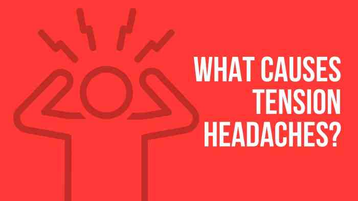
Facts about tension headaches: These common headaches, often described as a tight band around the head, affect millions worldwide. This...
 How to Lower Diastolic Blood Pressure
 Osmotic Laxatives for Constipation A Comprehensive Guide
 How Does Invisalign Work? A Comprehensive Guide
 Everything You Need to Know About Black Pepper
 Whats New in Mash A Deep Dive
 Food Recalls Lean Cuisine, Aldi Chomps
 Cholesterol Drug Helps Remove PFAS Study Insights
 Diabetes and Liver Disease A Deep Dive
 Inositol for PCOS Info A Comprehensive Guide