What is bone marrow edema? This condition, often detected on medical imaging, is a complex inflammatory response within the bone marrow. Understanding its various causes, locations, and imaging characteristics is crucial for accurate diagnosis and effective treatment. This exploration delves into the intricacies of bone marrow edema, offering insights into its definition, clinical significance, and the various pathologies it can accompany.
We’ll look at the underlying causes, the critical imaging characteristics, and the often subtle clinical presentations, as well as the treatment approaches and prognosis.
Bone marrow edema isn’t just a medical term; it represents a significant challenge in diagnosis. The variety of potential causes, from stress fractures to infections, makes precise identification crucial. Different imaging modalities, like MRI and CT scans, provide valuable insights into the nature and extent of the edema. This discussion will highlight the importance of distinguishing bone marrow edema from other bone lesions, emphasizing the importance of a comprehensive approach that combines imaging findings with patient history and physical examination.
Definition and Overview
Bone marrow edema is a condition characterized by an abnormal accumulation of fluid within the bone marrow. This fluid buildup, typically appearing as a bright signal on magnetic resonance imaging (MRI), isn’t a disease itself, but rather a sign of underlying pathology. Understanding its presence is crucial for diagnosing the root cause and guiding appropriate treatment.The clinical significance of bone marrow edema is substantial.
It often indicates stress, inflammation, or injury to the bone and surrounding tissues. Early detection can lead to prompt intervention and potentially prevent more severe complications. The location of the edema can offer clues about the affected area and the potential cause.
Clinical Significance of Bone Marrow Edema
Bone marrow edema is a critical imaging finding, often signaling ongoing inflammation or micro-trauma within the bone. This inflammatory response can be caused by a wide range of factors, from minor overuse injuries to more severe conditions like infections or tumors. The presence of bone marrow edema, coupled with the clinical presentation, aids in the differential diagnosis and helps guide appropriate management strategies.
Common Locations of Bone Marrow Edema
Bone marrow edema can be found in various locations throughout the skeleton. Its prevalence in certain areas reflects the high stress and mechanical loading those regions experience. Common sites include the:
- Pelvis and hip region:
- Lower extremities:
- Spine:
- Shoulders:
This area is prone to bone stress injuries, especially in athletes or individuals with repetitive stress activities.
The lower extremities bear significant weight and experience high impact forces, making them susceptible to edema from stress fractures, tendinopathies, and other overuse syndromes.
The spine is also susceptible to edema, often related to spinal conditions like spondylolysis or spondylolisthesis.
Repetitive overhead activities or trauma can lead to edema in the shoulder area.
Imaging Findings Associated with Bone Marrow Edema
The typical imaging finding associated with bone marrow edema is a hyperintense signal on MRI scans. This signal is usually seen on T2-weighted images and sometimes on STIR (short tau inversion recovery) sequences. The edema appears bright compared to normal bone marrow.
The specific appearance can vary depending on the severity and duration of the underlying pathology.
This characteristic signal is not specific to bone marrow edema, but it is highly suggestive and is frequently used in conjunction with other imaging findings and clinical history to arrive at a diagnosis.
Role of Bone Marrow Edema in Various Pathologies
Bone marrow edema can be a crucial indicator in various pathologies, from benign overuse injuries to serious conditions. It is essential to consider the location, extent, and presence of any accompanying symptoms when evaluating bone marrow edema.
- Stress fractures:
- Osteomyelitis:
- Tumors:
- Traumatic injuries:
- Inflammatory conditions:
Repeated stress on a bone can cause micro-fractures, leading to bone marrow edema.
Bone infection can result in bone marrow edema.
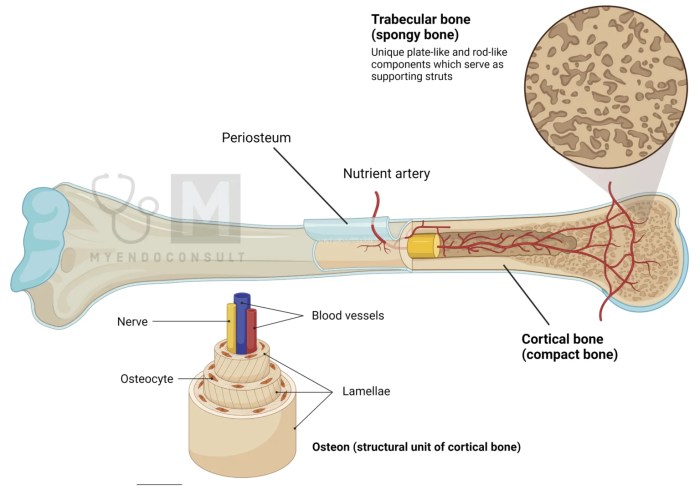
Bone marrow edema is basically swelling in the spongy inner part of your bones. It’s often a sign of something more serious, like an injury or infection. Sometimes, athletes experiencing discomfort might consider using silicone hydrogel contact lenses for eye strain, but that’s completely unrelated to bone marrow issues. The important thing is to understand the root cause of bone marrow edema to get proper treatment.
Both benign and malignant bone tumors can cause edema.
Trauma to the bone can lead to inflammation and edema.
Conditions like rheumatoid arthritis can cause edema in affected joints.
Table of Conditions Associated with Bone Marrow Edema
| Condition | Location of Edema | Imaging Findings | Clinical Presentation |
|---|---|---|---|
| Stress Fracture (Femur) | Distal femur | Hyperintense signal on T2-weighted MRI | Pain, tenderness, swelling, and sometimes limited range of motion. |
| Osteomyelitis (Tibia) | Tibia | Hyperintense signal on T2-weighted MRI, possible bone destruction | Fever, localized pain, swelling, and erythema. |
| Spinal Disc Herniation | Adjacent vertebral bodies | Hyperintense signal on T2-weighted MRI, possible disc bulge | Pain radiating down the leg, neurological symptoms. |
| Traumatic Injury (Rib) | Rib | Hyperintense signal on T2-weighted MRI, possible periosteal reaction | Localized pain, tenderness, and crepitus. |
| Osteoid Osteoma (Proximal Femur) | Proximal femur | Hyperintense signal on T2-weighted MRI, possible sclerotic rim | Pain that is often worse at night, localized tenderness. |
Etiology and Causes

Bone marrow edema, a condition characterized by swelling within the bone marrow, isn’t a disease itself but rather a symptom of underlying issues. Understanding the causes is crucial for proper diagnosis and treatment. Various factors can contribute to this inflammatory response, ranging from minor trauma to severe infections. Pinpointing the specific cause is often essential for effective management and recovery.
Frequent Causes of Bone Marrow Edema
Several factors frequently lead to bone marrow edema. These include stress fractures, infections, and various inflammatory conditions. The specific mechanisms and underlying pathology vary based on the causative agent.
Underlying Mechanisms of Bone Marrow Edema
The development of bone marrow edema involves a complex interplay of cellular and molecular processes. Trauma, infection, and inflammation all trigger a cascade of events leading to the accumulation of fluid within the bone marrow. This inflammatory response involves the release of cytokines and other signaling molecules, leading to increased vascular permeability and fluid leakage. The resultant swelling disrupts normal bone marrow function.
Pathophysiology of Bone Marrow Edema in Specific Conditions
Specific conditions influence the precise pathophysiology of bone marrow edema.
- Stress Fractures: Repetitive microtrauma to bone, such as seen in athletes or individuals with improper biomechanics, can initiate an inflammatory response. This response results in the recruitment of immune cells, leading to bone marrow edema. The inflammatory cascade is triggered by the release of inflammatory mediators, which ultimately lead to edema. For instance, a runner experiencing repeated stress on their tibia might develop bone marrow edema in that area.
- Infection: Infectious agents, such as bacteria or viruses, can directly or indirectly cause bone marrow edema. Direct invasion of the bone marrow by pathogens or the body’s immune response to infection can lead to edema. This inflammatory process results in fluid accumulation within the bone marrow. For example, osteomyelitis, an infection of the bone, is often associated with bone marrow edema.
Comparison of Causes in Different Age Groups, What is bone marrow edema
The causes of bone marrow edema can vary based on the age group. While stress fractures are more prevalent in young, active individuals, conditions like avascular necrosis or certain cancers might be more common in older age groups. The prevalence of certain infections can also vary depending on the age group.
Role of Inflammation in Bone Marrow Edema
Inflammation plays a pivotal role in the development of bone marrow edema. The inflammatory response is a complex cascade of events initiated by various stimuli. The release of cytokines, chemokines, and other inflammatory mediators leads to increased vascular permeability, resulting in fluid leakage into the bone marrow, thus contributing to the edema.
Table: Causes of Bone Marrow Edema and Clinical Features
| Cause | Clinical Features |
|---|---|
| Stress Fractures | Pain, swelling, tenderness localized to the affected bone. Symptoms often worsen with activity. |
| Infection (e.g., Osteomyelitis) | Pain, fever, chills, localized warmth, swelling, and tenderness in the affected area. Systemic symptoms may be present. |
| Tumors (e.g., Leukemia) | Depending on the specific tumor type, possible symptoms can include bone pain, swelling, anemia, fatigue, and others. |
| Avascular Necrosis | Pain, gradual onset of stiffness, and reduced range of motion in the affected joint. Symptoms can vary based on the location and severity. |
| Trauma | Pain, swelling, and ecchymosis (bruising) at the site of injury. |
Imaging Characteristics
Bone marrow edema, a common finding in various musculoskeletal conditions, presents distinct imaging characteristics that aid in diagnosis and differentiation from other pathologies. Accurate interpretation of these characteristics is crucial for appropriate management and treatment planning. Understanding the specific signal intensities observed on different imaging sequences and the subtle distinctions between bone marrow edema and other bone lesions is vital.Imaging modalities like MRI and CT play a significant role in evaluating bone marrow edema.
MRI, particularly, offers superior soft tissue contrast, allowing for detailed assessment of the affected bone marrow and surrounding structures. CT, while less sensitive to soft tissue changes, can provide crucial information regarding bone density and structural integrity, often complementing MRI findings.
MRI Characteristics of Bone Marrow Edema
MRI is the gold standard for evaluating bone marrow edema due to its superior soft tissue contrast. Different MRI sequences highlight various aspects of the condition, enabling a comprehensive assessment.
- T1-weighted images typically show low signal intensity within the affected bone marrow, reflecting the edema fluid’s reduced fat content. This low signal intensity is often subtle, and the surrounding normal bone marrow often appears as a high signal.
- T2-weighted images demonstrate high signal intensity within the affected bone marrow, indicating the presence of fluid or edema. The signal intensity is usually homogeneous within the affected area, though it can sometimes exhibit subtle heterogeneity.
- STIR (Short Tau Inversion Recovery) sequences and Proton Density Fat Suppression (PDFS) sequences are particularly useful for highlighting bone marrow edema. These sequences are sensitive to water and fluid content, providing enhanced visualization of the edematous area by suppressing the signal from fat. This enhances the visibility of the low signal on T1 and high signal on T2 in the affected area.
CT Characteristics of Bone Marrow Edema
While less sensitive to soft tissue changes compared to MRI, CT scans can provide valuable information about bone density and structural abnormalities associated with bone marrow edema. CT findings may be subtle, and in many cases, MRI is required for a more definitive diagnosis.
- CT scans might reveal subtle changes in bone density, like subtle cortical thinning or periosteal reaction in the affected area. However, these findings are often nonspecific and may not be apparent in all cases of bone marrow edema.
- CT scans are frequently used to assess for fracture or other structural damage, which may accompany or be the cause of bone marrow edema. This aspect is crucial in differentiating between bone marrow edema and other conditions like stress fractures.
Differential Diagnosis
Distinguishing bone marrow edema from other bone lesions is critical for appropriate treatment. The imaging characteristics, including signal intensity patterns and the distribution of the affected area, help in differential diagnosis.
| Condition | T1-weighted Signal | T2-weighted Signal | Additional Imaging Findings |
|---|---|---|---|
| Bone Marrow Edema | Low | High | Homogeneous signal intensity within the affected area, may involve a wide range of bone |
| Stress Fracture | Low/Variable | High | Linear or geographic zone of signal abnormality, potential for periosteal reaction, cortical disruption, often associated with specific stress or overuse patterns. |
| Tumor | Variable (can be low or high) | Variable (can be low or high) | Often heterogeneous signal intensity, possible bone erosion, and soft tissue mass |
| Infections | Variable (can be low or high) | Variable (can be low or high) | May show associated inflammatory changes in surrounding tissues, and possible bone destruction |
Role of MRI Sequences
Different MRI sequences play distinct roles in evaluating bone marrow edema.
- T1-weighted images are crucial for identifying the presence of low signal intensity, often subtle, within the affected bone marrow.
- T2-weighted images, STIR, and PDFS sequences are essential for detecting the characteristic high signal intensity of edema. The distribution and extent of the edema can be assessed.
- The combination of various sequences provides a more comprehensive picture of the lesion, aiding in differentiating bone marrow edema from other bone lesions.
Differential Diagnosis: What Is Bone Marrow Edema
Bone marrow edema, while a common finding on imaging, can mimic various other conditions. Accurately differentiating bone marrow edema from these mimics is crucial for appropriate diagnosis and treatment. Precise diagnosis hinges on a combination of imaging characteristics, clinical presentation, and a thorough understanding of the underlying pathology. A structured approach to differential diagnosis, incorporating both imaging and clinical data, is vital for avoiding misdiagnosis and ensuring optimal patient care.
Conditions Mimicking Bone Marrow Edema
Several conditions can present with imaging findings similar to bone marrow edema, leading to diagnostic challenges. These include stress fractures, tumors, infections, and inflammatory processes. Understanding the subtle distinctions between these entities is paramount for accurate diagnosis.
Bone marrow edema is basically swelling in the spongy inner part of your bones. It’s often a sign of something else going on, like stress fractures or infections. Interestingly, while dealing with such issues, you might also encounter situations where a “rule wipes medical debt credit score” rule wipes medical debt credit score could affect your financial well-being.
Regardless of the financial aspects, understanding the underlying causes of bone marrow edema is crucial for proper diagnosis and treatment.
Imaging Distinguishing Features
Imaging plays a critical role in differentiating bone marrow edema from other pathologies. While edema often shows as a low signal on T1-weighted images and high signal on T2-weighted images, the specific location, size, and shape of the affected area can provide clues. For instance, a localized area of edema adjacent to a fracture line strongly suggests a stress fracture.
The presence of a mass-like lesion or heterogeneous signal intensity, on the other hand, might point towards a tumor. Moreover, the presence of bone marrow edema in the absence of any apparent injury, or in association with fever and inflammatory markers, may point to an infection.
Clinical Presentation and Physical Examination
Clinical history and physical examination are indispensable for refining the differential diagnosis. Symptoms like pain, swelling, and limited range of motion can help distinguish bone marrow edema from other conditions. For example, severe pain that worsens with activity, often accompanied by localized tenderness, strongly suggests a stress fracture. Conversely, the presence of fever, chills, and systemic inflammatory response syndrome (SIRS) criteria might point to an infection.
A careful evaluation of the patient’s medical history, including any prior injuries, surgeries, or systemic illnesses, is also critical.
Structured Approach to Differential Diagnosis
A structured approach to differential diagnosis is crucial for minimizing diagnostic errors. This involves a systematic assessment of imaging findings, coupled with a detailed clinical evaluation. This approach typically begins with evaluating the location and extent of the edema on the MRI scans. Then, the presence of associated findings, such as fractures, bony abnormalities, or soft tissue swelling, is considered.
Next, the patient’s clinical history, including the nature and onset of symptoms, associated risk factors, and relevant past medical history, is examined. Finally, the combination of imaging findings and clinical presentation is used to generate a differential diagnosis list.
Comparison Table
| Condition | Imaging Findings | Clinical Presentation | Distinguishing Features |
|---|---|---|---|
| Bone Marrow Edema | Low signal on T1, high signal on T2 | Pain, possible swelling | Non-specific, often associated with trauma or overuse |
| Stress Fracture | Edema adjacent to fracture line | Pain that worsens with activity | History of trauma or repetitive stress |
| Tumor | Mass-like lesion, heterogeneous signal intensity | Pain, swelling, possible neurological symptoms | Mass effect, possible increased vascularity |
| Infection | Edema with possible surrounding soft tissue inflammation | Fever, chills, systemic inflammatory response | History of infection, elevated inflammatory markers |
Clinical Presentation and Symptoms
Bone marrow edema, a condition characterized by inflammation within the bone marrow, often presents with a range of symptoms that vary depending on the affected location and the underlying cause. Understanding these symptoms is crucial for accurate diagnosis and appropriate management. This section delves into the common clinical manifestations, highlighting the relationship between location and symptoms, and emphasizing the variability in presentation based on the etiology.
Common Clinical Symptoms
Bone marrow edema, while not always symptomatic, can lead to a variety of discomfort and pain. The symptoms often depend on the specific location and the extent of the inflammatory process within the bone. Pain is a common presenting complaint, often described as aching, throbbing, or sharp, and can vary in intensity. Other symptoms may include swelling, tenderness, and limited range of motion in the affected area.
Symptoms by Affected Body Regions
The location of bone marrow edema significantly influences the observed symptoms. Pain and tenderness are typically localized to the affected area. For example, bone marrow edema in the hip region may manifest as hip pain, whereas edema in the spine may lead to back pain. The severity of symptoms can range from mild discomfort to significant debilitating pain, impacting daily activities.
Bone marrow edema is basically swelling in the spongy inner part of a bone. It’s often a precursor to more serious issues, sometimes requiring significant intervention. Fortunately, there are now more options available for dealing with joint problems than just hip replacement. Exploring alternatives to hip replacement, like those discussed in this helpful resource on alternatives to hip replacement , is important for understanding potential less invasive treatments.
Ultimately, understanding bone marrow edema is key to making informed decisions about treatment options.
It’s important to note that the specific symptoms experienced can vary considerably, depending on the underlying cause of the edema.
Relationship Between Location and Symptoms
The location of bone marrow edema directly correlates with the location of the symptoms. Pain experienced in the knee, for example, suggests involvement of the bone marrow within the knee joint. This localization helps in guiding the diagnostic process and identifying the potential underlying cause.
Table of Common Symptoms and Potential Correlations with Location
| Affected Body Region | Common Symptoms | Potential Correlations |
|---|---|---|
| Hip | Hip pain, groin pain, limping, limited range of motion in hip | Possible involvement of the femoral head, acetabulum, or surrounding tissues |
| Knee | Knee pain, swelling, tenderness, stiffness, difficulty bearing weight | Possible involvement of the tibial plateau, femoral condyle, or menisci |
| Spine (vertebrae) | Back pain, radiating pain, numbness, weakness, muscle spasms | Possible involvement of the intervertebral discs, facet joints, or spinal cord |
| Foot/Ankle | Foot pain, ankle pain, swelling, tenderness, difficulty walking | Possible involvement of the tarsal bones, metatarsals, or surrounding tissues |
| Shoulder | Shoulder pain, limited range of motion, pain with overhead activities | Possible involvement of the humeral head, glenoid labrum, or surrounding tissues |
Variability of Symptoms Based on Underlying Cause
The specific symptoms associated with bone marrow edema can vary significantly depending on the underlying cause. For instance, trauma-induced bone marrow edema may present with immediate, intense pain, while edema related to infection might manifest with fever and systemic symptoms alongside the local pain. Careful consideration of the patient’s medical history, including any recent injuries, infections, or inflammatory conditions, can provide valuable insights into the potential cause of the edema.
Treatment and Management
Bone marrow edema, while often a symptom rather than a disease itself, requires appropriate management to address the underlying cause and alleviate associated pain and functional limitations. Effective treatment strategies often involve a combination of conservative approaches and, in some cases, surgical interventions. The primary goal is to reduce inflammation, promote healing, and restore normal function to the affected area.The management of bone marrow edema is highly individualized, taking into account the specific cause, location, severity, and overall health of the patient.
A multidisciplinary approach involving orthopedists, physical therapists, and other specialists is often crucial for optimal outcomes.
General Principles of Treatment
Effective treatment for bone marrow edema focuses on reducing inflammation, promoting healing, and restoring function. This often involves a multifaceted approach, targeting both the symptoms and the underlying cause. Strategies commonly include rest, physical therapy, and potentially, medication to manage pain and inflammation. Early intervention is key to minimizing long-term complications and maximizing functional recovery.
Conservative Management Strategies
Conservative management plays a significant role in the treatment of bone marrow edema, particularly in the initial stages. Rest and physical therapy are frequently used to reduce pain, inflammation, and stress on the affected area. Rest helps to allow the body to heal, reducing stress on the injured tissue. Physical therapy focuses on regaining range of motion, strengthening muscles surrounding the affected area, and improving overall function.
These strategies are particularly important in cases where the cause of the edema is related to overuse or repetitive strain.
Specific Treatment Modalities for Underlying Causes
The specific treatment modalities employed depend on the underlying cause of the bone marrow edema. For example, in cases of stress fractures, the primary treatment focuses on allowing the fracture to heal, potentially involving the use of casts or braces, and avoidance of activities that exacerbate the condition. In cases of osteomyelitis, the treatment focuses on addressing the infection through antibiotics, often in conjunction with surgical intervention to drain the affected area if necessary.
If the edema stems from a tumor, the approach will involve managing the tumor, which might involve chemotherapy, radiation therapy, or surgery.
Role of Surgical Intervention
Surgical intervention is reserved for severe or chronic cases of bone marrow edema where conservative management has failed to produce adequate results. Examples include cases of significant instability, persistent pain, or when there is a suspected underlying pathology requiring surgical intervention. Surgical procedures may involve debridement of damaged tissue, stabilization of the affected area, or removal of a tumor.
The specific surgical approach will depend on the nature of the underlying condition.
Monitoring Response to Treatment
Close monitoring of the patient’s response to treatment is crucial. This involves regular assessments of pain levels, range of motion, and functional capacity. Imaging studies, such as MRI scans, may be repeated to track the progression of edema resolution. Adjustments to the treatment plan may be necessary based on the observed response.
Treatment Approaches Based on Underlying Cause
| Underlying Cause | Treatment Approach |
|---|---|
| Stress fractures | Rest, immobilization (e.g., cast or brace), avoidance of aggravating activities, physical therapy to restore strength and mobility. |
| Osteomyelitis | Antibiotics, surgical drainage (if needed), pain management. |
| Tumors | Tumor-specific treatment (e.g., chemotherapy, radiation, surgery), pain management. |
| Repetitive strain injuries | Rest, activity modification, physical therapy, anti-inflammatory medications. |
| Traumatic injuries | Immobilization, pain management, physical therapy to restore function. Surgery may be necessary for significant injuries. |
Prognosis and Outcomes

Bone marrow edema, while often a temporary condition, can have varying impacts on patients, depending on the underlying cause and the individual’s overall health. Understanding the potential outcomes, including long-term effects and complications, is crucial for appropriate management and patient counseling. The prognosis for bone marrow edema is often tied to the resolution of the underlying cause, and successful treatment often leads to a favorable outcome.
Factors Influencing Outcome
Several factors significantly influence the prognosis of bone marrow edema. The severity of the underlying condition plays a pivotal role, as does the promptness and effectiveness of treatment. Patient age, overall health, and the presence of any other contributing medical conditions can also influence the recovery process. The location of the edema within the bone structure and the extent of the affected area are also critical factors in predicting the likelihood of a complete recovery.
Long-Term Effects
Bone marrow edema can result in a range of long-term effects, depending on the specific condition causing it. In some cases, edema may resolve completely without leaving any lasting consequences. However, in other situations, it can lead to persistent pain, decreased mobility, or even structural damage to the affected bone. Chronic conditions that trigger bone marrow edema can contribute to ongoing inflammation and potentially affect bone density and strength over time.
For example, in cases of stress fractures, long-term edema can result in delayed healing and potential for recurrent fractures.
Potential Complications
Potential complications associated with bone marrow edema vary depending on the underlying cause. In cases of infections, the edema may indicate a more severe systemic illness requiring aggressive treatment. In cases of tumors or cancers, the presence of bone marrow edema can be a marker of disease progression or recurrence. In some cases, prolonged edema may contribute to avascular necrosis, a condition where the bone tissue loses its blood supply, leading to potential bone death.
Furthermore, prolonged pain and discomfort can significantly impact quality of life.
Examples of Outcomes
Favorable outcomes often involve the complete resolution of edema and a return to normal function. For example, in cases of stress fractures effectively treated with rest and immobilization, edema usually resolves within a few weeks, and patients can return to their usual activities without significant long-term issues. Unfavorable outcomes, on the other hand, might involve persistent edema, pain, and reduced mobility.
For example, patients with underlying conditions like osteomyelitis or tumors might experience recurrent episodes of edema despite treatment, resulting in significant functional limitations.
Prognostic Factors Table
| Condition | Severity | Treatment Response | Potential Long-Term Effects | Prognosis |
|---|---|---|---|---|
| Stress Fracture | Mild | Good | Rare | Favorable |
| Osteomyelitis | Severe | Variable | High | Unfavorable |
| Tumor | Moderate to Severe | Variable | High | Uncertain |
| Trauma | Variable | Variable | Variable | Variable |
Note: This table provides a general overview and should not be considered a definitive prognostic tool. Individual cases require careful evaluation and personalized management strategies.
Closing Summary
In conclusion, bone marrow edema presents a multifaceted challenge to healthcare professionals. From its diverse causes and clinical manifestations to the crucial role of imaging in diagnosis, understanding this condition requires a thorough grasp of its underlying mechanisms and potential complications. This discussion has explored the various facets of bone marrow edema, offering a comprehensive overview of its definition, etiology, imaging characteristics, differential diagnosis, clinical presentation, treatment approaches, and prognosis.
We hope this detailed exploration has provided valuable insights into this intricate condition.


